pyruvic-acid-containing polysaccharide in the cell wall of bacillus polymyxa ahu 1385
TRANSCRIPT
Eur. J. Biochem. 174,255-260 (1988) 0 FEBS 1988
Pyruvic-acid-containing polysaccharide in the cell wall of Bacillus polymyxa AHU 1385 Naoya KOJIMA, Shunji KAYA, Yoshio ARAKI and Eiji IT0 Department of Chemistry, Faculty of Science, Hokkaido University
(Received December 22, 1987/March 21, 1988) - EJB 87 1421
Three acidic polymer fractions with molecular masses of about 16 kDa, 35 kDa and 70 kDa were isolated from lysozyme digests of N-acetylated cell walls of Bacilluspolymyxa AHU 1385 by ion-exchange chromatography and gel chromatography. These fractions, containing mannosamine, glucosamine and pyruvic acid in a molar ratio of about 1 : 1 : 1 together with glycopeptide components, were characterized as polysaccharide-linked glycopeptides with one, two and more polysaccharide chains. On the other hand, treatment of the cell walls with glycine/HCl buffer, pH 2.5, at 100°C for 10 min followed by separation of water-soluble products on ion-exchange chromatography gave three polysaccharide fractions, PS-I - 111, whch contained different amounts of pyruvic acid (0, 0.6 and 0.9 residue/mannosamine residue) along with equimolar amounts of mannosamine and glucosamine. Pyruvate-free polysaccharides similar to PS-I were also obtained from PS-11, PS-I11 and polysaccharide-linked glycopeptides by treatment with 10mM HCl at 100°C for 1 h. Results of analyses of these polysaccharide preparations by 'H-NMR and I3C-NMR measurement and methylation, together with data from characterization of fragments obtained by hydrogen fluoride hydrolysis, lead to the most likely structure, +3)[4,6-0-(1- carboxyethylidene)]ManNAc(~1-+4)GlcNAc(~1--f, for the acidic polysaccharide of this strain.
Pyruvic acid has been found in a ketal form in exocellular acidic polysaccharides of different bacteria [l] and cell wall teichoic acid of Brevibacterium iodium [2]. In connection with a study on distribution of mannosaminuronic acid and mannosamine among cell walls of gram-positive bacteria [3], the cell wall polysaccharide of Bacillus polymyxa AHU 1385 was shown to contain pyruvic acid as an acidic component, in addition to mannosamine and glucosamine. The present paper reports the structure of this polysaccharide, in which the pyruvic acid is attached to N-acetylmannosamine to form a six-membered ketal ring.
MATERIALS AND METHODS
Preparation of pyruvic-acid-containing polysaccharide and their glycopeptide complexes
The methods used for cultivation of B. polymyxa AHU 1385 (kindly given by Dr S. Takao, Hokkaido University) and for preparation and N-acetylation of cell walls were the same as those described in a previous paper [3]. For isolation of polysaccharide-linked glycopeptides, the N-acetylated cell walls (400 mg) were exhaustively digested with lysozyme (4 mg), and the polymer fraction obtained from the digest by dialysis and gel chromatography on Sephadex G-50 was subjected to chromatography on a DEAE-cellulose column (2 x 8 cm) equilibrated with 5 mM Tris/HCI buffer, pH 7.2 [4]. The column was eIuted with the same buffer, followed by a linear gradient of 0-0.4 M NaCl in the same buffer. A polymer fraction eluted without salt and fractions eluted with
Correspondence to E. Ito, Department of Chemistry, Faculty of Science, Hokkaido University, Kita-10-jyo, Nishi-8-chome, Kita-ku, Sapporo, Japan 600
Abbreviations. ManNAc, N-acetyl-D-mannosamine; PS-GP, polysaccharide-linked glycopeptide.
0.20 M and with 0.35 M NaCl were separately purified by gel chromatography on a Sephacryl S-200 column (1 x 100 cm) in 50 mM (NH4)2C03 and used as the polysaccharide-linked glycopeptides, PS-GP-I (30 mg), PS-GP-I1 (26 mg) and PS- GP-111 (35 mg), respectively. These preparations commonly contained peptidoglycan components, such as muramic acid, alanine, glutamic acid and diaminopimelic acid, together with the major components, glucosamine and mannosamine.
For isolation of polysaccharide chains, the cell wall prep- aration (400 mg) was treated in 320 ml 25 mM glycine/HCl buffer, pH 2.5, at 100°C for 10min and centrifuged at 20000 x g [4], and the resulting precipitate was treated three times in the same way. The supernatants were pooled and dialyzed, and nondialyzable material was subjected to chromatography on a DEAE-cellulose column (1.5 x 6 cm) equilibrated with 5 mM ammonium acetate buffer, pH 6.8. Elution of the column with the same buffer, followed by a linear gradient of 0 - 0.4 M NaCl in the same buffer, gave a neutral polysaccharide fraction (peak I) eluted without salt and two acidic polysaccharide fractions (peaks I1 and 111) respectively eluted with 0.10 M and 0.22 M NaCl. The poly- mer in the neutral polysaccharide fraction was purified by chromatography on Sephacryl S-200 (1 x 100 cm) in 50 mM (NH4)2C03 and used as PS-I (28 mg). The acidic poly- saccharide fractions in peaks I1 and 111 were individually purified by successive chromatography on columns of DEAE- cellulose and Sephacryl S-200, giving PS-II(96 mg) and PS-III (48 mg), respectively.
Removal of pyruvic acid
A portion of the PS-111 preparation (20 mg) was treated in 5 ml 10 mM HCl at 100°C for 1 h, and the product after lyophilization was fractionated by chromatography on a DEAE-cellulose column (1.5 x 3 cm) as described above.
256
Almost all of the sugar components were recovered in the neutral polymer fraction, which was used as the pyruvate-free PS-111 preparation after purification by chromatography on Sephacryl S-200.
N M R spectroscopy and methylation analysis
'H-NMR spectra were recorded in D 2 0 on a Jeol FX-500 spectrometer at 95 "C with sodium 3-trimethylsilylpropane sulfonate as an internal standard. The proton homonuclear- shift-correlated (COSY) NMR experiment was performed on the same instrument at 80°C. 13C-NMR spectra were record- ed on a Jeol FX-100 spectrometer at 25°C with methanol as an internal standard. For methylation analysis, pyruvate-free PS-I11 was permethylated by the method of Ciucanu and Kerek [5] and the permethylation product was hydrolyzed in 4 M HC1 at 100°C for 16 h. The partially methylated saccha- rides were converted to their alditol acetates and analyzed by gas-liquid chromatography/mass spectrometry (GC-MS) on a Jeol DX-300 mass spectrometer using a glass column packed with 2% OV-17lUniport HW. In the case of intact PS-111, the sample was first reduced by treatment with carbodiimide/ NaBH, [6] and then permethylated as above.
Partial hydrolysis of polysaccharide
The pyruvate-free PS-111 preparation (8 mg) was treated in 1 ml 55% hydrogen fluoride (HF) at 37°C for 18 h. After evaporation by an air flush followed by N-acetylation with acetic anhydride, the product was subjected to chromatog- raphy on a Cellulofine GCL-25-m column (1 x 147 cm) in 50 mM (NH4)2C03. Fractions (1 ml) were collected and de- termined for total hexosamine and reducing groups.
Other materials and analytical methods
Unless otherwise stated, analytical methods were essen- tially the same as those described in previous papers [4, 71. Standard dextrans, T-10, T-20, T-40 and T-70, were purchased from Pharmacia Fine Chemicals. Total hexosamine was de- termined by the method of Tsuji et al. [8] after N-deacetylation of samples by acid hydrolysis (2 M HCI, 100°C, 1 h); pyruvic acid, by the enzymatic method using L-lactate dehydrogenase [9] after treatment of samples with mild acid (0.01 M HC1, 1OO"C, 1 h) followed by neutralization with 0.1 M NaOH; reducing groups, by the method of Park and Johnson [lo]. Hexosamine and hexosaminitol were analyzed by gas-liquid chromatography after acid hydrolysis (4 M HCl, 100°C, 4 h) of samples followed by N-acetylation and trimethylsilylation [Ill. Amino acids, amino sugars and muramic acid 6-phos- phate were determined with an amino acid analyzer as de- scribed previously [12]. NaIO, oxidation and Smith degrada- tion were carried out under the conditions similar to those described previously [4]. Descending paper chromatography was performed on Toyo no.50 filter paper in 1-butanol/ pyridine/water (6:4: 3, by vol.).
RESULTS
Isolation and analysis of polysaccharide-glycopeptide complexes and polysaccharide chains
The N-acetylated cell wall preparation of B. polymyxa AHU 1385 contained mannosamine, pyruvic acid and an amount of glucosamine exceeding the amount of muramic
acid derivatives in a molar ratio of about 1:1:1, together with peptidoglycan components (Table 1). From the polymer fraction obtained by lysozyme digestion of the N-acetylated cell walls, three preparations of polysaccharide-linked glycopeptide, PS-GP-I, PS-GP-I1 and PS-GP-111, were isolat- ed by successive chromatography on columns of DEAE-cellu- lose and Sephacryl S-200. All preparations contained mannosamine, excess glucosamine and pyruvic acid in the same molar ratios as did cell wall preparation (Table 1). The apparent molecular masses of PS-GP-I, PS-GP-I1 and PS- GP-I11 were 16 kDa, 35 kDa and 70 kDa, respectively, when estimated by gel filtration on columns of Sephacryl S-200 and S-300 with dextrans T-20, T-40 and T-70 as standards. In addition, these three complexes and the cell wall preparation showed similar molar ratios of mannosamine/muramic acid 6-phosphate of about 20. By analogy with the teichoic-acid - glycopeptide complex of B. cereus AHU 1030 [7], PS-GP-I, PS-GP-I1 and PS-GP-111 seem to have one, two and three or more similar polysaccharide chains, respectively, which were attached to either a single glycopeptide strand or two (or more) glycopeptide strands joined by cross linkages. The PS- GP-I preparation was used for further structural studies.
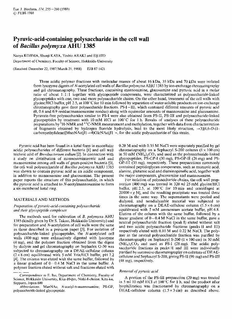
It has been reported that heating cell walls at pH 2.5 causes specific cleavage of the linkages between terminal sugar residues of polymer chains and the phosphoryl groups of muramic acid 6-phosphate residues of peptidoglycan [4, 71. When the N-acetylated cell walls were treated with 25 mM glycine/HCl buffer, pH 2.5, at 100°C for 10 min, the majority of mannosamine and pyruvic acid was released as components of water-soluble polymers. Under these conditions, about 25% of the pyruvate was concomitantly liberated from the polysaccharide chains. The resulting water-soluble polymer was separated into three fractions (peaks I - 111) of hexosamine-containing materials by chromatography on a column of DEAE-cellulose (Fig. 1). The major polymers in peaks I - I11 were individually purified by successive chroma- tography on columns of DEAE-cellulose and Sephacryl S-200, giving polysaccharide preparations PS-I, PS-I1 and PS-111, respectively. The apparent molecular masses of PS-I, PS-I1 and PS-111 were about 8 kDa, 11 kDa and 12 kDa, respect- ively, when estimated by chromatography on Sephacryl s-200 with dextrans as standards.
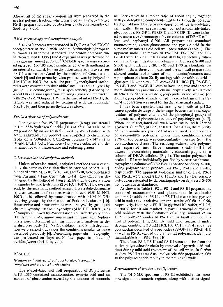
As shown in Table 1, PS-I, PS-I1 and PS-I11 preparations contained mannosamine and glucosamine in equimolar amounts. In addition, PS-I1 and PS-111 also contained pyruvic acid in molar ratios relative to mannosamine of 0.60 and 0.90, respectively. Heating of PS-111 in glycine/HCl buffer, pH 2.5, at 100°C for 10 min resulted in partial removal of pyruvic acid residues with the formation of a large amount of an anionic polymer similar to PS-I1 and a small amount of a neutral polymer (Fig.2A). By mild acid treatment under stronger conditions (10 mM HCl, lOO"C, 1 h), PS-I1 and three polysaccharide-linked glycopeptides (PS-GP-I to PS-GP-111) as well as PS-111 yielded only a neutral polysaccharide indis- tinguishable from PS-I (Fig. 2 B).
Therefore, PS-I, PS-I1 and PS-111 seem to arise from the native polysaccharide chain by removal of pyruvic acid resi- dues during mild acid treatment of the cell walls. In further studies, PS-111 was used as a polysaccharide preparation akin to the polysaccharide moiety in the native cell walls.
Determination of anomeric configuration
The 'H-NMR spectrum of PS-111 exhibited rather com- plex signals in anomeric regions, along with distinct signals

257
Table 1. Composition of N-acetylated cell walls, PS-GP-I, PS-I, PS-II and PS-III Each preparation was analyzed for components after acid hydrolysis (4 M HCl, lOO"C, 4 h). Pyruvic acid was assayed by the enzymatic method [9] after treatment of samples with mild acid (10 mM HCI, lOO"C, 1 h). Excess glucosamine is the difference between the amount of glucosamine and the total amount of muramic acid derivatives
Component N-Acetylated cell walls PS-GP-I" PS-I PS-I1 PS-111
nmol/mg dry material
Muramic acid 6-phosphate 32 62 0 0 0 Muramic acid 450 297 0 0 0 Glucosamine 1190 1550 2380 1960 1790 Excess glucosamine 710 1190 Mannosamine 669 1250 2300 1890 1680 Pyruvic acid 700 1240 trace 1130 1490 Phosphorus 40 68 0 0 0 Glutamic acidb 484 368 0 0 0
a Similar results were obtained with PS-GP-I1 and PS-GP-111. Each preparation of N-acetylated cell walls and PS-GP-1-111 contained alanine, diaminopimelic acid and glutamic acid in a molar
ratio of about 1 : 1 : 1. The values for glutamic acid are shown as representative data for these amino acid components.
II I - I
/
, b' I 0
0 10 20 30 40 Froction number
Fig. 1. Separation ofpolysaccharide chains. The water-soluble fraction, obtained from N-acetylated cell walls by heating at pH 2.5, was sub- jected to chromatography on a DEAE-cellulose column. Fractions ( 5 ml) were collected and analyzed for total hexosamine. Fractions indicated by bars were pooled, purified by chromatography on Sephacryl S-200, and used as the PSI, PS-I1 and PS-111 preparations
for two acetamide groups of N-acetylhexosamines (6 = 2.040 ppm, 6 H) and about one methyl group of pyruvic acid (6 = 1.486 ppm, 2.7 H). On the other hand, the spectrum of pyruvate-free PS-111 exhibited two anomeric signals [6 = 4.895 ppm, J1,2 = 1.2 Hz (semidoublet), 1 H; 6 = 4.582 ppm,
= 7.81 Hz (doublet), 1 HI. The signals at 4.582 and 4.895 ppm are assumed to be ascribable to P-N-acetyl- glucosaminide and D-N-acetylmannosaminide, respectively. An additional signal (6 = 4.632 ppm, pseudo-quartet, 1 H) observed in the anomeric region of the spectrum arises from H-2 of the j-N-acetylmannosamine residue. This assignment was supported by the connectivity between the signals at 4.895 ppm and 4.632 ppm in the COSY NMR experiment. The anomeric configuration proposed for the above two sugars was strongly supported by the 13C-NMR spectrum (Fig. 3 and Table 2). The 'H-coupled 13C-NMR spectrum gave two anomeric carbon signals at 101.9 ppm (P-N- acetylglucosamine) and 103.3 ppm (P-N-acetylmannosamine)
0.8
0.4 1 0
E,
5
- al .r 0
07 1.c 2 al I
0.5
C
' I
Fraction number
Fig. 2. Chromatography of PS-111 after treatment with mild acid. The PS-I11 preparation (3 mg) was treated either in glycinelHC1 buffer, pH 2.5, at 100°C for 10 min (A) or in 10 mM HC1 at 100°C for 1 h (B). Each product was chromatographed on DEAE-cellulose under the same conditions as in Fig. 1. When each of PS-I1 and PS-GP-I to PS-GP-111 was treated in 10 mM HCl at 100°C for 1 h, an elution profile similar to that in (B) was also obtained
with lJCH values lower than 170 Hz, namely 155 Hz and 164 Hz, respectively [13, 141. Furthermore, both hexosamine residues of the pyruvate-free PS-111 preparation were com- pletely destroyed by chromium trioxide oxidation. Thus, the polysaccharide chain of PS-111 is believed to consist of p-N- acetylmannosamine and p-N-acetylglucosamine.
The presence of ketal-linked pyruvic acid
The 'H-NMR spectrum given by PS-111 exhibited a sharp singlet signal (6 = 1.486 pprn), indicating the presence of ketal-linked pyruvic acid residues. This signal completely dis- appeared from the 'H-NMR spectrum of the same prep- aration after treatment with mild acid. From a comparison of

the 13C-NMR spectra between PS-111 and pyruvate-free PS-I11 (Fig.3), the signals at 6 = 176.5 ppm, 102.4 ppm and 27.1 ppm are assigned to carboxyl, ketal and methyl carbons of the pyruvic acid moiety, respectively. A comparison of the chemical shift for the ketal carbon (6 = 102.4ppm) with the previous spectral data [15, 161 strongly suggests that the pyruvic acid was present in a six-membered ring rather than a five-membered ketal ring, which gives higher chemical shift values (1 1 1 - 1 16 ppm).
Determination of linkage position
NaIO, oxidation of the pyruvate-free PS-I11 preparation caused no change in the hexosamine residues, indicating that both species of hexosamine residues were substituted at C-3 or C-4. When the oxidation was carried out after the same preparation had been N-deacetylated by treatment with alkali
A
180 160 HO 120 100 80 60 40 20 0 s (PPM)
Fig.3. I3C-NMR spectra of PS-III and pyruvate-free PS-Ill. The spectra of PS-111 (A) and pyruvate-free PS-I11 (B) were recorded at 25°C with a 100-MHz instrument
(3.3 M NaOH, IOOT, 5 h), almost all the glucosamine residues were degraded, whereas the mannosamine residues were fully recovered. Therefore, the polysaccharide backbone chain is assumed to have consisted of 4-substituted glucosamine and 3-substituted mannosamine residues. The assumption is also supported by the I3C-NMR spectral data. The signals at 6 = 81.4 ppm and 79.3 ppm (or 79.2 ppm) in the 13C-NMR spectrum of pyruvate-free PS-111 as well as of PS-I11 (Fig. 3 and Table 2) are ascribable to C-4 of the N-acetylglucosamine residues and C-3 of the N-acetylmannosamine residues, re- spectively. The downfield shifts (about 6-9 ppm) of the re- spective signals from the literature values for unsubstituted p- N-acetylglucosaminide and P-N-acetylmannosaminide [13, 141 may be explained by the glycosylation effect on the corresponding carbon atoms.
A Comparison of the I3C-NMR spectra between the PS-111 and the pyruvate-free PS-111 preparation indicates that the signal ascribable to C-3 of the N-acetylmannosamine residues as well as the signals ascribable to C-3 and C-6 of the N- acetylglucosamine residues was unchanged by the removal of pyruvate. On the other hand, the signals ascribable to C-4 and C-6 (76.3 ppm and 69.2 ppm, respectively) of the N- acetylmannosamine residues in the untreated PS-111 prep- aration are largely shifted to downfield regions as compared to those in the pyruvate-free preparation (6 = 67.3 ppm and 62.5 ppm) (Table 2). Therefore, the pyruvic acid residues seem to be linked to the N-acetylmannosamine residues at C-4 and C-6 in a ketal form.
Methylation analysis
To confirm the substitution positions of sugar com- ponents, the carboxyl-reduced PS-111 and pyruvate-free PS-I11 preparations were subjected to permethylation, and partially methylated sugar components were analyzed by GC/MS as methylated alditol acetates. When the carboxyl groups had not been reduced, PS-111 was incompletely methylated, probably owing to the presence of the carboxyl groups. In the case of pyruvate-free PS-111, 3,6 - di - 0 - methyl - N - methyl - N - acetylglucosaminitol acetate and 4,6-di-O-rnethyl-N-methyl- N-acetylmannosaminitol acetate were found in approximately equimolar amounts, whereas in the case of carboxyl-reduced PS-111, 3,6-di-O-rnethyl-N-methyl-N-acetylglucosaminitol ac- etate and N-methyl-N-acetylmannosaminitol acetate were detected (Table 3). This result indicates the presence of 4- substituted N-acetylglucosamine and 3,4,6-substituted N- acetylmannosamine residues in the native polysaccharide chain and that the latter residues were converted to 3-substi- tuted ones by the treatment with mild acid. These results
Table 2. '3C-NMR spectra of PS-Ill andpyruvatelfree PS-II Each spectrum also contained signals at 24.5 ppm and 176.5 ppm ascribable to methyl (2C) and carbonyl carbons (2C) of acetamide groups
Sample Group 6 for
c - I c -2 c - 3 c - 4 c- 5 C-6
PS-I11 +4)GlcNAc(bl+ 101.9 57.4 74.6 81.4 78.5 62.5 -+3)ManNAc(pl+ 103.3 52.3 79.3 16.3 76.7 69.2 pyruvic acid 176.5 102.4 27.1
Pyruvate-free PS-I11 +4)GlcNAc(fiI + 101.5 57.5 74.6 81.4 78.4 62.1 +3)ManNAc(pl+ 103.3 52.2 79.2 67.3 76.7 62.5

259
Table 3. Methylation analysis of PS-III and pyruvatelfree PS-III Data are shown in molar ratios to 3,6-di-0-methyl-N-methyl-N- acetylghcosaminitol acetate. Retention time, t R , is measured relative to 3,4,6-tri-O-methyl-N-methyl-N-acetylglucosaminitol acetate
~
Compound tR Molar ratio in
PS-111 pyruvate- free PS-IT1
~
3,6-Di-O-methyl-N-methyl-N-
4,6-Di-O-methyl-N-methyl-N-
N-Methyl-N-acetylmannos-
acetylglucosaminitol acetate 1.52 1.00 1 .00
acetylmannosaminitol acetate 1.79 0.08 0.76
aminitol acetate 3.62 0.73 trace
Fraction number
Fig.4. Chromatography of pyruvate:free PS-IZI after treatment with hydrogen fluoride. The pyruvate-free PS-111 preparation (8 mg) wdS
treated in 1 ml of 55% HF at 37°C for 18 h. The product was subjected to chromatography on a Cellulofine GCL-25-m column. Fractions (1 ml) were collected and analyzed for total hexosamine ( 0 ) and reducing groups (0). Fractions indicated by bars were pooled and purified by rechromatography on the same column followed by paper chromatography. Arrows 1 , 2 and 3 show the elution positions of the monomer, dimer and trimer of N-acetylglucosamine
are consistent with the above inference that the pyruvic acid residues were most likely linked to C-4 and C-6 of the N- acetylmannosamine residues in a ketal form.
Isolation and characterization of oligosaccharides
In order to obtain ohgosaccharide fragments, the pyruvate-free PS-I11 preparation was treated with 55% HF at 37°C for 18 h. After N-acetylation with acetic anhydride, the products were separated into five hexosamine-containing saccharides (peaks 1 - 5) by chromatography on a Cellulofine GCL-25-m column (Fig.4). Saccharides in peaks 1, 2, 3 and 4 were purified by rechromatography on the same column followed by paper chromatography, giving saccharides 1, 2, 3 and 4, respectively (1.2, 2.0, 0.55 and 3.3 pmol as total
hexosamine, respectively). Peak 5 consisted of free N- acetylmannosamine and N-acetylglucosamine in a molar ratio ofabout 1.5:l.
From results of analysis involving NaBH, reduction, Smith degradation and chromium trioxide oxidation, saccha- ride 4 was characterized to be ManNAc(fll+4)GlcNAc. In a similar procedure, saccharide 2 was shown to be a linear tetrasaccharide composed of equimolar amounts of N- acetylglucosamine and N-acetylmannosamine with the former sugar at the reducing end. Hydrolysis (4 M HCl, 100 "C, 4 h) of products from Smith degradation of samples of reduced saccharide 2 yielded glycerol, mannosamine, glucosamine and xylosaminitol in a molar ratio of about 1 : 1 : 1 : 1. When the same samples were treated in a similar way after N-deacetyl- ation by alkaline treatment (3.3 M NaOH, 100°C, 5 h), only mannosamine was yielded together with glycerol. Therefore, the most probable structure of saccharide 2 is ManNAc(P1 4 4)GlcNAc(fl1 + 3)ManNAc(pl + 4)GlcNAc. Saccharide 1 was a linear hexasaccharide composed of equimolar amounts of glucosamine and mannosamine and seems to be a trimer of saccharide 4.
The minor product, saccharide 3, seemed to be a mix- ture of GlcNAc-ManNAc-GlcNAc and ManNAc-GlcNAc- ManNAc, but it was not further characterized. The yields of saccharides I, 2 and 4 account for about 60% of the total amount of glucosamine present in the starting pyruvate-free PS-I11 preparation. Thus, it is most probable that the polysaccharide backbone chain was made up of repeating units, ManNAc(P1 -t4)GlcNAc, of which the N-acetylglucos- amine residues were linked P-glycosidically to C-3 of the N- acetylmannosamine residues of neighboring units.
DISCUSSION
The results described here lead to the most likely struc- ture, + 3)[4,6-0-(1 -carboxyethylidene)]ManNAc(fll + 4)- GlcNAc(P 1 +, for the repeating units of the pyruvic-acid- containing polysaccharide of B. polymyxa AHU 1385. From the molar ratio of mannosamine/muramic acid 6-phosphate in PS-GP-I and in the starting cell wall preparation, each polysaccharide chain seems to be composed of about 20 re- peating disaccharide units. As calculated from this number, the molecular mass for the unsubstituted polysaccharide chain was about 8 kDa, roughly coincident with the apparent molec- ular masses of the isolated polysaccharide chains (8 kDa, 1 2 kDa and 12 kDa for PS-I, PS-I1 and PS-111, respectively). The difference in the apparent molecular masses between PS-I, PS-I1 and PS-111 may be ascribable to variation in their con- tent of pyruvic acid (Table l), whose negative charge seems to cause an extra increase in apparent molecular sizes mea- sured by gel chromatography. The production of the polysac- charides with different charges by heating of the walls at pH 2.5 seems to be explained by concomitant elimination of ketal-linked pyruvic acid residues.
The most probable structure for repeating units deduced from the results of chemical analysis is consistent with the 'H- NMR and 13C-NMR spectral data (Table 2). The spectral data are coincident with those previously reported for the polysaccharide [ +3)ManNAc(fl1-+4)GlcNAc(fil +In which was derived from the Haemophilus injluenzae type-e capsular polysaccharide by alkaline treatment followed by carboxyl- reduction [17]. The anomeric configuration for saccharide residues was primarily deduced from the values of '.IcII for anomeric carbons (155 Hz and 164 Hz, for N-acetylglucos-

amine and N-acetylmannosamine, respectively), and support- ed by the result of chromium trioxide oxidation of pyruvate- free PS-111. Furthermore, the chemical shift for ketal carbons (6 = 102.4 ppm) indicates that the pyruvic acid residues are attached to mannosamine residues in a six-membered ring form.
Pyruvic acid is known to occur in extracellular poly- saccharides of Streptococcus pneumoniae [ 181, Coryne- bacterium irisidiosum [19], Mycobacterium salivarium [20], Escherichia coli, Aerobacter [21, 221, Klebsiella [23 - 321, Xanthomonas [33,34] and Rhizobium [35 - 371. In addition, Mycobacterial glycolipids [38, 391 and wall mannitol teichoic acid [2] were also shown to contain pyruvic acid. In these polysaccharides, pyruvic acid is attached to various saccharide residues, such as glucose [22, 23, 35-37], mannose [27, 30, 33, 341, galactose [18, 19, 21, 28, 31, 371, rhamnose [25, 261, 3-0-methylglucose [38, 391, glucuronic acid [20, 24, 291, and mannitol [2] in the form of either a five-membered or a six- membered ring. The pyruvic-acid-containing polysaccharide of B. polymyxa AHU 1385 reported in the present paper is unique in possessing 4,6-0-(1 -carboxyethylidene)N-acetyl- hexosamine as a constituent of the backbone chain. A prelimi- nary study showed that two other strains of B. polymyxa and three strains of Bacillus circulans also contain similar pyruvic- acid-linked polysaccharides in their cell walls.
REFERENCES 1. Sutherland, 1. W. (1985) Annu. Rev. Microbiol. 39,243 - 270. 2. Anderton, W. J. & Wilkinson, S. G. (1985) Biochem. J . 226,587-
599. 3. Yoneyama, T., Koike, Y., Arakawa, H., Yokoyama, K., Sasaki,
Y., Kawamura, T., Araki, Y., Ito, E. & Tdkao, S. (1982) J . Bacteriol. 144, 15 - 21.
4. Kojima, N., Araki, Y. & Ito, E. (1985) J . Bacteriol. 161, 299- 306.
5. Ciucanu, I. & Kerek, F. (1984) Carbohydr. Res. 131, 209-217. 6. Taylor, R. L. & Conrad, H. E. (1972) Biochemistry 11, 1383-
1388. 7. Sasaki, Y., Araki, Y. & Ito, E. (1983) Eur. J . Biochem. 132,207-
213. 8. Tsuji, A., Kinoshita, T. & Hoshino, M. (1969) Chem. Pharm.
Bull. (Tokyo) 17, 217-218. 9. Lamprecht, W. & Heinz, F. (1983) in Methods of enzymatic analy-
sis (Bergmeyer, H. U., ed.) vo1.6, pp.570-577, VCH Verlagsgesellschaft, Weinheim.
10. Park,J.T.&Johnson,M. J.(1949)J. Biol. Chem. 181,149-151. 11. Kojima, N., Araki, Y. & Ito, E. (1985) Eur. J . Biochem. 148,29 -
12. Amano, K., Hazama, S., Araki, Y. & Ito, E. (1977) Eur. J . 34.
Biochem. 75, 51 3 - 522.
13. Bock, K. & Thoegersen, H. (1982) Ann. Rep. N M R spectrosc. 13,
14. Jennings, H. J. & Smith, I. C. P. (1978) Methods Enzymol. 50,
15. Gorin, P. A. J., Mazurek, M., Duarte, H. S., lacomini, M. & Duatre, J. H. (3982) Curbohydr. Res. 100, 1-15.
16. Buchanan, J. G., Edgar, A. R., Rawson, D. I., Shahidi, P. & Wightman, R. H. (1982) Carbohydr. Res. ZOO, 75-86.
17. Tsui, F.-P., Schneeson, R., Boykins, R. A., Karpas, A. B. & Egan, W. (1981) Carbohydr. Res. 97,293-306.
18. Jannson, P.-E., Lindberg, B. & Lindquist, U. (1981) Carbohydr. Res. 95, 73 - 80.
19. Gorin, P. A. &Spencer, J. F. T. (1980) Curbohydr. Rex 79,313- 315.
20. Sviridov, A. F., Shashkov, A. S., Kochetokov, N. K., Botvinko, I. V. & Egorov, N. A. (1982) Bioorg. Khim. 8,1242-1251.
21. Lawson, C. J., McCleary, C. W., Nakada, H. I., Rees, D. A,, Sutherland, I. W. & Wilkinson, J. F. (1969) Biochem. J . 115,
22. Kamei, A,, Takeuchi, N., Akashi, S. & Kagabe, K . (1978) Chem.
23. Thurow, H., Choy, Y.-M., Frank, N., Niemann, H. & Stirm, S.
24. Erbiny, C., Kenne, L., Lindberg, B., Lonngren, J. & Sutherland,
25. Dutton, G. G. S. & Mackie, K. L. (1978) Carhohydr. Res. 62,
26. Bebault, G. M., Dutton, G. G. S., Funnell, N. A. & Mackie, K.
27. Elsaesser-Beile, U., Friebolin, H. & Stirm, S. (1978) Carbohydr.
28. Dutton, G. G. S. & Savage, A. V. (1980) Carbohydr. Res. 83,
29. Dutton, G. G. S. & Savage, A. V. (1980) Carbohydr. Res. 84,
30. Okutani, K. & Dutton, G. G. S. (1980) Carbohydr. Res. 86,259-
31. Dutton, G. G. S. & Paulin, M. (1980) Carbohydr. Res. 87, 119-
32. Dutton, G. G. S., Parolis, H. &Paroh , L. A. S. (1985) Curbohydr.
33. Jansson, P.-E., Kenne, L. & Lindberg, B. (1975) Curbohydr. Res.
34. Melton, L. D., Mindt, L., Rees, D. A. & Sanderson, G. R. (1976) Carhohydr. Res. 46, 245 - 257.
35. Jansson, P.-E., Kenne, L., Lindberg, B., Ljunggren, H., Lonngren, J., Ruden, U. & Svensson, S. (1977) J. Am. Chem.
36. Aman, P., McNeil, M., Franzen, L.-E., Darvill, A. G. &
37. Robertsen, B. K., Aman, P., Darvill, A. G., McNeil, M. &
38. Brennan, P. J., Aspinall, G. 0. & Nam-Shin, J. E. (1981) J . Biol.
39. Kamisango, K., Saadat, S., Dell, A. & Ballou, C. E. (1985) J .
1-57.
39 - 50.
947 - 958.
Pharm. Bull. (Tokyo) 26, 3395 - 3403.
(1975) Carbohydr. Res. 41, 241 -256.
I . W. (1 976) Curbohydr. Res. 50, 11 5 - 120.
321 - 335.
L. (1978) Curbohydr. Res. 63, 183-192.
Res. 65, 245 - 249.
351 -362.
297 - 305.
271.
127.
Res. 140, 263-275.
45,275 -282.
SUC. 99,3812-3815.
Albersheim, P. (1981) Carbohydr. Res. 95,263-282.
Albersheim, P. (1981) Plant Physiol. 67, 389-400.
Chem. 256,6817-6822.
Biol. Chem. 260,4117-4121.