q5: what is the added advantage of doing an ... · role of eeg in management of convulsive epilepsy...
TRANSCRIPT

Role of EEG in management of convulsive epilepsy
1
Q5: What is the added advantage of doing an electroencephalography (EEG) in people with convulsive epilepsy in non-
specialist settings in low and middle income countries?
Background
The EEG is a technical instrument that can be used to support the diagnosis of seizures, classify epilepsy and to provide information on risk factors for seizure recurrence. Epilepsy is, by definition, a clinical condition characterized by two or more unprovoked seizures 24 hours apart (ILAE, 1993). However, this diagnostic and prognostic aid has several limitations which need to be emphasized in light of the available literature. An evidence-based approach is thus needed to put the EEG into a correct perspective. For this reason, the main indications of the EEG (i.e., use in support for the diagnosis of seizures and epilepsy, recognition of specific epilepsy syndromes to predict efficacy and tolerability of treatment, and prognostic value) must be assessed separately. In this light, the validity, reliability and overall utility of the EEG must be assessed with reference to different settings, especially LAMIC.
Population/Intervention(s)/Comparison/Outcomes (PICO)
Population: children and adults with epilepsy from population-based and clinic-based settings
Interventions: EEG
Comparison: not applicable
Outcomes: appropriate diagnosis
management (seizure recurrence, mortality, adverse effects)
sensitivity, specificity and predictive values of EEG for the diagnosis of epilepsy
risk of seizure recurrence after the first unprovoked seizure
Search strategy

Role of EEG in management of convulsive epilepsy
2
A Medline search was made for the diagnostic appropriateness and management separately; the diagnosis was addressed with the following key-words: EEG &
epilepsy & appropriate diagnosis or diagnostic accuracy or misuse or useful tool; EEG & epilepsy & recurrence or seizure recurrence or risk of recurrence; the
mortality was addressed with the key-words EEG & epilepsy & mortality or SUDEP; adverse effects of interventions were assessed with the key-words EEG &
epilepsy & adverse effects. The abstract for the papers were examined first. Papers for which the abstract was not available were examined only if the title
indicated an original article in line with the scoping question. Full text articles were examined when the corresponding abstracts were judged suitable and in
accordance with the aims of the search. In addition, relevant papers included in the reference list of examined articles but not traced through the Medline
search were retrieved and examined.
Inclusion and exclusion criteria: Included were studies done in humans; excluded were studies done in neonates, narrative reviews, case-reports, editorials,
comments and papers without abstract and with title not fitting with the scoping question (see above). Only papers having these requisites were retained:
study design; main demographic; diagnosis; EEG findings at least categorized as normal, slow or epileptiform.
Narrative description of the studies that went into the analysis
EEG and the diagnosis of epileptic seizure(s) / epilepsy (Table 1)
The first and foremost contribution of EEG in epilepsy to be evaluated is its utility for diagnostic purposes (i.e. the ascertainment of true epilepsy versus non-
epileptic events and, to a lesser extent, the diagnosis of a specific epileptic syndrome). Most of the articles on diagnostic utility of EEG are from high income
countries. These articles consider a standard EEG as a useful tool for diagnosis of a first seizure, provided that on the basis of a previous careful examination
obvious non-epileptic events (syncope for instance) are ruled out. Indeed, EEG is not useful in the diagnosis of true syncope, while it could be positive in 30-50%
of cases of true unprovoked first seizure, mainly due to the detection of epileptiform abnormalities (EA). Repeat EEG or activation procedures
(hyperventilation, photic stimulation, sleep and sleep deprivation) may enhance EEG sensitivity up to 60-90 %. However, the additional diagnostic yield of EEG
is virtually null after four tracings. The percentage of positive EEGs decreases with age. While EEG is not considered helpful in the management of febrile
seizures, some guidelines recommend it in children with an apparent first unprovoked seizure; EEG abnormalities are more common in those with remote
symptomatic seizures. Specificity of EEG can be considered high: false positive findings are detected only in 0.5-3.5% of cases, mostly in children. In summary,
EEG could be recommended to support the diagnosis of epilepsy and help in defining epilepsies and epilepsy syndromes. Although epileptiform abnormalities
tend to occur only in about 40-50% of children with status epilepticus, they help determine the nature and location of precipitating event.
Author Title Reference Study design Demographic
features of
examined
Clinical
features of
examined
EEG
evaluator EEG results Diagnostic yield

Role of EEG in management of convulsive epilepsy
3
sample sample
Ajmone Marsan
C, Zivin LS
(1970)
Factors Related to the
Occurrence of Typical
Paroxysmal Abnormalities in
the EEG Records of Epileptic
Patients
Epilepsia,11:
361-381
Retrospective
EEGs of epileptic
patients selected
from the first
fourth of the
alphabetically
arranged files.
Unquestionable
epileptic seizure
disorder and
repeated (at least
3) EEGs
Sensitivity of EEG
EA in the first EEG
55.5%
EA after>1 year
92%
Percentage of positive EEGs
decreased with age
At least 1 positive EEG:
<10 y 71.5% 10-19 69.5% 20-30 67.5% 31-40 55% >40 26.5%
Beghi E (2008)
Management of a first
seizure: General conclusions
and recommendations
Epilepsia,
49(Suppl. 1):58–
61
Review
50% abnormal EEG
after a first seizure
The diagnostic yield of the EEG in
patients with a first seizure is
moderate
Berg AT et al
(2000)
How Well Can Epilepsy
Syndromes Be Identified at
Diagnosis?: A Reassessment
2 Years After Initial Diagnosis
Epilepsia,
41(10):1269-
1275
Prospective cohort:
613 children
recruited at the
time of the initial
diagnosis with
epilepsy,
reassessed after 2
years of follow-up
Median age at
diagnosis 5.3y
Etiology at
diagnosis:
Idiopathic
185
Cryptogenic
317
Remote
symptomatic
111
At follow-up:
Etiology
Syndrome
reassessment was
due to a subsequent
EEG in 45 cases
(75%)
Syndrome reclassification due to
EEG was mostly from a partial
syndrome (e.g., cryptogenic
localization-related or
undetermined) to a more specific
syndrome

Role of EEG in management of convulsive epilepsy
4
reassessed
28
Syndrome
reassessed
84
(syndrome
evolution 24
true
reassessment
60)
Berg AT et al
(1999)
Classification of Childhood
Epilepsy Syndromes in Newly
Diagnosed Epilepsy: Inter-
rater Agreement and
Reasons for Disagreement
Epilepsia,
40:439-44
Prospective,
community-based
study.
613 children with
newly diagnosed
epilepsy.
3 pediatric
neurologists
independently
classified epilepsy
syndromes. Inter-
rater agreement
assessed with K.
statistic
Median age at
diagnosis 5.3y
Epilepsy
syndrome:
Localization-
related 359
Generalized
178
Undetermined
76
Inter-rater
agreement was
extremely good,
with K scores > 0.80
for almost all
comparisons.
Discrepancies
between EEG and
seizure information
were associated with
a tendency for more
disagreement
Inter-rater agreement in EEG reading
is more likely to be complete when
raters indicate that the EEG is clearly
contributory to a specific diagnosis;
the K are higher than when raters
indicate that there were problems
with the EEG

Role of EEG in management of convulsive epilepsy
5
Binnie CD,
Stefan H (1999)
Modern
electroencephalography: its
role in epilepsy management
Clinical
Neurophysiology,
110:1671-97..
Review
EEG sensitivity:
wake
49%
sleep
81%
repeat wake and
sleep 92%
EEG specificity:
false positive
0.4-3%
Proposed use of the EEG in epilepsy
1. support for the diagnosis of
epilepsy
2. classification of epilepsies and
epilepsy syndromes
3. excluding specific epilepsy
syndromes with particular EEG
abnormalities
4. classifying seizures (may require
use of intensive monitoring)
5. differential diagnosis between
epilepsy and other episodic
phenomena
Camfield P et al
(1995)
EEG results are rarely the
same if repeated within six
months in childhood epilepsy
Canadian Journal
of Neurological
Sciences, 22:297-
300.
Retrospective
EEG results of all
children diagnosed
in Nova Scotia with
epilepsy onset
between 1977-85.
The results of the
EEG at time of
diagnosis (EEG1)
were compared
with those of a
second EEG (EEG2)
within 6 months
EEG1 and EEG2 were
both normal in 23%.
If EEG1 was
abnormal, there was
a 40-70%
discordance for the
type of abnormality
on EEG2
Inter-ictal EEG in childhood epilepsy
appears to be an unstable test. A
repeat EEG within 6 months of a first
EEG may yield different and
conflicting information
Carpay JA et al
(1997)
The Diagnostic Yield of a
Second EEG After Partial
Sleep Deprivation: A
Prospective Study in Children
with Newly Diagnosed
Seizures
Epilepsia,
38:595-9.
Prospective,
multicenter study:
to assess the
diagnostic yield of
a repeated
EEG after partial
sleep deprivation
552 children and
adolescents,
mean age 6y
(range 1 month
to 16 y)
Newly
diagnosed
idiopathic or
remote
symptomatic
seizures
Standard EEG (552):
with EA
309(56%)
without EA
243(44%)
Repeated EEG after
Standard EEG shows EA in about fifty
percent of patients with newly
diagnosed seizures; sleep deprived
EEG adds another 10%.

Role of EEG in management of convulsive epilepsy
6
when previous
standard EEG does
not show
EA
sleep deprivation
(177):
with EA
61(35%)
Flink R et al
(2002)
Guidelines for the use of EEG
methodology in the diagnosis
of epilepsy
Acta Neurologica
Scandinavica,
106:1-7
Review
Routine EEG can detect EA in 50% of
epileptic patients
Using activation procedures
(hyperventilation, photic
stimulation, sleep and sleep
deprivation) EA are found in almost
90%
Fountain NB,
Freeman JM
(2006)
EEG Is an Essential Clinical
Tool: Pro and Con
Epilepsia,
47(Suppl. 1):23–
25
Expert opinion
Not considered
(opinions not
adequately
supported by
evidence)
Hamiwka LD et
al (2007)
Diagnostic Inaccuracy in
Children Referred with “First
Seizure”: Role for a First
Seizure Clinic
Epilepsia,
48:1062–66
Prospective cohort
study: to
determine the
range of diagnoses,
and the prevalence
of previous
seizures in children
from a first seizure
clinic.
127 children (67
M, 60 F; mean
age 5, range 1-
17)
94 children
had epileptic
events (58
first seizure,
36 at least 1
previous
seizure)
EEG of 94 children
with an epileptic
event was abnormal
in 44 cases (37 EA, 7
non EA). No
difference between
early and late (>48
h) EEG
EEG sensitivity 0.39
Hirtz D et al
(2000)
Practice parameter:
Evaluating a first non-febrile
seizure in children
Neurology,
55:616–23
AAN Quality
Standard on the
basis of available
evidence
The majority of
evidence from Class I
and Class II studies
confirms that an EEG
helps in
determination of
seizure type,
epilepsy syndrome
and may be helpful
in determining the
need for imaging
EEG is recommended as part of the
diagnostic evaluation of the child
with an apparent first unprovoked
seizure (Standard)

Role of EEG in management of convulsive epilepsy
7
studies
Jan MMS (2002)
Assessment of the utility of
pediatric
electroencephalography
Seizure, 11:99–
103
Prospective:
to examine the
relationship
between clinical
indications and EEG
results, and assess
the predictability of
normal results
438 consecutive
pediatric EEGs
performed in
438 children
(mean age 5y,
SD 4.2, range 1-
17)
Established
epilepsy 187
Probable
seizure or
154
seizures of
new onset
Non-epileptic
40
paroxysmal
event
Acute CNS
disorder 29
Chronic CNS
disorder 28
45% of all EEGs were
normal: 44/187
(24%) in established
epilepsy; 95/154
(62%) in probable
seizures or seizures
of new onset; 39/40
(98%) in non-
epileptic paroxysmal
events
A normal EEG is highly predictable in
children with non-epileptic
paroxysmal events
King MA et al
(1998)
Epileptology of the first-
seizure presentation: a
clinical,
electroencephalographic,
and magnetic resonance
imaging study of 300
consecutive patients
Lancet,
352:1007-11
Prospective:
300 consecutive
adults and children
with unexplained
seizures submitted
to EEG within 24h.
If EEG was
negative, a sleep-
deprived EEG was
done
A generalized
or partial epilepsy
syndrome was
clinically diagnosed
in 141 (47%)
patients.
EEG data enabled to
diagnose an epilepsy
syndrome in 232
(77%) patients. EEG
within 24 h was
more useful in
diagnosis of EA than
later EEG (51 vs
34%).
The sensitivity of the EEG is fairly
low when epilepsy must be
confirmed; however, the diagnostic
yield of an EEG done within 24h is
higher and, when positive, the
tracing helps diagnosing an epilepsy
syndrome

Role of EEG in management of convulsive epilepsy
8
Krumholz A et al
(2007)
Practice Parameter:
evaluating an apparent
unprovoked first seizure in
adults (an evidence-based
review): report of the Quality
Standards Subcommittee of
the American Academy of
Neurology and the American
Epilepsy Society.
Neurology,
69:1996-2007
AAN Quality
Standard on the
basis of available
evidence
For adults
presenting with a
first seizure, a
routine EEG revealed
EA in approximately
23% of patients, and
these were
predictive of seizure
recurrence.
EEG should be considered as part of
the routine neurodiagnostic
evaluation of adults presenting with
an apparent unprovoked first seizure
(Level B).
Kuyk J et al
(1997)
The diagnosis of psychogenic
non-epileptic seizures: a
review
Seizure, 6:243-53 Review
Not considered: few
data about the
specific question
(value of EEG in the
differential diagnosis
of ES vs PNES)
Neufeld MY et al
(2000)
The diagnostic aid of routine
EEG findings in patients
presenting with a presumed
first-ever unprovoked seizure
Epilepsy
Research,
42:197–202
Retrospective
Patients >15 y
admitted through
the emergency
room during 1991–
1995 with first-ever
unprovoked
seizure submitted
to EEG
91 patients (age
50y±24; 52 M)
66% with
seizure of
unknown
origin
34% with
presumed
remote
symptomatic
seizures
Abnormal EEGs
69%
EA
21%
Slowing
58%
EA and slowing
10%
Specific EA particularly helpful in
supporting the clinical diagnosis of
epileptic event mainly in younger
patients with seizures of unknown
origin
Panayiotopoulos
CP (1998)
Significance of the EEG after
the first afebrile seizure
Archives of
Disease in
Childhood,
78:575–7
Expert opinion
Not considered:
review of studies
already present in
the list

Role of EEG in management of convulsive epilepsy
9
Perrig S, Jallon P
(2008)
Management of a first
seizure: Is the first seizure
truly epileptic?
Epilepsia,
49(Suppl. 1):2–7 Review
Not considered:
review of studies
already present in
the list
Pillai J, Sperling
MR (2006)
Interictal EEG and the
Diagnosis of Epilepsy
Epilepsia,
47(Suppl. 1):14–
22
Review
Sensitivity
First EEG 29%-55%
Repeat EEG
80%–90%
Sleep deprivation
increases the chance
by 30%–70%
Hyperventilation
increases the yield
<10%
Specificity
EA normal children
1.9–3.5%
EA normal adults
0.2–0.5%
Sensitivity of first EEG is at best
moderate but increases with repeat
tracings, especially if sleep deprived;
mild risk of false positive tracings
especially in children
Pohlmann-Eden
B, Newton M
(2008)
First seizure: EEG and
neuroimaging following an
epileptic seizure
Epilepsia,
49(Suppl. 1):19–
25
Review
(summary of
studies already
present in the list)
The reported yield of
EA in routine EEG
ranged from 12% to
27% and increased
to 23–50% in sleep
EEG
EA on the first EEG
was significantly
EEG provides valuable information
with regard to syndrome
classification and seizure recurrence
rates

Role of EEG in management of convulsive epilepsy
10
greater in children
compared to
patients >16 years
Riviello JJ Jr et al
(2006)
Practice parameter:
diagnostic assessment of the
child with status epilepticus
(an evidence-based review)
Neurology,
67:1542-50.
AAN Quality
Standard on the
basis of available
evidence
EA occurred in 43%
of EEGs of children
with SE and helped
determine the
nature and location
of precipitating
electroconvulsive
events (8%
generalized, 16%
focal, and 19%
both).
Although EA tend to occur only in
about 40-50% of children with SE,
they help determine nature and
location of precipitating event
Salinsky M et al
(1987)
Effectiveness of Multiple
EEGs in Supporting the
Diagnosis of Epilepsy: An
Operational Curve
Epilepsia,
28:331-4
Retrospective
To determine the
probability of
finding EA with
serial EEGs
reviewing data
from 1,201 EEGs
504 patients
followed at
tertiary epilepsy
center referred
for the
evaluation of
probable
epilepsy
Mean age 45
(18-86)
EA are present in
50% of epileptic
patients on the first
record, in 84% by
the third EEG, and in
92% by the fourth
The diagnostic yield of first EEG is
modest but tends to increase with
repeated tracings up to the fourth;
little gain beyond this point
Shinnar S et al
(1994)
EEG Abnormalities in
Children with a First
Unprovoked Seizure
Epilepsia,
35:471-6
347 children,
198 M-149 F
Mean age 6.8y
at time of first
seizure
Mean follow-up
47 month
291 (84%)
with a first
idiopathic
seizure
56 (16%) with
a first remote
symptomatic
seizure
EEGs available in
321/347 children
(93%)
268/291 (92%) with
an idiopathic seizure
53/56 (95%) with a
remote symptomatic
seizure
Results:
Abnormal EEG
EEG abnormalities are more
common in children with partial
seizures and those with remote
symptomatic seizures.
Abnormal EEGs occur at a higher
rate after age 3 years than before

Role of EEG in management of convulsive epilepsy
11
135
Idiopathic
103(38%)
Remote
symptomatic
32(60%)
Non-EA
33
Idiopathic 20
(7%)
Remote
Symptomatic
13(25%)
Stores G (1991)
When does an EEG
contribute to the
management of febrile
seizures?
Archives of
Disease in
Childhood, 66:
554-7
Expert opinion
An early postictal EEG will not be
helpful in the following respects:
(a) it will not distinguish between
clinically simple and atypical seizures
(b) it will not particularly help in the
identification of a cerebral infective
aetiology
(c) EEG findings lack predictive value
for the later occurrence of later
febrile or afebrile seizures.
Storzbach D et
al (2000)
Improved Prediction of
Nonepileptic Seizures with
Combined MMPI and EEG
Measures
Epilepsia,
41:332-7
Prospective
Consecutive
patients referred
for video-EEG
underwent
standard EEG and
167 pt >18y
referred to a
tertiary epilepsy
center for
suspected PNES
or mixed
Overall classification
accuracy was 74%
for standard EEG, 71
% for MMPI-2 Hs
scale, and 77% for
MMPI-2 Hy scale.
The model that best
EEG in combination with MMPI and
symptom duration is a useful
diagnostic tool. This model may be
useful in order to distinguish ES and
PNES and for screening candidates
for video-EEG
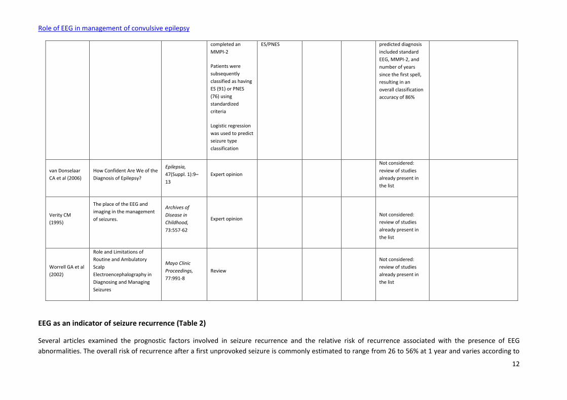
Role of EEG in management of convulsive epilepsy
12
completed an
MMPI-2
Patients were
subsequently
classified as having
ES (91) or PNES
(76) using
standardized
criteria
Logistic regression
was used to predict
seizure type
classification
ES/PNES predicted diagnosis
included standard
EEG, MMPI-2, and
number of years
since the first spell,
resulting in an
overall classification
accuracy of 86%
van Donselaar
CA et al (2006)
How Confident Are We of the
Diagnosis of Epilepsy?
Epilepsia,
47(Suppl. 1):9–
13
Expert opinion
Not considered:
review of studies
already present in
the list
Verity CM
(1995)
The place of the EEG and
imaging in the management
of seizures.
Archives of
Disease in
Childhood,
73:557-62
Expert opinion
Not considered:
review of studies
already present in
the list
Worrell GA et al
(2002)
Role and Limitations of
Routine and Ambulatory
Scalp
Electroencephalography in
Diagnosing and Managing
Seizures
Mayo Clinic
Proceedings,
77:991-8
Review
Not considered:
review of studies
already present in
the list
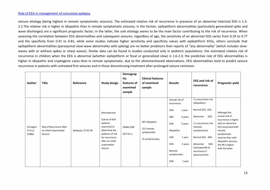
EEG as an indicator of seizure recurrence (Table 2)
Several articles examined the prognostic factors involved in seizure recurrence and the relative risk of recurrence associated with the presence of EEG
abnormalities. The overall risk of recurrence after a first unprovoked seizure is commonly estimated to range from 26 to 56% at 1 year and varies according to
Role of EEG in management of convulsive epilepsy
13
seizure etiology (being highest in remote symptomatic seizures). The estimated relative risk of recurrence in presence of an abnormal interictal EEG is 1.3-
2.2.The relative risk is higher in idiopathic than in remote symptomatic seizures; in the former, epileptiform abnormalities (particularly generalized spike and
wave discharges) are a significant prognostic factor; in the latter, the sole etiology seems to be the main factor contributing to the risk of recurrence. When
assessing the correlation between EEG abnormalities and subsequent seizures, regardless of age, the sensitivity of an abnormal EEG varies from 0.29 to 0.77
and the specificity from 0.41 to 0.81; while some studies indicate higher sensitivity and specificity values with epileptiform EEGs, others conclude that
epileptiform abnormalities (paroxysmal slow-wave abnormality with spiking) are no better predictors than reports of “any abnormality” (which includes slow-
waves with or without spikes or sharp waves). Similar data can be found in studies conducted only in pediatric populations: the estimated relative risk of
recurrence in children when the EEG is abnormal (whether epileptiform or focal or generalized slow) is 1.6-2.3; the predictive role of EEG abnormalities is
higher in idiopathic and cryptogenic cases than in remote symptomatic, due to the aforementioned observations. EEG abnormalities tend to predict seizure
recurrence in patients with untreated first seizures and in those discontinuing treatment after prolonged seizure remission.
Author Title Reference Study design
Demograp
hic
features of
examined
sample
Clinical features
of examined
sample
Results EEG and risk of
recurrence Prognostic yield
Annegers
JF et al
(1986)
Risk of Recurrence After
an Initial Unprovoked
Seizure
Epilepsia, 27:43-50
Retrospective
Cohort of 424
patients
examined to
determine the
patterns of risk
for recurrence
after an initial
unprovoked
seizure
204M,220F
247>55 y
287 idiopathic
122 remote
symptomatic
15 cerebral palsy
Overall risk of
recurrence:
36% 1 year
48% 3 years
56% 5 years
Idiopathic:
26% 1 year
45% 5 years
Remote
symptomatic:
56% 1 year
1-y recurrence risk
(idiopathic):
Normal EEG 16%
Abnormal 26%
1-y recurrence risk
(remote
symptomatic):
Normal EEG 40%
Abnormal 56%
Estimated RR of
recurrence if
abnormal EEG:
Although the
overall risk of
recurrence is higher
with an abnormal
EEG associated with
remote
symptomatic
seizures than with
idiopathic seizures,
the RR is higher
with the latter

Role of EEG in management of convulsive epilepsy
14
77% 5 years
Cerebral palsy:
92% 1 year
Idiopathic:
2.2 (1.1-4.3)
Remote
symptomatic: 1.3
(0.6-3.0)
Arts WFM
et al
(1999)
The Early Prognosis of
Epilepsy in Childhood: The
Prediction of a Poor
Outcome. The Dutch
Study of Epilepsy in
Childhood
Epilepsia, 40:726-34
Prospective
Hospital-based
cohort of 466
children with
newly diagnosed
epilepsy
followed to
determine
good/poor
outcome and to
develop models
to predict it
225M,241F
Median age
5.5 y
Etiology:
Idiopathic 235
Remote symptomatic
136
Cryptogenic 95
OR (CI 95%) for
poor outcome
(remission<6 mo):
EEG at intake: EA vs
normal
1.33 (0.71- 2.48)
EEG at 6 mo
EA vs normal
2.21 (1.12-4.36)
EA at six months are
associated with a
higher risk of
seizure recurrence
in children with
newly diagnosed
epilepsy
Beghi E
(2008)
Management of a first
seizure
General conclusions and
recommendations
Epilepsia, 49(Suppl.
1):58–61 Review
Not considered:
review of studies
already present in
the list
Berg AT,
Shinnar S
(1991)
The risk of seizure
recurrence following a
first unprovoked seizure:
A quantitative review.
Neurology, 41:965-72 Meta-analysis of
16 reports
The average
recurrence risk
across the 16
studies was 51%
(40% and 52% in
prospective and
retrospective
studies)
At 2 years following
the first seizure,
the recurrence risk
2-y seizure
recurrence was
24% with normal
EEG and unknown
etiology, 48% with
abnormal EEG and
unknown etiology,
and 65% with
abnormal EEG and
documented
etiology
Seizure etiology and
the EEG were the
strongest predictors
of recurrence of a
first unprovoked
seizure

Role of EEG in management of convulsive epilepsy
15
was 36% and 47%
in prospective and
retrospective
studies
Berg AT et
al (2001)
Two-Year Remission and
Subsequent Relapse in
Children with Newly
Diagnosed Epilepsy
Epilepsia, 42:1553-62
Cohort of 613
children with
newly diagnosed
epilepsy
Minimum
follow-up (2
years) in 594
(96.9%).
Median age at
onset 5.3
Partial epilepsies 345
Idiopathic 57
Symptomatic 189
Cryptogenic 99
Generalized epilepsies
176
Idiopathic 124
Symptomatic 9
Cryptogenic 43
442 (74%) children
achieved a 2-year
remission in a
median time of 2.3
years (2-6)
After achieving a 2-
year remission, 107
(24.2%) children
experienced a
relapse
Predictors
associated with a
decreased chance
of attaining a 2-
year remission:
Any slowing on
initial EEG: rate
ratio 0.71 (0.54–
0.93)
Predictors
associated with an
increased risk of
relapse after a 2-
year remission:
Focal slowing on
initial EEG: rate
ratio 2.13 (1.19–
3.87)
Slowing of the EEG
tracing is a marker
for poor outcome in
children with newly
diagnosed epilepsy
Berg AT
(2008)
Risk of recurrence after a
first unprovoked seizure
Epilepsia, 49(Suppl.
1):13–8 Review
Not considered:
review of studies
already present in
the list
Camfield
P,
Camfield C
(2008).
Special considerations for
a first seizure in childhood
and adolescence
Epilepsia, 49(Suppl.
1):40–4 Review
Not considered:
review of studies
already present in
the list
Camfield
PR et al
(1985)
Epilepsy after a first
unprovoked seizure in
childhood
Neurology, 35:1657–60
Overall, 51.8%
recurred, and of
those with a
recurrence, 79%
had additional
Children with
generalized
seizures, normal
EEG and no
neurological
The recurrence risk
is increased by focal
versus generalized
seizures, spike
discharge on EEG,

Role of EEG in management of convulsive epilepsy
16
seizures. deficits have
approximately a
20% chance of
recurrence;
children with focal
seizures, abnormal
EEGs, and
concomitant
neurological
deficits have about
an 80% risk of
recurrence
and presence of
concomitant
neurological
deficits.
Das CP et
al (2000)
Risk of recurrence of
seizures following Single
Unprovoked Idiopathic
seizure
Neurology India,
48:357-360
Prospective
76 pt with a first
seizure
excluding
symptomatic
and those with
an abnormal CT
scan were
randomized to
AED/not AED
56 M-20 F
22 (M=16, F=6) of
the 76 patients
(M=56, F=20) had a
recurrence of
seizure.
Recurrence (22/76)
EEG normal
12
EEG abnormal
10
No recurrence
(54/76)
EEG normal
50
EEG abnormal
4
P<0.001
Abnormal EEG
predicts risk of
recurrence of a first
unprovoked seizure
Dooley J
et al
(1996)
Discontinuation of
anticonvulsant therapy in
children free of seizures for 1
year: A prospective study
Neurology, 46:969-74
Prospective
97 children with
2 or more
afebrile seizures,
on AED
monotherapy,
seizure free for 1
year (excluded
50 M-47 F;
mean seizure
onset 65.9
±45.89
months; mean
age at
attaining
seizure control
86.95±49.2
The overall
probability of
remaining seizure
free was 78% at 3
months (95% CI,
70, 87), 71% at 6
months (95% CI,
61, 81), 66% at 12
months (95% CI,
Pt without
recurrence:
EEG normal
45
EEG abnormal
5
The only EEG factor
slightly predictive of
recurrence is
abnormal
background activity

Role of EEG in management of convulsive epilepsy
17
those with
juvenile
myoclonic
epilepsy) were
followed for
32.4±13.1
months or until
seizure
recurrence
months.
57, 75), and 61% at
24 months (95% CI,
51, 71)
Pt with recurrence:
EEG normal
27
EEG abnormal
8
p 0.074
Fisher LS,
Leppik I
(2008)
Debate: When does a
seizure imply epilepsy?
Epilepsia, 49(Suppl.
9):7–12 Review
Not considered:
review of studies
already present in
the list
Gilbert DL,
Buncher
CR (2000)
An EEG should not be
obtained routinely after
first unprovoked seizure
in childhood
Neurology, 54:635-41 Systematic
review
Not considered:
review of studies
already present in
the list
Gilbert DL
et al
(2003)
Meta-analysis of EEG test
performance shows wide
variation among studies
Neurology, 60:564–70
Meta-analysis of
variation of EEG
sensitivity and
specificity in
predicting future
seizures
4,288 patients
and EEGs (19
publications)
relating
epileptiform
EEG and
subsequent
seizures.
1,856 patients
and EEGs (12
publications)
relating
abnormal EEG
and
subsequent
seizures
Risk of seizure
recurrence:
Epileptiform EEG
Sensitivity
0.20–0.81
Specificity
0.41–0.99
Abnormal EEG
Sensitivity
0.29–0.77
Specificity
0.42–0.81
The prognostic yield
of EEG in predicting
seizure recurrence
is fairly low and
varies across
studies
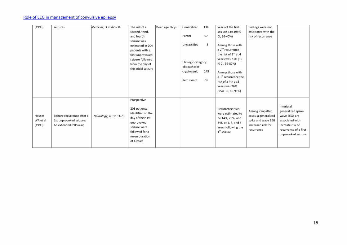
Hauser
WA et al
Risk of recurrent seizures
after two unprovoked New England Journal of
Prospective 142 M-62 F Type of seizure:
Risk of 2nd
recurrence within 5
In the univariate
analysis EEG
Role of EEG in management of convulsive epilepsy
18
(1998) seizures Medicine, 338:429-34 The risk of a
second, third,
and fourth
seizure was
estimated in 204
patients with a
first unprovoked
seizure followed
from the day of
the initial seizure
Mean age 36 ys Generalized 134
Partial 67
Unclassified 3
Etiologic category:
Idiopathic or
cryptogenic 145
Rem sympt 59
years of the first
seizure 33% (95%
CI, 26-40%)
Among those with
a 2nd recurrence
the risk of 3rd at 4
years was 73% (95
% CI, 59-87%)
Among those with
a 3rd recurrence the
risk of a 4th at 3
years was 76%
(95% CI, 60-91%)
findings were not
associated with the
risk of recurrence
Hauser
WA et al
(1990)
Seizure recurrence after a
1st unprovoked seizure:
An extended follow-up
Neurology, 40:1163-70
Prospective
208 patients
identified on the
day of their 1st
unprovoked
seizure were
followed for a
mean duration
of 4 years
Recurrence risks
were estimated to
be 14%, 29%, and
34% at 1, 3, and 5
years following the
1st seizure
Among idiopathic
cases, a generalized
spike and wave EEG
increased risk for
recurrence
Interictal
generalized spike-
wave EEGs are
associated with
increate risk of
recurrence of a first
unprovoked seizure
Role of EEG in management of convulsive epilepsy
19
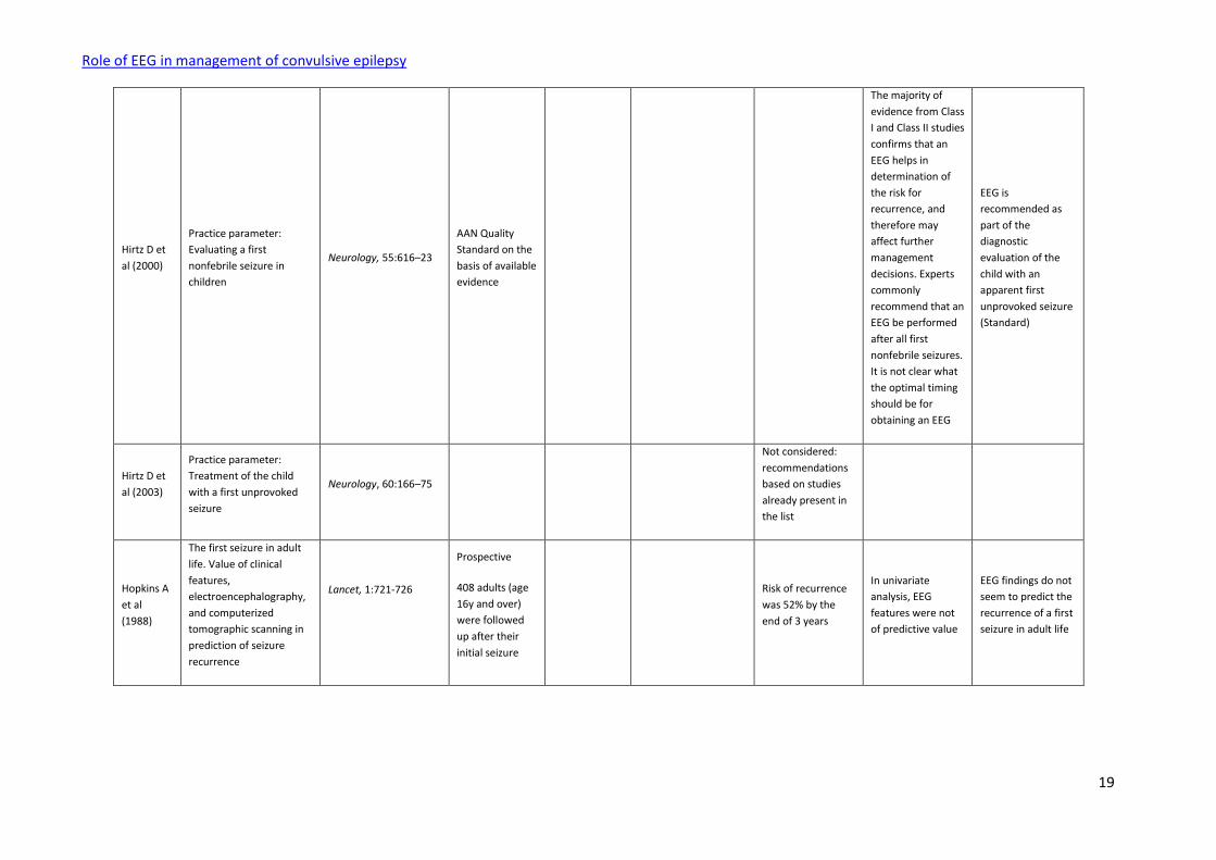
Hirtz D et
al (2000)
Practice parameter:
Evaluating a first
nonfebrile seizure in
children
Neurology, 55:616–23
AAN Quality
Standard on the
basis of available
evidence
The majority of
evidence from Class
I and Class II studies
confirms that an
EEG helps in
determination of
the risk for
recurrence, and
therefore may
affect further
management
decisions. Experts
commonly
recommend that an
EEG be performed
after all first
nonfebrile seizures.
It is not clear what
the optimal timing
should be for
obtaining an EEG
EEG is
recommended as
part of the
diagnostic
evaluation of the
child with an
apparent first
unprovoked seizure
(Standard)
Hirtz D et
al (2003)
Practice parameter:
Treatment of the child
with a first unprovoked
seizure
Neurology, 60:166–75
Not considered:
recommendations
based on studies
already present in
the list
Hopkins A
et al
(1988)
The first seizure in adult
life. Value of clinical
features,
electroencephalography,
and computerized
tomographic scanning in
prediction of seizure
recurrence
Lancet, 1:721-726
Prospective
408 adults (age
16y and over)
were followed
up after their
initial seizure
Risk of recurrence
was 52% by the
end of 3 years
In univariate
analysis, EEG
features were not
of predictive value
EEG findings do not
seem to predict the
recurrence of a first
seizure in adult life

Role of EEG in management of convulsive epilepsy
20
Kho LK et
al (2006)
First seizure presentation:
Do multiple seizures
within 24 hours predict
recurrence?
Neurology, 67:1047–49
Prospective
prognosis of 72
adults with first-
ever multiple
discrete seizures
within 24 hours
compared to
425 patients
presenting with
a single seizure
The overall seizure
recurrence rate at
1 year was 38%
The only variable
independently
predictive of
seizure recurrence
at 1 year on
stepwise logistic
regression was
remote
symptomatic
etiology (OR 2.2, CI
1.4- 3.4; p= 0.0006)
EEG findings do not
seem to predict the
recurrence of a first
seizure in adult life
Kim LG et
al (2006)
Prediction of risk of
seizure recurrence after a
single seizure and early
epilepsy: further results
from the MESS trial
Lancet Neurology, 5:
317–22
Prospective
to assess the
role of patients’
characteristics
and treatment in
the prediction of
seizure
recurrence
1420 patients
were randomly
assigned either
immediate
treatment with
an AED or
delayed
treatment
Median follow-
up 4·4 years
815 M - 605 F
Mean age at
randomisation
31.2y (SD
19.1)
Neurological disorder
254 (18%)
Abnormal EEG 618
(44%)
Epilepsy syndrome 63
(4%)
Individuals with 2
or 3 seizures, a
neurological
disorder, or an
abnormal EEG were
identified as the
medium-risk group,
those with 2 of
these features or
more than 3
seizures as the
high-risk group,
and those with a
single seizure only
as the low-risk
group
No significant
difference was
observed between
treatments for low-
risk individuals
(Log-rank test
χ²=1·7, p=0·2), but
there was an
indication of
Hazard ratio for
recurrence if
abnormal EEG
(defined as specific
focal or generalised
epileptiform or
slow wave
abnormality) 1·54
(1·27–1·86)
p<0·0001
EA on the EEG
(paroxysmal slow-
wave abnormality
with spiking) added
no greater
specificity than the
variable “any
abnormality”,
which included also
slow-wave
disturbance
An abnormal EEG
(regardless of the
presence of EA)
indicates an
increased risk of
recurrence in
patients with a first
unprovoked seizure
and call for early
treatment

Role of EEG in management of convulsive epilepsy
21
(3·0–6·3) improvement with
immediate AED
treatment for
medium-risk and
high-risk
individuals (7·0,
p=0·008; 21·9,
p<0·0005,
respectively)
without spikes or
sharp waves
Lindsten H
et al
(2001)
Remission of Seizures in a
Population-Based Adult
Cohort with a Newly
Diagnosed Unprovoked
Epileptic Seizure
Epilepsia, 42:1025-30.
Prospective
107 pts with a
newly diagnosed
unprovoked
epileptic seizure
in 1985 through
1987 were
followed up until
the date of
death or to the
end of 1996. The
proportion of
cases that
attained a 1-y, 3-
y, 5-y remission
was calculated
by actuarial
analyses
Median age at
diagnosis
52 ys (17-83)
Etiologic groups:
Cryptogenic
42 (39%)
Remote symptomatic
65 (61%)
Cumulative 1-yr
remission
calculated from
epilepsy diagnosis
68%
Cumulative 5-yr
remission
calculated from
epilepsy diagnosis
58%
Focal spikes (25
patients) or
generalized spike–
wave activity (9
patients) on the
EEG at inclusion
had a tendency to
be a good predictor
of not achieving a
5-year remission (p
= 0.06)
Focal spikes or
generalized spike-
wake activity are
negative prognostic
predictors for 5-y
remission in newly
diagnosed epileptic
seizures in adults
Marson A
et al
Immediate versus
deferred antiepileptic
drug treatment for early
Lancet, 365: 2007–13 Prospective:
722 pt were
Immediate AED
treatment reduces
the occurrence of
No information
about EEG data and

Role of EEG in management of convulsive epilepsy
22
(2005) epilepsy and single
seizures: a randomised
controlled trial
assigned
immediate
treatment with
AEDs and 721
were assigned
deferred
treatment
seizures in the next
1–2 years, but does
not affect long-
term remission in
individuals with
single or infrequent
seizures
risk of recurrence
Martinovic
Z, Jovic N
(1997)
Seizure recurrence after a
first generalized tonic-
clonic seizure in children,
adolescents and young
adults
Seizure, 6:461-465
Prospective
78 patients who
had a first
unprovoked
generalized
tonic-clonic
seizure between
the age of 3 and
21y
Sex (M/F) 46 /
32
Age at first
GTCS:
Mean 9.3 ys
(3-21)
Follow-up
period:
Mean 4.1 ys
(2-10)
Seizure aetiology:
Idiopathic 50 (64. 1 %)
Cryptogenic 16
(20.5%)
Remote symptomatic
12 (15.4%)
Number of patients
with recurrent
seizures 54 (68.3%)
Seizure recurrence:
Idiopathic 32
41.0%
Cryptogenic 12
15.4%
Symptomatic 10
12.8%
Seizure recurrence
(54)
Epileptiform EEG
51
Normal EEG
3
No recurrence (24)
Epileptiform EEG
14
Normal EEG
10
p<0.001
The presence of
epileptiform EEG
patterns in the first
two EEGs (including
prolonged
monitoring and/or
sleep after sleep
deprivation in 22
patients) is
predictive for
seizure recurrence
Musicco
M et al
(1997)
Treatment of first tonic-
clonic seizure does not
improve the prognosis of
epilepsy. First Seizure
Trial Group (FIRST Group)
Neurology, 49:991-8
Prospective
419 randomized
to evaluate the
probability of
achieving 1 or 2
years of
complete seizure
control in pts
treated at first
seizure (215) vs
those (204)
treated only in
the event of
236 M, 183 F
In treated pts the
cumulative
probability of
recurrence was
17% after 1 year
and 26% after 2
years; the
corresponding
probabilities in
initially untreated
patients
were 37% and 45%.
An epileptiform
EEG was one of the
significant
predictors of the
risk of recurrence.
However it did not
influence the
probability of
remaining seizure
free for 1 or 2 years
Epileptiform
interictal EEG
findings predict
early seizure
recurrence but do
not affect long-term
prognosis of a first
unprovoked seizure

Role of EEG in management of convulsive epilepsy
23
seizure
recurrence
Pohlmann-
Eden B et
al (2006)
The first seizure and its
management in adults
and children
British Medical Journal,
332:339–42 Review
Not considered:
review of studies
already present in
the list
Ramos
Lizana J et
al (2000)
Seizure Recurrence After
a First Unprovoked
Seizure in Childhood: A
Prospective Study
Epilepsia, 41:1005-13
Prospective
Consecutive
patients aged
less than 14
years with one
or more
unprovoked
seizures
followed to
study the risk of
recurrence
217 children
133 M- 84 F
Mean age 7.4
y
Age:
0-3y 53
3-10y 107
10-14y 57
Etiology:
Symptomatic 47
Idiopathic 48
Cryptogenic 122
Recurrence risk
was 58% (±3.4),
65% (±3.3), 74%
(±3.1), 74% (±3.1),
76% (±3.1), and
79% (±3.2) at 6
months, 1 year, 2
years, 3 years, 4
years, and 5 years,
respectively
RR of recurrence in
different etiologic
groups if EEG
abnormal
Overall cohort
1.6 (1-2.7)
p=0.057
Idiopathic/cryptoge
nic
2 1.(1-3.5)
p=0.0262
Symptomatic
0.7 (0.3-1.9)
p=0.5186
An abnormal EEG is
an important
predictor of
recurrence risk, but
only in patients
with
idiopathic/cryptoge
nic seizures
Schreiner
A,
Pohlmann-
Eden B
(2003)
Value of the early
electroencephalogram
after a first unprovoked
seizure
Clinical
Electroencephalograph
y, 34:140-4
Prospective
The predictive
value of the
standard EEG
and EEG with
sleep
deprivation for
seizure relapse
was studied in
157 adult
patients
The standard EEG
was abnormal in
70.7% and
significantly
associated with an
increased risk of
seizure recurrence
(risk ratio 4.5, 95%
CI 1.8-11.3,
p=0.001).
Subgroup analysis
The abnormal EEG
is a highly
significant predictor
for seizure
recurrence. An
additional EEG with
sleep deprivation is
helpful in cases
when standard EEG
does not reveal
epileptiform

Role of EEG in management of convulsive epilepsy
24
presenting with
a first
unprovoked
seizure. EEGs
were performed
within the first
48 hours of the
first seizure
revealed the
highest recurrence
rates for patients
with focal EA (risk
ratio 2.2, CI 95%
1.2-4.2, p=0.01).
discharges.
Shafer SQ
et al
(1988)
EEG and Other Early
Predictors of Epilepsy
Remission: A Community
Study
Epilepsia, 29:590-600
Retrospective
From 1935
through 1982,
731 new cases of
epilepsy among
residents of
Rochester were
followed, with
432 with at-least
5 years of
follow-up
306/432 had an
EEG after the
first seizure and
before
remission.
Outcome:
remission (5
years seizure-
free with or
without
prescription of
AED)
Of the 432 new
cases, 283 (66%)
achieved 5-y
remission
Hazard ratio for 5-y
remission if no
generalized spike-
wave: 1.58, p<0.01
Absence of
generalized spike-
wave activity in the
interictal EEG
predicts 5-y
remission in
patients with newly
diagnosed epilepsy

Role of EEG in management of convulsive epilepsy
25
Shinnar S
et al
(1994b)
Discontinuing
antiepileptic drugs in
children with epilepsy: a
prospective study
Annals of Neurology,
35:534-45
Prospective
AED were
discontinued in
264 children
with epilepsy
after a mean
seizure-free
interval of 2.9
years and then
followed for a
mean of 58
months to
ascertain
recurrence
Seizures recurred
in 95 (36%) of the
children
Slowing on the EEG
prior to medication
withdrawal was a
significant
predictor of
recurrence in the
idiopathic group
(RR = 2.4)
Slowing of interictal
EEG predicts
recurrence after
treatment
discontinuation in
children in long-
term remission with
AED
Shinnar S
et al
(1994a)
EEG Abnormalities in
Children with a First
Unprovoked Seizure
Epilepsia, 35(3):471-6
Prospective
347 children
with a first
unprovoked
afebrile seizure
347 children,
198 M - 149 F
Mean age 6.8y
at the time of
first seizure
Mean follow-
up 47 month
291 (84%) with an
idiopathic first seizure
56 (16%) with a
remote symptomatic
first seizure
EEGs were
available in
321/347 children
(93%)
Risk of recurrence:
Normal EEGs
42/165 (25%)
Abnormal EEG
56/103(54%)
p<0.001
EEG abnormalities
were more
common in children
with partial
seizures and those
with remote
symptomatic
seizures.
Abnormal EEGs
occur at a higher
rate after age 3
years than before
Abnormal EEG
predicts recurrence
of a first
unprovoked seizure
in children
Shinnar S
et al
(2000)
Predictors of Multiple
Seizures in a Cohort of
Children Prospectively
Followed from the Time
of Their First Unprovoked
Seizure
Annals of Neurology,
48:140–7
Prospective
407 children
followed from
the time of their
first unprovoked
seizure to assess
the risk of
407 children
234 M - 173 F
Mean age at
first seizure
6.8y
Etiology:
Cryptogenic/Idiopathi
c 342
Remote symptomatic
182/407 (45%)
experienced a
recurrence
The overall risk of
recurrence was
29% at 1 year (25-
33%), 37% at 2
An abnormal EEG
was associated
with an increased
recurrence risk
after a first and a
second seizure, but
not with an
increased risk of
Abnormal interictal
EEG predicts
recurrence after a
first and a second
unprovoked seizure
but not multiple
seizures

Role of EEG in management of convulsive epilepsy
26
multiple
recurrences
after an initial
seizure
Mean follow-up
after first seizure
9.6 (yr)
65 years (33-42%),
43% at 5 years (38-
48%), and 46% at
10 years (41-51%)
The risk of a third
seizure was 57%,
63%, and 72% at 1,
2, and 5 years,
respectively, after
the second seizure
After a third
seizure, the
cumulative risk of
another seizure
was 66%, 70%, and
81% at 1, 2, and 5
years, respectively.
multiple seizures
after a second
seizure (RR = 1.08;
95% CI: 0.81,1.67; p
= 0.41).
Risk of 2nd seizure
with abnormal EEG
was 2.16 (1.60-
2.91, p<0.0001),
and risk of 3rd
seizure was 2.15
(1.51-3.05,
p<0.0001)
Shinnar S
et al
(1996)
The Risk of Seizure
Recurrence After a First
Unprovoked Afebrile
Seizure in Childhood: An
Extended Follow-up
Paediatrics, 98;216-25
Prospective
407 children
who presented
with a first
unprovoked
seizure were
then followed to
assess the long-
term recurrence
risks after a first
unprovoked
seizure
Mean follow-up
period was 6.3
years
407 children
234 M - 173 F
Mean age at
first seizure
6.8y
Etiology:
Cryptogenic/Idiopathi
c 342
Remote symptomatic
65
171/407 (42%) had
recurrences
The overall
estimate of
recurrence was
22% at 6 months
(18-26%), 29% at 1
year (25-33%), 37%
at 2 years (32-
42%), 42% at 5
years (37-47%),
and 44% at 8 years
(39-49%)
Abnormal EEG and
risk of recurrence:
Overall RR 2.3 (1.7-
3.2, p <.0001)
Cryptogenic RR 2.6
(1.8-3.7, p <.0001)
Remote
Symptomatic RR
1.1 (0.6-2.0, p=0
.78
Abnormal EEG
predicts long-term
recurrence of a first
unprovoked afebrile
seizure but only in
children with
cryptogenic seizures

Role of EEG in management of convulsive epilepsy
27
Stroink H
et al
(1998)
The first unprovoked,
untreated seizure in
childhood: a hospital
based study of the
accuracy of the diagnosis,
rate of recurrence, and
long term outcome after
recurrence. Dutch study
of epilepsy in childhood
Journal of Neurology,
Neurosurgery &
Psychiatry, 64:595–600
Prospective
156 children
aged 1 month to
16 years after a
first seizure
followed up to
assess the
accuracy of the
diagnosis, the
recurrence rate
within two
years, the risk
factors for
recurrence, and
the long term
outcome two
years after
recurrence
156 children
70 M – 86 F
median age at
intake 6.9y;
range 0.2–
15.6y
Etiology:
Idiopathic/cryptogenic
129
Remote symptomatic
27
The overall
recurrence rate
was 40% (33–48%)
at six months; 46%
(38–53%) at one
year; and 54% ( 46–
62%) at two years
Children with
epileptic discharges
in their first EEG
(n=68; 44%) had a
recurrence rate of
71% at two years
(60–81%); in
children with a
normal first EEG
(57) the rate was
40% (28–53%)
An epileptiform EEG
is the most
important
predictive factor for
seizure recurrence
in children with a
first unprovoked
seizure in a full
model multivariate
analysis
van
Donselaar
CA et al
(1992)
Value of the
Electroencephalogram in
Adult Patients With
Untreated Idiopathic First
Seizures
Archives of Neurology,
49:231-7
Prospective
To assess the
reliability and
accuracy of the
EEG as a
predictor of the
risk of
recurrence
within 2 years in
157 patients
with untreated
idiopathic first
seizures
The finding of
epileptic discharges
was associated
with a risk of
recurrence of 83%
(CI 95% , 69% to
97%) vs 41% (CI
95%, 29% to 53%)
in patients with
nonepileptic
abnormalities
EA predict
recurrence in adults
with a first
untreated
idiopathic seizure

Role of EEG in management of convulsive epilepsy
28
Wiebe S et
al (2008)
Management of a first
seizure: An evidence-
based approach to the
first seizure
Epilepsia, 49(Suppl.
1):50–7 Review
Not considered:
review of studies
already present in
the list
EEG as a predictor of mortality (Table 3)
Another important issue is the overall risk of mortality in epilepsy and the incidence of sudden unexplained death in epilepsy (SUDEP). Some clinical factors are
commonly identified as potentially increasing the risk of death: symptomatic etiology (both remote and acute), comorbid neurologic disease and, particularly
for SUDEP, seizure severity and high frequency. On the contrary, no detailed data are available for a possible contribution of EEG to the definition of mortality
risk, with a notable exception: few articles deal with the possible role of cardiac asystolia provoked by epileptic seizures and ictal hypoxemia and hypercapnia
(possibly implied in SUDEP).
Author Title Reference Study design
Demographic
features of
examined
sample
Clinical
features of
examined
sample
Results EEG and risk of
SUDEP/mortality
Prognostic
yield
Bateman
LM et al
(2008)
Ictal hypoxemia in
localization-related
epilepsy: analysis of
incidence, severity
and risk factors
Brain, 131(Pt
12):3239-45
Prospective:
to determine the
incidence and severity of
ictal hypoxemia in patients
with localization-related
epilepsy undergoing
video-EEG
304 seizures with
accompanying
oxygen saturation
data were
recorded in 56
consecutive
patients with
intractable
localization-related
epilepsy
Desaturations below 90%
were significantly correlated
with seizure localization (p =
0.005; OR of temporal versus
extratemporal = 5.202;
[1.665, 16.257]), seizure
lateralization (p = 0.001; OR
of right versus left = 2.098;
[1.078, 4.085]), contralateral
spread of seizures (p =
0.028; OR of contralateral
spread versus no spread =
2.591; [1.112, 6.039])
Ictal hypoxemia
occurs often in
patients with
localization-
related epilepsy
and may be
pronounced and
prolonged.
Ictal hypoxemia and hypercapnia may contribute to SUDEP

Role of EEG in management of convulsive epilepsy
29
Berg AT et
al (2004)
Mortality in
Childhood-Onset
Epilepsy
Archives of
Pediatric
&Adolescent
Medicine,
158:1147-52
Prospective:
to evaluate mortality in
children with newly
diagnosed epilepsy, to
determine the risk of
death, and to identify
predictors of death from
the point of diagnosis.
Median follow-up 7.9
years
613 children were
recruited 307 M –
306 F The median
age at study entry
was 6.1 years (age
range, 1 month to
16 years).
13 had died 7
months to 7 years
(median, 4.2 y)
after diagnosis
In a multivariable Cox
proportional hazards model,
remote symptomatic
etiology (RR, 10.2;2.1-49.6;
P=.004) and epileptic
encephalopathy (RR, 13.3;
3.4-51.7; P<.001) were
independently associated
with mortality
No data on possible
contribution of EEG to
the definition of
mortality risk
Brodie MJ,
Gregory LH
(2008)
Should all patients be
told about sudden
unexpected death in
epilepsy (SUDEP)?
Pros and Cons
Epilepsia, 49(Suppl.
9):99–101 Review
Factors associated with the
greatest risk of SUDEP are
generalized tonic–clonic
seizures, high seizure
frequency, concomitant
learning disabilities, AED
polypharmacy, and frequent
changes in dosing
No data on possible
contribution of EEG to
the definition of
mortality risk
Callenbach
PMC et al
(2001)
Mortality Risk in
Children With
Epilepsy: The Dutch
Study of Epilepsy in
Childhood
Pediatrics,107:
1259 –63
Prospective:
472 children, aged 1
month to 16 years, with 2
or more newly diagnosed
unprovoked seizures were
enrolled. All children were
followed for 5 years or
9 children died during follow-
up, amounting to a mortality
rate of 3.8/1000 person-
years, sevenfold higher than
expected (95% CI 2.4 –11.5).
All deceased children had
epilepsy that was caused by a
static or progressive
neurologic disorder
No data on possible
contribution of EEG to
the definition of
mortality risk

Role of EEG in management of convulsive epilepsy
30
until death (mortality risk = 22.9; CI 95%
7.9 –37.9). None of them
died from SUDEP
Camfield P,
Camfield C
(2008)
Special
considerations for a
first seizure in
childhood and
adolescence
Epilepsia, 49(Suppl.
1):40–4 Review
Not considered: review
of studies already
present in the list
Camfield
CS et al
(2002)
Death in children
with epilepsy: a
population-based
study
Lancet, 359:1891–5
Prospective:
population-based cohort
study including all children
who developed epilepsy
during 1977–85 to assess
the frequency and causes
of death of children with
epilepsy
Children with epilepsy have
more than five times the
risk of dying than the
general population in the
first 15–20 years after
diagnosis. Most deaths are
related to comorbid
neurological disorders
sufficient to cause
functional neurological
deficit and not to the
epilepsy
No data on possible
contribution of EEG to
the definition of
mortality risk
Forsgren L
et al (2005)
Mortality of Epilepsy
in Developed
Countries: A Review
Epilepsia, 46(Suppl.
11):18–27 Review
Not considered: review
of studies already
present in the list
Hauser
WA, Beghi
E (2008)
First seizure
definitions and
worldwide incidence
and mortality
Epilepsia, 49(Suppl.
1):8–12 Review
Not considered: review
of studies already
present in the list
Hauser WA
et al (1980)
Mortality in Patients
with Epilepsy
Epilepsia, 21:399-
412
Retrospective:
618 residents with first
diagnosis of epilepsy
between 1935 and 1974
were observed for 8,233
person-years.
During the period of follow-
up, there were 187 deaths.
SMR for the total group was
2.3 (1.9 – 2.6 through 29
years of follow-up)
No data on possible
contribution of EEG to
the definition of
mortality risk

Role of EEG in management of convulsive epilepsy
31
Hitiris N et
al (2007b) Mortality in epilepsy
Epilepsy &
Behavior, 10 : 363–
76
Review
Not considered: review
of studies already
present in the list
Hitiris N et
al (2007a)
Sudden unexpected
death in epilepsy: A
search for risk factors
Epilepsy &
Behavior, 10: 138–
41
Retrospective:
case–control study of
SUDEP within a large
cohort of adult patients
followed up over a 23-year
period to investigate the
association between
clinical characteristics and
SUDEP, focusing on likely
risk factors
No data on possible
contribution of EEG to
the definition of risk of
SUDEP
Jehi L,
Najm IM (
2008)
Sudden unexpected
death in epilepsy:
Impact, mechanisms,
and prevention
Cleveland Clinical
Journal of
Medicine,75 (suppl
2): S66-S70
Review
No data on possible
contribution of EEG to
the definition of risk of
SUDEP
Langan Y et
al (2005)
Case-control study of
SUDEP
154 cases with a
postmortem examination
were investigated. Each
case had 4 controls with
epilepsy from the
community. To examine
the influence of various
factors on the risk of
SUDEP in epilepsy OR for
risk and protection were
determined
No data on possible
contribution of EEG to
the definition of risk of
SUDEP

Role of EEG in management of convulsive epilepsy
32
Langan Y
(2000)
Sudden unexpected
death in epilepsy
(SUDEP): risk factors
and case control
studies
Seizure, 9:179–83 Review
Not considered: review
of studies already
present in the list
Lhatoo SD
et al (2001)
Mortality in Epilepsy
in the First 11 to 14
Years after Diagnosis:
Multivariate Analysis
of a Long-Term,
Prospective,
Population-Based
Cohort
Annals of Neurol
ogy, 49:336–44
Prospective, population-
based study of newly
diagnosed epilepsy. A
cohort of 792 patients was
followed for up to 14 years
(median follow-up 11.8
years, range 10.6 –11.7
years), a total of 11,400
person-years
214 deaths occurred in the
entire cohort (SMR 1.9; CI
95% CI = 1.6, 2.2; p< 0.001)
In the idiopathic group SMR
1.3 (CI 95% = 0.9,1.9)
In the remote symptomatic
SMR 3.7 (CI 95% 2.9, 4.6)
and in the acute
symptomatic group SMR 3.0
(CI 95% 2.0, 4.3).
No data on possible
contribution of EEG to
the definition of
mortality risk
Lindsten H
et al (2000)
Mortality Risk in an
Adult Cohort with a
Newly Diagnosed
Unprovoked Epileptic
Seizure: A
Population-Based
Study
Epilepsia, 41:1469-
73
Prospective:
107 patients >17 yr with
newly diagnosed
unprovoked epileptic
seizures were followed
until the date of death or
the end of 1996. SMR was
analyzed. The influences
on the SMR of time since
diagnosis, sex, age at
diagnosis, seizure cause,
seizure type, and cause of
death were also
investigated
The total cohort mortality
risk was significantly
increased (SMR, 2.5; 1.2-
3.2)
Patients with remote
symptomatic cause had a
significantly elevated risk of
death (SMR, 3.3; 2.4-4.5)
No data on possible
contribution of EEG to
the definition of
mortality risk

Role of EEG in management of convulsive epilepsy
33
Loiseau P
et al (2005)
One-Year Mortality in
Bordeaux Cohort: the
value of syndrome
classification
Epilepsia, 46(Suppl.
11):11–4
Retrospective:
Date and cause of death in
an incidence study of first
afebrile seizures were
classified by epilepsy
syndrome. 804 pts were
included. SMR were
calculated
Cryptogenic SMR = 1.7, 95%
CI 0.1–9.7
Remote symptomatic SMR =
6.4, 95% CI 3.6–10.3
Acute symptomatic
SMR=10.3, 95% CI 8.3–12.7
No specific data on
possible contribution of
EEG to the definition of
mortality risk
Although a
syndromic
diagnosis is
important for
treatment
decisions and
some prognostic
aspects of seizure
disorders, its
value in mortality
studies is limited
Montè CP
et al (2007)
Sudden unexpected
death in epilepsy
patients: Risk factors
A systematic review
Seizure, 16: 1-7 Review
No data on possible
contribution of EEG to
the definition of risk of
SUDEP
Olafsson E
et al (1998)
Long-Term Survival of
People with
Unprovoked Seizures:
A Population-Based
Study
Epilepsia, 39:89-92
Prospective:
224 incidence cases of
unprovoked seizures in
Iceland.
Survivorship status and
date of death was
collected.
SMR was calculated
30 years after diagnosis,
there were 45 deaths
among the index cases
(SMR=1.6,1.2-2.1)
In the remote symptomatic
group SMR was 2.3,1.4-3.5
In the idiopathic group SMR
was 1.3, 0.8-1.9
No data on possible
contribution of EEG to
the definition of
mortality risk
Rocamora
R et al
(2003)
Cardiac Asystole in
Epilepsy: Clinical and
Neurophysiologic
Features
Epilepsia, 44:179–
85
Retrospective analysis of
the clinical records of
hospitalized patients who
underwent long-term
video-EEG monitoring to
determine the frequency
of cardiac asystole
provoked by epileptic
seizures (possibly implied
Seizure-induced asystole is a
rare complication. The
event appeared only in 5
cases of focal epilepsy (3
frontal and 2 left-temporal)
EEG may contribute to
identify ictal patterns at
risk for seizure-induced
asystolia

Role of EEG in management of convulsive epilepsy
34
in SUDEP) and to analyze
the correlation between
EEG, ECG, and clinical
features obtained from
long-term video-EEG
monitoring.
Schuele SU
et al (2007)
Video-electrographic
and clinical features
in patients with ictal
asystole
Neurology,69:434-
41
Retrospective:
Ictal asystole (IA) is a rare
event mostly seen in
patients with temporal
lobe epilepsy (TLE) and a
potential contributor to
SUDEP
A database search was
performed of 6,825
patients undergoing long-
term video-EEG
monitoring for episodes of
IA
IA was recorded in 0.27% of
all patients with epilepsy, 8
with TLE, 2 with
extratemporal (XTLE), and
none with generalized
epilepsy
Clinical predisposing
factors for IA, including
cardiovascular risk
factors or ECG or EEG
abnormalities were not
identified
Tèllez-
Zenteno JF
et al (2005)
Sudden unexpected
death in epilepsy:
Evidence-based
analysis of incidence
and risk factors
Epilepsy Research,
65:101–15 Evidence-based review
No data on possible
contribution of EEG to
the definition of risk of
SUDEP
Tomson T
et al (2008)
Sudden unexpected
death in epilepsy:
current knowledge
and future directions
Lancet Neurology,
7: 1021–31 Review
No data on possible
contribution of EEG to
the definition of risk of
SUDEP
Tomson T
et al (2005)
Sudden Unexpected
Death in Epilepsy: A
Review of Incidence
and Risk Factors
Epilepsia, 46(Suppl.
11):54–61 Review
No data on possible
contribution of EEG to
the definition of risk of
SUDEP
Role of EEG in management of convulsive epilepsy
35
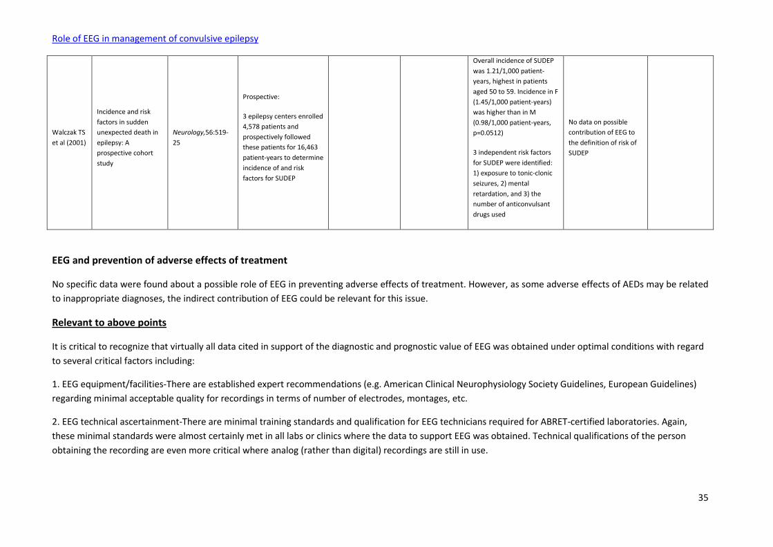
Walczak TS
et al (2001)
Incidence and risk
factors in sudden
unexpected death in
epilepsy: A
prospective cohort
study
Neurology,56:519-
25
Prospective:
3 epilepsy centers enrolled
4,578 patients and
prospectively followed
these patients for 16,463
patient-years to determine
incidence of and risk
factors for SUDEP
Overall incidence of SUDEP
was 1.21/1,000 patient-
years, highest in patients
aged 50 to 59. Incidence in F
(1.45/1,000 patient-years)
was higher than in M
(0.98/1,000 patient-years,
p=0.0512)
3 independent risk factors
for SUDEP were identified:
1) exposure to tonic-clonic
seizures, 2) mental
retardation, and 3) the
number of anticonvulsant
drugs used
No data on possible
contribution of EEG to
the definition of risk of
SUDEP
EEG and prevention of adverse effects of treatment
No specific data were found about a possible role of EEG in preventing adverse effects of treatment. However, as some adverse effects of AEDs may be related
to inappropriate diagnoses, the indirect contribution of EEG could be relevant for this issue.
Relevant to above points
It is critical to recognize that virtually all data cited in support of the diagnostic and prognostic value of EEG was obtained under optimal conditions with regard
to several critical factors including:
1. EEG equipment/facilities-There are established expert recommendations (e.g. American Clinical Neurophysiology Society Guidelines, European Guidelines)
regarding minimal acceptable quality for recordings in terms of number of electrodes, montages, etc.
2. EEG technical ascertainment-There are minimal training standards and qualification for EEG technicians required for ABRET-certified laboratories. Again,
these minimal standards were almost certainly met in all labs or clinics where the data to support EEG was obtained. Technical qualifications of the person
obtaining the recording are even more critical where analog (rather than digital) recordings are still in use.
Role of EEG in management of convulsive epilepsy
36
3. EEG interpretation-The data cited in support of EEGs value was almost certainly obtained in setting where the EEG was interpreted by an epileptologist (i.e. a
neurologist with additional training in epilepsy/clinical neurophysiology).
**It is important to note that there is a reasonable body of data to suggest that in fact when EEG is obtained under suboptimal conditions, the value of EEG for
diagnostic purposes is substantially less. There is some limited data to indicate that EEG can be harmful and lead to overdiagnosis and unnecessary treatment
when used for diagnostic confirmation by less experienced readers. There is a general agreement among epileptologists that over-reading is more harmful than
under-reading and that less experienced readers are more likely to over-interpret benign patterns and variants as epileptic(Engel 1984; Chadwick 1990)**
See below for details of evidence
Williams et al, 1985: This study took a random sample of 100 active electroencephalographers in the US and showed them selected EEGs to determine inter-
rater variability and correct responses. Reader characteristics such as board certification, number of EEGs reviewed annually and percent of time in EEG work
determined reader responses.
Gilbert et al, 2003: A meta-analysis of 25 studies involving 4,912 EEGs revealed a wide range of inter-reader variation in terms of sensitivity and specificity with
regard to whether or not the EEG could predict seizure recurrence. The largest proportion of this variability (37% of variability) was accounted for by the
interpretation threshold of the neurologist reader. Higher specificity readers had greater diagnostic accuracy.
Benbadis & Lin, 2008: This retrospective review from a single epilepsy center looked at EEGs from patients referred for epilepsy management who were
ultimately determined NOT to suffer from epilepsy. The EEGs of interest were those obtained by non-epilepsy experts and interpreted as having epileptiform
discharges by the local EEG reader who was not expert in EEG interpretation. The retrospective review identified 37 patients who met the following criteria—
(1) had been referred for epilepsy management (2) had an outside EEG available for review that had been reported as having epileptiform discharges, and (3)
the patient was ultimately determined NOT to have epilepsy. Expert review of these 37 EEGs determined that none of them actually had epileptiform
discharges (EDCs) but in fact had benign patterns commonly mistaken for EDCs by naive readers.
Benbadis & Tatum, 2003: This is a retrospective review of the EEG tracings (obtained and interpreted by the referring physician) of patients referred to an
epilepsy center with a diagnosis of epilepsy who were ultimately determined to have psychogenetic, non-epileptic seizures (PNES). Subjects identified with
PNES from January 1999-June 2001 were included. EEGs obtained and interpreted by the referring physicians were reviewed by a board-certified
electroencephalographer specializing in epilepsy. The referring physicians were general neurologists. Of 127 PNES patients, 41 had a history of epileptiform
discharges on their referring EEGs—and 15 of these 41 EEGs were available for expert review. On review, 0/15 EEGs showed epileptiform discharges. Normal
variants and benign wave patterns mistaken for EDCs were identified.

Role of EEG in management of convulsive epilepsy
37
Smith et al, 1999: This retrospective analysis of 184 patients referred to a single epilepsy center with a diagnosis of intractable epilepsy identified an overall
misdiagnosis rate (meaning the patient did not have epilepsy) of 26.1% (46/184). Nineteen of the 46 misdiagnoses were based upon EEGs obtained and
interpreted by non-epileptologists that were interpreted as having epileptiform discharges but which on expert review had no EDCs evident but only normal
variants. Essentially, poor EEG interpretation by a general neurologist contributed to almost half of the misdiagnosed cases. Importantly, most of these patients
had been treated unnecessarily with AEDs for long periods of time.
Tuan et al, 2008: This field survey done in northern Vietnam identified 1,338 positive responses to a questionnaire on seizure disorders. On clinical
examination, 190 patients met the criteria for active epilepsy. All considered to fulfil the criteria for active epilepsy, were offered an EEG examination (not
feasible in 10 patients). EEG was without abnormalities in 149 (78%) of 190 cases (artifact in 6 patients), whereas epileptiform activity was recorded in only 21
patients (11%).
Tekle-Haimanot et al, 1997: This community-based study identified 139 incident cases in a door-to-door survey in a rural area of central Ethiopia. This
corresponded to an annual incidence of 64 in 100,000 (95% CI 44-84). EEG was recorded in 53 patients; it was normal in 38%, and demonstrated epileptiform
activity in 34%, non-specific abnormalities in 18%, and paroxysmal rhythms in 11%. Of those with abnormal EEG recordings, 20% had focal unilateral or
nonfocal unilateral abnormalities suggestive of a lateralized lesion (partial epilepsy). In some subjects who clinically had only generalized convulsive seizures,
the EEG showed focal epileptiform activity. These patients were classified as having partial seizures secondarily generalized.
Given the above (i.e. that all data cited in support of EEG’s utility was obtained under ideal circumstances in terms of equipment, technical support and
interpretators’ qualifications) recommendations can only be made in support of EEG under similar circumstances/conditions. Furthermore, there should be
some notes of caution issued about the use of EEG in the absence of well-qualified technicians and interpreters.
Engel 1984: This study looks at the EEG being an aid for the diagnostic process of epilepsy and not a diagnosis itself. When used to help differentiate between
generalized and partial epileptic conditions and to Identify benign epileptic syndromes, the EEG has prognostic and therapeutic implications. Also, the EEG can
be used to determine whether a patient is deteriorating due to increased seizure activity or increased side effects. The study also discussed how healthy young
adults with medically intractable partial complex seizures with unilateral or bilaterally independent interictal anterior temporal EEG spike foci are the best
candidates for resective surgical therapy while those with multifocal or bilaterally synchronous interictal EEG spikes combined with mental retardation are less
likely to benefit from resective surgery.
Benbadis SR, Tatum WO (2003). "Overintepretation of EEGs and misdiagnosis of epilepsy." Journal of Clinical Neurophysiology, 20(1): 42-4. The overinterpretation of EEGs is a known problem that has not been reported specifically. The authors report a series of EEGs on patients who were diagnosed
eventually with psychogenic nonepileptic seizures and who had an EEG read as epileptiform. Of the 15 actual records available for review, the overread
patterns were wicket spikes (n = 1), hypnagogic hypersynchrony (n = 1), and hyperventilation-induced slowing (n = 1). In the other 12 records, the overread

Role of EEG in management of convulsive epilepsy
38
patterns were simple fluctuations of sharply contoured background rhythms or fragmented alpha activity. Rather than well-described normal variants, the
overinterpreted patterns tend to be normal fluctuations of background activity
Benbadis SR and Lin K, 2008: "Errors in EEG interpretation and misdiagnosis of epilepsy. Which EEG patterns are overread?" European Neurology, 59(5): 267-71. The overinterpretation of EEGs is common and is an important contributor to the misdiagnosis of epilepsy. The authors reviewed their experience in order to
clarify which EEG patterns are commonly overread as epileptiform. They identified patients who were seen at our epilepsy clinic and were ultimately diagnosed as having conditions other than epilepsy. They selected those who had previously had an EEG read as showing epileptiform discharges and whose EEG was available for their own re-review. 37 patients met the above criteria. Eventual diagnoses were psychogenic nonepileptic seizures (10), syncope (7), other miscellaneous diagnoses (5) and unexplained nonspecific symptoms (15). None of the EEGs had epileptiform discharges. The descriptions of the abnormalities included 'temporal sharp waves' in 30, 'frontal sharp waves' in 2 and 'generalized spike-wave complexes' in 2. Three had no reports available to identify the alleged abnormality. The benign patterns mistaken for temporal (30) and frontal (2) sharp waves were simple fluctuations of background activity with temporal phase reversals. The authors concluded that by far the most common patterns overread as epileptiform are nonspecific fluctuations of background in the temporal regions, which are misread as temporal sharp waves.
Any statistical summaries
The best review of evidence available about the contribution of EEG to diagnosis is AAN Quality Standard (Hirtz et al, 2000) about evaluation of first nonfebrile
seizures in children, concluding that the majority of evidence from Class I and Class II studies confirms that an EEG helps in determination of seizure type,
epilepsy syndrome and may be helpful in determining the need for imaging studies. A number of meta-analyses deal with EEG and the risk of seizure
recurrence following a first unprovoked seizure: Berg & Shinnar, 1991, (meta-analysis of 16 reports) concludes that seizure etiology and the EEG are the
strongest predictors of recurrence; Gilbert & Buncher, 2000 (meta-analysis of 31 publications relating epileptiform or abnormal EEG and subsequent seizures)
indicates a wide range of values for both sensitivity (0.20-0.81) and specificity (0.41-0.99) in a previous systematic review of 4 studies involving 831 children.
Gilbert & Buncher, 2000 stated that the pretest probability of recurrence was less than the lower limit of the range for rational testing, concluding that EEG
should be ordered selectively, not routinely, after first unprovoked seizure in childhood. On the contrary, the aforementioned AAN Quality Standards on the
basis of available evidence (Hirtz et al, 2000; Krumholz et al, 2007) recommends EEG as part of the diagnostic evaluation of the child with an apparent first
unprovoked seizure.
Methodological limitations
Methodological limitations have to be considered, some of them intrinsic to the EEG recording and interpretation and some depending on the quality of the
study designs. The major intrinsic limitations are the low sensitivity of interictal EEG, which can be moderately increased only by prolonging the duration of the
recording (generally through repeat testing), and the poor interrater reliability. Besides the well known subjectivity of EEG reporting, some publications support

Role of EEG in management of convulsive epilepsy
39
the concept that interictal EEG in childhood epilepsy appears an unstable test per se, so a repeated EEG could yield different and conflicting information; on the
contrary, others (dealing with interrater agreement in classification of childhood epilepsy syndromes) conclude that when an abnormal EEG is clearly
contributory to a specific diagnosis, interrater agreement is extremely good. A possible interpretation is that some EEG patterns are unequivocal and less prone
to differing interpretations and others, perhaps the majority, are ill-defined and subjected to differing interpretations. However, these apparently conflicting
results also reflect the limitations of the EEG technology, which in spite of standard recording and interpretation techniques, is time-dependent and may be
profoundly affected by the variability of the bioelectric patterns in the epileptic brain. For this reason, repeat tracings and use of activation techniques may
explain at least in part the differing results across studies and the increasing diagnostic yield with a more intensive investigation. Another important flaw is that
EEG readers are often unblinded. This is even more important, given the fair and sometimes poor reliability of the EEG readings. Other limitations include the
retrospective design, the small sample size, the differing study populations, and the duration of the follow-up.
Directness (in terms of population, outcome, intervention and comparator)
The largest body of information on the diagnostic and prognostic yield of EEG has been obtained from clinic-based observational studies done in selected
referral populations (mostly children and adolescents and – to a lesser extent – adults) which were investigated using different technologies and modalities for
recording and interpretation. Lesser studies were performed in population-based samples. Most of these studies have been carried out in high income
countries. However, given the purposes of these latter surveys (mostly interested in broad EEG findings), there were few or no attempts in assessing the
diagnostic contribution of the EEG tracings depending on the intensity of the investigation and in classifying patients into precise syndromic categories. As well,
regarding the prognostic yield of interictal EEG, population-based studies only assessed the results of the first tracing disregarding the possible contribution of
repeat EEGs. Although the results of electrophysiological investigations and neuroimaging have been repeatedly compared in surgical candidates to assess the
outcome of epilepsy surgery and the comparative value of EEG and neuroimaging has been assessed in patients with a first seizure, to our knowledge there are
no reports on the evidence-based diagnostic contribution of the EEG tracings compared to other diagnostic techniques. Only indirect comparisons are available
between EEG and other prognostic indicators in studies on the risk of seizure relapse and mortality of epilepsy. There are virtually no reports on the diagnostic
yield of interictal EEG in resource-poor countries. In this light, given the fairly low sensitivity of this diagnostic technique, about 50% of cases with true epileptic
seizures may go undetected unless repeat tracings and/or activation procedures are performed.
Reference List
Ajmone Marsac C, Zivin LS (1970). Factors Related to the Occurrence of Typical Paroxysmal Abnormalities in the EEG Records of Epileptic Patients. Epilepsia, 11:361-381.

Role of EEG in management of convulsive epilepsy
40
Annegers JF et al (1986). Risk of Recurrence After an Initial Unprovoked Seizure. Epilepsia, 27:43-50.
Arts WFM et al (1999). The Early Prognosis of Epilepsy in Childhood: The Prediction of a Poor Outcome. The Dutch Study of Epilepsy in Childhood. Epilepsia,
40:726-34.
Bateman LM, Li CS, Bayer M (2008). Ictal hypoxemia in localization-related epilepsy: analysis of incidence, severity and risk factors. Brain, 131(Pt 12):3239-45.
Beghi E (2008). Management of a first seizure. General conclusions and recommendations. Epilepsia, 49(Suppl. 1):58–61.
Benbadis SR, Lin k (2008). Errors in EEG interpretation and misdiagnosis of epilepsy. Which EEG patterns are overread? European Neurology, 59:267-71. Epub
2008 Feb 8.
Benbadis SR, Tatum WO (2003). Overinterpretation of EEGs and misdiagnosis of epilepsy. Journal of Clinical Neurophysiology, 20:42-4.
Berg AT (2008). Risk of recurrence after a first unprovoked seizure. Epilepsia, 42:1553-62.
Berg AT, Shinnar S (1991). The risk of seizure recurrence following a first unprovoked seizure: A quantitative review. Neurology, 41:965-72.
Berg AT et al (1999). Classification of Childhood Epilepsy Syndromes in Newly Diagnosed Epilepsy: Inter-rater Agreement and Reasons for Disagreement. Epilepsia, 40:439-4. Berg AT et al (2000). How Well Can Epilepsy Syndromes Be Identified at Diagnosis?: A Reassessment 2 Years After Initial Diagnosis. Epilepsia, 41:1269-75. Berg AT et al (2001). Two-Year Remission and Subsequent Relapse in Children with Newly Diagnosed Epilepsy. Epilepsia, 42:1553-62.
Berg AT et al (2004). Mortality in Childhood-Onset Epilepsy. Archives of Pediatric &Adolescent Medicine, 158:1147-52.
Binnie CD, Stefan H (1999). Modern electroencephalography: its role in epilepsy management. Clinical Neurophysiology, 110 :1671-97. Brodie MJ, Gregory LH (2008). Should all patients be told about sudden unexpected death in epilepsy (SUDEP)? Pros and Cons. Epilepsia, 49(Suppl. 9):99–101.
Callenbach PMC et al (2001). Mortality Risk in Children With Epilepsy: The Dutch Study of Epilepsy in Childhood. Pediatrics,107:1259 –63.
Camfield P, Camfield C (2008). Special considerations for a first seizure in childhood and adolescence. Epilepsia, 49(Suppl. 1):40–4.
Camfield CS, Camfield PR, Veugelers PJ (2002). Death in children with epilepsy: a population-based study. Lancet, 359:1891–5.

Role of EEG in management of convulsive epilepsy
41
Camfield PR et al (1985). Epilepsy after a first unprovoked seizure in childhood. Neurology, 35:1657–60.
Camfield P et al (1995). EEG results are rarely the same if repeated within six months in childhood epilepsy. Canadian Journal of Neurological Sciences, 22:297-300. Carpay JA et al (1997). The Diagnostic Yield of a Second EEG After Partial Sleep Deprivation: A Prospective Study in Children with Newly Diagnosed Seizures.
Epilepsia, 38:595-9.
Chadwick D (1990). Diagnosis of Epilepsy. Lancet, 336:291-5.
Das CP et al (2000). Risk of recurrence of seizures following Single Unprovoked Idiopathic seizure. Neurology India, 48:357-60.
Dooley J et al (1996). Discontinuation of anticonvulsant therapy in children free of seizures for 1 year: A prospective study. Neurology, 46:969-74.
Engel J Jr (1984). A practical guide for routine EEG studies in epilepsy. Journal of Clinical Neurophysiology, 1:109-42.
Fisher LS, Leppik I (2008). Debate: When does a seizure imply epilepsy? Epilepsia, 49(Suppl. 9):7–12.
Flink R et al (2002). Guidelines for the use of EEG methodology in the diagnosis of epilepsy. Acta Neurologica Scandinavica, 106:1-7.
Forsgren L et al (2005). Mortality of Epilepsy in Developed Countries: A Review. Epilepsia, 46(Suppl. 11):18–27.
Fountain NB, Freeman JM (2006). EEG Is an Essential Clinical Tool: Pro and Con. Epilepsia, 47(Suppl. 1):23–25.
Gilbert DL, Buncher CR (2000). An EEG should not be obtained routinely after first unprovoked seizure in childhood. Neurology, 54:635-41.
Gilbert DL et al (2003). Meta-analysis of EEG test performance shows wide variation among studies. Neurology, 60:564–70.
Hamiwka LD et al (2007). Diagnostic Inaccuracy in Children Referred with “First Seizure”: Role for a First Seizure Clinic. Epilepsia, 48:1062–66.
Hauser WA, Beghi E (2008). First seizure definitions and worldwide incidence and mortality. Epilepsia, 49(Suppl. 1):8–12.
Hauser WA, Hannegers JF, Elveback LR (1980). Mortality in Patients with Epilepsy. Epilepsia, 21:399-412.
Hauser WA et al (1990). Seizure recurrence after a 1st unprovoked seizure: An extended follow-up. Neurology, 40:1163-70.

Role of EEG in management of convulsive epilepsy
42
Hauser WA et al (1998). Risk of recurrent seizures after two unprovoked seizures. New England Journal of Medicine, 338:429-34.
Hirtz D et al (2000). Practice parameter: Evaluating a first non-febrile seizure in children. Neurology, 55:616–23.
Hirtz D et al (2003). Practice parameter: Treatment of the child with a first unprovoked seizure. Neurology, 60:166–75.
Hitiris N et al (2007a). Sudden unexpected death in epilepsy: A search for risk factors. Epilepsy & Behavior, 10:138–41.
Hitiris N et al (2007b). Mortality in epilepsy. Epilepsy & Behavior, 10:363–76.
Hopkins A et al (1988). The first seizure in adult life. Value of clinical features, electroencephalography, and computerized tomographic scanning in prediction
of seizure recurrence. Lancet, 1:721-6.
International League Against Epilepsy (ILAE) (1993). Guidelines for Epidemiologic studies on Epilepsy. Epilepsia, 34:592-6. Jan MM (2002). Assessment of the utility of paediatric electroencephalography. Seizure, 11:99–103. Jehi L, Najm IM (2008). Sudden unexpected death in epilepsy: Impact, mechanisms, and prevention. Cleveland Clinical Journal of Medicine, 75 (suppl 2): S66-
S70.
Kho LK et al (2006). First seizure presentation: Do multiple seizures within 24 hours predict recurrence? Neurology, 67:1047–49.
Kim LG et al (2006). Prediction of risk of seizure recurrence after a single seizure and early epilepsy: further results from the MESS trial. Lancet Neurology,
5:317–22.
King MA et al (1998). Epileptology of the first-seizure presentation: a clinical, electroencephalographic, and magnetic resonance imaging study of 300 consecutive patients. Lancet, 352:1007-11. Krumholz A et al (2007). Practice Parameter: evaluating an apparent unprovoked first seizure in adults (an evidence-based review): report of the Quality Standards Subcommittee of the American Academy of Neurology and the American Epilepsy Society. Neurology, 69:1996-2007. Kuyk J et al (1997). The diagnosis of psychogenic non-epileptic seizures: a review. Seizure, 6:243-53. Langan Y (2000). Sudden unexpected death in epilepsy (SUDEP): risk factors and case control studies. Seizure, 9:179–83.
Role of EEG in management of convulsive epilepsy
43
Langan Y, Nashef l, Sander JW (2005). Case-control study of SUDEP. Neurology, 64:1131–3.
Lhatoo SD et al (2001). Mortality in Epilepsy in the First 11 to 14 Years after Diagnosis: Multivariate Analysis of a Long-Term, Prospective, Population-Based
Cohort. Annals of Neurol ogy, 49:336–44.
Lindsten H, Nystrom L, Forsgren L (2000). Mortality Risk in an Adult Cohort with a Newly Diagnosed Unprovoked Epileptic Seizure: A Population-Based Study.
Epilepsia, 41:1469-73.
Lindsten H, Stenlund H, Forsgren L (2001). Remission of Seizures in a Population-Based Adult Cohort with a Newly Diagnosed Unprovoked Epileptic Seizure.
Epilepsia, 42:1025-30.
Loiseau P, Loiseau J, Picot MC (2005). One-Year Mortality in Bordeaux Cohort: the value of syndrome classification. Epilepsia, 46(Suppl. 11):11–4.
Marson A et al (2005). Immediate versus deferred antiepileptic drug treatment for early epilepsy and single seizures: a randomized controlled trial. Lancet, 365:
2007–13.
Martinovic Z, Jovic N (1997). Seizure recurrence after a first generalized tonic-clonic seizure in children, adolescents and young adults. Seizure, 6:461-465.
Montè CP et al (2007). Sudden unexpected death in epilepsy patients: Risk factors. A systematic review. Seizure, 16: 1-7.
Musicco M et al (1997). Treatment of first tonic-clonic seizure does not improve the prognosis of epilepsy. First Seizure Trial Group (FIRST Group). Neurology,
49:991-8.
Neufeld MY et al (2000). The diagnostic aid of routine EEG findings in patients presenting with a presumed first-ever unprovoked seizure. Epilepsy Research,
42:197–202.
Olafsson E, Hauser WA, Gudmundsson G (1998). Long-Term Survival of People with Unprovoked Seizures: A Population-Based Study. Epilepsia, 39:89-92.
Panayiotopoulos CP (1998). Significance of the EEG after the first afebrile seizure. Archives of Disease in Childhood, 78:575–7.
Perrig S, Jallon P (2008). Management of a first seizure: Is the first seizure truly epileptic? Epilepsia, 49(Suppl. 1):2–7.
Pillai J, Sperling MR (2006). Interictal EEG and the Diagnosis of Epilepsy. Epilepsia, 47(Suppl. 1):14–22.
Pohlmann-Eden B, Newton M (2008). First seizure: EEG and neuroimaging following an epileptic seizure. Epilepsia, 49(Suppl. 1):19–25.

Role of EEG in management of convulsive epilepsy
44
Pohlmann-Eden B et al (2006). The first seizure and its management in adults and children. British Medical Journal, 332:339–42.
Ramos Lizana J et al (2000). Seizure Recurrence After a First Unprovoked Seizure in Childhood: A Prospective Study. Epilepsia, 41:1005-13.
Riviello JJ Jr et al (2006). Practice parameter: diagnostic assessment of the child with status epilepticus (an evidence-based review). Neurology, 67:1542-50.
Rocamora R et al (2003). Cardiac Asystole in Epilepsy: Clinical and Neurophysiologic Features. Epilepsia, 44:179–85.
Salinsky M, Kanter R, Dasheiff RM (1987). Effectiveness of Multiple EEGs in Supporting the Diagnosis of Epilepsy: An Operational Curve. Epilepsia, 28:331-4.
Schreiner A, Pohlmann-Eden B (2003). Value of the early electroencephalogram after a first unprovoked seizure. Clinical Electroencephalography, 34:140-4.
Schuele SU et al (2007). Video-electrographic and clinical features in patients with ictal asystole. Neurology,69:434-41.
Shafer SQ et al (1988). EEG and Other Early Predictors of Epilepsy Remission: A Community Study. Epilepsia, 29:590-600.
Shinnar S et al (1994a). EEG Abnormalities in Children with a First Unprovoked Seizure. Epilepsia, 35:471-6.
Shinnar S et al (1994b). Discontinuing antiepileptic drugs in children with epilepsy: a prospective study. Annals of Neurology, 35:534-45.
Shinnar S et al (1996). The Risk of Seizure Recurrence After a First Unprovoked Afebrile Seizure in Childhood: An Extended Follow-up. Paediatrics, 98;216-25.
Shinnar S et al (2000). Predictors of Multiple Seizures in a Cohort of Children Prospectively Followed from the Time of Their First Unprovoked Seizure. Annals of
Neurology, 48:140–7.
Smith D, Defalla BA, Chadwick DW (1999). The misdiagnosis of epilepsy and the management of refractory epilepsy in a specialist clinic. Monthly Journal of the
Association of Physicians, 92:15-23.
Stores G (1991). When does an EEG contribute to the management of febrile seizures? Archives of Disease in Childhood, 66:554-7.
Storzbach D et al (2000). Improved Prediction of Nonepileptic Seizures with Combined MMPI and EEG Measures. Epilepsia, 41:332-7.
Stroink H et al (1998). The first unprovoked, untreated seizure in childhood: a hospital based study of the accuracy of the diagnosis, rate of recurrence, and long
term outcome after recurrence. Dutch study of epilepsy in childhood. Journal of Neurology, Neurosurgery & Psychiatry, 64:595–600.
Tekle-Haimanot R, Forsgren L, Ekstedt J (1997). Incidence of epilepsy in rural central Ethiopia. Epilepsia, 38:541-6.

Role of EEG in management of convulsive epilepsy
45
Tèllez-Zenteno JF, Ronquillo LH, Wiebe S (2005). Sudden unexpected death in epilepsy: Evidence-based analysis of incidence and risk factors. Epilepsy Research,
65:101–15.
Tomson T et al (2005). Sudden Unexpected Death in Epilepsy: A Review of Incidence and Risk Factors. Epilepsia, 46(Suppl. 11):54–61.
Tomson T, Nashef L, Ryvlin P (2008). Sudden unexpected death in epilepsy: current knowledge and future directions. Lancet Neurology, 7: 1021–31.
Tuan NA et al (2008). The prevalence of epilepsy in a rural district of Vietnam: a population-based study from the EPIBAVI project. Epilepsia, 49, 1634-7. Epub
2008 May 20.
van Donselaar CA et al (1992). Value of the Electroencephalogram in Adult Patients With Untreated Idiopathic First Seizures. Archives of Neurology, 49:231-7.
van Donselaar CA et al (2006). How Confident Are We of the Diagnosis of Epilepsy? Epilepsia, 47(Suppl. 1):9–13.
Verity CM (1995). The place of the EEG and imaging in the management of seizures. Archives of Disease in Childhood, 73:557-62.
Walczak TS et al (2001). Incidence and risk factors in sudden unexpected death in epilepsy: A prospective cohort study. Neurology,56:519-25.
Wiebe S et al (2008). Management of a first seizure: An evidence-based approach to the first seizure. Epilepsia, 49(Suppl. 1):50–7.
Williams GW et al (1985). Interobserver variability in EEG interpretation. Neurology, 35:1714-9.
Worrell GA et al (2002). Role and Limitations of Routine and Ambulatory Scalp Electroencephalography in Diagnosing and Managing Seizures. Mayo Clinic
Proceedings, 77:991-8.
From evidence to recommendations
Factor Explanation
Narrative summary of the evidence
base
Diagnosis – Seven prospective and four retrospective studies, usually descriptive, plus reviews.
It is important to use clinical judgement in the diagnosis of possible epileptic events – EEG is not
helpful in the diagnosis of, for example, syncope. Initial EEG is likely to be positive in up to half of

Role of EEG in management of convulsive epilepsy
46
cases of unprovoked seizures and this yield may be improved by repeat EEG or activation
procedures (although there is little to be gained beyond the fourth EEG). The percentage of
positive EEG recordings obtained decreases with increasing age, but EEGs are not useful in the
management of febrile seizures. They may be useful in determining the nature and location of
precipitating events in children with status epilepticus.
It is critical to recognize that virtually all data cited in support of the diagnostic and prognostic
value of EEG was obtained under optimal conditions with regard to several critical factors
including:
a) EEG equipment/facilities-There are established expert recommendations (e.g. American
Clinical Neurophysiology Society Guidelines, European Guidelines) regarding minimal acceptable
quality for recordings in terms of number of electrodes, montages, etc.
b) EEG technical ascertainment-There are minimal training standards and qualification for EEG
technicians required for ABRET-certified laboratories. Again, these minimal standards were
almost certainly met in all labs or clinics where the data to support EEG was obtained. Technical
qualifications of the person obtaining the recording are even more critical where analog (rather
than digital) recordings are still in use.
c) EEG interpretation-The data cited in support of EEGs value was almost certainly obtained in
setting where the EEG was interpreted by an Epileptologist (i.e. a Neurologist with additional
training in epilepsy/clinical neurophysiology).
When EEG is obtained under suboptimal conditions, the value of EEG for diagnostic purposes is
substantially less. There is some limited data to indicate that EEG can be harmful and lead to
overdiagnosis and unnecessary treatment when used for diagnostic confirmation by less
experienced readers. There is a general agreement among epileptologists that over-reading is
more harmful than under-reading and that less experienced readers are more likely to over-
interpret benign patterns and variants as epileptic.
Risk of recurrence. Nineteen prospective and two retrospective studies contribute to the

Role of EEG in management of convulsive epilepsy
47
evidence, plus reviews. Usually internal comparisons.
The overall risk of recurrence after a first unprovoked seizure is about 26 to 56% at one year and
is highest among those with remote symptomatic seizures. Overall the presence of an abnormal
EEG is associated with an increased relative risk of recurrence, but the RR is greater in people
with idiopathic epilepsy than in those with remote symptomatic seizures. Most studies shows
that recurrence, or poor outcome, is predicted by an abnormal EEG but the results are
heterogeneous and some studies show no predictive value while others show positive findings
only in people with certain seizure types.
Mortality. Only 3 studies considered the evidence. Two were retrospective. The prospective
study used internal comparisons.
Most studies did not look at evidence regarding EEG and mortality. A prospective study involving
EEG and oxygen saturation data found desaturations significantly correlated with seizure location
– this may be of relevance to SUDEP. A retrospective study found that EEG may sometimes
contribute to identification of ictal patterns at risk for asystole, while another retrospective study
found no associations.
Summary of the quality of evidence Most of these studies are from high income countries done in specialist settings - LOW or VERY
LOW quality evidence
Balance of benefits versus harms Most published evidence in support of the diagnostic and prognostic value of EEG was obtained
under optimal conditions regarding facilities, technical procedures and interpretation. There are
limited data to suggest that EEG can be harmful and lead to overdiagnosis and unnecessary
treatment when used for diagnostic confirmation by less experienced people.
Values and preferences including any
variability and human rights issues
Diagnostic accuracy may contribute to the treatment and management of people with epilepsy.
It may help to differentiate those whose risk of recurrence is lower. It may also contribute to the
correct treatment of those whose risk of recurrence is higher, thus increasing the chances of
seizure freedom and reducing stigma.

Role of EEG in management of convulsive epilepsy
48
Costs and resource use and any other
relevant feasibility issues
Whilst standard EEG may in itself be a fairly inexpensive treatment aid, it is important that certain
technical and interpretive standards are met so that over-diagnosis and treatment are avoided.
The expertise required may be expensive. Multiple retesting is resource intensive.
Final recommendation(s)
EEG should not be used routinely for diagnosis of epilepsy in non-specialized health care in low and middle income countries. If there is clinical evidence for the diagnosis of convulsive epilepsy, treatment should be started without EEG. EEG may be a useful tool to support diagnosis and classify epileptic syndromes particularly in young, but should be done in specialized facilities under optimum technical conditions and with adequate expertise for interpretation of the data and results. Strength of recommendation: STRONG
Update of the literature search – June 2012
In June 2012 the literature search for this scoping question was updated. No new systematic reviews were found to be relevant.