quality assurance in dental...
TRANSCRIPT
Quality Assurancein DentalRadiography
KODAK Dental Radiography Series
DENTAL PRODUCTS

Quality Assurance in Dental Radiography
A complete quality assurance program is easy to establish and maintain.Quality assurance will pay for itself, not only in dollars and cents, but byreducing exposure to patients and personnel, and by helping the dentalcommunity provide better patient care.
Quality assurance is a plan of action to ensure that a diagnostic x-ray facilitywill produce consistent, high-quality images with a minimum of exposure topatients and personnel.
Quality assurance means regular testing to detect equipment malfunctions,planned monitoring and scheduled maintenance for consistent film processing,and regular assessment of other variables that affect image quality.
The benefits derived from a quality assurance program far outweigh implemen-tation efforts and costs. In addition to aiding in diagnosis, a well carried outquality assurance program results in minimized radiation dosage to patientsbecause radiographs are produced under the most favorable conditions.
Fewer repeat radiographs mean time and cost savings for both patients andoperators; Regular testing also helps ensure compliance with state and federalregulations.
Quality Control
One of the most important facets of quality assurance is quality control; Qualitycontrol refers to the tests used for routine assessment of x-ray systems; Theseinclude tests of the basic performance of x-ray units, of manual and automaticprocessing, of image receptors, and viewing conditions;
Accurate calibration of x-ray machines promotes consistent equipment perfor-mance, and reduces the number of film retakes due to equipment malfunctions.All machines, both new and old, must be recalibrated at regular intervals by aqualified technician.
Quality Assurance in Dental Radiography 1

Annual Testing
In addition to regular calibration, the American Academy of Oral andMaxillofacial Radiology recommends that the following eight tests ofx-ray generating equipment be conducted annually.
Annual X-ray Unit Tests• X-ray Output
• Kilovoltage (kVp) Calibration
• Half Value Layer
• Timer
• Milliamperage (mA)
• Collimation
• Beam Alignment
• Tube Head Stability
All of these tests can be performed by dental practitioners, and require only filmand test logs to provide written records of quality control checks and somebasic testing materials. The tests can also be completed by the equipmentmanufacturers service representative.
Step-by-step procedures for performing each of these quality control tests aredescribed in KODAK Publication No. ME-504A1, "Quality Control Tests forDental Radiography" (see the inside back cover for ordering instructions for allKodak publications mentioned in this booklet).
2 Quality Assurance in Dental Radiography

Film ProcessingOne of the most critical areas for quality control, and most frequently over-looked, is film processing. Even the best exposure conditions can be compro-mised with poor processing technique.
Manual ProcessingFor manual processing, it is crucial to adhere to the times and temperaturesrecommended by the film manufacturer. The chart below shows those forKODAK Intraoral and Extraoral Dental Films with KODAK GBX Chemicals.
Intraoral and Extraoral Manual Processing Recommendations
KODAK Intraoral Films
EKTASPEED PlusULTRA-SPEED
KODAK Extraoral Films
EKTAVISIONT-MAT GT-MAT LT-MAT H
EKTAMAT GRapid Process CopyX-OMAT 2
Duplicating
X-OMAT RP
Develop
KODAK GBX Developerand Replenisher
68°F (20°C)70°F (21°C)72°F (22°C)
76°F (24.5°C)80°F (26.5°C)
Develop
KODAK GBX Developerand Replenisher
72°F (22°C)80°F (26.5°C)
68°F (20°C)80°F (26.5°C)
68°F (20°C)
5 min4 1/2 min4 min3 min
21/2 min
7 min4 min
5 min2 min
7 min
Rinse
Fresh running water
60-85°F(15.5-29.5°C)
Rinse
Fresh running waler
60-85°F(15.5-29.5°C)
60-85°F(15.5-29.5°C)
60-85°F
30 sec.agitate
continuously
30 sec
30 sec
30 sec:
KODAK GBX Fixerand Replenisher
60-85°F(15.5-29.5°C)
Fix
KODAK GBX Fixerand Replenisher
60-85°F(15.5-29.5°C)
60-85°F(15.5-29.5°C)
60-85°F
2-4 min ortwice the
clearing time,intermittent
agitation
2-4 min
2-4 min
2-4 min
Fix Wash
Clean running water(approx 8 volumechanges per hour)
60-85°F(15.5-29.5°C)
WashClean running water
(approx 8 volumechanges per hour)
60-85°F(15.5-29.5°C)
60-85°F(15.5-29.5°C)
60-85°F
10 min
5 min
5 min
5 min
Quality Assurance in Dental Radiography 3
The process below is used for intraoral films when rapid processing is desirable;for example, during endodontic procedures;
Special Rapid Process Recommendations for Intraoral Films
SoIution/Step
KODAK Rapid AccessDental Developer
KODAK Rapid AccessDental Fixer
Wash
Temp
72°F (22°C)
60-85°F(15.5-29.5°C)
60-85°F(15.5-29.5°C)
Time
15 sec.
1 5 sec. or until clear
Quick rinse
Agitation
Continuous
Continuous
Running Water
Note: Discard developer and fixer after processing the equivalent of 60 intraoral films (No. 2 sizepackets) in 5 ounces (148 mL) of solution or after 3 days, whichever comes first. Keep developercovered when not in use to reduce the rate of oxidation and evaporation and to prevent contami-nation.
If radiographs are to be retained, refix for 2 to 4 minutes in KODAK GBX Fixer or KODAK RapidAccess Dental Fixer, and rewash in running water for 1 0 minutes. Dry film in a dust-free area atroom temperature or in a suitable drying cabinet (temperature not to exceed 120°F [49°C]).
A common error in manual processing is failing to check the temperature ofthe solution before development and making appropriate adjustments.
4 Quality Assurance in Dental Radiography

Unless temperature control units are used, solution temperature may varysignificantly during the day because it is influenced by incoming water and bythe surrounding environment. Even a slight increase in temperature can causefilms to darken.
Dark images, like the one represented by this radiograph, may becaused by:
• Development time too long.
• Over-replenishment.
• Developer solution too hot.
Light images, like the one represented by this radiograph, can resultfrom:
• Development time too short.
• Underactive or exhausted solutions.
• Developer solution too cold.
Fresh or replenished chemicals designed for manual processing should be usedin manual systems. KODAK GBX Developer and Replenisher, and KODAK GBXFixer and Replenisher are available in a combination twin pack.
The concentration and purity of the chemical solutions in your manual process-ing tanks are important to the image quality of the resulting radiograph. Alwayskeep separate, labeled, capped bottles of developer replenisher and fixerreplenisher readily available.
For optimal image quality, we suggest that you add 8 ounces of the appropriatereplenisher (236 mL) per one gallon (3.8 L) of developer and fixer workingsolutions every morning, regardless of the solution levels visible in the tanks.This will help ensure that the correct processing solution strength is present forthe radiographs to be processed that day. It may be necessary to remove someof the existing solution before adding the replenisher, so that the processingtanks do not overflow. To guard against possible cross-contamination, alwaysuse separate paddles to mix each type of processing solution. Label the paddlesto indicate the solution they are to be used for; always wash and dry thepaddles after each use.
Quality Assurance in Dental Radiography 5

Automatic ProcessingDuring automatic processing, the film is transported through the processingsequence mechanically. It moves through the developing, fixing, and washingstages at a controlled speed. With proper care, most mechanical processors canbe efficient, reliable and consistent.
The chemicals used in automatic processing are specially designed to be usedwith mechanical transport systems. They operate at higher temperatures and donot require film rinsing between development and fixing.
Follow the chemical manufacturer's recommendations for mixing chemicalsolutions. Chemicals are available mainly in two forms: concentrate and ready-to-use. The concentrate requires mixing with water. Ready-lo-use chemicals,such as KODAK READYMATIC Dental Developer and Replenisher and KODAKREADYMATIC Dental Fixer and Replenisher, do not require any mixing.
In some roller transport systems, the temperature of the chemicals is thermo-statically controlled and the processing time is regulated by controlling thespeed of the rollers in ihe transport mechanism.
The amount of time required to complete the processing cycle and the tempera-ture selling for the developer varies from processor to processor and is set bythe manufacturer. In today's equipment, the time required to complete theprocessing cycle for KODAK Intraoral Films, for example, ranges from 4 min-utes at 85°F to 6 1/2 minutes at room temperature. Temperature and time indeveloper are inversely related; i.e., the higher the temperature, the shorter thetime in the developer. KODAK READYMATIC Chemicals can be used in slowersystems at room temperature as well as in faster systems at higher temperatures.
Some film processors employ an Endo Cycle, typically a 2 1/2-minute processingcycle, that provides a quick-look for non-archival quality periapical films. Thisprocess is used for dental surgical procedures where rapid processing is desired.For best image quality and archival results, use the full processing cycle orprocess manually using the guidelines stated in the Manual Processing section.
While some automatic processors will provide replenishment automatically,other equipment requires operator intervention to maintain the correctchemistry levels and strengths.
For those intraoral film processors without automatic replenishment, it isrecommended that 8 ounces (236 mL) of replenisher be added to each workingsolution daily, based on an average daily run of 20 to 30 intraoral films. Even ifno films are processed for a period of time, replenishment is necessary. If youprocess more than 30 intraoral films per day, you should increase the amount of
6 Quality Assurance in Dental Radiography

daily replenisher solution at the rate of 0.25 fluid ounce (7 mL) per additionalfilm processed. For example, 50 intraoral films per day would require that 5additional ounces (148 mL) be added to the daily 8 ounces, for a total of 13ounces (384 mL).
All replenished solutions should be changed every 3 to 4 weeks under normalconditions. (Normal use is defined as 30 intraoral films per day.) Manufacturersof automatic non-roller type processors recommend that all non-replenishedsolutions be changed every two weeks.
For those processors incorporating daylight film-loading features, it is importantthat the processing unit be placed in subdued lighting (no direct, bright light-ing). This will help to prevent film fogging through the filter of the loading units.
Quality Control in Film ProcessingThe majority of problems in radiography can be traced to improper film pro-cessing procedures. Quality control of processing is the most effective way toavoid these problems, and the most effective way to achieve high-qualityradiographic images consistently.
Effective quality control for automatic processingbegins at each solution change:
1. Wearing rubber gloves, a rubber apron and pro-tective goggles/glasses, clean the tanks and rollersthoroughly to remove chemical deposits and con-taminants. For normal weekly or monthly cleaning,Kodak recommends using warm water and a non-abrasive brush to prevent scratching of ihe rollers.Periodically (2-4 times a year), use the systemscleaner recommended by your processor manufac-turer to remove heavier buildup.
2. After cleaning with a systems cleaner, rinse for twice as long as the manufac-turer recommends to remove any trace of cleaner. If not completely rinsedoff, residual systems cleaner will contaminate your fresh chemicals resultingin poor image quality.
Note: soaking rollers in water for a period of time is not an effective means ofcleaning or rinsing.
3. Inspect the rollers, gears, and turning mechanism for signs of wear, andreassemble.
4. Always fill the fixer tank first. Rinse out the empty developer tank in case anyfixer splashed into it while filling the fixer tank. Then, fill the developer tank.This prevents contamination of the developer.
5. After filling the processor, turn it on. If the processor has a thermostat toadjust temperature, let it reach the pre-deterrnined operating temperature.Periodically double-check the temperature of the unit by inserting a ther-mometer into each tank.
Quality Assurance in Dental Radiography 7

Simple quality control procedures assure high-quality patientradiographs:When large numbers of radiographs are being processed; optimum qualitycontrol can be achieved using a sensitometer, which measures light, and adensitometer, which measures film density. A dental office, in general, does notprocess enough film to justify purchase of these tools.
In the dental office, there are three simple and inexpensive procedures that canbe used for quality control of processing.
1. Create an intraoral or stepwedge reference radiograph and tape it to theviewbox.
2. Use an aluminum stepwedge and periapical or larger film to create testmonitor strips.
3. Use a Dental Radiographic Normalizing and Monitoring Device.
1. Reference radiographThe reference radiograph can easily be created in the dental office. The imagethat appears on the reference radiograph is a personal choice. It could be of anintraoral image that you feel reflects the quality you seek or it could be of astepwedge where the range of densities from white to black is recorded.
Whatever choice you make, it is very important to produce the referenceradiograph under optimum conditions:
• Fresh, unexpired film that was properly stored.
• Correct exposure under the proper conditions. (Intraoral and extraoral expo-sure information is available from Kodak as a wall chart, KODAK PublicationN-412. Exposure information for a single image of a stepwedge appears onthe next page).
• Freshly-mixed chemicals.
• Chemical temperatures at recommended settings.
8 Quality Assurance in Dental Radiography

An intraoral or a stepwedge radiograph processed under these conditionswould yield a quality image that could be used as a comparative/referenceradiograph.
The radiograph should be viewed only when surrounded by an opaque frameto prevent incidental light from diminishing the overall intensity of the black,gray and white tones. Each day, as you process x-ray film, select one of theradiographs and compare the new image to the range of densities (the black,gray and white tones) that appear on the reference radiograph. Minor or majorchanges in densities would indicate problems with exposure, processing orsolutions, and corrective action should be taken (see page 11).
To help you isolate the problem, use the appropriate publication referred to onthe inside back cover of this booklet.
2. Stepwedge monitor stripsTo prepare monitoring strips, expose about 20 films with a dental aluminumstepwedge using the same exposure factors and focal film distance. The expo-sure factors for a molar projection (kV 90, mA 10, 20 impulses) should producea satisfactory image of the wedge.
Save 19 films by placing them in a cool, dry place protected from radiation.Process one film in your recently cleaned processor containing fresh chemicals.Use this film as the standard. View the film and select a density approximatelyin the middle of the step for monitoring purposes.
The following day, after replenishing the solutions and allowing the chemicalsto reach their proper operating temperature, take out one monitoring strip andallow it to reach room temperature. Then, process the pre-exposed monitoringfilm strip. Compare the processed film with the standard processed the daybefore.
The films should be very close in appearance and density, since they wereexposed under identical conditions. If the films look different, their differencecan be attributed to film fog or faulty processing conditions. Repeat this proce-dure daily. If the daily film strips differ from the standard by more than twosteps on the stepwedge, corrective action should be taken (see page 11).
Quality Assurance in Dental Radiography 9

3. Dental Radiographic Normalizing and Monitoring DeviceA third procedure is to expose a "D" or "E" speed intraoral film, using yourusual settings, while inserted under a copper filter provided with the "DentalRadiographic Normalizing and Monitoring Device".
After processing in fresh solutions, the film is compared with a 7-step film strip,also provided with the Device. If the processed film matches the mid-range ofthe film strip, skin exposure per film is within an acceptable range, If the expo-sure is below or above the mid-range, suggestions for correcting the exposureor processing are provided with the Device.
10 Quality Assurance in Dental Radiography

This simple monitoring method helps maintain a level of consistency in theimaging process. The drawing below represents films used to monitor a proces-sor for five days. The matching film densities indicate that processing is affectingall the films in a consistent way.
However, between days 4 and 5 there was a drastic reduction in film density.After careful inspection, it was found that someone had changed the tempera-ture setting. The temperature was adjusted and a second strip was processed; amatching density was achieved.
Ordering information for the Normalizing and Monitoring Device recom-mended by the American Academy of Oral and Maxillofacial Radiology can befound on the inside back cover of this booklet.
Corrective ActionsShould any of the quality control checks indicate a problem, here are some ofthe corrective actions which can be taken:
• Check operating temperature.
• Add replenisher daily.
• Change the processing solutions completely.
• Check darkroom for light leaks.
The advantage of performing daily processing checks is that the changes areoften detected before they affect patient radiographs.
Quality Assurance in Dental Radiography 11

Daily Routine
To maintain the stability and consistency of the processing chemicals, replenishthe developer and fixer daily. Regardless of whether or not films are processed,chemicals should be replenished daily to compensate for oxidation. If theprocessing volume is heavy, more replenishment may be needed. Consult theliterature provided by the chemical manufacturers for recommendations. Also,see the Kodak pamphlet "Exposure and Processing for Dental Radiography,"KODAK Publication No. N-413.
First thing in the morning, processing solutions should be allowed to reachworking temperature. To remove any residual gelatin or dirt on the rollers, runa piece of KODAK Roller Transport Cleanup Film through each side of theprocessor.
At the end of the day, remove the main cover and the developer and fixercovers, unless otherwise stated by the manufacturer, and allow chemical fumesto escape. This prevents condensation droplets from forming under the covers ofautomatic processors. These droplets soil the rollers, corrode metal parts andcreate film artifacts. In manual systems, cover the chemicals to reduce theoxidation process.
Maintain a log that lists the date that the processing solutions were changed,problems that were encountered, and any corrective action taken. Consistentcare and preventive action promotes a reliable, predictable, high-quality workenvironment.
12 Quality Assurance in Dental Radiography
Film Storage & Handling
It is extremely important to store and handle film properly. Unexposed and un-processed film should be kept in a cool, dry place. Ideally, film should bestored at temperatures ranging between 50°F (10°C) and 70°F (21 °C), andbetween 30% and 50% relative humidity. Films kept at very low temperaturesand brought out into room temperature can cause condensation on the film.High temperatures decrease contrast and increase gross fog. Film should beused before its date of expiration. Use the oldest film first. Observe this rule ofthumb for using film: FIRST IN, FIRST OUT.
Improper film handling can result in artifacts such as streaks, lines, and marksthat may interfere with the diagnostic value of the radiograph. Films should behandled with care. Avoid bending the film or touching it with wet hands.Handle film by the edges, and always make sure it is protected from potentialsources of fogging.
Screens and Cassettes
Proper care of screens is also important. They should becleaned with KODAK Intensifying Screen Cleaner andAntistatic Solution, and inspected on a regular basis toassess film-screen contact and to check for light leaksand artifacts.
Because some cleaning agents produce residues whichseriously affect the emission of the screen, use onlydenatured ethyl alcohol if KODAK Intensifying ScreenCleaner and Antistatic Solution is not available.The following procedure is suggested for cleaning acassette:
1. Open the cassette.
2. Look for worn or stained areas.
3. Dampen a clean gauze square or lint-free cloth with KODAK IntensifyingScreen Cleaner and Antistatic Solution.
4. Wipe one screen at a time. Wipe in one direction only.
5. After cleaning, wipe each screen with a dry gauze square or lint-free cloth.
6. Leave the cassette open until the screens are thoroughly dry.
Quality Assurance in Dental Radiography 13
To test for screen-film contact, you will need a 1/8-inch brass or copper piece ofwire mesh cut to fit on top of the cassette as the subject. Since the wire meshwill be handled frequently and you will want to protect it from damage, mountit between 2 sheets of cardboard or composition board. The following proce-dure is suggested:
1. Place the wire mesh on top of the cassette on a flat surface; check forevenness.
2. Position the tube at a 40-inch focal film distance, making the central rayperpendicular to the wire mesh and cassette.
3. Make an exposure using about 10 mA, 1 5 impulses at 70 kVp.
4. Process the film.
5. View the processed radiograph in a dimly lit room on an x-ray viewbox at adistance of about 6 feet.
6. Cassettes that exhibit areas of poor contact in the central position should berepaired or replaced.
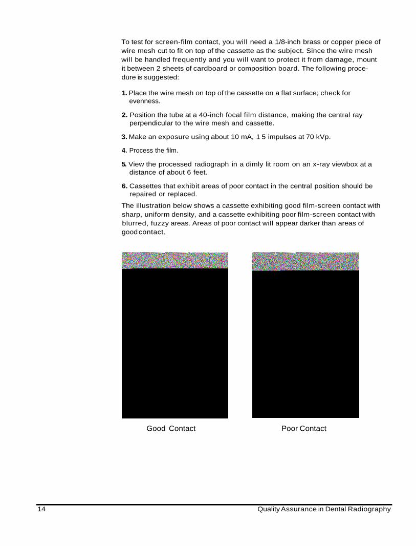
The illustration below shows a cassette exhibiting good film-screen contact withsharp, uniform density, and a cassette exhibiting poor film-screen contact withblurred, fuzzy areas. Areas of poor contact will appear darker than areas ofgood contact.
Good Contact Poor Contact
14 Quality Assurance in Dental Radiography

Viewbox Maintenance
The condition of the viewbox or illuminator can have an effect on the perceiveddensity and contrast of images. Variations or distortions can result from:
• Dirt on the Plexiglas.
• Discoloration of the Plexiglas front.
• Type or age of the bulb.
As a bulb approaches the end of its useful life, it will begin to blacken. If thishappens, the lamp should be replaced before it burns out totally. The entirebank of lights should be replaced simultaneously and the illuminator frontscleaned regularly.
Suggested Guidelines for Quality Assurance
A well-planned and well-executed quality assurance program can increase thelikelihood of producing the best possible radiographs with the lowest possibleradiation exposure to patients, the lowest cost, and the least number of retakes.Dental offices can very easily establish and maintain quality control proceduresas part of their overall management system. Remember that your Kodak serviceand sales representative is always available for further consultation aboutquality control measures.
Quality control tests should be performed annually on dental x-ray generatingequipment owned and operated by dental practitioners.
Quality Assurance in Dental Radiography 15

The best quality control procedures are invalid, however, without carefulmonitoring of film processing conditions. The highest quality images are usuallyproduced under optimum processing conditions. To complement the qualitycontrol tests recommended for dental x-ray generating equipment, follow thesuggested routines that follow:
Processor Maintenance:• Clean processors and work areas at regular intervals.
• Change solutions at scheduled time.
• Replenish fixer as well as developer daily.
• Monitor time and temperature daily.
• Check the operating time and temperature of processors and the temperatureof solutions daily.
• Perform quality control every morning (after replenishment and chemicalsreach proper temperature).
• Use one of the three quality control methods described on pages 8 through11 daily.
Film Storage and Handling:• Store and handle film properly.
• Rotate film stock regularly.
Darkroom:• Use KODAK GBX-2 Safelight Filters.
• Position safelight at no less than 4 feet (1.2m) from the film.
• Use a 15-watt frosted bulb.
• Check safelighting conditions every six months.
• Check for light leaks each month.
• Make sure ventilation is adequate.
Radiographic Screens:• Clean with KODAK Intensifying Screen Cleaner and Antistatic Solution
monthly.
• Inspect screens monthly.
Viewbox Maintenance:• Clean the Plexiglas weekly.
• Inspect the fluorescent bulbs and replace them when necessary.
16 Quality Assurance in Dental Radiography

There is a great deal of helpful information about common film processingerrors contained in two publications available from Eastman Kodak Company."Successful Intraoral Radiography" (KODAK Publication No. N-418) containshelpful information on how to identify and correct common errors in intraoralradiography. "Successful Panoramic Radiography" (KODAK Publication No.N-406) details the common errors made in panoramic radiography and how tocorrect them. See below for information on how to order your copy free ofcharge.
To obtain a free copy of any of the Kodak publications referred to inthis pamphlet:• In the U.S.A., please call Kodak at 1-800-233-1650 or write:
Eastman Kodak CompanyAdvertising DistributionOrder Entry, Dept. 454343 Slate StreetRochester, New York 14650-3009
Order the publication by name and code number.
• Outside the U.S.A., all publication requests should be directed to thenearest Kodak Company, or write:
Dental BusinessHealth Imaging DivisionEastman Kodak Company343 State StreetRochester, New York 14650-1122 U.S.A.
To obtain the Dental Radiographic Normalizing and Monitoring Devicerecommended by the American Academy of Oral and Maxillofacial Radiology,order from:
Dental Radiographic DevicesP. O. Box 9294Silver Spring, Maryland 20916(301)598-6543
To talk to a Kodak representative about quality control in dental radiography:
• In the U.S.A., phone (800) 933-8031
• In Canada, phone (800) GO-KODAK
• Elsewhere, phone the local Kodak subsidiary or (716) 724-5631
To contact Kodak via the Internet:
http://www.kodak.com/go/dental
EASTMAN KODAK COMPANYDental BusinessHealth Imaging Division343 State StreetRochester, NY 14650-1122
KODAK CANADA INC.Health Imaging Division3500 Eglinton Ave. WToronto, Ontario MGM IV3CANADA
Kodak,Ultra-Speed,Ektaspeed,T-Mat, Ektamat,Ektavision,Readymatic & X-Omat are trademarks.
N-416 Printed in the U.S.A. 8/98 © Eastman Kodak Company, 1998