quinone methide precursors as potential therapeutics towards the
TRANSCRIPT

Quinone Methide Precursors as Potential Therapeutics Towards the Realkylation
of Aged Acetylcholinesterase
Research Thesis
Presented in Partial Fulfillment of the Requirements for graduation with research distinction in the undergraduate colleges of The Ohio State University
by
Travis Glenn Blanton
The Ohio State University April 2015
Project Advisors: Dr. Ryan J. Yoder and Dr. Christopher S. Callam
Department of Chemistry and Biochemistry

i
Abstract Acetylcholinesterase (AChE) is an essential enzyme required by the human body. It hydrolyzes
the neurotransmitter acetylcholine (ACh) into two products, choline and acetate, at neurosynaptic
junctions. Organophosphorous (OP) nerve agents such as sarin, tabun, and VX are covalent inhibitors
of AChE. After a period of time that varies with each OP compound, the inhibition of AChE becomes
irreversible due to aging, which is when a de-alkylation of the OP compound in the AChE active site
occurs. This aging process results in ACh accumulation in the peripheral and central nervous system.
While current pharmaceuticals containing an oxime functional are used to reverse inhibition of AChE
by OP compounds, they are not effective in reversing inhibition of AChE after the OP compound has
aged. A lack of pharmaceuticals to reverse this pathological aged state was the subject of intense
research in the early 1970s, but quickly came to a halt due to the limitations of technology during that
era. Now, with far more powerful technology and computational algorithms, research into drug
discovery to reactivate aged AChE can be more efficient and effective. The focus of our research is to
reactivate aged AChE. Previous reports from the literature demonstrated the capability of Quinone
Methides (QMs) to alkylate phosphodiesters and other biological molecules. As a result, this class of
compounds has the potential to reverse the aging process and restore function to aged AChE by
alkylating the phosphonylated Serine-203 residue in the aged AChE active site. Using the
computational methods of Molecular Docking and Molecular Dynamics, a library of Quinone Methide
Precursors (QMPs) were tested and evaluated to determine which compounds from the library spent
the most time in the aged AChE active site. Additionally, other residues of interest in the active site
were examined with the goal of steering design efforts to produce QMPs that have a higher likelihood
of potential alkylation.

ii
Acknowledgements Throughout my time spent at Ohio State, I have met many individuals who have influenced the
course of my life significantly enough to warrant mention in this section. Unfortunately only a few will
be mentioned; however, I extend my gratitude and appreciation to all – listed or unlisted.
Firstly, I would like to extend my gratitude to Dr. Jeffery Turner for instilling a love of
chemistry into a highly impressionable twenty-something. As a result of his influence, I explored
research opportunities in the field of organic chemistry and found an opening with a new tenure-track
faculty member…
Who happened to be Dr. Ryan J. Yoder, my current research advisor. I would like to thank
Ryan for imparting some of his computational chemistry knowledge onto me, as well as for going
above and beyond the call of an advisor. Many times, adversity strikes when one is on the cusp of
achieving something great. To Ryan, this adversity was always an indication to overcome and advance
through in order to reap the rewards that often lie on the other side of struggle. For that, a debt of
gratitude is owed. Thank you for believing in me.
I would also like to thank Dr. Christopher Callam for his support, guidance, sharp wit, and
excellent sense of humor. Never doubt the power of a good joke (or five.)
I must also thank Dr. Christopher Hadad for using the depth and breadth of his knowledge to
help drive the project forward at critical crossroads. Without him, I feel that this thesis may never have
been conceptualized. For that, I thank you greatly.
I find it necessary to thank The Ohio State University at Marion for providing the opportunities
to explore a breadth of subjects and meet the faculty who teach them. Providing those opportunities
allowed me to meet and learn from the following staff and faculty who have forever positively
impacted my life: Dr. John Emens, Dr. Robert Klips, Dr. Gary Maul, Professor Emeritus Dr. Brian
McEnnis, Dr. Donna Bobbitt-Zeher, Dr. John Maharry Dr. Zakaria Nyongesa, Dr. Chris and Amy

iii
Friesen, Dr. Ben McCorkle, Dr. Terry Pettijohn, Dr. Daniel Wojta, Dr. Christopher Daddis, Dr. Nikole
Huffman, Leslie Beary, Lynda Behan, and the staff of Maynard Hall. Sometimes, pleasant
conversation and smiling faces are what will take you the distance. Thank you for everything.
Lastly, I would like to thank my friends, family, and those I care deeply about for supporting
me during my time as an undergraduate and throughout the medical school application process.

iv
List of Figures Figure Page Figure 1.1. Structures of OPs used as chemical warfare agents along with less toxic pesticides, amiton
and paraoxon.. ........................................................................................................................................... 1
Figure 1.2. The use of QMPs to reactivate aged AChE in order to reverse the aging process of the
enzyme upon exposure to nerve agents. ................................................................................................... 3
Figure 2.1. Initial Library of Quinone Methide Precursor Compounds ................................................... 6
Figure 2.2. Compound E interacting with residues of interest within AChE ......................................... 16
Figure 2.3. A second view of Compound E interacting with residues of interest within AChE ............ 17
Figure 2.4. A third view of Compound E interacting with residues of interest within AChE. .............. 18
Figure 3.1. QMP Structures used as base scaffolds for an expanded library of new variants ................ 21
Figure 3.2. Second Library of Compounds ............................................................................................ 22
Figure 3.3 Second Library of Compounds ............................................................................................. 23
Figure 3.4 Second Library of Compounds ............................................................................................. 24
Figure 3.5 Second Library of Compounds ............................................................................................. 25
Figure 3.6. Compounds with highest active site affinity ........................................................................ 29
Figure 3.7. Compounds with lowest active site affinity ......................................................................... 30
Figure 4.1. Compound A3DM with its dimethylammonium leaving group sitting close to W86. ........ 33
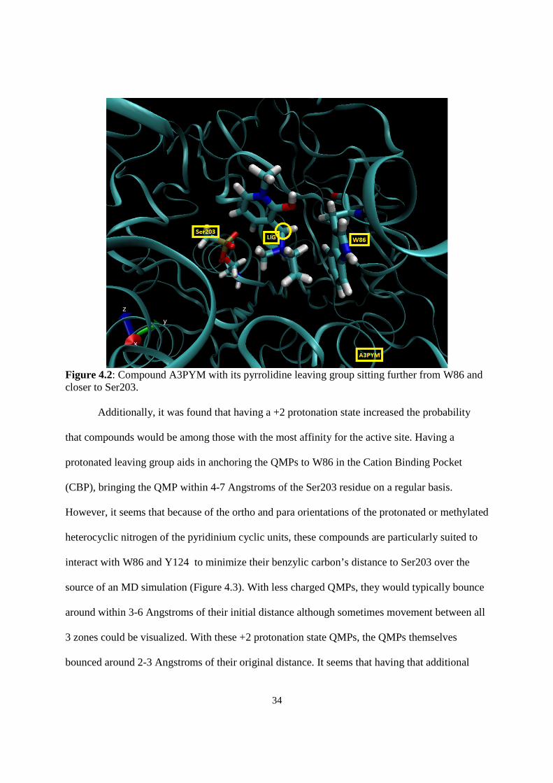
Figure 4.2. Compound A3PYM with its pyrrolidine leaving group sitting further from W86 and closer
to Ser203. ................................................................................................................................................ 34
Figure 4.3. An illustration of the interactions between a +2 Protonation State Methylpyridinium QMP
and W86, Y124, and Ser203. .................................................................................................................. 35
Figure 4.4. Compound A3MN, its unprotonated leaving group, and identification of interactions which
might facilitate its poor affinity for the active site. ................................................................................ 36

v
List of Tables
Table Page Table 2.1. Molecular Docking Results for First Library ........................................................................ 10
Table 2.2. AChE Zones (All bounds closed except “Between AS and BN” and “Other”) .................... 13
Table 2.3. MD Results for First Library ................................................................................................. 14
Table 2.4. MD Statistics Measuring Distance between Phosphonate Oxygen and QMP Benzylic
Carbon for First Library .......................................................................................................................... 14
Table 3.1. MD Results for Second Library............................................................................................. 29
Table 3.2. MD results for compounds with highest active site affinity (high to low) ............................ 30
Table 3.3. MD results for compounds with lowest active site affinity (low to high) ............................. 31

vi
Table of Contents Abstract ...................................................................................................................................................... i
Acknowledgements ................................................................................................................................... ii
List of Figures ........................................................................................................................................... ii
List of Tables ........................................................................................................................................... v
1. Introduction ........................................................................................................................................... 1
1.1 OP Exposure and the Role of Quinone Methides as Potential Therapeutic Agents .................. 1
1.2 References .................................................................................................................................. 5
2. Initial Library ........................................................................................................................................ 6
2.1 Introduction ................................................................................................................................. 6
2.2 Preparation of Compounds for Molecular Docking .................................................................... 7
2.3 Molecular Docking ..................................................................................................................... 8
2.4 Molecular Docking Results ....................................................................................................... 10
2.5 Molecular Dynamics ................................................................................................................. 12
2.6 Molecular Dynamics Results .................................................................................................... 14
2.7 Compound E Analysis .............................................................................................................. 16
2.8 References ................................................................................................................................. 20
3. Second Library .................................................................................................................................... 21
3.1 Introduction .............................................................................................................................. 21
3.2 Molecular Dynamics Results ................................................................................................... 27
3.3 References ................................................................................................................................ 32
4. Conclusions and Future Work ............................................................................................................ 33
4.1 Conclusions .............................................................................................................................. 33
4.2 Future Work ............................................................................................................................. 37

1
1. Introduction 1.1 OP Exposure and the Role of Quinone Methides as Potential Therapeutic Agents
Organophosphorus compounds (OPs) are a class of compounds that contain a phosphoryl
group. Their origin lies in the development of pesticides, when tabun was accidentally
discovered by Dr. Gerhard Schrader in Germany in 1936.1 This extreme toxicity to human life
did not go unnoticed, leading to tabun’s development into a potential chemical weapon.2 Since
then, many more OP compounds have been created. Of these, the most frequently recognized
compounds are sarin (GB), soman (GD), and VX (Figure 1.1).
Figure 1.1: Structures of OPs used as chemical warfare agents along with less toxic pesticides,
amiton and paraoxon.
In recent history, these OP compounds have been used on military and civilian
populations in several countries. Between the months of April and July 1988, the Iraqi army led
by Saddam Hussein launched several rockets containing OP nerve agent near the towns of al-
Faw, Mehran, and Dehloran in Iraq, killing hundreds to thousands of Iranian solders.3 In 1995,
sarin gas was released into the Japanese subway system by Aum Shinrikyo, a terrorist
organization. At least 7 people died and thousands more were hospitalized by the exposure.4 On

2
August 21st 2013, the Syrian government launched rockets with sarin-containing warheads into
suburbs of Damascus.5 While there is no official death toll, Médecins Sans Frontières (Doctors
Without Borders) has said that at least 3,600 civilians were treated for symptoms consistent with
sarin poisoning - vomiting, respiratory issues, and kidney failure at the area’s three hospitals
within 3 hours of the attack.6 Records collected by Human Rights Watch show that 734 civilians
in the suburb of Zamalka died as a result of the sarin gas attacks, with most victims being
children.7 With casualties primarily being civilians, there has never been a greater need to
develop treatments for OP exposure.
OP nerve agents are neurotoxins that are extremely potent. They owe their biological
activity to their ability to inactivate the catalytic triad found in acetylcholinesterase (AChE), an
enzyme that hydrolyzes the excitatory neurotransmitter acetylcholine. This catalytic triad
consists of Ser-His-Glu residues found in the active site. The serine residue of this triad is
modified by OPs after they covalently bind to it, effectively inhibiting the enzyme's function. At
this time, the treatment protocol for OP exposure is the concurrent administration of 2-
Pralidoxime (2-PAM), atropine, and diazepam (Valium) to exposed individuals. 2-Pralidoxime
(2-PAM) is a strong nucleophile capable of displacing the OP moiety from the OP-AChE
complex. It is the powerhouse of this protocol that restores catalytic function to the inhibited
enzyme. If untreated, the OP-AChE complex can undergo a process called aging, where the OP
compound is dealkylated.8 This aging process leaves only the OP’s phosphate group attached to
the serine residue, irreversibly inhibiting the AChE enzyme (Figure 1.2). The speed at which
aging occurs depends solely on the OP compound being used9; regardless, the outcome of aging
is irreversible inhibition of AChE. After aging occurs, there are no pharmaceuticals currently in

3
existence that are capable of displacing the OP moiety. The lack of therapeutic agents to reverse
the dealkylation of aged AChE is the focus of our research.
Quinone Methides (QMs) are high-energy electrophiles that are used in important
biological processes, like lignin biosynthesis in plants, DNA alkylation, and cross-linking of
structural proteins.10,11 The motivation to explore this class of compounds towards reversing the
aging process of OP-inhibited AChE comes from two studies. The first, a study from Modica et
al.12, determined that QMPs had the ability to alkylate many nucleophiles, specifically amino
acids. The second study, from Bakke et al.13 reported that QMPs can re-alkylate phosphodiesters
in aqueous conditions. The ability of QMP compounds to re-alkylate phosphodiesters and amino
acids encourages us in the pursuit of these compounds as a solution to reversing aged AChE,
because the OP compounds inhibiting AChE form a phosphodiester bond with the serine residue
which results in the OP-AChE complex.
Figure 1.2: The use of QMPs to reactivate aged AChE in order to reverse the aging process of
the enzyme upon exposure to nerve agents.

4
Due to the sheer number of QMPs that could be created, a high-throughput process was
required to narrow down the potential candidates for laboratory testing. As a result,
computational methods were employed to guide synthetic efforts to produce a QMP that was
selective and specific for the active site of AChE.

5
1.2 References
1. Ballantyne, B. and Marrs, T. C. (1992). Overview of the biological and clinical aspects of organophosphates and carbamates, in Clinical and Experimental Toxicology of Organophosphates and Carbamates, Ballantyne, B. and Marrs, T. C., Eds., Butterworth, Oxford, England, 1. 2. Dacre, J. C. (1984). Toxicology of some Anticholinesterases used as chemical warfare agents - a review, in Cholinesterases, Fundamental and Applied Aspects, Brzin, M., Barnard, E. A. and Sket, D., Eds., de Gruyter, Berlin, Germany, 415. 3. Human Rights Watch, Iraq’s Crime Of Genocide: The Anfal Campaign against the Kurds (Human Rights Watch, 1994), http://www.hrw.org/reports/1994/05/01/iraq-s-crime-genocide-anfal-campaign-against-kurds. 4. Seto, Dr. Yasuo: The Sarin Gas Attack in Japan and the Related Forensic Investigation. Org. for Prohibition of Chemical Weapons, June 2001, http://www.opcw.org/news/article/the-sarin-gas-attack-in-japan-and-the-related-forensic-investigation/ (accessed September 16th, 2014) 5. Rogin, J. Exclusive: U.S. to Bring Chemical Weapons Witnesses Out of Syria. The Daily Beast, May 2013, http://www.thedailybeast.com/articles/2013/05/22/exclusive-u-s-to-bring-chemical-weapons-witnesses-out-of-syria.html (accessed May 27, 2013) 6. Médecins Sans Frontières, “Syria: Thousands Suffering from Neurotoxic Symptoms Treated in Hospitals Supported by MSF,” August 24, 2013. 7. Human Rights Watch, Attacks on Ghouta.(Human Rights Watch, 2013), http://www.hrw.org/node/118724/section/4 http://www.hrw.org/reports/1993/iraqanfal/. 8. Michel, H. O., Hackley B. E. Jr, Berkowitz, L., List, G., Hackley, E. B., Gilliam, W. and Paukan, M. (1967) Aging and dealkylation of soman (pinocolylmethyl- phosphonofluoridate)- inactivated eel cholinesterase. Arch. Biochem. Biophys. 121, 29–34. 9. Worek, F., Szinicz, L., Eyer, P. and Thiermann, H. (2005) Evalulation of oxime efficacy in nerve agent poisoning: Development of a kinetic-based dynamic model, Toxicol. Appl. Pharmacol. 209, 193-202. 10. Peter, M.G. Angew. Chem. Int. Ed. Engl. 1989, 28, 555. 11. Veldhuyen, W.F.; Shallop, A. J.; Jones, R. A.; Rokita, S. E. J. Am. Chem. Soc. 2001, 123, 11126. 12. Modica, E.; Zanaletti, R.; Freccero, M.; Mela, M. J. Org. Chem. 2001, 66, 41. 13. Bakke, B. A.; McIntosh, M. C.; Turnbull, K. D. J. Org. Chem. 2005, 70, 4338-4345.

6
2. First Library
2.1 Introduction
An initial library of seven para-Quinone Methide Precursors (p-QMPs) (Figure 2.1) was
produced during discussion with collaborators. These seven compounds use vanillin as a
scaffold, where variations between them are the result of group substitutions at only the phenolic
position. Compound E represents this base vanillin scaffold. For the amine leaving group, each
QMP was held constant using dimethyl ammonium.
Figure 2.1: Initial Library of Quinone Methide Precursor Compounds

7
2.2 Preparation of Compounds for Molecular Docking
In order to dock these compounds into the active site of aged AChE, they first required
preparation in order to provide meaningful results. Initially, all seven p-QMPs were built in
GaussView 4.1.21. Several different conformations of each compound were manually generated
by altering bond angles and distances in order to give a best attempt at discovering each
compound’s global energy minimum. Then, all seven compounds and their alternate
conformations were optimized with Gaussian 092 using the B3LYP/6-311+G** level of theory
with a Polarizable Continuum Model3 of Solvation of water. After the optimizations, the lowest
energy conformation for each compound was determined and underwent vibrational frequency
analysis at B3LYP/6-311+G** to look for the absence of imaginary frequencies, which would
confirm that the geometry was a minimum on the potential energy surface. After confirmation of
the absence of imaginary frequencies, then Merz-Kollman charge calculations were performed at
the B3LYP/6-311+G** level of theory on each compound’s lowest energy conformation to
complete their preparation for molecular docking.

8
2.3 Molecular Docking
Molecular Docking is a computational technique that can give insight into the possible
orientations of ligands inside receptors by examining the electrostatic interactions between the
two. Typically, the ligand is treated as flexible with no rotational or torsional restrictions whereas
the receptor is treated as rigid and no torsion or rotation is permitted. For this research, p-QMPs
were docked inside the active site of thirteen snapshots of artificially aged human
acetylcholinesterase (AChE). The thirteen artificially aged AChE snapshots (frames) were
produced by Dr. Jeremy Beck as a portion of his dissertation.4 The frames are stills taken from a
5 nanosecond molecular dynamics simulation of aged AChE with a generic QMP in the active
site. The benefit of using these frames for our docking calculations is that induced fit effects on
AChE from QMPs could be considered while still being able to treat AChE as rigid, which has a
lower computational cost. Dr. Jeremy Beck sourced the structure of AChE from an available
crystal structure in the Protein Data Bank (PDB id 1B415). He removed waters, cofactors, and an
associated fasciculin-II peptide using UCSF Chimera.6 He then restored any missing residues
using electric ray AChE homology (PDB id 1C2B7) and then added a phosphonate to the
catalytic Serine-203 residue (Ser203) in order to produce human aged AChE.
For the molecular docking simulations on these seven compounds, all compounds were
docked into each of the 13 receptor frames using Autodock 4.0.8 A 50 x 50 x 50 Å3 grid box was
produced and centered at a moiety known as the oxyanion hole in order to limit the calculation to
the active site and associated gorge to reduce computational expense. For each compound, 200
poses (snapshots of the compounds inside AChE) were generated for every receptor frame. This
resulted in a total of 2600 poses per compound.

9
After completion of the molecular docking simulations, the positioning of these
compounds within aged AChE was evaluated. Each docking simulation output contained 200
poses organized in a histogram from lowest binding energy to highest. The histogram associated
poses with very similar binding energies into one bin. Each bin was examined and the distance
measured for every pose within it. A bin with 75% or more of its poses falling into one of the
three categories, which will be explained below had all of its poses designated as that category.
This reduced the amount of manpower hours necessary to score each compound. Lastly, the
poses associated with each category were summed and divided by the total number of poses per
compound to give percentages.
Each pose was evaluated and classified into two categories: “within 3.7 Å” and “other”.
A “within 3.7 Å” pose was considered to be one that had a distance equal or less than 3.7 Å
between the p-QMP benzylic carbon and phosphonate oxygen on Ser203. An “other” pose
covers any pose that has a benzylic carbon to phosphonate oxygen distance greater than 3.7 Å. A
sub-category of “within 3.7 Å”, titled “within 3.7 Å, reactive orientation” was created to describe
poses satisfying the distance requirement but also having the QMP’s amine leaving group
oriented away from the phosphonylated serine oxygens. This orientation would result in minimal
steric interference if the desired alkylation reaction were to occur via SN2 direct displacement.

10
2.4 Molecular Docking Results
After completing the manual scoring process as outlined in Section 2.3, a table of the
results was produced (Table 2.1).
Compound Within 3.7 Å, reactive
orientation (%)
Within 3.7 Å, unreactive orientation
(%) Other (%)
A 31.9 4.6 63.5
B 37.4 15.9 46.7
C 29.7 30.3 40
D 23.2 40.9 35.9
E 17.6 28.3 54.1
F 15.6 43.3 41.1
G 38.8 11.1 50.1
Table 2.1: Molecular Docking Results for First Library
Of these seven compounds, those with the highest proportion of poses within 3.7 Å were C, D,
and F, which have fluorine, no functional, and acetal substitutions at the phenolic position,
respectively. This was a rather interesting finding, as A, E, and G are the three worst performing
compounds by this same metric and chemical intuition suggests that these compounds might
preferentially undergo an SN1 dissociation rather than a direct displacement reaction.
Additionally, A, E, and G had the highest percentages of their poses exceeding 3.7 Å in distance
between their benzylic carbons and the phosphonylated serine oxygens. Further experimental
data regarding these ligands and enzyme itself would be ideal so that plausible, meaningful
explanations of these trends are better supported. Therefore, Molecular Dynamics simulations

11
were performed on the library of seven compounds to simulate the interactions between the
ligands and the aged AChE active site and obtain more information.

12
2.5 Molecular Dynamics (MD)
Molecular Dynamics is a computational method that permits one to simulate how a
ligand might interact with a receptor or other molecule in a variety of solvents (or the gas phase)
over time. In this calculation, both the ligand and the enzyme are treated as flexible, which
permits a better approximation than molecular docking of what could be happening on an atomic
level during experiments. For this application, the three lowest energy clusters from the previous
molecular docking calculations were used as input geometry for the MD simulations. These input
geometries placed the p-QMPs at varying distances from the phosphonylated serine residue
within AChE’s active site. All 13 prepared aged AChE receptor frames were used in these
calculations, making a maximum total of 39 MD calculations per ligand. These were performed
using the Sander module in AMBER11.9 The simulated system was neutralized with Na+ ions
and solvated with explicit water. After this preparation, the simulated system underwent two
minimizations. The first set of minimizations involved 500 steps of a calculation known as
Steepest Descent Minimization10, where the overall energy of the system is pushed away from
any local maxima as quickly as possible while permitting the calculations to have larger error in
their approximations. Next, the simulated system underwent 500 steps of a calculation known as
Conjugate Gradient Minimization10, which lowers the system’s energy at a much slower rate so
that it can be approximated with more accuracy. This allows the system to be brought closer to
its global minimum. For the second set of minimizations, the system underwent another 1000
steps of Steepest Decent Minimization followed by 1500 steps of Conjugate Gradient
Minimization. After these minimizations, the system was heated linearly from 0 Kelvin to 300
Kelvin over 20 picoseconds (10,000 steps of 2 femtoseconds) at 1 atm. After heating, MD

13
calculations were performed on this system over 1 nanosecond (500,000 steps of 2
femtoseconds).
After completion of the MD calculations, it became readily apparent that characterization
of the enzyme AChE would be necessary in order to derive meaning from the results. For this
characterization, the gorge to the active site of AChE was labeled based upon the distance
between the phosphonate oxygens on Ser203 in the active site to certain residues in specific
regions in the gorge (Table 2.2).
Zone Distance from
Ser203
Active Site (AS) 0 - 5 Å
Between AS and BN
5- 7 Å
Bottleneck (BN) 7 - 9.5 Å
Gorge Mouth (GM)
10 - 15 Å
Other 15 Å+
Table 2.2: AChE Zones (All bounds closed except “Between AS and BN” and “Other”)

14
2.6 Molecular Dynamics Results
Using the previously defined zones (Table 2.3), a table of results was produced by
measuring the distance between the benzylic carbon of each QMP and the phosphonylated Serine
oxygens in the active site for every still frame in the MD calculation (500 per 1ns MD). Every
still frame was classified as one of the five zones. After every still frame was classified (using
Excel � Filter) a fraction of the total number of still frames per ligand (typically 19500) was
taken and a fraction of total poses present in a particular zone was then determined (Table 2.4).
Name % Active Site % Between AS and
BN % Bottleneck % Gorge Mouth % Other
A 47.5 18.5 11.9 20.0 20.7 B 44.3 22.6 9.2 22.2 24.3 C 46.6 16.3 10.6 24.5 18.2 D 45.8 27.2 7.1 18.1 29.0 E 44.7 23.2 6.0 22.6 26.7 F 58.2 12.9 10.4 16.6 14.8
G 49.8 10.9 14.2 20.7 15.3 Table 2.3: MD Results for First Library
Compound Mean Median Mode Max Min A 6.45 5.18 3.30 15.46 2.77 B 6.47 5.33 3.64 16.05 2.66 C 6.83 5.19 5.41 15.65 2.73 D 6.09 5.17 3.35 16.97 2.68 E 6.63 5.18 6.66 16.00 2.73 F 5.72 3.82 3.23 15.35 2.73 G 6.62 5.02 3.28 15.22 2.78
Table 2.4: MD Statistics Measuring Distance between Phosphonate Oxygen and QMP Benzylic
Carbon for First Library
The 3 compounds with highest affinity for the active site after examining the MD results
are A, F, and G. Compound F (acetal) also happens to be one of the best performers in the
Molecular Docking studies. However, these MD results should be compared with Molecular

15
Docking results very carefully, as it is important to acknowledge that the upper boundary for the
“active site” in MD is 5.0 Å, whereas that bound is 3.7 Å in Molecular Docking results.
Focusing back onto the MD results in Figure 2.4, a curious question is posed – why does
Compound E (hydroxyl at the phenolic position) perform several percentage points lower than
Compounds A and G, which should form the same QM intermediate if they undertake SN2
dissociation? A closer look was taken at Compound E to suggest an explanation for the deviance.

16
2.7 Analysis of Compound E
In an attempt to understand the deviance from expected values, one MD video from each
of the 13 receptor frames were viewed in an attempt to explain why this particular compound
preferred to orient itself further away from the phosphonylated serine-203 residue (S203-OP
adduct).
In most of the videos observed, the ligand (Compound E) was visualized interacting with
Y124 and W86. Y124 is an aromatic residue (tyrosine) located in the Bottleneck zone. It is
capable of hydrogen bond donation and acceptance, as well as cation-pi interactions. W86 is an
aromatic residue (tryptophan) that is capable of only hydrogen bond acceptance and cation-pi
interactions. Three labeled images (Figures 2.2, 2.3, and 2.4) are displayed below and explained.
Figure 2.2: Compound E interacting with residues of interest within AChE

17
The first image (Figure 2.2) suggests that the ligand Compound E’s hydroxyl group at the
phenolic position may be involved in unique hydrogen bonding reciprocity with Y124 not
demonstrated by the rest of the seven compounds. The image also suggests that cation-pi
interactions between the dimethylammonium leaving group of the ligand and W86 may also play
a role in tethering the ligand to residues outside of the upper bound for the active site. These
residues and the suggested interactions are indicated in yellow.
Figure 2.3: A second view of Compound E interacting with residues of interest within AChE
The second image (Figure 2.3) above does not show interactions between the ligand and
Y124. However, it does imply hydrogen bond donation and cation-pi interactions between the
ligand and W86.

18
Figure 2.4: A third view of Compound E interacting with residues of interest within AChE
The third view (Figure 2.7) suggests that hydrogen bonding reciprocity between Y124
and the ligand are present, as well as the implication of potential cation-pi interactions with Y124
from the ligand. In this third image, there are no interactions with W86.
These three images highlight three different orientations of Compound E near AChE’s
gorge bottleneck. They represent the majority of Compound E’s simulated interactions with the
enzyme. The most plausible explanation of Compound E’s inferior performance in comparison
to Compounds A and G is that the presence of a hydroxyl group at the phenolic position on
Compound E allows it to both donate and accept hydrogen bonding with Y124. In combination
with its protonated dimethylammonium leaving group permitting cation-pi interactions with
residues near the active site, this added hydrogen bond acceptance-donor reciprocity uniquely

19
enables Compound E to select Y124 and reduce the amount of time it spends within the active
site zone.

20
2.8 References
1. GaussView, Version 4.1.2, Dennington, Roy; Keith, Todd; Millam, John. Semichem Inc., Shawnee Mission, KS, 2009. 2. Gaussian 09, Revision D.01, Frisch, M. J.; Trucks, G. W.; Schlegel, H. B.; Scuseria, G. E.; Robb, M. A.; Cheeseman, J. R.; Scalmani, G.; Barone, V.; Mennucci, B.; Petersson, G. A.; Nakatsuji, H.; Caricato, M.; Li, X.; Hratchian, H. P.; Izmaylov, A. F.; Bloino, J.; Zheng, G.; Sonnenberg, J. L.; Hada, M.; Ehara, M.; Toyota, K.; Fukuda, R.; Hasegawa, J.; Ishida, M.; Nakajima, T.; Honda, Y.; Kitao, O.; Nakai, H.; Vreven, T.; Montgomery, J. A., Jr.; Peralta, J. E.; Ogliaro, F.; Bearpark, M.; Heyd, J. J.; Brothers, E.; Kudin, K. N.; Staroverov, V. N.; Kobayashi, R.; Normand, J.; Raghavachari, K.; Rendell, A.; Burant, J. C.; Iyengar, S. S.; Tomasi, J.; Cossi, M.; Rega, N.; Millam, N. J.; Klene, M.; Knox, J. E.; Cross, J. B.; Bakken, V.; Adamo, C.; Jaramillo, J.; Gomperts, R.; Stratmann, R. E.; Yazyev, O.; Austin, A. J.; Cammi, R.; Pomelli, C.; Ochterski, J. W.; Martin, R. L.; Morokuma, K.; Zakrzewski, V. G.; Voth, G. A.; Salvador, P.; Dannenberg, J. J.; Dapprich, S.; Daniels, A. D.; Farkas, Ö.; Foresman, J. B.; Ortiz, J. V.; Cioslowski, J.; Fox, D. J. Gaussian, Inc., Wallingford CT, 2009. 3. Mennucci, B. (2012), Polarizable continuum model. WIREs Comput Mol Sci, 2: 386–404. doi: 10.1002/wcms.1086 4. Beck, J. M., Ph.D. Dissertation, The Ohio State University, 2011. 5. Kryger, G.; Harel, M.; Giles, K.; Toker, L.; Velan, B.; Lazar, A.; Kronman, C.; Barak, D.; Ariel, N.; Shafferman, A.; Silman, I.; Sussman, J. L.Acta Crystallogr., Sect. D 2000, 56, 1385-1394 6. Pettersen, E. F.; Goddard, T. D.; Huang, C. C.; Couch, G. S.; Greenblatt, D. M.; Meng, E. C.; Ferrin, T. E. J. Comput. Chem. 2004, 25, 1605-1612. 7. Bourne, Y.; Grassi, J.; Bougis, P. E.; Marchot, J. J. Biol. Chem. 1999, 274, 30370-30376 8. Morris, G. M., Huey, R., Lindstrom, W., Sanner, M. F., Belew, R. K., Goodsell, D. S. and Olson, A. J. (2009) Autodock4 and AutoDockTools4: automated docking with selective receptor flexiblity. J. Computational Chemistry 2009, 16: 2785-91. 9. Case, D. A.; Darden, T. A.; Cheatham, T. E., III; Simmerling, C. L.; Wang, J.; Duke, R. E.; Luo, R.; Crowley, M.; Walker, R. C.; Zhang, W.; Merz, K. M.; Wang, B.; Hayik, S.; Roitberg, A.; Seabra, G.; Kolossváry, I.; Wong, K. F.; Paesani, F.; Vanicek, J.; Wu, X.; Brozell, S. R.; Steinbrecher, T.; Gohlke, H.; Yang, L.; Tan, C.; Mongan, J.; Hornak, V.; Cui, G.; Mathews, D. H.; Seetin, M.G.; Sagui, C.; Babin, V.; Kollman, P. A. AMBER 11, University of California, San Francisco, 2008. 10. Adcock, S. A., & McCammon, J. A. Molecular Dynamics: Survey of Methods for Simulating the Activity of Proteins. Chemical Reviews 2006, 106(5), 1589–1615. doi:10.1021/cr040426m

21
3. Second Library
3.1 Introduction
After examination of the MD data shown in the previous chapter, three compounds with
the highest affinity for the active site were selected as scaffolds to produce a new library of
compounds to evaluate. An additional six compounds were selected from MD calculations
performed by fellow undergraduates Keegan Fitzpatrick and Andrew Franjesevic on the same
basis. Based on their origins, one letter codes were assigned to each compound to produce three
“classes” of QMPs – T, A, and K (Figure 3.1).1
Figure 3.1: QMP Structures used as base scaffolds for an expanded library of new variants.

22
After discussion with collaborators, two variations in the base scaffolds evaluated for
their effects on active site affinity. The first variation was a substitution of the amine leaving
group, substituting dimethylammonium with dimethylammonia, pyrrolidine, piperidine, or
morpholine functionals. The second variation was a change in the protonation state of the leaving
group and heterocyclic nitrogen (if applicable) to neutral, protonated, or methylated analogs.
Using these variations, ninety-nine analogs were designed. From those, seventy-eight were
selected for evaluation (Figure 3.2, 3.3, 3.4, 3.5) using the molecular docking and molecular
dynamics procedures described in Chapter 2. The remaining compounds were given to fellow
undergraduates to perform the same calculations.
Figure 3.2: Second Library of Compounds

23
Figure 3.3: Second Library of Compounds

24
Figure 3.4: Second Library of Compounds

25
Figure 3.5: Second Library of Compounds

26
The naming scheme for this library is relatively straightforward. As mentioned earlier in
this chapter, the first two characters in the name indicate the class that the scaffold belongs to,
and which specific scaffold was used. The remaining characters indicate which amine leaving
group is present on the compound, followed by what the protonation state of the compound is.
For leaving groups, dimethylammonia is indicated by D, pyrrolidine is indicated by PY,
piperidine by PI, and morpholine by M. For protonation states, neutral is indicated by N,
protonated by P, and methylated by M. The order of the codes is the same for every compound in
this library.

27
3.2 Molecular Dynamics Results
Using the same zoning criteria from the previous chapter, tables of results were produced
from the MD calculations (Table 3.1).
Compound Name % Active
Site % Between AS and BN
% Bottleneck
% Gorge Mouth
% Other
A1DN 20.88 35.66 26.87 11.95 4.65 A1MN 21.31 32.82 30.84 12.32 2.72 A1PIN 27.06 38.07 24.28 9.05 1.55 A1PYN 37.10 31.64 22.59 5.23 3.45 A2DN 36.77 25.85 18.70 13.95 4.73 A2MN 9.12 29.42 22.55 33.19 5.71 A2PIN 25.71 46.33 15.36 10.69 1.90 A2PYN 30.37 40.46 20.32 6.39 2.45 A3DN 40.58 27.17 23.42 7.01 1.82 A3MN 11.68 42.33 25.32 17.29 3.38 A3PIN 27.01 34.93 27.32 9.42 1.33
A3PYN 30.39 31.36 28.09 9.23 0.93
A1DM 37.54 32.41 7.22 17.64 5.20
A1MM 45.72 15.71 2.74 30.65 5.17
A1MP 41.15 23.10 5.49 25.79 4.48
A1PIM 53.88 14.54 1.59 27.21 2.78
A1PIP 67.66 14.06 2.78 8.20 7.30
A1PYM 45.54 23.09 5.28 17.39 8.70
A1PYP 65.76 16.43 1.76 9.59 6.46
A2DM 61.47 10.11 5.42 14.83 8.17
A2MM 21.48 36.23 11.69 25.11 5.49
A2MP 49.66 16.47 7.37 20.21 6.29
A2PIM 23.84 42.04 8.49 22.29 3.35
A2PIP 57.36 21.55 7.86 7.04 6.18
A2PYM 41.59 32.70 5.47 14.41 5.83
A2PYP 62.85 17.58 1.95 13.38 4.24
A3DM 73.75 5.21 3.53 13.46 4.05
A3MM 24.64 18.00 9.15 39.31 8.89
A3MP 40.97 15.79 9.92 28.71 4.60
A3PIM 64.57 7.18 5.87 17.98 4.39
A3PIP 68.51 6.51 4.83 15.31 4.85

28
Compound Name % Active
Site % Between AS and BN
% Bottleneck
% Gorge Mouth
% Other
A1DM 37.54 32.41 7.22 17.64 5.20
A1MM 45.72 15.71 2.74 30.65 5.17
K1DN 44.44 23.06 16.73 10.52 5.25
K1MN 28.85 41.55 16.21 9.14 4.25
K1PIN 49.81 29.08 13.66 5.88 1.57
K1PYN 41.57 33.18 15.70 8.22 1.33
K2DN 29.86 25.24 21.72 17.98 5.20
K2MN 24.16 29.72 20.76 21.82 3.53
K2PIN 38.46 17.23 22.88 15.38 6.05
K2PYN 34.83 32.55 16.02 12.76 3.85
K3DN 34.32 22.46 34.33 6.82 2.07
K3MN 17.82 34.34 25.76 16.90 5.18
K3PIN 16.56 29.96 30.23 19.81 3.45
K3PYN 25.36 30.37 31.24 10.87 2.16
K1DM 63.06 7.36 2.48 16.23 10.87
K1MM 36.54 17.38 2.66 37.17 6.24
K1MP 53.95 12.46 7.29 16.71 9.59
K1PIM 58.09 9.74 4.60 22.63 4.94
K1PIP 56.53 17.83 4.93 16.76 3.94
K1PYM 71.29 5.65 2.43 18.26 2.37
K1PYP 68.65 10.57 2.00 15.33 3.45
K2DM 42.06 20.92 5.51 21.01 10.51
K2MM 44.92 18.35 1.93 28.52 6.28
K2MP 40.17 14.75 3.41 27.74 13.93
K2PIM 44.93 17.86 9.48 24.45 3.28
K2PIP 67.17 8.26 4.04 15.23 5.31
K2PYM 65.86 12.62 3.43 16.08 2.02
K2PYP 56.40 9.46 6.00 23.10 5.04
K3DM 55.48 15.09 6.81 18.42 4.21
K3MM 35.37 12.06 7.23 37.23 8.11
K3MP 42.07 18.10 0.78 28.78 10.27
K3PIM 46.40 14.62 6.62 27.97 4.39
K3PIP 56.53 8.39 3.68 15.76 15.63
K3PYM 55.61 15.35 5.64 20.27 3.14
K3PYP 64.69 15.78 4.70 9.04 5.78

29
Compound Name % Active
Site % Between AS and BN
% Bottleneck
% Gorge Mouth
% Other
T1DM 50.4 14.1 11.0 20.7 3.8
T1MM 25.7 18.1 23.2 29.0 4.0
T1PIM 47.5 21.9 9.8 18.1 2.8
T1PYM 44.3 11.6 18.2 23.8 2.0
T2DM 51.7 8.3 12.3 20.9 6.9
T2MM 25.4 15.2 25.0 31.3 3.2
T2PIM 49.7 11.7 15.5 20.5 2.6
T2PYM 54.7 20.5 9.3 13.8 1.7
T3DM 63.2 9.5 8.5 14.2 4.5
T3MM 30.0 11.9 19.6 34.0 4.5
T3PIM 49.1 28.1 6.2 14.2 2.4
T3PYM 35.3 36.2 6.9 17.8 3.8 Table 3.1: MD Results for Second Library
Of these seventy-eight compounds, ten compounds with the highest and lowest affinities
for the active site were identified, and their data was assimilated (Figure 3.3, Table 3.2, Figure
3.4, Table 3.3).
Figure 3.6: Compounds with highest active site affinity

30
Compound Name % Active
Site % Between AS and BN
% Bottleneck
% Gorge Mouth
% Other
A3DM 73.70 5.20 3.50 13.50 4.10 A3PYM 71.40 4.60 1.90 17.60 4.50 K1PYM 71.30 5.70 2.40 18.30 2.40 K1PYP 68.60 10.60 2.00 15.30 3.50 A3PIP 68.50 6.50 4.80 15.30 4.80 A1PIP 67.70 14.10 2.80 8.20 7.30 K2PIP 67.20 8.30 4.00 15.20 5.30
K2PYM 65.90 12.60 3.40 16.10 2.00 A1PYP 65.80 16.40 1.80 9.60 6.50
T3DM 63.20 9.50 8.50 14.20 4.50 Table 3.2: MD results for compounds with highest active site affinity (high to low)
Figure 3.7: Compounds with lowest active site affinity

31
Compound Name % Active
Site % Between AS and BN %
Bottleneck % Gorge Mouth % Other
A2MN 9.12 29.42 22.55 33.19 5.71 A3MN 11.68 42.33 25.32 17.29 3.38 K3PIN 16.60 30.00 30.20 19.80 3.40 K3MN 17.80 34.30 25.80 16.90 5.20 A1DN 20.88 35.66 26.87 11.95 4.65 A1MN 21.31 32.82 30.84 12.32 2.72 A2MM 21.50 36.20 11.70 25.10 5.50 T2MM 25.40 15.20 25.00 31.30 3.20 T1MM 25.73 18.06 23.22 28.99 4.00
A2PIN 25.71 46.33 15.36 10.69 1.91 Table 3.3: MD results for compounds with lowest active site affinity (low to high)
Comparing the ten compounds with highest active site affinity, it is readily apparent that
the majority of these compounds had pyrrolidine and piperidine leaving groups. In fact, the only
leaving group that did not have any representation in the top ten was morpholine. All but one of
the top performers had pyridinium or methylpyridinium cyclic units, with the compound falling
into tenth place containing a phenyl ring. Additionally, all but one of the top performers had
protonation states of +2 with a 5-to-4 split of methylation to protonation.
Looking at the ten compounds with the lowest active site affinity of the library, seven of
the ten compounds had morpholine leaving groups, suggesting that morpholine’s absence from
the highest affinity list is likely not a coincidence. Additionally, nine of the ten compounds had
protonation states of +1. Most of these compounds have pyridinium or methylpyridinium rings,
with a slightly larger proportion of the phenyl ringed vanillin compounds being present in this
short list.

32
3.3 References
1. Fitzpatrick, Keegan. Computational Insights into the Alkylation Reactions of Pyridine and Pyridinium Quinone Methide Precursors: Studies Towards the Realkylation of Aged Acetylcholinesterase. Thesis, The Ohio State University, 2013.

33
4. Conclusions and Future Work
4.1 Conclusions
The data shows that the best performing leaving groups from the expanded library of
seventy-eight were pyrrolidine and piperidine functional groups that also were protonated or
methylated at the heterocyclic nitrogen. After observing MD simulation videos of these
compounds, it was seen that because of the size of these leaving groups, they were not as close to
W86 as the library of 7 dimethylammonium leaving group compounds were as discussed in
Chapter 2 (Figure 4.1). While these larger leaving groups were still close enough to undergo
cation-pi interactions, it appears that the conformation of those leaving groups prevented them
from being flatter and therefore sitting closer to W86 (Figure 4.2). This inability to sit closely
resulted in the benzylic carbon being closer in proximity to the Ser203 phosphonate oxygens,
resulting in these compounds having better rankings.
Figure 4.1: Compound A3DM with its dimethylammonium leaving group sitting close to W86.
34
Figure 4.2: Compound A3PYM with its pyrrolidine leaving group sitting further from W86 and closer to Ser203. Additionally, it was found that having a +2 protonation state increased the probability
that compounds would be among those with the most affinity for the active site. Having a
protonated leaving group aids in anchoring the QMPs to W86 in the Cation Binding Pocket
(CBP), bringing the QMP within 4-7 Angstroms of the Ser203 residue on a regular basis.
However, it seems that because of the ortho and para orientations of the protonated or methylated
heterocyclic nitrogen of the pyridinium cyclic units, these compounds are particularly suited to
interact with W86 and Y124 to minimize their benzylic carbon’s distance to Ser203 over the
source of an MD simulation (Figure 4.3). With less charged QMPs, they would typically bounce
around within 3-6 Angstroms of their initial distance although sometimes movement between all
3 zones could be visualized. With these +2 protonation state QMPs, the QMPs themselves
bounced around 2-3 Angstroms of their original distance. It seems that having that additional

35
charge in the ring permits H-bond acceptance from Y124 as well as alternating cation-pi
interactions between the protonated leaving group and the pyridinium unit, reducing the distance
that these compounds will move when they are in the active site.
Figure 4.3: An illustration of the interactions between a +2 Protonation State Methylpyridinium
QMP and W86, Y124, and Ser203.
Regarding the compounds with the lowest active site affinity, a +1 protonation state and a
morpholine leaving group were the most commonly observed variations. A compound that has
only a protonated amine leaving group, using the hypothesis suggested above, should have more
variance when interacting with W86 in the CBP. For one of the worst performing compounds
A3MN, interactions with residues in the bottleneck zone were commonly observed (Figure 4.4).
Seemingly, an unprotonated amine leaving group may result in having the compounds “hang up”
in the bottleneck . Regarding the morpholine leaving group performing poorly, it appears the

36
presence of the heterocyclic oxygen in the morpholine leaving group permits interactions with
Y124, which are demonstrated by these compounds having such a high proportion of their still
poses between the active site and the bottleneck.
Figure 4.4: Compound A3MN, its unprotonated leaving group, and identification of interactions
which might facilitate its poor affinity for the active site.

37
4.2 Future Work
With this larger library, we have shown the tunability of these molecules with regard to
their affinity for the aged AChE active site. At this point, these results should be passed along to
our experimental collaborators to focus their synthesis on QMPs with the structural
characteristics that make binding possible. Additionally, the exploration of other scaffolds,
namely quinolone and isoquinoline, should be considered.
Additionally, QM/MM calculations should be performed on some select QMPs in the
active site of AChE to determine whether a quinone methide or an intermediate can be generated
there. This is of significant importance to the project, as all of the computational work thus far
has been associated with designing QMPs to be specific for the active site and evaluating the
energetics of QMPs. Starting with QM/MM calculations is part of a progression towards
determining the actual mechanism of alkylation and beginning design efforts relating to
alkylation.