r in children current pathologic and classification
TRANSCRIPT

20
Review papeR
RhabdomyosaRcoma in childRen – cuRRent pathologic and moleculaR classification
Ireneusz DzIuba1,2*, Paweł Kurzawa1,3*, MIchał DoPIerała3,4, ana b. Larque5, Danuta JanuszKIewIcz-LewanDowsKa4,6
*First co-authors, authors with equal first contribution in preparation of the manuscript
1�Pathology�Department,�University�Hospital�of�Lord’s�Transfiguration,�Partner�of�Poznan�University�of�Medical�Sciences,�Poznan,�Poland�
2Pomeranian�Medical�University,�Szczecin,�Poland3Clinical�Pathology�Department,�Poznan�University�of�Medical�Sciences,�Poland4�Department�of�Paediatric�Oncology,�Haematology,�and�Transplantology,�Poznan�University�of�Medical�Sciences,�Poland
5Pathology�Department,�Massachusetts�General�Hospital,�MA,�USA6Medical�Diagnostics�Department,�Poznan,�Poland
The�last�25�years�have�brought�significant�progress�in�the�treatment�of�sarcomas�in� children,� especially� rhabdomyosarcoma� (RMS).� Nevertheless,� treatment� fail-ure� in� some�patients� results� from�considerable�biological�heterogeneity�noted� in�these� tumours.�RMS,� the�most�common�malignant�soft� tissue�neoplasm� in�chil-dren,�includes�two�main�subtypes:�embryonal�(ERMS)�and�alveolar�(ARMS).�Due�to�greater�aggressiveness�and�worse�prognosis�of�ARMS�in�comparison�to�ERMS,�discrimination�between�different� rhabdomyosarcoma� subtypes� is� of� crucial� clini-cal�importance.�This�paper�presents�the�current�histological�classification�of�RMS,�up-to-date�immunohistochemical�and�biological�research�regarding�RMS,�and�its�associated�clinical�and�prognostic�significance.�
Key words:�rhabdomyosarcoma,�children,�pathologic�and�molecular�classification.
DoI: httPs://DoI.org/10.5114/PJP.2018.75333 PoL J PathoL 2018; 69 (1): 20-32
Introduction
Rhabdomyosarcoma� (RMS)� is� the� most� common�malignant�solid�tumour�in�children�after�neuroblas-toma�and�nephroblastoma�(Wilms�tumour).�This�tu-mour�accounts� for�5%�to�10%�of� all� childhood� tu-mours� [1,� 2,� 3,� 4,� 5].� For� all� soft� tissue� sarcomas,�RMS�accounts� for�19%�of� such� cases� in� adults� and�45%�of�cases�in�children.�Rhabdomyosarcoma�is�the�most�common�soft�tissue�malignant�neoplasm�in�the�latter�age�group.�Rhabdomyosarcoma�is�derived�from�primary�mesenchymal�cells�that�show�skeletal�mus-cle�differentiation. It�was�first�described�by�Weber�in�1854.� About� 90%� of� all� RMS� presentations� are� in�individuals�under�25�years�of�age,�and�almost�70%�
are�in�children�under�10�years�of�age�[1,�2,�3,�6].�The�most�common�RMS�location�is�in�the�head�and�neck�region�(35-40%),�followed�by�the�urogenital�system,�extremities,�and�torso�[1,�2,�3,�6,�7].
Rhabdomyosarcoma aetiology and pathogenesis
The� predisposing� factors� for� the� development� of�soft� tissue� sarcoma� are� unknown.� Increased� RMS�morbidity� is� observed� in� individuals� with� genetic�syndromes� predisposing� to� carcinogenesis,� such� as�Li-Fraumeni� syndrome,� Gardner� syndrome,� neu-rofibromatosis� type� I,� and� Beckwith-Wiedemann�syndrome.�The�common�co-occurrence�of�RMS�with�

21
Pathologic and molecular classification of rhabdomyosarcoma in children
defects�of�the�central�nervous�system,�urogenital,�gas-trointestinal,� and� circulatory� systems,� and� melano-cytic�nevi�has�been�reported�[3,�5,�8,�9,�10,�11,�12].
Nomenclature and pathologic classification
In� formerly� used� pathologic� classifications,� RMS�was�divided� into� two�main� types:� alveolar� and�em-bryonal� [3,�5,�7,�13].�The�embryonal� type� includes�the�botryoides�and�spindle�cell�subtypes.�However,�in�the�current�WHO�classification� (World�Health�Or-ganisation;�WHO�2013)�[14],�four�histological�RMS�types�are�recognised�and�classified�as�follows:
1.�Embryonal�rhabdomyosarcoma:a.�botryoides�variant,b.�anaplastic�variant.
2.�Alveolar�rhabdomyosarcoma:a.�solid�variant,b.�anaplastic�variant.
3.�Pleomorphic�rhabdomyosarcoma.4.�Spindle�cell/sclerosing�rhabdomyosarcoma.It�should�be�noted�that�pleomorphic�RMS�occurs�
most�commonly� in�adults,�but�rare�cases�have�been�observed�in�children�[14].
The�College�of�American�Pathology�(CAP)�classi-fication�of�skeletal�muscle-derived�tumours�also�rec-ognises�ectomesenchymoma�as�part�of�this�group�of�malignant�neoplasms.�Ectomesenchymoma�is�a�neo-plasm� consisting� of� rhabdomyoblasts� resembling�the� myosarcoma� of� both� embryonal� and� alveolar�type,�with�a�distinctly�higher�presence�of�ERMS�el-ements�observed.�The�histology�of� the� tumour�also�shows�the�presence�of�ganglion�cells�and�foci�of�neu-roblastomatous�dedifferentiation.�The�current�WHO�classification�of�soft�tissue�and�bone�tumours�places�ectomesenchymoma�in�the�same�group�with�neuro-genic�neoplasms�[14].�
The�American�group�of�physicians�specialising�in�RMS,�“Children’s�Oncology�Group� (COG)”,�distin-
guishes� the� classical� and� solid� ARMS� variant,� and�typical,�botryoid,�spindle�cell,�sclerosing,�dense,�and�epithelioid� ERMS.� Additionally,� rare� mixed-type�RMS� is� listed.� The� rhabdomyosarcoma,� not� other-wise� specified� (RMS-NOS)� category� is� restricted� to�tumours�that�are�small�and�showing�sampling�or�fix-ation�artefact,�or�are�necrotic,�making�a�specific�clas-sification�impossible�[15].
Pathologic appearance of rhabdomyosarcoma
Rhabdomyosarcoma�diagnosis�should�be�based�not�only�on�the�histopathological�appearance�but�also�on�the� immunohistochemical� and� molecular� profiles.�The�histology�of�RMS� shows� cellular� elements� that�are� related� to� the� structures� resembling�the�cells�of�developing�striated�muscle�(Fig.�1).�Rhabdomyoblasts�with�a�diverse�level�of�atypia�are�the�key�cells�in�RMS�diagnosis.�The�highly�differentiated�rhabdomyoblast�is�a�round�or�oval�cell�that�contains�abundant�acido-philic�granular�or�fibrillar�cytoplasm,�with�eccentric�or� centrally-located� circular� nuclei.� Binucleation� is�commonly�noted.�Occasional� large�nucleoli� are� vis-ible�within�nuclei�[16]�(Fig.�2).�Vacuoles�containing�glycogen� are� sometimes� visible� in� the� cytoplasm.�Rhabdomyoblasts� can� take� many� different� forms� –ribbon� or� tadpole-like,� appearing� similar� to� tennis�rackets�or�spiders�(Fig.�3).�In�less�than�30%�of�diag-nosed�RMS,�rhabdomyoblasts�with�distinct�striations�are�observed.�Detection�of�such�a�characteristic�RMS�cytological�feature�is�difficult�using�standard�haema-toxylin�and�eosin�staining.�The�striations�can�be�eas-ily�visualised�using�a�tricolour�histochemical�staining�method,�such�as�phosphotungstic�acid�haematoxylin�(PTAH)�(Fig.�4).�However,�most�of�the�tumours�are�composed� of� less� differentiated� or� undifferentiated�rhabdomyoblasts�with�scant�cytoplasm,�and�round�or�oval�nuclei.�The�cellular�edges�of�such�myoblasts�are�star�shaped.�Sometimes�RMS�cells�fuse�and�generate�
Fig. 1. Embryonal� rhabdomyosarcoma.� Neoplastic� cell�resembles� developing� striated� skeletal� muscle� (=>)� (HE�stain,�magnification�400�×)
Fig. 2. Embryonal�rhabdomyosarcoma.�Rhabdomyoblasts:�round�and�oval�shapes�of�neoplastic�cells�(=>)�(HE�stain,�magnification�400�×)

22
Ireneusz DzIuba, Paweł Kurzawa, MIchał DoPIerała, et al.
Fig. 5. Rhabdomyosarcoma,�pleomorphic�type.�Numerous�cells�with�multiple�nuclei�resembling�multinucleated�giant�cells�(=>)�(stain�HE,�magnification�600�×)
Fig. 3. Embryonal�rhabdomyosarcoma.�Rhabdomyoblasts:�various�shapes�of�neoplastic�cells�(>,�=>,�→)�(HE�stain,�magnification�400�×)
Fig. 4. Embryonal�rhabdomyosarcoma.�Cross-striations�in�cytoplasm�of�rhabdomyoblasts�(=>)�(PTAH�stain,�magni-fication�400�×)
Fig. 6. Embryonal�rhabdomyosarcoma.�Hypercellular�zones�around�blood�vessels�(HE�stain,�magnification�400�×)
polynuclear�cells�that�resemble�multinucleated�giant�cells�(Fig.�5)�[17].�
Embryonal rhabdomyosarcoma
�The�microscopic�appearance�of�embryonal�rhab-domyosarcoma� (ERMS)� shows� rhabdomyoblasts� of�heterogeneous� appearance.� Undeveloped,� round�cells� with� a� hyperchromatic� nucleus� and� basophilic�cytoplasm� are� common� in� low-cell-density� regions�embedded� in� a� myxoid� submucosa.� High-cell-con-centration�regions�are�present�around�vessels�and�are�organised� in� characteristic� perivascular� thickenings�(Fig.� 6).� In� association� with� poorly� differentiated�cells,�better�differentiated�rhabdomyoblasts�showing�acidophilic� cytoplasm,� sometimes� with� cross-stri-ation,� are� commonly� observed.� Overall,� ERMS� his-tology�resembles�a�combination�of�the�stages�of�the�embryonal�development�of�striated�muscle:�from�the�small,� round,� undifferentiated� cells,� through� tad-
pole-like� cells,� ribbon-shaped� striated� cells,� to� fully�differentiated�rhabdomyoblasts�(Fig.�7).
Embryonal rhabdomyosarcoma – botryoid variant
In�the�botryoid�variant�of�ERMS,�a�so-called�com-pact�cambium�layer�(appearance�analogous�to�the�lay-er�of�plant�cells�that�are�present�between�xylem�and�phloem�and�cause�the�thickening�of�the�plant)�can�be�seen.� These� cells� form� the� group� of� densely� packed�undifferentiated�neoplastic�cells�just�under�the�epithe-lium.�The�more�hypocellular�and�mucoid�areas�of�the�neoplasm�are�observed�under�this�layer�(Figs.�8,�9).
Embryonal rhabdomyosarcoma – anaplastic variant
The� anaplastic� variant� of� ERMS� is� composed� of�large,� anaplastic� rhabdomyoblasts� with� hyperchro-matic�nuclei.�These�cells�are�often�present�as� single�cells� between� rhabdomyoblasts� with� various� types�

23
Pathologic and molecular classification of rhabdomyosarcoma in children
Fig. 7. Embryonal� rhabdomyosarcoma.� Characteristic�pleomorphic� rhabdomyoblasts� (HE� stain,� magnification�400�×)
Fig. 11.�Alveolar�rhabdomyosarcoma,�cells�with�big�nuclei�and�scant�cytoplasm�(HE�stain,�magnification�400�×)
Fig. 8. Embryonal�rhabdomyosarcoma,�botryoides�variant.�Epithelium� covering� the� cambial� layer� (=>)� with� loose-ly�arranged�cells�underneath�(→)�(HE�stain,�magnification�200�×)
Fig. 9. Embryonal�rhabdomyosarcoma,�botryoides�variant.�A�hypercellular�cambial� layer�with�epithelial� layer�under-neath�(HE�stain,�magnification�400�×)
Fig. 10.� Embryonal� rhabdomyosarcoma,� anaplastic� vari-ant.�Single�cells�with�features�of�anaplasia�(=>)�(HE�stain,�magnification�200�×)
of� atypia� (Fig.� 10).� They� are� highly� polymorphous�and�contain�a�small�amount�of�cytoplasm.�This�last�characteristic�is�distinct�from�the�pleomorphic�type,�where�cells�often�contain�abundant�acidophilic�cyto-plasm.�Atypical�mitotic�figures�are�also�present.
Alveolar rhabdomyosarcoma
The�alveolar�RMS�(ARMS)�is�characterised�by�the�presence� of� poorly� differentiated� rhabdomyoblasts,�which� are� slightly� larger� than� the� undifferentiated�cells�in�ERMS.�These�cells�are�characterised�by�scant�cytoplasm�and� large�nuclei� (Fig.�11).�The�histology�of�ARMS�also�shows�differentiated�rhabdomyoblasts�with�abundant�acidophilic�cytoplasm�and�character-istic�neoplastic�multinucleated�giant�cells.�The�neo-plastic�cells�cover�the�thick�strands�of�connective�tis-sue�with�regressive�changes�in�the�form�of�sclerosis.�

24
Ireneusz DzIuba, Paweł Kurzawa, MIchał DoPIerała, et al.
Fig. 12.�Alveolar�rhabdomyosarcoma,�cells�forming�a�pat-tern�resembling�pulmonary�alveoli�(=>)�(HE�stain,�mag-nification�400�×)
The�neoplastic�cells�aggregate�in�areas�at�the�edges�of�fibrous� septa� to�which� the� rhabdomyoblasts� adhere�in�a�single�layer.�In�the�central�portion�of�such�lesion,�the� cells� lose� this� connection,� undergoing� necrosis�and�degeneration.�Such� foci�can�resemble�a�pulmo-nary�alveolar�structure�(Figs.�12,�13).�
Alveolar rhabdomyosarcoma – solid variant
In�this�variant�of�the�tumour,�the�neoplastic�cells�have� no� connective� tissue� submucosa,� and� rhabdo-myoblasts� are� present� in� various� stages� of� differen-tiation�forming�extensive�lobular�structures.�The�cy-tological�features�of�the�cells�are�the�same�as� in�the�classic�form�[14] (Fig.�14).�
The�WHO�classification�of�bone�and�soft�tissue�tu-mours�additionally�lists�rare�ARMS�variants�without�detailed�characteristics�including�mixed�alveolar�and�embryonal�rhabdomyosarcoma�and�anaplastic�alveo-lar�rhabdomyosarcoma [14].
Fig. 13.�Alveolar� rhabdomyosarcoma.�Alveolar-like� spaces�formed�by�primitive�round�cells�separated�with�connective�tissue�septa�(silver�impregnation�stain,�magnification�400�×)
Fig. 14.�Alveolar�rhabdomyosarcoma,�solid�variant.�Sheets�of�small�round�blue�cells�(HE�stain,�magnification�400�×)
Fig. 15.� Rhabdomyosarcoma,� pleomorphic� type.� Chaoti-cally�arranged�bizarre�cells�in�the�bottom�left-hand�corner�of�the�Fig.�(HE�stain,�magnification�400�×)
Pleomorphic rhabdomyosarcoma
Pleomorphic� rhabdomyosarcoma� is� composed� of�the� following�cells:�polymorphic,� spindle,� and�mul-tinucleated�giant�cells�with�abundant�acidophilic�cy-toplasm.�Sometimes�cells�with�bizarre�atypia�are�hap-hazardly�arranged�in�the�connective�tissue�submucosa�(Fig.�15).�Highly�differentiated�striated�rhabdomyo-blasts�are�rarely�observed.
Spindle cell/sclerosing rhabdomyosarcoma
The� spindle-cell� variant� is� characterised� by� ex-tensive� histologic� heterogeneity.� Cells� can� be� rib-bon-shaped,� embedded� in� sclerotic� submucosa,� or�can�form�fascicular�or�elongated�interwoven�arrange-ments� composing� a� so-called� herring-bone� pattern�(Figs.�16,�17).�The�sclerosing�subtype�rarely�occurs�in�children�[18].�There�is�evidence�that�suggests�a�dif-ferent�biology�and�prognosis�for�the�spindle-cell�RMS�subtype�in�children�in�comparison�with�ERMS�[19].�

25
Pathologic and molecular classification of rhabdomyosarcoma in children
Fig. 17.�Rhabdomyosarcoma,� spindle� cell/sclerosing�vari-ant.�Spindle�cells�forming�herringbone�pattern�(HE�stain,�magnification�400�×)
Fig. 16.�Rhabdomyosarcoma,� spindle� cell/sclerosing�vari-ant.�Cords�of�cells�in�hyalising�stroma�(HE�stain,�magnifi-cation�400�×)
Fig. 18.� Alveolar� rhabdomyosarcoma.� Positive� nuclear�staining�for�MyoD1�(magnification�400�×)
Therefore,� distinguishing� between� these� two� RMS�groups�is�of�crucial�importance�(Table�I)�[20,�21].
Immunohistochemical diagnostics
The�diversity� in�RMS�morphology�often� leads� to�significant�difficulty� in�correct�diagnosis.�Therefore,�utilisation�of�integrated�diagnostic�methods,�includ-ing�immunohistochemical�and�molecular�methods,�is�necessary.
The� use� of� immunohistochemical� methods� for�RMS�diagnosis�in�order�to�identify�rhabdomyoblasts�is�a�routine�procedure.�In�cases�of�less�differentiated�tumours,� the� easiest� method� to� detect� rhabdomyo-blastic�differentiation�of� the� sarcoma� is� the�demon-stration� of� expression� of� MyoD1� protein� and� myo-genin�(Myf4)�(Figs.�18,�19).�Positive�nuclear�staining�in�both�markers�is�an�important�diagnostic�criterion�for�RMS�and�is�the�gold�standard�in�differential�diag-nosis�with�other�neoplasms.�
Furthermore,� MyoD1� and� myogenin� have� addi-tional� practical� significance� for� distinguishing� the�ARMS�from�other�RMS�subtypes.�Myogenin�expres-
Table I. The�morphological�differences�between�ERMS�and�spindle-cell�RMS�[20,�21]
moRphological chaRacteRistic eRms spindle-cell Rms
Cell�shape Oval,�star-shaped,�spindle-like Regular,�elongated,�spindle-like
Cell�patterns Lobes�with�various�cellular�density,�from�loose,�low-cell-concentration�to�
high-cell-concentration
fascicles,�cords,�storiform�patterns
Rhabdomyoblasts Frequently�present,�but�in�varying�amounts
Can�be�present,�usually�in�small�amounts
Mitotic�figures Frequent Frequent
Submucosa Often�loose,�myxoid Various�amount�of�collagenised�submucosa�organised�in�fascicles
sion�obtained�in�more�than�50%�of�the�neoplastic�cells�is�highly�suggestive�of�a�diagnosis�of�ARMS�[22,�23,�24].�Myogenin�and�MyoD1�are�useful�for�distinguish-ing�the�classical�ARMS�and�sclerosing�RMS.�Myogen-in�expression�in�sclerosing�RMS�is�weak�and�focal,�and�

26
Ireneusz DzIuba, Paweł Kurzawa, MIchał DoPIerała, et al.
Fig. 19.� Alveolar� rhabdomyosarcoma.� Positive� nuclear�staining�for�myogenin�(Myf-4)�(magnification�400�×)
Fig. 20.� Alveolar� rhabdomyosarcoma.� Positive� cytoplas-matic�staining�for�desmin�(magnification�400�×)
in�the�case�of�ARMS,�strong�and�diffuse.�In�contrast,�the�MyoD1�expression�is�strong�and�diffuse�in�scleros-ing�RMS.�ARMS,�however,�exhibits�variable�MyoD1�expression�[15].�Cytoplasmic�expression�of�vimentin�and�desmin�can�be�observed�in�poor�or�undifferentiat-ed�cells�(Fig.�20),�and�expression�of�muscle�actin�and�myoglobin,�in�differentiated�rhabdomyoblasts.�
Recently� the� importance� of� novel� immunohisto-chemical�markers�as�prognostic�factors�for�RMS�has�been�reported.�Among�others,�the�role�of�p53,�bcl-2,�MDR-1,�and�MIB1�(Ki67)�expression�is�highlighted.�The�DNA�ploidy� status� in� cerralion�with� the� clini-cal�course�of�RMS�was�also�analysed�–�hyperdiploid�ERMS�tumours�showing�a�more�favourable�prognosis�[25,�26].
Expression�of�other�proteins�evaluated�the�use�of�immunohistochemical� techniques� such� as�AP2i� and�P-cadherin�appeared� to�be�a� selective�ARMS�mark-er.�On� the�other�hand,� the� expression�of� epidermal�growth� factor� receptor (EGFR)� and� fibrillin-2� are�characteristic�for�ERMS.�EGFR�and�fibrillin-2�expres-sion�are�correlated�with�the�favourable�course�of�the�disease,�while�the�presence�of�AP2i�and�P-cadherin�is�associated�with�a�poor�prognosis�[27].
Differential diagnosis
The� differential� diagnosis� with� other� tumours�showing�small�cells�with�round�and�spindled�config-urations�is�based�not�only�on�the�morphology�of�the�cells�but,�above�all,�also�on�additional�tests,�particular-ly�immunophenotyping�[21]�(Tables�II,�III).�Various�neoplasms�can�exhibit�differentiation�towards�skele-tal�muscle,�which�can�additionally�hamper�obtaining�a�correct�diagnosis�[28,�29,�30].�The�differential�di-agnosis�in�this�group�of�tumours�includes:�malignant�Triton�tumour,�spindle�cell�carcinomas,�sarcomatoid�carcinomas,�melanoma,�liposarcoma,�malignant�tera-toma,�teratocarcinosarcoma,�salivary�carcinosarcoma,�
anaplastic� thyroid� carcinoma,� nephroblastoma,� and�some�tumours�of�the�central�nervous�system.
Molecular diagnostics
The�traditional�clinical�and�pathological�parame-ters�are�sometimes�not�sufficient�to�adequately�define�the�clinical�course�and�prognosis.�Additionally,�deter-mining�the�RMS�subtype�is�not�always�possible�based�on� the�pathologic�examination�alone.�Hence,�much�recent�attention�has�been�devoted� to� the�molecular�distinctions� of� rhabdomyosarcoma.� The� analyses� of�cytogenetic� changes�have� shown� their� usefulness� in�distinguishing�subtypes�of�RMS.�The�most�common�translocations� that� are� selectively� characteristic� for�ARMS� are� t(2;13)(q35;q14)� and� t(1;13)(p36;q14),�which�lead�to�the�generation�of�fusion�genes,�PAX3 (2q35)�or�PAX7 (1p36),�respectively,�with�the�gene�encoding� fork-head-region� transcription� factor� –�foxo1�(previous�name�fkhr)�(13q14)�[31].�The�trans-locations�mentioned�above�are�present,� respectively,�in�56%�to�85%,�and�6%�to�10%�of�all�ARMS-type�tumours,� and� these� are� either� not� observed� or� are�only�occasionally�present�in�ERMS�[31,�32].�Prelim-inary�data�shows�that�ARMS�tumours�with�a�PAX7/FKHR�translocation�exhibit�a�less�aggressive�course�of�disease�in�comparison�with�tumours�having�PAX3/FKHR�translocation.�However,�ERMS-type�tumours�show�changes�in�a�number�of�chromosome�pairs�–�2,�7,�8,�11,�12,�13,�and�20�–�in�as�much�as�25%�to�50%�of� cases.�The� loss�of�9�and�10�chromosome�pairs� is�described�in�20%�to�30%�of�cases.�Genome�amplifi-cation�is�another�change�observed�relatively�often�in�RMS�–�in�as�many�as�16%�to�56%�of�tumours.�In�case�of�ERMS,�the�amplification�covers�the�12q13-q15�re-gion,�and�in�the�case�of�ARMS�it�involves�the�1p36�(PAX7-FOXO1),�2p24�(MYCN),12q13-q14,�13q14�(PAX7-FOXO1),�and�13q31�(MIR17HG,�encoding�miR-17-92�microRNA)�regions.�RMS-type�tumours�

27
Pathologic and molecular classification of rhabdomyosarcoma in children
Tabl
e II
.�Diff
eren
tiat
ion�
of�s
mal
l,�ro
und,
�blu
e�ce
ll�tu
mou
rs�(t
able
�mod
ified
)�[21
]
tu
mo
uR
ty
pic
al
mo
Rp
ho
log
ica
l c
ha
Ra
ct
eRis
tic
sK
eRa
tin
cd
99n
b84
cd
45d
esm
inm
yo
gen
in
syn
ap
to
ph
ysi
n
ot
heR
ma
RK
eRs
an
d R
ema
RK
s
Alv
eola
r�R
MS
Pres
ence
�of�r
habd
omyo
blas
ts+
/––
––
++
++
++
+/–
Myo
D1(
+)
Des
mop
last
ic�
smal
l�rou
nd�c
ell�
tum
our
Smal
l,�m
onom
orph
ic�c
ells
�org
anis
ed�in
�ne
sts,
�cor
ds,�s
picu
les,
�lobe
s�w
ith�
prom
inen
t�de
smop
last
ic�s
ubm
ucos
a
++
++
/–+
/––
++
+�
(dot
)–
+/–
WT
1(+
)�if�t
he�a
ntib
ody�
reco
gnis
es�c
arbo
xy�
term
inus
;�ver
y�ra
re��
in�a
dult
s
Ewin
g�sa
rcom
aA
reas
�of�s
mal
l�mon
omor
phic
�cel
ls;�H
omer
�W
righ
t�ro
sett
es+
/–+
++
+/–
––
–+
/–FL
I-1(
+);�
nota
ble�
nega
tive
�CD
56;�r
arer
�in�
adul
ts
Extr
aren
al�
rhab
doid
�tum
our
Poly
gona
l,�va
riou
s�in
�siz
e,�r
hom
boid
al�c
ells
�w
ith�
wea
k�co
hesi
on,�o
rgan
ised
�in�lo
bes�
or�
solid
�spi
cule
s;�fr
eque
nt�n
ecro
sis�
++
++
/––
––
–+
/–C
hara
cter
isti
c�lo
ss�o
f�nu
clea
r�IN
I1;�v
ery�
rare
��in
�adu
lts
Lym
phom
aD
iffer
ent�
mor
phol
ogic
al�c
hara
cter
isti
cs�
depe
nden
t�on
�the
�typ
e�of
�lym
phom
a–
+/–
–+
++
––
–T
dT(+
)�to�
help
�exc
lude
�C
D45
(-)�l
ymph
obla
stic
�ly
mph
omas
Mel
anom
aC
ells
�var
ious
�in�s
ize�
and�
pleo
mor
phis
m�w
ith�
wea
k�co
hesi
on,�s
omet
imes
�wit
h�pr
esen
ce�o
f�br
own�
pigm
ent
––
––
––
–S1
00(+
),�SO
X10
(+);�
vari
able
�mel
anoc
ytic
Neu
robl
asto
ma
Pres
ence
�of�n
eura
l�diff
eren
tiat
ion,
�e.g
.��so
-cal
led�
neur
opile
––
++
+–
––
++
+N
otab
le�C
D99
�neg
ativ
ity
Poor
ly�
diffe
rent
iate
d�sy
novi
al�s
arco
ma
The
�arr
ange
men
ts�w
ith�
high
�cel
l�co
ncen
trat
ion�
of�c
ells
�wit
h�hi
gh�
pleo
mor
phis
m�a
nd�n
umer
ous�
mit
oses
+/–
++
+–
––
––
TLE
1(+
);�al
so�c
omm
only
�ex
pres
ses�
CD
56
Smal
l�cel
l�ne
uroe
ndoc
rine
�ca
rcin
oma
Salt
-and
-pep
per�
nucl
ei�
++
+–
––
––
++
+T
TF-
1(+
),�ve
ry�r
are�
in�
child
ren
(++
+)
– ty
pica
lly p
ositi
ve; (
+/–
) –
vari
ably
pos
itive
or
nega
tive;
(–)
– ty
pica
lly n
egat
ive

28
Ireneusz DzIuba, Paweł Kurzawa, MIchał DoPIerała, et al.
Tabl
e II
I.�D
iffer
enti
atio
n�of
�spi
ndle
-cel
l�tum
ours
�(tab
le�m
odifi
ed)�[
21]
tu
mo
uR
ty
pic
al
mo
Rp
ho
log
ica
l c
ha
Ra
ct
eRis
tic
sK
eRa-
tin
s100
p
Ro
tei
n
sma
des
min
cd
34o
th
eR m
aR
KeR
s
Spin
dle-
cell�
RM
SPr
esen
ce�o
f�rha
bdom
yobl
asts
,�cel
ls�o
rgan
ised
�in�
stor
iform
�or�
herr
ing-
bone
�pat
tern
––
+/–
++
+–
Myo
geni
n(+
),�M
yoD
1(+
)
Cel
lula
r�an
giof
ibro
ma
Cel
ls�w
ith�
bipo
lar�
nodu
les,
�sm
all�a
nd�m
ediu
m-s
ize�
vess
els�
pres
ent�
wit
h�ch
arac
teri
stic
�hya
linis
ated
�w
all
––
––
++
+Lo
ss�o
f�nuc
lear
�Rb�
prot
ein�
expr
essi
on;�
typi
cally
�in�p
opul
atio
n�of
�mid
dle-
aged
�and
�ol
der�
adul
ts�
Der
mat
ofib
rosa
rcom
a�pr
otub
eran
s�(D
FSP)
Hig
h-ce
ll-de
nsit
y�hi
stol
ogy�
wit
h�po
orly
�def
ined
�bo
rder
s–
––
–+
++
CD
34�e
xpre
ssio
n�de
crea
sed�
or�a
bsen
t�in
�fib
rosa
rcom
atou
s�D
FSP;
�can
�be�
pres
ent�
in�
child
ren,
�eve
n�as
�a�c
onge
nita
l�les
ion
Fibr
omat
osis
�–
–+
/––
–N
ucle
ar�β
-cat
enin
�exp
ress
ion�
in�m
ajor
ity�
of�
tum
ours
Infla
mm
ator
y�m
yofib
robl
asti
c�tu
mou
rM
yofib
robl
asts
�and
�infla
mm
ator
y�in
filtr
atio
n�w
ith�
vari
ous�
degr
ee�o
f�sev
erit
y�+
/––
++
++
/––
ALK
(+)�i
n�>
�50%
�of�c
ases
Leio
myo
sarc
oma�
Cel
ls�o
rgan
ised
�in�fa
scic
les�
or�c
ords
,�fre
quen
t�ne
cros
is–
–+
++
+/–
–C
alde
smon
(+);�
occa
sion
al�fo
cal�k
erat
in(+
);�ra
re�in
�chi
ldre
n
Low
-gra
de�
myo
fibro
blas
tic�
sarc
oma
Hig
h-ce
ll-de
nsit
y�hi
stol
ogy�
wit
h�ce
lls�o
rgan
ised
�in
�fasc
icle
s�an
d�st
orifo
rm�p
atte
rns;
�col
lage
nise
d�su
bmuc
osa
––
+/–
+/–
–D
esm
in(+
)�and
�abs
ence
�of�n
ucle
ar�
beta
-cat
enin
�favo
rs�t
his�
diag
nosi
s�ov
er�
fibro
mat
osis
;�rar
e�in
�chi
ldre
n
Low
-gra
de�fi
brom
yxoi
d�sa
rcom
aTy
pica
lly�p
rese
nt�z
ones
�of�f
ibro
us�a
nd�m
ixoi
d�hi
stol
ogy
––
+/–
––
MU
C4�
mos
t�sp
ecifi
c�m
arke
r;�fo
cal�E
MA
�ex
pres
sion�
com
mon
;�abo
ut�2
0%�o
f�cas
es�in
�pa
tient
s�<
�18�
year
s�ol
d
Mal
igna
nt�p
erip
hera
l�ne
rve�
shea
th�t
umou
rVa
riab
le�c
ellu
lari
ty�w
ithi
n�th
e�le
sion
,�cel
ls�
orga
nise
d�in
�fasc
icle
,�who
rled
,�her
ring
-bon
e�pa
tter
ns�
–+
++
––
+/–
S100
(+)�u
sual
ly�fo
cal�o
r�pa
tchy
;�des
min
�ex
pres
sion
�see
n�in
�rha
bdom
yobl
asti
c�el
emen
ts�(m
alig
nant
�Tri
ton�
tum
our)
;�can
�oc
cur�
in�v
ario
us�a
ge
Mam
mar
y-ty
pe�
myo
fibro
blas
tom
aVa
riab
le�in
�siz
e�fa
scic
les�
of�fr
agile
�spi
ndle
�cel
ls–
––
++
++
++
Loss
�of�n
ucle
ar�R
b�pr
otei
n�ex
pres
sion
;�pr
esen
t�in
�4th
-7th�d
ecad
e�of
�life
Neu
rofib
rom
aFe
w�d
iffer
ent�
type
s�of
�org
anis
atio
n:�s
prea
d,�
loca
lised
,�ple
xifr
om–
++
+–
–+
++
Con
tain
s�m
ixtu
re�o
f�S10
0(+
)�Sch
wan
n�ce
lls;�
can�
be�p
rese
nt�in
�chi
ldre
n�C
D34
(+)�s
trom
al�
cells
�and
�EM
A/c
laud
in-1
(+)�p
erin
eura
l�cel
ls
Nod
ular
�fasc
iitis
Loos
e�fa
scic
les�
and�
stor
ifrom
�pat
tern
s,�o
ften
�wit
h�ex
trav
asat
ed�e
ryth
rocy
tes
––
++
+–
–R
are�
foca
l�des
min
(+);�
can�
occu
r�in
�chi
ldre
n
PEC
oma
Nes
ts,�s
picu
les,
�and
�lobe
s�of
�epi
thel
ioid
�and
�sp
indl
e-sh
ape�
cells
––
++
++
/––
Myo
mel
anoc
ytic
�imm
unop
heno
type
�wit
h�ad
diti
onal
�var
iabl
e�ex
pres
sion
�of�H
MB
-45,
�m
elan
-A,�a
nd/o
r�M
ITF

29
Pathologic and molecular classification of rhabdomyosarcoma in children
tu
mo
uR
ty
pic
al
mo
Rp
ho
log
ica
l c
ha
Ra
ct
eRis
tic
sK
eRa-
tin
s100
p
Ro
tei
n
sma
des
min
cd
34o
th
eR m
aR
KeR
s
Peri
neur
iom
aSl
ight
ly�e
long
ated
�cel
ls�in
�var
ious
�pat
tern
s:�
stor
iform
,�who
rled
,�lam
ella
r,�fa
scic
ular
––
––
+/–
EMA
(+),�
clau
din-
1(+
);�ra
re�in
�chi
ldre
n
Schw
anno
ma
Pres
ence
�of�t
wo�
cellu
lar�
patt
erns
:�Ant
oni�A
�and
�A
nton
i�B;�V
eroc
ay�b
odie
s–
++
+–
–+
/–C
D34
�exp
ress
ion�
usua
lly�s
ubca
psul
ar;�f
ocal
�ke
rati
n(+
)�in�
retr
oper
iton
eal�s
chw
anno
mas
;�ca
n�oc
cur�
in�c
hild
ren
Solit
ary�
fibro
us�t
umou
rC
lass
ic�p
atte
rn�w
itho
ut�c
hara
cter
isti
c�va
riou
s�ce
llula
rity
––
––
++
+N
ucle
ar�S
TAT
6(+
)�is�
high
ly�s
peci
fic;�c
an�
occu
r�in
�chi
ldre
n
Spin
dle�
cell�
lipom
aId
enti
cal,�
frag
ile�c
ells
�wit
h�th
e�te
nden
cy�t
o�fo
rm�
“sho
als”
,�dev
elop
ed�a
dipo
cyte
s�pr
esen
t–
––
–+
++
Loss
�of�n
ucle
ar�R
b�pr
otei
n�ex
pres
sion
;��ve
ry�r
are�
<�2
0�ye
ars�
of�li
fe
Syno
vial
�sar
com
aC
an�o
ccur
�as�
a�bi
phas
ic�(s
pind
le�c
ells
�and
�epi
thel
ial�
elem
ents
)�or�
mon
opha
sic�
lesi
on��
(spi
ndle
�cel
ls)
++
++
/––
––
TLE
1(+
);�al
so�e
xpre
sses
�CD
56�a
nd�
calr
etin
in;�m
ost�
com
mon
ly�b
etw
een�
10�a
nd�
40�y
ears
�of�a
ge(+
++
) –
typi
cally
pos
itive
; (+
/–)
– va
riab
ly p
ositi
ve o
r ne
gativ
e; (–
) –
typi
cally
neg
ativ
e
Tabl
e II
I.�C
ont.
also�show�relatively�common�loss�of�heterozygosity�of�the�region�located�on�chromosome�11p15.5�[33,�34].
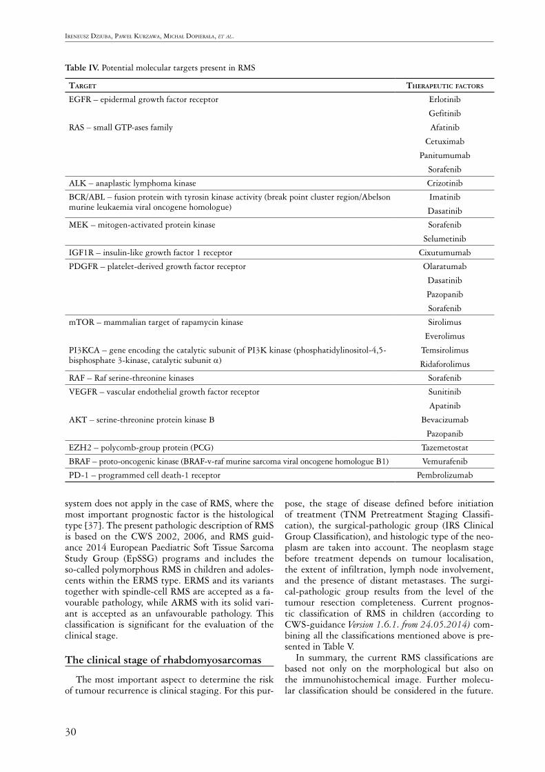
Analysis�of�gene�mutations�have�shown�their�com-mon�occurrence�in�RMS,�potentially�indicating�their�contribution�in�neoplastic�pathogenesis.�As�many�as�28%�of�ERMS�tumours�are�found�to�have�point�mu-tations� in� KRAS, TP53, FGFR4, EGRF, PIK3CA, CTNNB1, CDKN2A, BRAF,�and�PTPN11�genes,�in-dicating�frequent�occurrence�in�this�tumour�[33,�34].�However,� in�ARMS�tumours� these�mutations�occur�only�occasionally.�Mutation�of�the�MyoD1�–�myogen-ic differentiation 1 –�gene�was�observed� in� the� spin-dle-cell�subtype�localised�in�head�and�neck�and�limb�regions.�This�mutation�is�present�in�the�DNA-bind-ing�element�of�the�MyoD1�transcription�factor,�which�leads� to� the�generation�of� a�protein�product� acting�as�a�MYC�oncogene.�The�mutation�in�MyoD1� is�as-sosiated� with� poor� prognosis� [33,� 34].� Although�chemotherapy� remains� the� primary� treatment� for�child�patients,� the� identification�of�point�mutations�in�the�previously�mentioned�genes�may�in�the�future�be�a�useful�diagnostic�element�in�the�targeted�ther-apy� of� RMS.� These� therapies� offer� a� new� approach�to�increase�the�efficacy�of�RMS�treatment.�The�most�important�of�these�are�those�blocking�the�signalling�pathways� of� the� epidermal� growth� factor� receptor�(EGFR, HER-1, ERBB1),�which�is�a�member�of�the�group�of�tyrosine�kinase�receptors,�consisting�of�three�additional�receptors�that�are�similar�in�structure:�
EGFR2/HER2/HER-2-NEU/ERBB2,� EGFR3/HER-3/ERBB3,�and�ERBB4/HER4.�Phosphorylated�tyrosine�kinase� stimulates� intracellular� signal� trans-duction�by�a�cascade�of�other�pathways�such�as�RAS-RAF-MEK-MAPK-PI3K-AKT-JAK-STAT,� which�regulate�processes�of�proliferation,�apoptosis,�and�an-giogenesis.�Others� are� those� that� participate� in� the�mTOR�pathway�distorting�the�integration�of�signals�from�proteins�such�as�PI3K, AKT,�and�PTEN.�This�pathway�harmonises�with�other�IGF1-R-PI3K/AKT-mTOR�and�MAPK�pathways.�
The� summary�of�molecular� changes� in�RMS�and�possible�targeted�therapies�can�be�seen�in�Table�IV.
Clinico-surgical-pathologic classification
All�types�of�RMS�should�be�accepted�as�high-grade�sarcomas�[35].�The�exception�is�pleomorphic�RMS�in�adults,�for�which�the�grading�system�was�established�by�Fédération�Nationale�des�Centres�de�Lutte�Con-tre� le�Cancer/American�Joint�Committee�on�Cancer�(FNCLCC/AJCC).�This�system�is�based�on�the�anal-ysis�of�the�histological�type,�the�mitotic�activity,�and�the� level�of�necrosis� in�the�tumour�tissue�[36].�The�Paediatric� Oncology� Group� (POG)� introduced� its�own� system� for� assessment� of� the�malignancy� level�based�on�the�histological�type,�presence,�and�amount�of�necrosis,�and�mitotic�activity.�However,�the�POG�
30
Ireneusz DzIuba, Paweł Kurzawa, MIchał DoPIerała, et al.
Table IV.�Potential�molecular�targets�present�in�RMS�
taRget theRapeutic factoRs
EGFR�–�epidermal�growth�factor�receptor
RAS�–�small�GTP-ases�family
Erlotinib
Gefitinib
Afatinib
Cetuximab�
Panitumumab
Sorafenib
ALK�–�anaplastic�lymphoma�kinase Crizotinib
BCR/ABL�–�fusion�protein�with�tyrosin�kinase�activity�(break�point�cluster�region/Abelson�murine�leukaemia�viral�oncogene�homologue)
Imatinib
Dasatinib
MEK�–�mitogen-activated�protein�kinase� Sorafenib
Selumetinib
IGF1R�–�insulin-like�growth�factor�1�receptor Cixutumumab
PDGFR�–�platelet-derived�growth�factor�receptor Olaratumab
Dasatinib
Pazopanib
Sorafenib
mTOR�–�mammalian�target�of�rapamycin�kinase
PI3KCA�–�gene�encoding�the�catalytic�subunit�of�PI3K�kinase�(phosphatidylinositol-4,5-bisphosphate�3-kinase,�catalytic�subunit�α)
Sirolimus
Everolimus
Temsirolimus
Ridaforolimus
RAF�–�Raf�serine-threonine�kinases Sorafenib
VEGFR�–�vascular�endothelial�growth�factor�receptor
AKT�–�serine-threonine�protein�kinase�B�
Sunitinib�
Apatinib
Bevacizumab
Pazopanib
EZH2�–�polycomb-group�protein�(PCG) Tazemetostat
BRAF�–�proto-oncogenic�kinase�(BRAF-v-raf�murine�sarcoma�viral�oncogene�homologue�B1) Vemurafenib
PD-1�–�programmed�cell�death-1�receptor Pembrolizumab
system�does�not�apply�in�the�case�of�RMS,�where�the�most� important�prognostic� factor� is� the�histological�type�[37].�The�present�pathologic�description�of�RMS�is� based� on� the�CWS�2002,� 2006,� and�RMS�guid-ance�2014�European�Paediatric�Soft�Tissue�Sarcoma�Study� Group� (EpSSG)� programs� and� includes� the�so-called�polymorphous�RMS�in�children�and�adoles-cents�within�the�ERMS�type.�ERMS�and�its�variants�together�with�spindle-cell�RMS�are�accepted�as�a�fa-vourable�pathology,�while�ARMS�with�its�solid�vari-ant� is� accepted� as� an� unfavourable� pathology.� This�classification� is� significant� for� the� evaluation� of� the�clinical�stage.�
The clinical stage of rhabdomyosarcomas
The�most�important�aspect�to�determine�the�risk�of�tumour�recurrence�is�clinical�staging.�For�this�pur-
pose,� the� stage� of� disease� defined� before� initiation�of� treatment� (TNM� Pretreatment� Staging� Classifi-cation),� the� surgical-pathologic� group� (IRS� Clinical�Group�Classification),�and�histologic�type�of�the�neo-plasm� are� taken� into� account.� The� neoplasm� stage�before� treatment� depends� on� tumour� localisation,�the�extent�of� infiltration,� lymph�node� involvement,�and� the� presence� of� distant� metastases.� The� surgi-cal-pathologic� group� results� from� the� level� of� the�tumour� resection� completeness.� Current� prognos-tic� classification� of� RMS� in� children� (according� to�CWS-guidance�Version 1.6.1. from 24.05.2014) com-bining�all�the�classifications�mentioned�above�is�pre-sented�in�Table�V.
In� summary,� the� current� RMS� classifications� are�based� not� only� on� the� morphological� but� also� on�the� immunohistochemical� image.� Further� molecu-lar�classification�should�be�considered� in�the�future.�

31
Pathologic and molecular classification of rhabdomyosarcoma in children
Table V. Current�prognostic�classification�of�RMS�in�children�(according�to�CWS-guidance�for�risk-adapted�treatment�of�soft�tissue�sarcoma�and�soft�tissue�tumours�in�children,�adolescents,�and�young�adults�Version�1.6.1.�from�24.05.2014)
RisK gRoup subgRoup pathology iRs localisation involvement of the lymph
nodes
size of the tumouR and age
of the patient
Low-risk� A Favourable I Any N0 ≤�5.0�cm�and�≤�10�years�of�age
Standard� B Favourable I Any N0 >�5.0�cm�or�>�10�years�of�age
C Favourable II,�III Favourable N0 any
D Favourable II,�III Unfavourable N0 ≤�5.0�cm�and�≤�10�years�of�age
High-risk� E Favourable II,�III Unfavourable N0 >�5.0�cm�or�>�10�years�of�age
F Favourable II,�III Any N1 Any
G Unfavourable I,�II,�III Any N0 Any
Very�high-risk� H Unfavourable II,�III Any N1 AnyHistology favourable = ERMS, spindle cell and botryoid variant; unfavourable = ARMS, solid variant IRS – clinical stage according to Intergroup Rhabdomyosarcoma Study Group I – primary complete resection (R0); group II – microscopically non-radical resection (R1) or primary radical resection but with N1 stage; group III – macro-scopically non-radical resection (R2)
Localisation: Favourable – orbital, urogenital, excluding urinary bladder and prostate (peritesticular or vagina/uterus) Unfavourable – all others (periosteum, extremities, urogenital – urinary bladder and prostate) Lymph node involvement according to the TNM classification N0 – no clinical and pathologic involvement characteristics; N1 – clinical or pathologic involvement of the lymph nodes
The�development�of�molecular�diagnostics�gives�the�opportunity�not�only�to�confirm�the�RMS�diagnosis,�but�also�to�monitor�residual�disease�during�treatment�and,�more� importantly,�offers�the�possibility� for�the�application�of�targeted�therapy.�
Part of the pathology slide photography is published thanks to the kindness and consent of Professor G. Petur Nielsen. These photographs are part of his archival collec-tion.
The authors declare no conflict of interest.
References1.�Koscielniak� E,� Morgan� M,� Treuner� J.� Soft� tissue� sarcoma� in�
children:� prognosis� and� management.� Paediatr� Drugs� 2002;�4:�21-28.
2.�Malempati� S,� Hawkins� DS.� Rhabdomyosarcoma:� review� of�the� Children’s� Oncology� Group� (COG)� Soft-Tissue� Sarcoma�Committee�experience�and�rationale�for�current�COG�studies.�Pediatr�Blood�Cancer�2012;�59:�5-10.�
3.�Tarnowski�M,�Grymuła�K,�Tkacz�M,�et�al.�Molekularne�me-chanizmy�regulacji�przerzutowania�komórek�nowotworowych�na�przykładzie�mięsaka�prążkowanokomórkowego�(rhabdomy-osarcoma).�Postepy�Hig�Med�Dosw�2014;�68:�258-257.�
4.�Wachtel�M,�Runge�T,�Leuschner�I,�et�al.�Subtype�and�prognos-tic�classification�of�rhabdomyosarcoma�by�immunohistochem-istry.�J�Clin�Oncol�2006;�5:�816-822.�
5.�Barr�FG.�Molecular�genetics�and�pathogenesis�of�rhabdomyo-sarcoma.�J�Pediatr�Hematol�Oncol�1997;�19:�483-491.�
6.�Gurney�JG,�Severson�RK,�Davis,�et�al.�Incidence�of�cancer�in�children� in� United� States.� Sex-race-,� and� 1-year� age-specific�rates�by�histologic�type.�Cancer�1995;�75:�2186-2195.�
7.�Dagher�R,�Helman�L.�Rhabdomyosarcoma:�an�overview.�On-cologist�1999;�4:�34-44.�
8.�Villani�A,�Tabori�U,�Schiffman�J,� et� al.�Biochemical� and� im-aging� surveillance� in� germline� TP53� mutation� carriers� with�Li-Fraumeni�syndrome:�a�prospective�observational�study.�Lan-cet�Oncol�2011;�12:�559-567.�
9.�Knapke�S,�Zelley�K,�Nichols�KE,�et�al.�Identification,�manage-ment,�and�evaluation�of�children�with�cancer-predisposition�syn-dromes.�Am�Soc�Clin�Oncol�Educ�Book�2012;�2012:�576-584.
10.�Bennicelli�JL,�Advani�S,�Schäfer�BW,�et�al.�PAX3�and�PAX7�exhibit� conserved� cis-acting� transcription� repression�domains�and�utilize�a�common�gain�of�function�mechanism�in�alveolar�rhabdomyosarcoma.�Oncogene�1999;�18:�4348-4356.
11.�Scrable�H,�Witte�D,�Shimada�H,�et�al.�Molecular�differentia�pathology�of�rhabdomyosarcoma.�Genes�Chromosomes�Cancer�1989;�1:�23-25.
12.�Slominski�A,�Wortsman�J,�Carlson�A,�et�al.�Molecular�pathol-ogy�of�soft�tissue�and�bone�tumors.�A�review.�Arch�Pathol�Lab�Med�1999;�123:�1246-1259.�
13.�Merlino�G,�Helman�LJ.�Rhabdomyosarcoma�–�working�out�the�pathways.�Oncogene�1999;�18:�5340-5348.
14.�Fletcher�CD,�Bridge�JA,�Hogendoorm�PC,�et�al.�WHO�Classi-fication�of�tumours�of�soft�tissue�and�bone.�World�Health�Or-ganization�Classification�of�Tumours.�IARC�Press,�Lyon�2013;�125-135.
15.� Rudzinski� E.� Histology� and� fusion� status� in� rhabdomyosar-coma.�Am�Soc�Clin�Oncol�Educ�Book�2013;�2013:�425-428.�
16.�Kodet�R,�Fajstavr�J,�Kabelka�Z,�et�al.�Is�fetal�cellular�rhabomy-oma�an�entity�or�a�differentiated�rhabdomyosarcoma?�A�study�of� patients� with� rhabdomyoma� of� the� tongue� and� sarcoma�

32
Ireneusz DzIuba, Paweł Kurzawa, MIchał DoPIerała, et al.
of� the� tongue� enrolled� in� the� intergroup� rhabdomyosarcoma�studies�I,�II�and�III.�Cancer�1991;�67:�2907-2913.
17.�Parham�DM,�Ellison�DA.�Rhabdomyosarcomas�in�adults�and�children:�an�update.�Arch�Pathol�Lab�Med�2006;�130:�1454-1465.
18.�Rudzinski�ER,�Anderson�JR,�Hawkins�DS,�et�al.�The�World�Health�Organization�Classification�of�Skeletal�Muscle�Tumors�in� Pediatric� Rhabdomyosarcoma:� A� Report� From� the� Chil-dren’s� Oncology� Group.� Arch� Pathol� Lab� Med� 2015;� 139:�1281-1287.
19.�Rekhi�B,�Upadhyay�P,�Ramteke�MP,�et�al.�MYOD1�(L122R)�mutations�are�associated�with�spindle�cell�and�sclerosing�rhab-domyosarcomas�with�aggressive�clinical�outcomes.�Mod�Pathol�2016;�29:�1532-1540.
20.�Carroll� SJ,�Nodit�L.� Spindle� cell� rhabdomyosarcoma:�a�brief�diagnostic�review�and�differential�diagnosis.�Arch�Pathol�Lab�Med�2013;�137:�1155-1158.
21.�Matthew�R.�Lindberg.�Soft�Tissue�Immunohistochemistry.�Tu-mors�of�Sceletal�Muscle�Malignant.�In:�Diagnostic�Pathology:�Soft�Tissue�Tumors.�2nd�Edition.�Tumors�of�skeletal�muscles,�malignant.�Elsevier�2017;�10-15,�372-397.
22.�Dias�P,�Chen�B,�Dilday�B,�et�al.�Strong� immunostaining�for�myogenin� in� rhabdomyosarcoma� is� significantly� associated�with�tumors�of�the�alveolar�subclass.�Am�J�Pathol�2000;�156:�399-408.
23.�Hostein� I,�Andraud-Fregeville�M,�Guillou�L,� et� al.�Rhabdo-myosarcoma:�value�of�myogenin�expression�analysis�and�mo-lecular� testing� in� diagnosing� the� alveolar� subtype:� an� analy-sis� of� 109� paraffin-embedded� specimens.� Cancer� 2004;� 101:�2817-2824.
24.� Morotti� RA,� Nicol� KK,� Parham� DM� et� al.� An� immunohis-tochemical�algorithm�to�facilitate�diagnosis�and�subtyping�of�rhabdomyosarcoma:� the� Children’s� Oncology� Group� experi-ence.�Am�J�Surg�Pathol�2006;�30:�962-968.
25.�Wachtel�M,�Runge�T,�Leuschner�I,�et�al.�Subtype�and�prognos-tic�classification�of�rhabdomyosarcoma�by�immunohistochem-istry.�J�Clin�Oncol�2006;�24:�816-822.
26.�Leuschner�I,�Langhans�I,�Schmitz�R,�et�al.�p53�and�mdm-2�ex-pression�in�Rhabdomyosarcoma�of�childhood�and�adolescence:�clinicopathologic�study�by�the�Kiel�Pediatric�Tumour�Registry�and�the�German�Cooperative�Soft�Tissue�Sarcoma�Study.�Pedi-atr�Dev�Pathol�2003;�6:�128-136.
27.�Jain�S,�Xu�R,�Prieto�VG,�et�al.�Molecular�classification�of�soft�tissue� sarcomas� and� its� clinical� applications.� Int� J� Clin� Exp�Pathol�2010;�3:�416-428.
28.�Bishop�JA,�Thompson�LD,�Cardesa�A,�et�al.�Rhabdomyoblastic�differentiation�in�head�and�neck�malignancies�other�than�rhab-domyosarcoma.�Head�Neck�Pathol�2015;�9:�507-518.
29.� Alexandrescu� S,� Akhavanfard� S,� Harris� MH,� et� al.� Clinical,�pathologic,�and�genetic�features�of�Wilms�tumors�with�WTX�gene�mutation.�Pediatr�Dev�Pathol�2017;�20:�105-111.
30.�Homma�T,�Hemmi�A,�Ohta�T�et�al.�A�rare�case�of�a�pineoblas-toma� with� a� rhabdomyoblastic� component.� Neuropathology�2017;�37:�227-232.
31.�Sorensen�PH,�Lynch�JC,�Qualman�SJ,�et�al.�PAX3-FKHR�and�PAX7-FKHR�gene�fusions�are�prognostic�indicators�in�alveo-lar�rhabdomyosarcoma:�a�report�from�the�children’s�oncology�group.�J�Clin�Oncol�2002;�20:�2672-2679.
32.�El�Demellawy�D,�McGowan-Jordan�J,�de�Nanassy�J,�et�al.�Up-date�on�molecular� findings� in�rhabdomyosarcoma.�Pathology�2016;�49:�238-246.�
33.�Hawkins�DS,�Gupta�AA,�Rudzinski�ER.�What�is�new�in�the�biology�and�treatment�of�pediatric�rhabdomyosarcoma?�Curr�Opin�Pediatr�2014;�26:�50-56.�
34.� Parham� DM,� Barr� FG.� Classification� of� rhabdomyosarcoma�and�its�molecular�basis.�Adv�Anat�Pathol�2013;�20:�387-397.
35.�Hornick�JL.�Biologic�Potential,�Grading,�Staging,�and�Report-ing�of�Sarcomas�in:�Practical�Soft�Tissue�Pathology:�Diagnos-
tic�Approach;�Biological�Potential,�Grading,�Staging�and�Re-porting�of�Sarcomas,�Hornick�JL.�(ed.),�Elsevier,�Philadelphia�2013;�7-10.
36.� Amin� MB,� Greene� FL,� Edge� SB,� et� al.� The� Eighth� Edition�AJCC�Cancer�Staging�Manual:�Continuing�to�build�a�bridge�from�a�population-based�to�a�more�“personalized”�approach�to�cancer�staging.�CA�Cancer�J�Clin�2017;�67:�93-99.
37.�Coindre�JM.�Grading�of�soft�tissue�sarcomas,�review�and�up-date.�Arch�Pathol�Lab�Med�2006;�130:�1448-1453.�
Address for correspondenceIreneusz Dziuba Pathology�DepartmentUniversity�Hospital�of�Lord’s�TransfigurationSzamarzewskiego�82/8460-569�Poznan,�Poland�e-mail:�[email protected]