r.. - vfu · suum jarvae without any conspicuous changes in the concenirjlions of plasma proteins,...
TRANSCRIPT
ACTA VET. BRNO 2000, 69: 201 207
PATHOGENESIS OF ASCARIS SUUM IN REPEATED INFECTION OF LAMBS
P. DUBINSK'i' I, E. SVICK'i'2., G. KovAC!. L. LENHARD~,1. KRUPICER1, z. VASILKovAI, E. DVOROWAKOvAl. M. LEVKlIT 2, I. PAPAJovAI, D. J . MONCOLJ
IParasitological lnstitute SAS, Ko~ice, Slovak Republic. 2Uni versity of Veterinary Medicine, Ko~ice, Slovak Republic, JCollege of Veterinary Medicine, North Carolina State University, Raleigh, USA ,
Received March 27. 2000 AcCl'p/ed July 27, 2000
Abstract
Dubin skj P., E . Svi c k y, G. Ko vit . r.. L e nhardt, I. Krupice r. Z. Vasilkovi. E . Dv o roUiikovi M . Levkut, I . Papaj ovi , D. J . M oncol: Palhogerusiso/ Ascarissuum ill Repealed II1/ecliol1 ojwmbs. Acta Vel. Bmo2000. 69: 201 - 207.
After a repeated long-teon infection of lambs with the dose of 100 and I 000 eggs for 23 days only sporadical larvae were detected in the intestines, but they migrnted into the liver and the lungs. Clinical signs and biochemical changes were l illie expressive. Numerous small disseminated greyish white nodules were fOl1lling in the liver during infection. the space around the bi le ducts and vesse ls was infiltrated with lymphocytic cells and developing foci were made up of lymphocytes and polymorphonuclear cells. 'The changes in the lung parenchyma occurred later than in the liver, appearing as disseminated greyish blue nodules. The peribronchial area and interalveolar septa were infiltrated with lymphocytic cell s. After infection has been ceased, the restorntion of changes in the liver was very rapid, while in the lungs it was slower. An increased alkaline phosphatase and acid phosphatase activities in the intestinal wall induced by larval penetration were persisting and suggested a long-ternl malabsorption. Despite the mentioned lesions, the infected lambs were able to compensate for the negative influence of migrdting Ascari.! suum Jarvae without any conspicuous changes in the concenirJlions of plasma proteins, albumins. total immunoglobulins, bilirubin and marker enzyme activities.
Lnmbs. I1OI1-SpeCijic hrJ51S, Ascaris suum, pathogenesis
Postinfective larvae of some nematode species of the suborder Ascaridata are able to migrate also in the organism of non-specifi c hosts (McDona ld and Chevi s 1965; Bore ll a et al. 1966). Thi s ability is also possessed by larvae of Ascaris suum, which cause pathomorphological changes in the liver and lungs of lambs and calves (A i Iken and Sanford 1968) . Some larvae in sheep are even able to reach their maturity . The migration ability of Ascaris suum larvae in ruminants was also confirmed experimentally, when lambs and calves were infected with a single large dose ofinfective eggs (Fitzgerald 1962; McCraw 1973, 1975). Such an infection, however, occurs only rarely in practice. Non~specific hosts usually come into contaci with infective Ascaris eggs injoint enc los ures or on pasture grounds manured with pig slurry (Bor l and et al. 1980; Gunn 1980; Mit c he l and Linklal e r 1980; Gibson and Lanning 1981), or when pigs and cattle are grazed on the same paslure grounds (Than sborg et a1 . 1999). The eggs in turf and soil develop. survive, and remain infective for a long time (J urHe k et al. 1993). Grazing animals are therefore repeatedly infected usually with small numbers of eggs.
Changes in the organism of lambs were studied after their repeated long~lerm infection with two different doses of Ascaris suum eggs.
Materials a nd Mdhods
Experiments were conducted on four-nJonth-old lambs of improved Val a~ka breed. The animals were divided into three groups. Each of six lambs in group I was daily infected with 100 eggs and each of 13 lambs in group 2
""""'" +4~1 ~ 6JJ.«". +421'" 6J J14 11 Pu:"'~19'6J)1'1' I!·mail: <IIol>insl<y.~ hnp1 ..... · .. · ... .sask"e.otJ _p.llO ......... ·/paII.hlmJ
202
with I 000 eggs for 23 days. Control group consisting of five lambs was not infected. The lambs were housed separately in pens and fed with an optimal feeding dose with free access to water.
The eggs were isolated from distal portions of ASCOT;! suum uteri. They were sedimented and placed in 0.5 N NaOH for 15 min, repeatedly wa,hed with distilled water and incubated in 0.1 N H2S0~ at 26 °C for 30 days.
PriorlO the experiment and then at 7-<1ay intervals until day 56, the lambs were we ighed, thcirbody temperature and resp iration rate taken and blood and fecal samplescollOCIed. On days 7, 14, 21, 28 and 35, one animal of group 2 infe<:ted with 1 OOOA. 5uum eggs was sacrificed. On days 42 and 56 experiment, i . e. on days 19 and 33 afterthe last infe<:tion. each time three lambs of group 1 and four lambs of group 2 were sacrificed. On day 2Sof experiment one lamb and on days 42 and 56 two Iambs from con trol uninfecled grou p were killed each time.
Levels of total proteins . albumins, immunoglobulins and total bilirubin as well as activities of aspanate aminotr.msferase (AST). alanine aminotransfer.l);(: (ALT). ganuna glutamyltransferase (GGT) and alkaline phospbatase (ALP) were determined speetrophotometrically using Bio-Lachema tests. Samples from the intestine, liver and lungs of dissected animals were taken for histopathological, histochemical and parasitological examination. The intestinal coment and mucosal scr.tpings from the intestine and samples of liver and lungs were exami ned by the Baennan method for the presence of A. swum larvae. F¥CCs and the intestinal content were examined by a flotation method (Manual of Veterinary Parasitological Techniques 1986). A histopathological e~aminatioo was performed on paraffm sections stained with haellUlloxylin-eosin.
In testinal samples for histochemical e~amination were frozen within 10 min and cut imo 7 I-Lm thick sections on cryostat. Enzyme activities in the intestinal wall were determined by the V.QCopulation method, with a 30 min incubation at room temperature for alkaline phosphatase and a 15 min incubation at 37 °C for acid phosphatase (Loj d a et al. 1979). The enzyme activities were evaluated on an integrating microdensitometer VICKERS-M-86 (Germany). The reaction product de nsity was measured with standard use of objective ,..20 and screen with diameter of 2I-Lm at optimum length wave (alkaline phosphatase at 480 nm and acid phosphatase at 520 om). Each enzyme was measured on fourinteSlinal section from each lamb, on 10 intestinal villi and at 10points. The measured values were evaluated by a one-way ANOV A. Signi ficance of differences was evaluated by Tukey's test.
Results
Changes in weight and other parameters of the clinical and immunological status of infected and control lambs were published earlier (Krll picer et aJ. 1999: Levk ut et at. 1999).
Examinat io n of th e co ntent and scrapi ngs of th e d igestive sys tem and of fece s Two infective larvae A. suum were found in the ruminal content on day 28 of experiment.
In the content and scraping from the small and the large intestine and in feces sporadical larvae and eggs A. suum were observed from day 7 to day 28 of experiment. No higher developmental stages of A. suum were found in the intestines.
Table J Number of Ascaris suum larvae per gram (LPG) in liver and lungs of lambs infected for 23 days with
I ()(X) Ascaris suum eggs a day
Days of Liver Lungs experiment (LPO) (LPG)
7 2.4 ± 0.69 1.43 ± 0.75
14 1.2 ± 0.26 1.21 ±0.38
21 0 .78±0.39 1.02 ± 0.07
28 0 0 l5 0 0 42 0 0
" 0 0
Exa mina ti on of l ive r a nd lu ngs Samples of the li ver and the lungs weighing 3 g. from lambs of experimental group 2
infected daily with I 000 larvae, were examined by Bacnnan method in three repetitions. The results (Tab. I) suggest that the maximum LPG (larvae per gram) value was recorded on day 7 of experiment. The number of larvae in the I.iver was decreasing more rapidly than
203
in the lungs. Larvae were detected only on day 7, 14 and 2 1 of experiment. Control animals showed no presence of larvae.
Table 2 Statistical significance of increased al kali ne phosphatase (ALP) and acid phosphatase: (ACP) activities in Ihe small inte5line of lambs inf«led wilh 100 and I OOOA$caris $ .. lIm eggs for 23 days compared with control on
I days 42 and 56 after the last infection.
Dose of Ascaris .luwn eggs ALP ACP
Day42 Day 56 Day 42 Da)'56
100 P>O.OS P > O.OS P<O.OI P>O.OS
1000 P<O.OOI P>O.OS P<O.OOI P<O,OI
Bioc hem ica l examin ati o n of se rum Biochemical parameters in the blood plasma of control and infected lambs showed no
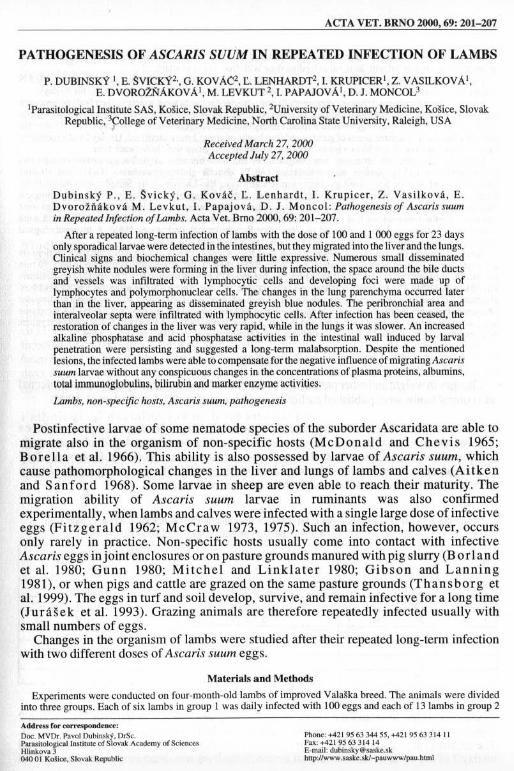
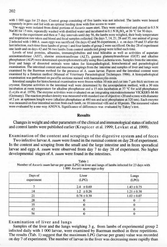
significant changes. Lnfecrion of lambs with 100 and I OOOA. sWIm eggs per day for 23 days neither influenced 10lai proteins which were within the lower range of the standard, nor the levels of albumins and 100a i immunoglobulins. Total bilirubin was standard in all anima1s (Fig. I), but in infected lambs higher than in control. Among enzymes studied (ALT, AST, GGT and ALP), infection influenced only ALP activity. Compared with control, the group infected with 100 eggs exhibited statistically insignificantly lower ALP acti vity (Fig. 2) from day 14to day 42, but a statistically significant decrease in the activity was observed on days 49 and 56 (P < 0.0 1 and P < 0.05, respectively), Administration of I 000 eggs had no effect on ALP activity in lambs.
Patho logica l examin a ti o n of live r and lun gs The liver parenchyma of lambs infected with I 000 eggs and necropsied on days 7 and 14
after infection showed small, inconspicuous greyish white nodules 0. 1-0.2 mm in size. However, on days 2 1 and 28 even more pronounced greyiSh white foci. 2 x 3 mm large, were observed. On day 35 only sporadical almost invisible greyish white foci were recorded. In
8 .... 100 eggs .......
7 ____ 1 000 eggs
1 6 --+- Control group J 5
I j .... .
~ ....
4 .......... .... (; L
.......... ."" E 3 / p "-
2
1 Infection of A suum ~
0 14 28 35 42 4. 56
Days
Fig. I. Total bilirubin in the plasma of lambs iflrecled for 23 days with Asrnris slIlIm eggs
204
6
5
4
" 3 ~
... " • .. ,' 100 eggs
-----.- 1 000 eggs
-+- Control group
"- 2 I ~::::r ...... ........ ...... . ~. . .. ,., ... " ..... ,. ,, " T . • ...........• ..... . ..... ~
1 Infection of A.suum ............. •
0+------,-----,------,-----,------,------. 14 28 35
Days 42 49
Fig. 2. Alkaline phosphatase in the plasma of lambs infected for 2J days with Ascaris $uum eggs
56
most lambs of both experimental groups examined on days 42 and 56 the liver parenchyma was without visible macroscopical changes. only one animal showed the presence of conspicuous narrow bands, 5-\ 0 mm long.
The lungs of lambs infected with 1 ()(X) eggs were without any macroscopical changes on day 7. On day 14, sporadicaJ greyish blue nodules, 0.2-0.5 mm large, were disseminated in the parenchyma. On days 14 and 28, the nodules were more conspicuous, more abundant and disseminated all over the lung parenchyma. From day 35 the nodules were becoming smaller, fewer and less visible, but persisted in both experimental groups for as late as day 42 when they assumed greyish white colour. On day 56, the lungs of one lamb from each experimental group showed the presence of greyish white foci, characteristic of verminous pneumonia.
Hi stopa thologi ca l examination of liver and lung s From day 7, largely lymphocytic cell infiltrates were appearing around the bile ducts in
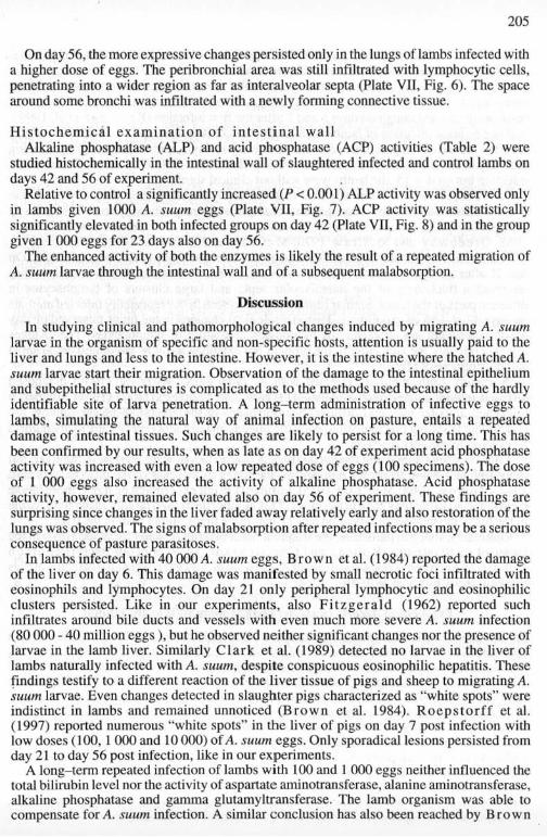
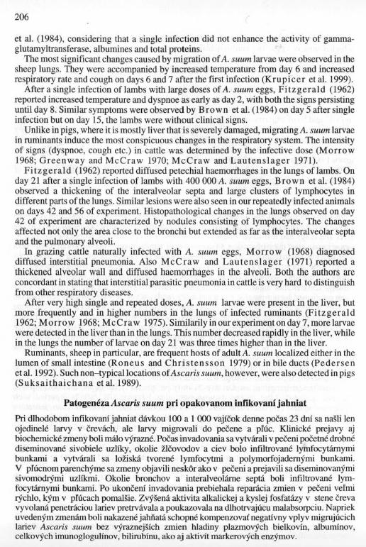
animals given 1 000 eggs of A. .mum. On day 14, the infiltrates also appeared around some vessels. Day 28 showed the presence of nodules around the bile ducts, changing into foci consisting of lymphocytes and polymorphonuclear cells. From day 35, connective tissue started to form around some bile ducts. On day 42, inconspicuous eosinophilic infiltrates were still present around some bile ducts and larger vessels in 60 % of lambs from both experimental groups (Plate VI. Fig. 3). Only in a single animal nodules detected in the liver were composed mainly oflymphocytes. On day 49, the region around some liver vessels and bile ducts was permeated by the forming connective tissue (Plate VI, Fig. 4).
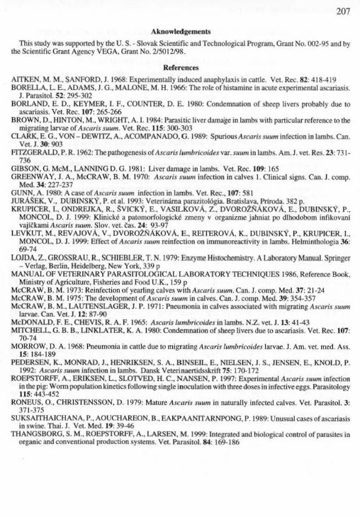
On day 14. the lungs were infiltrated with lymphocytic cells around the bronchi and sporadically also around the interalveolar sepIa. On day 2 1, the peribronchial lymphocytic infiltrates were more conspicuous and sporadical nodules with a cluster of lymphocytes were observed (Pl ate VII. Fig. 5). On day 35, the peribronchiallymphocYlic infiltrate was spreading over a wider area, reaching as for as interalveolar septa. Sporadical clusters of lymphocytes occurred around the bronchi. On day 42 the lung alveoli and bronchi at sites without infiltration and nodules were airy and empty in lambs of both experimental groups. The nodules consisted mostly of lymphocytes, less of polymorphonuclear cells.
205
On day 56, the more expressive changes persistcdonly in the lungs oflambs infected wi th a higher dose of eggs. The peribronchial area was still infiltrated with lymphocytic cells, peneLrating into a wider region as far as interalveolar septa (Plate vn, Fig. 6). The space around some bronchi was infiltrated with a newly fonning conncclive tissue.
Hi stochem ical examination of i n te s tina l wall Alkaline phosphatase (ALP) and acid phosphatase (ACP) activities (Table 2) were
studied histochemically in the intestinal wall of slaughtered infected and control lambs on days 42 and 56 of experiment. .
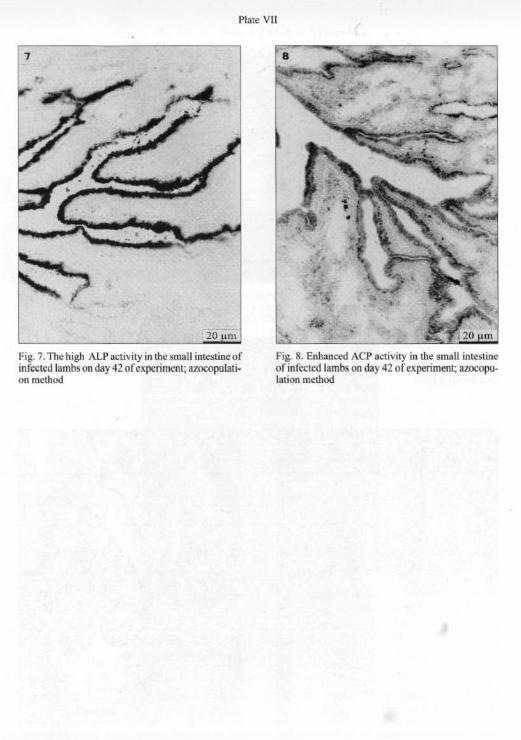
Relative 10 control a significantly increased (P < 0.001) ALP activity was observed only in lambs given 1000 A. suum eggs (Plate vn, Fig. 7), ACP activity was stalistically significan tly elevated in both infected groups on day 42 (Plate VII, Fig. 8) and in the group given I 000 eggs for 23 days also on day 56.
The enhanced activity of both the enzymes is likely the result of a repeated migration of A. slIlIm larvae through {he intestinal wall and of a subsequent malabsorption.
Discussion
In studying clinical and pathomorphological changes induced by migrating A. slIlIm
larvae in the organism of specific and non-specific hosts, attention is usually paid to the liver and lungs and less to the intest ine. However, it is the intesti ne where the hatched A. SlIW1I larvae start their migration. Observatio n of the damage to the intestinal epithelium and subepithelial structures is complicated as to the methods used because of the hardly identifiable site of larva penetration. A l ong~term admin istration of infective eggs to lambs, simulating the natural way of animal infection on pasture, entails a repeated damage of intestinal tissues. Such changes are likely to persist for a long lime. This has been confinned by our results, when as late as on day 42 of experiment acid phosphatase activity was increased with even a low repeated dose of eggs ( 100 specimens). The dose of I 000 eggs also increased the activity of alkaline phosphatase. Acid phosphatase activi ty, however, remained elevated also on day 56 of experiment. These findings are surprising since changes in the liver faded away relatively early and also restoration of the lungs was observed. The signs of malabsorption after repeated infect ions may be a serious consequence of pasture parasi toses.
In lambs infected with 40 000 A. sUllm eggs, Brown et al. (1984) reported the damage of the liver on day 6. This damage was manifested by small necrotic foc i infiltrated with eosinophils and lymphocytes. On day 21 on ly peripheral lymphocytic and eosinophi lic clusters persisted. Like in our experiments. also Fi tzgerald (1962) reported such infi ltrates around bile ducts and vessels with even much more severe A. slIum infection (80000 - 40 million eggs), but he observed neither Significan t changes nor the presence of larvae in the lamb liver. Similarly Clark et al. (1989) detected no larvae in the liver of lambs naturally infec ted wi th A. slIum. despite conspicuous eosinophilic hepatitis. These find ings testify to a different reaction of the liver tissue of pigs and sheep to migrating A. SUUIn larvae. Even changes detected in slaughter pigs characterized as "while spots" were indistinct in lambs and remained unnoticed (Brow n el al. 1984). Roep sto rff et al. ( 1997) reponed numerous "white spots" in the liver of pigs on day 7 post infection with low doses (100. I 000 and 10000) of A. SU UIII eggs. Only sporadical lesions persisted from day 21 to day 56 post infection, like in our experiments.
A long- tenn repeated infect ion of lambs with 100 and I 000 eggs neither influenced the total bilirubin level nor the activity of aspanate aminotransferase. alanine aminotransferase. alkaline phosphatase and gamma glutamyltransferase. The lamb organism was able to compensate for A. SUUIII infection. A similar conclusion has also been reached by Brown
206
ct aI. (1984), considering that a si ngle infection did not enhance the activity of gammaglutamyltransferase, albumines and total prolcins.
The most significanl changes caused by migration of A. sUlllll larvae were observed in the sheep lungs. They were accompanied by increased temperature from day 6 and increased respiratory rate and cough on days 6 and 7 after the first infection (K ru p i cer et al. 1999).
After a single infection of lambs with large doses of A. suum eggs, Fitzgerald (1962) reported increased temperature and dyspnoe as early as day 2, with both the signs persisting until day 8. Similar symptoms were observed by Brown et aI. ( 1984) on day 5 after single infec tion but on day 15, the lambs were without clinical signs.
Unlike in pigs, where it is mostly liver that is severe ly damaged, migrating A. suum larvae in ruminants induce the most conspicuous changes in the respiratory system . The intensity of signs (dyspnoe. cough etc.) in cattle was detennined by the infective dose (M o rrow 1968; Gree nw ay and McCraw 1970; McCraw and Laut e ns lager 197 1).
Fi tzgera ld (1962) reponed d iffused petechial haemorrhages in the lungs of Iambs. On day 2 1 after a single infeclion of lambs with 400 OOOA. Sllum eggs. Brown et al. ( 1984) observed a thickening of the interalveolar septa and large clusters of lymphocytes in d ifferent parts of the lungs. Similar lesions were also seen in our repeatedly infected animals on days 42 and 56 of experiment Histopatho logical changes in the lungs observed on day 42 of experiment are characterized by nodules consisting of lymphocytes. The changes affected not only the area close to the bronchi but extended as far as the interalveolar septa and the pulmonary alveoli.
In grazing cattlc naturally infected with A. SU/l11I eggs, M o rrow (1 968) d iagnosed d iffused intersti tial pneumonia. Also McCraw and La ut e ns lager (197 1) reponed a thickened alveolar wall and diffused haemorrhages in the alveoli. Both the authors arc concordant in stating that interstitial parasitic pneumonia in cattle is very hard to distinguish from other respiratory diseases.
After very high single and repeated doses. A. SUiun larvae were present in the liver. but more frequently and in higher numbers in the lungs of infected ruminants (Fi t zgera ld 1962; M o rro w !968; McC raw 1975). Similarily in our expcriment on day 7, more larvae were detected in the liver than in the lungs. This number decreased rapidly in the liver. while in the lungs the number of larvae on day 2 1 was three times higher than in the liver.
Ruminants, sheep in particular, are frequent hoslS of adult A. suum localized either in the lumen of small intestine (R one us and C hri s te n sso n 1979) or in bi le ducts (Pede rsen et al. 1992). Such non-typical locations of Ascaris S Ill/I1l . however, were also detected in pigs (S uk sa ithaicbana el al. 1989).
Patogeneza Ascaris suum pri opakovanom infikovani jahniat
Pri dlhodobom infikovani jahniat davkou 100 a I 000 vajfcok denne poCas 23 dol sa n~li lcn ojedinele larvy v crevach, ale larvy migrovali do peeene a pfUc. KJinickc prejavy aj biochemicke zmcny boli mAlo vyrw..ne. Pocas invadovania sa vytvarali v !>OCeni pOCetne drobnc discmioovane sivobicle uzliky. okolie t1covodov a ciev bolo infiltrovane Iymfocytamymi bunkami a vytvarali sa lo! iska tvorene lymfocytmi a polymorfojadcmymi bunkami. V prucnom parenchyme sa zmcny objavili neskOr ako v ~eni a prcjavi li sa diseminovanyrni sivomodrymi uzllkmi. Oko lie bronchov a intcralvCQlamc septa boli infiltrovanc lymfocytAmymi bunkami. Po ukonceni invadovania prebiehala reparficia zrnien v ~eni vermi rjchlo. kym v pfucach pomaB:ie. Zvy~na aktivita alkalickej a kyslcj fosfat:izy v stene creva vyvolanfi pcneuticiou lariev prctrvfi.vala a pouk:v.ovala na dLhotrvajucu malabsorpciu . Naprick uvedenym zmenfi.m boli nak:v.ene jaMatfi schopnt kompcnzova( negallvny vplyv migrujucich lanev Ascaris suum bcz vyrazncj~fch zmicn hladiny plazmovych bielkovfn, a1buminov. celkovych imunoglogulinov, bilirublnu, ako aj aktivlt markcrovych enzymov.
207
Akno,,'ledgt mtnts
This study was supported by ttle U. S.· Slovak Scientific and Technological Program, Grant No. 002·95 and by the Sck:ntific Granl ""geoc)' VEGA. Grant NO.1J50 I2J98.
Keferencal
AITKEN, M. M., SANFORD,]. 1968: Blpcrimentally induccdanaphyln is in clllie. Vel. R~, 82: 418419 BORELLA, L. E .. ADAMS, 1. G., MALONE. M. H. 1966: TIle role of histamine in acule experimental ascariasis.
1. Parasitol, 52: 295·302 BORLAND. E. D., K£Y~1ER. I. F .. COUNTER, D. E. 1980: Condemnation of sheep livers probably due 10
ascariasis. Vel. Ree. 107: 265·266 BROWN. D .• HINTON. M .. WRIGHT, A.I. 1984: Parasitic liver damage in lambs with particular rcfcI'I:rM;e 10 the
migrating larvae of Ascaris $lIum. Vet. Re<:. 115: 300-303 CLARK. E. G .. VON - DEWITZ. A., ACOMPANAOO. O. 1989: SpurioosAscaris swum infection in lambs. Can.
Vet. 1. 30: 903 FJTZ.GERALD. P. R. 1962: The pathogenesis of Ascarislwnbricoidu var,s""", in lambs. Am. J. vel. Res. 23: 731-
736 GI.BSON. G. MeM., LANNING D.G. 1981 : Uverdamage in lambs. Vet. Rec. 109: 165 GREENWAY. J. A., McCRAW. B. M. 1970: Ascaris SUrtm illfectioll in calves l. Clinical signs. Can. J. compo
Me<!. 3-1: 227·237 GUNN, A. 1980: A cascof Ascaris slIlIm infection in lambs. Vel. Rce .. 107: 581 JURASEK, V., DUBINSK'i' , P. e( a1. 1993: VeterinMna parazitolOgia. 8rati~lava. Prfroda. 382 p. KRUPICER,I., ONDREJKA, R .. SVICK~, E .. VASILKovA. z.. DVOROlNAKovA. E.. DUBINSKY, P.,
MONCOL. D. J. 1999; KHnickt a patomorfologk:kt 7Jlleny v organizme jahnial po dlilodobom infikovanl vajr~k.ami Ascaris luum. Slov. VCI. ~as. 14: 93-97
LEVKUT. M .. REVAJovA. V .. DVOR02.~AKovA. E., RElTERovA, K" DUBINSKY. P .. KRUPICER. I .. MONCOL. D. J. 1999: Effect of Ascaris IUum reinfectioll 011 immunoreactivity in lambs. Helminthologia 36: 69·14
LOJDA, z..GROSSRAU. R., SCHlEBLER, T, N. 1979: Enzyme Histochemistry. A Labor.ItOl)' Manual.Springer - Verlag. Berlin, Heidelberg, New York, 339 p
MANUAL OF VETERINARY PARASlTOLOCilCAL LABORATORY TECHNIQUES 1986. Reference Book. Ministry of Agriculture. Fisheries and Food U. K .. 159 P
McCRAW. B. M. 1913: Reinfection of yearling calves with Ascaris suum. Can. J. compo Mod. 37: 21·24 McCRAW. B. M. 1975: The development of Ascaris SIIIW/ in calves. Can. J. comp. Mod. 39: 354-357 McCRAW. B. M .. LAUTENSLAGER, J. P. 1971 : Pneumonia in calves associated with migrating Ascaris suum
larvae. Can. Ve(. l . 12: 87-90 McDONALD. F. E .. CHEVIS. R. A. F. 1965: ...uc-arislumbricoUks in lambs. N.Z. vel. J. 1]:41-4) MITCHELL. G. B. B .. LlNKLATER. K, A. 1980: Condemnation of sheep liven due 10 ascariasis. Vet. Re<: . 107:
70-74 MORROW. D. A. 1968: Pneumonia in canle due to migrating Ascari.f Iumbricoides larvae. 1. Am. vel. med. Ass.
15: 184· 189 PEDERSEN. K., MONRAD, J .. HENRIKSEN. S. A .. BINSEIL, E, NIELSEN, J. S., JENSEN, e., KNOLD, P.
1992: Ibalris suum infoction in lambs. Dansk Vetetinaenidsskrifi 15: 170- 112 ROEPSTORFF, A., ERIKSEN. L. SLO'IVED, H. C .• NANSEN, P. 1991: Experimental A.KarisJuum infection
in the pig: Worm population kinetics following single inoculation with three do.se5 in infecli\'C eggs. Parasitology 115: 44)-452
RONEUS. 0., CHRISTENSSON. D. 1979: Mature Ascaru SUU'" in naturally infected calves. Vel. ParasitoL 3: )71·)75
SUKSAITHAICHANA, P., AOUCHAREON. 8 .. EAKPAANITARNPONG. P. 1989: Unusual CasesOfa.scarillliis in swine. 1bai. J. Vet. Med. 19:39-46
THANGSBORG.S. M., ROEPSTORFF.A., LARSEN. M. 1999: Integrated and biological control ofparuite5 in organic and conventional production systems. Vet. Parasitol. 84: 169-186
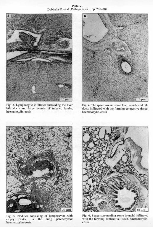
Plate VI Dubinsky P. el a1. : Pathogenesis ... pp. 20 1-207
Fig. 3. Lymphocytic i nfi ltr~les surrouding the liver bile ducls and large l'c!ISCls of infcctcd lambs. haemaloll'l lin-e<>sin
Fig. 5, Nodules consisting cmply center, in the hacmatollylin.cosin
of lymphocytes wi th lung parcnchyma;
!'ig. 4. The space around some liver vesse ls and bile d UCIS infillr.lted with Ihe fonning connectivc tissuc; hacmatOll'llin-cosin
Fig. 6. Spacc surrounding $Orne bronchi infiltrated with thc fonnin g connccti,'c tissuc, hacmatoxylineosin
Plale VII
1
Fig. 7. The high Al.P acth'ity in the small intestine of infetted lambs on day 42 ofc:xpcrimcnt; azocopulati · OIl method
fig. 8. Enhanced ACP act ivity in the small intestine ofinfcctcd lambs on day 42 of experiment: 1ZOOOp1llal ion method