radar · 6 departmentof physiotherapy, paulista stateuniversity (unesp), presidenteprudente,...
TRANSCRIPT

RADAR Research Archive and Digital Asset Repository Alvarez, MPB., Silva, TDd., Favero, FM., Valenti, VE., Raimundo, RD., Vanderlei, LCM., Garner, DM. and Monteiro, CBM. (2017) 'Autonomic Modulation in Duchenne Muscular Dystrophy during a Computer Task: A Prospective Control Trial', PLoS ONE, 12 (1):e0169633 DOI: https://doi.org/10.1371/journal.pone.0169633 This document is the Version of Record. License: https://creativecommons.org/licenses/by/4.0 Available from RADAR: https://radar.brookes.ac.uk/radar/items/43fc5b2a-3a87-4020-9774-26655d92019a/1/ Copyright © and Moral Rights are retained by the author(s) and/ or other copyright owners unless otherwise waved in a license stated or linked to above. A copy can be downloaded for personal non-commercial research or study, without prior permission or charge. This item cannot be reproduced or quoted extensively from without first obtaining permission in writing from the copyright holder(s). The content must not be changed in any way or sold commercially in any format or medium without the formal permission of the copyright holders.
RESEARCH ARTICLE
Autonomic Modulation in Duchenne Muscular
Dystrophy during a Computer Task: A
Prospective Control Trial
Mayra Priscila Boscolo Alvarez1, Talita Dias da Silva2, Francis Meire Favero3*, Vitor
Engracia Valenti4, Rodrigo Daminello Raimundo5, Luiz Carlos Marques Vanderlei6, David
M. Garner7, Carlos Bandeira de Mello Monteiro1,8
1 Physical Therapy, Speech and Occupational Therapy Department, School of Medicine, University of São
Paulo, São Paulo, SP, Brazil, 2 Federal University of São Paulo, Paulista School of Medicine, São Paulo, SP,
Brazil, 3 Federal University of São Paulo, Department of Neurology/Neurosurgery, Paulista School of
Medicine, São Paulo, SP, Brazil, 4 Autonomic Nervous System Center Study, Speech Therapy Department
Faculty of Sciences, Paulista State University (UNESP), Marılia, SP, Brazil, 5 Laboratory Design and
Scientific Writing, Department of Community Health, ABC Medical School, Santo Andre, SP, Brazil,
6 Department of Physiotherapy, Paulista State University (UNESP), Presidente Prudente, São Paulo, SP,
Brazil, 7 Cardiorespiratory Research Group, Department of Biological and Medical Sciences, Faculty of
Health and Life Sciences, Oxford Brookes University, Gipsy Lane, Oxford OX3 0BP, United Kingdom,
8 School of Arts, Sciences and Humanities, University of São Paulo, São Paulo, SP, Brazil
Abstract
Introduction
Duchenne Muscular Dystrophy (DMD) is characterized by progressive muscle weakness
that can lead to disability. Owing to functional difficulties faced by individuals with DMD, the
use of assistive technology is essential to provide or facilitate functional abilities. In DMD,
cardiac autonomic dysfunction has been reported in addition to musculoskeletal impairment.
Consequently, the objective was to investigate acute cardiac autonomic responses, by
Heart Rate Variability (HRV), during computer tasks in subjects with DMD.
Method
HRV was assessed by linear and nonlinear methods, using the heart rate monitor Polar
RS800CX chest strap Electrocardiographic measuring device. Then, 45 subjects were
included in the group with DMD and 45 in the healthy Typical Development (TD) control
group. They were assessed for twenty minutes at rest sitting, and five minutes after under-
going a task on the computer.
Results
Individuals with DMD had a statistically significant lower parasympathetic cardiac modula-
tion at rest when compared to the control group, which further declined when undergoing
the tasks on the computer.
PLOS ONE | DOI:10.1371/journal.pone.0169633 January 24, 2017 1 / 14
a1111111111
a1111111111
a1111111111
a1111111111
a1111111111
OPENACCESS
Citation: Alvarez MPB, Silva TDd, Favero FM,
Valenti VE, Raimundo RD, Vanderlei LCM, et al.
(2017) Autonomic Modulation in Duchenne
Muscular Dystrophy during a Computer Task: A
Prospective Control Trial. PLoS ONE 12(1):
e0169633. doi:10.1371/journal.pone.0169633
Editor: Carlos E. Ambrosio, Faculty of Animal
Sciences and Food Engineering, University of São
Paulo, BRAZIL
Received: March 8, 2016
Accepted: December 20, 2016
Published: January 24, 2017
Copyright: © 2017 Alvarez et al. This is an open
access article distributed under the terms of the
Creative Commons Attribution License, which
permits unrestricted use, distribution, and
reproduction in any medium, provided the original
author and source are credited.
Data Availability Statement: All relevant data are
within the paper and its Supporting Information
file.
Funding: The authors received financial support
from the FAPESP (Fundacão de Amparo àPesquisa do Estado de São Paulo, process number
2012/16970-6).
Competing Interests: The authors have declared
that no competing interests exist.
Conclusion
DMD patients presented decreased HRV and exhibited greater intensity of cardiac auto-
nomic responses during computer tasks characterized by vagal withdrawal when compared
to the healthy TD control subjects.
Introduction
Muscular dystrophies consist of a group of genetic disorders characterized by muscle weakness
and atrophy [1, 2], particularly of early onset and of a progressive nature [3, 4].
Amongst all types of the muscular dystrophies, Duchenne Muscular Dystrophy (DMD) is
considered the most widespread [5], with recessive genetic inheritance [3], and affecting
approximately 1:3500 male births [6]. DMD occurs by a mutation of the gene encoding the
dystrophin enzyme which is located on the short arm of the X chromosome [7] in the Xp21
region [1, 8]. DMD is categorized by the progressive loss of movement, which initially affects
the lower limbs and then the upper limbs, with pseudo-hypertrophy of the affected muscles,
interstitial increase of connective tissue and in the advanced stages significant increase of fat
tissue in the muscles [9, 10].
On account of the functional difficulties presented by individuals with DMD; to enable the
capability in social activities and performance; the practice of assistive technology or resources
are needed. They achieve functional abilities of individuals with disabilities and thus promote
greater independence and social inclusion [11]. According to Neistadt and Crepeau [12], assis-
tive technology can be defined as any item or product, equipped for use, adapted or custom-
ized, that maintains or improves functional capabilities of individuals with a disability.
Recently the advances in computational assistive technology and the provision of rehabilita-
tion programs using computer equipment during treatment allow the patient with DMD to
undertake tasks in challenging situations by means of simple technology and achieving rapid
responses. Additionally, it is possible to provide interactions with targets, through logical cog-
nition and different reaction times associated with movement, allowing the repetition of mus-
cle contractions and enhancing performance [13–20].
Moreover, the researched deficiencies in the musculoskeletal system [5] and cardiac auto-
nomic dysfunction have been previously well researched in DMD [21, 22].
Thus, amongst the techniques applied to analyze the ANS, Heart Rate Variability (HRV)
has emerged as a simple, reliable, inexpensive and non-invasive measure of the autonomic
impulses. It represents one of the most promising quantitative markers of autonomic balance
[23].
The wide use and cost-effectiveness of the technique and ease of data acquisition make
HRV a capable choice for the interpretation of ANS functioning and a promising clinical tool
to assess and identify physiological deficiencies [23]. Fluctuations in HRV patterns provide an
early and sensitive diagnosis of the physiological behavior of the human body and health status
of the individual [24].
Likewise, Thomas et al. [21] studied heart rate autonomic dysfunction in DMD. These
authors assessed HRV in DMD and in a healthy control group by 24-hour Holter monitoring.
They found that the control group demonstrated a higher maximum heart rate on Holter
monitoring than in DMD patients. Dittrich et al. [22] assessed cardiac autonomic regulation
in DMD, and the analytical value of the diagnostic procedures in clinical settings. Both of the
abovementioned studies investigated the cardiac autonomic dysfunction in DMD.
Autonomic Modulation in DMD during a Computer Task: A Prospective Control Trial
PLOS ONE | DOI:10.1371/journal.pone.0169633 January 24, 2017 2 / 14

Consequently, the autonomic impairment is recognized for patients with DMD at rest. How-
ever, the effect of the computational task is not fully understood. At present, computer tasks
are required for the groups independence and has been widely adopted in persons with DMD.
It is especially important when assessing the influences on the autonomic system.
Whilst recent literature has revealed lesser HRV in DMD [21, 22], the specific cardiac auto-
nomic response of this population is unclear when undergoing stimulation through computer
tasks. There is little research on virtual technology procedures in DMD. Despite the use of
computers in rehabilitation, we did not find any published research on the physiological
changes that these tasks cause in individuals with DMD. To enable an understanding of these
problems, this study evaluated these physiological adaptations by assessing the Autonomic
Nervous System (ANS). Therefore, we aimed to investigate acute cardiac autonomic responses
during computer tasks in individuals with DMD versus healthy people. If the HRV responses
of DMD patients during computer tasks are improved comparing with the responses at rest, it
provides new pathways for research using computational tasks that may be therapeutic by
improving autonomic dysfunction in this group of subjects.
Materials and Methods
Participants
This is a prospective controlled trial. In this study were 90 age matched male subjects divided
into equal groups with diagnosis of DMD and those healthy Typically Developed (TD) indi-
viduals without DMD. All individuals diagnosed with DMD were confirmed by molecular
methods and/or protein expression in skeletal muscle.
Subjects were excluded with severely dilated myocardium, other associated diseases and
individuals with changes in cognitive functions that would impede the simple cognition of
commands in the proposed activities.
The research project (number 236/13) was approved by the research ethics committee of
the University of São Paulo and undertaken after the signing of the Terms of Free and
Informed Consent by the participants or legal guardian. Research participants aged 17 years or
younger also submitted the research consent form.
To achieve the characterization of individuals with DMD, the Vignos scale was enforced
[25]. This characterizes the disease severity according to pathological progression. They were
classified from patient at stage 1 (walk and climb stairs without assistance) to 10 (permanently
confined to bed).
Data collection instruments
Data collection forms from the medical records were completed in individuals with DMD. It
was used to obtain relevant information regarding patients’ care, such as associated diseases
and usage of medications.
A Premium Aneroid Sphygmomanometer (Model S82, Prestige Medical, Northridge, Cali-
fornia, USA) and a BIC stethoscope (CBEMED, Itupeva, Brazil) were required to undertake
systolic blood pressure (SBP) and diastolic blood pressure (DBP) measurements. Starting and
final heart rate (HR) was verified by the investigator through their radial pulse and beats calcu-
lated for one minute.
HRV was recorded using the Polar RS800CX chest strap ECG measuring device (Polar
Electro Oy, Kempele, Finland) previously validated to capture beat-to-beat HR (RR intervals),
that represents the interval between each beat) [24].
Autonomic Modulation in DMD during a Computer Task: A Prospective Control Trial
PLOS ONE | DOI:10.1371/journal.pone.0169633 January 24, 2017 3 / 14
Collection procedures
SBP, DBP and HR measurements were assessed and recorded following the first minute of sit-
ting and at the conclusion of twenty minutes of rest; then, before the start and at the end of the
five minutes of the computer task.
Following the initial assessment, the capture strap was placed on the chest of volunteers and
the HR receptor was placed on the wrist.
After strap placement and the computer screen startup, the individuals from both groups
remained at rest and sitting in a chair (walkers, TD- and DMD-group) or in their own wheel-
chair (non-walkers, DMD-group), with spontaneous breathing for twenty minutes. Following
this period, the computer screen was restarted, and the individuals remained seated with a
notebook computer to enable them to perform a maze task on the computer for five minutes.
HRV was analyzed during two time periods: the period before (20 minutes) and then dur-
ing the cognitive computer task (5 minutes).
The computer task undertaken was a maze paradigm used for its cognitive requirements
with ease and adaptability for use in individuals with DMD. To complete the tasks, the research-
ers selected a computer program developed by the Department of Mathematics of the Federal
University of Rio Grande do Sul, presented by Souza et al. [26]. The task comprised of different
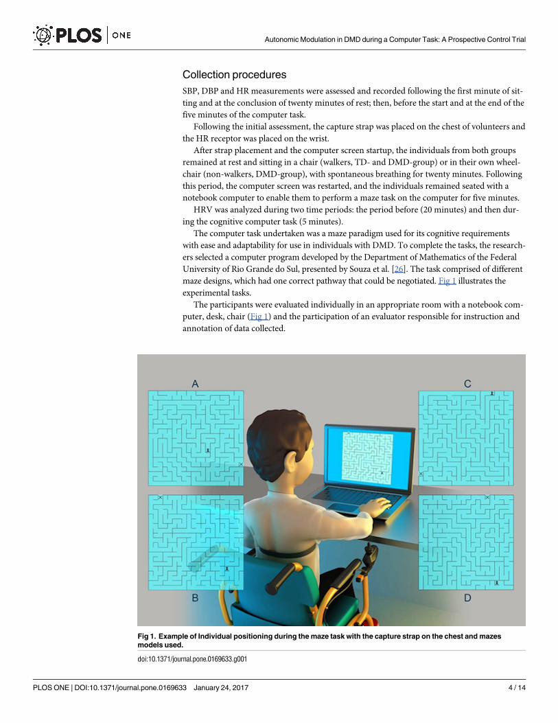
maze designs, which had one correct pathway that could be negotiated. Fig 1 illustrates the
experimental tasks.
The participants were evaluated individually in an appropriate room with a notebook com-
puter, desk, chair (Fig 1) and the participation of an evaluator responsible for instruction and
annotation of data collected.
Fig 1. Example of Individual positioning during the maze task with the capture strap on the chest and mazes
models used.
doi:10.1371/journal.pone.0169633.g001
Autonomic Modulation in DMD during a Computer Task: A Prospective Control Trial
PLOS ONE | DOI:10.1371/journal.pone.0169633 January 24, 2017 4 / 14
Each individual was well positioned and the task was elucidated concurrently with the pre-
sentation of the maze, along which the individual should devise the path with the digital chess
piece (pawn) character (pointed to, on the screen, by the evaluator) until the exit of the maze
identified by an "x" (pointed to, on the screen, by the evaluator). The individual was instructed
to perform the task as quickly as possible using the arrow buttons on the keyboard identified
by arrows up, down, right and left.
A 20-by-20 cm maze was presented while the individuals undertook the task as many times
as necessary to remain active at the computer for five minutes.
Data analysis
The dependent variables (HRV indexes) were submitted to a 2 (group: DMD, TD) by 2
(Task: Rest, Computer) ANOVA with repeated measures on the last factor for each HRV
index. Post-hoc comparisons were undertaken by Tukey-HSD (Honest Significant Differ-
ence) test (p < 0.05). For the physiologically independent variables such as Systolic Blood
Pressure, Diastolic Blood Pressure and Heart Rate, the comparisons were made by using
Student t test for unpaired data. The software package operated was SPSS, 20.0 (Chicago,
Illinois, USA).
HRV analysis
HRV analysis was completed under guidelines from the Task Force of the European Society of
Cardiology and North American Society of Pacing and Electrophysiology [27]. The RR inter-
vals were recorded using the portable Polar RS800CX heart rate (HR) monitor (Polar Electro,
Finland) with a sampling rate of 1 kHz. They were downloaded to the Polar Precision Perfor-
mance program (v.3.0). The software enabled the visualization of HR and the extraction of a
cardiac period (RR interval; the variation of beat-to-beat separations) file in “txt” format. For
analysis of HRV data in the sitting position, we analyzed 1000 consecutive RR intervals, and
for HRV analysis for the computational task, the greatest number of consecutive RR intervals
obtained was used, but with a minimum number of 256 RR intervals. Digital filtering comple-
mented by manual filtering was performed to eliminate artifacts and only series with more
than 95% of sinus beats were included in the study [28].
HRV analysis was assessed by linear methods, in the time (Dt) and frequency (Df) domains,
and then by nonlinear methods (Poincare plot). We chose to use linear and nonlinear meth-
ods, considering that both are shown to be complementary to each other, providing additional
information [29].
Linear methods
Time domain. In the Time domain Dt, the time interval between successive heart beat
intervals, was determined by statistical and geometric methods [27].
The necessary statistical methods to assess ANS were SDNN (index of standard deviation of
all normal-to-normal RR intervals), rMSSD (root mean square of successive differences between
adjacent normal RR intervals), pNN50 (percentage of adjacent RR intervals with a difference
longer than 50 milliseconds) [27].
For geometrical methods we enforced RR Tri (Total number of all NN intervals divided by
the height of the histogram of all NN intervals) and TINN (baseline width of the minimum
square difference triangular interpolation of the highest peak of the histogram of all NN inter-
vals) [27].
Frequency Domain. For HRV analysis in Frequency domain, Df, low frequency (LF) and
high frequency (HF) spectral components were appointed in absolute values of power (ms2) or
Autonomic Modulation in DMD during a Computer Task: A Prospective Control Trial
PLOS ONE | DOI:10.1371/journal.pone.0169633 January 24, 2017 5 / 14

in normalized units (n.u.). The ratio between these components in absolute values (LF/HF)
represents the relative value of each spectral component in relation to the total potential minus
the very low frequency (VLF) components [27].
The index Total (or Total Power) is the variance of NN intervals over the approximate tem-
poral segment [27].
Nonlinear methods
For HRV analysis by nonlinear methods, we applied the Poincare plot (SD1 components—
standard deviation of instantaneous beat-to-beat variability, SD2—standard deviation of long-
term continuous RR intervals and relation SD1/SD2) [27].
The Poincare plot enables each RR interval to be plotted against the next interval. For quan-
titative analysis of the plot, the indices for SD1, SD2 and the relation SD1/SD2 were calculated
[30]. According to Hoshi et al., [31] “the Poincare plot for heart rate variability analysis is a
technique considered geometrical and non-linear, that can be used to assess the dynamics of
heart rate variability by a representation of the values of each pair of R–R intervals into a sim-
plified phase space that describes the system’s evolution.”
For more information regarding the HRV indexes, see S1 File in the Supporting
Information.
Results
The age, anthropometric variables and medications taken by DMD group are stated in
Table 1.
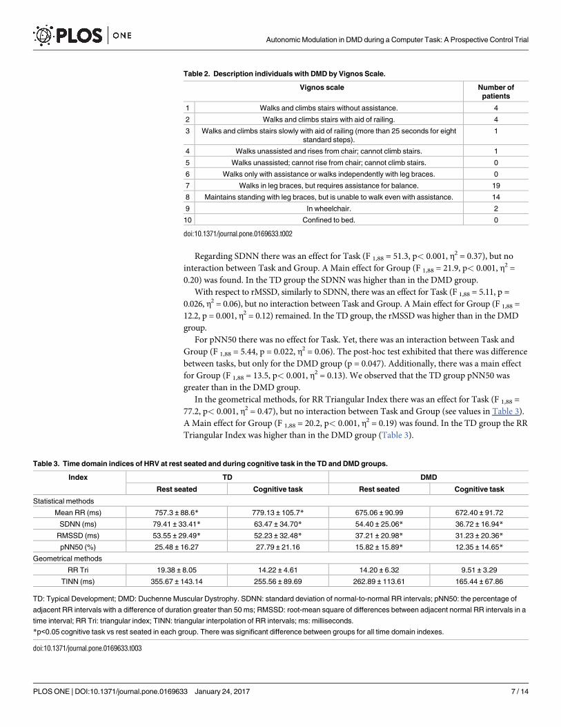
Individuals with DMD were classified by Vignos Scale, as described in Table 2.
Heart rate variability
Time domain. For statistical methods, concerning the mean RR interval there was no
effect of the computer Task. However, there was an interaction between Task and Group
(F 1,88 = 4.44, p = 0.038, η2 = 0.05). The post-hoc test illustrated that there was difference
between tasks just for TD group (p = 0.009) (Table 3). There was a main effect for Group
(F 1,88 = 24.5, p< 0.001, η2 = 0.22). This implied that the TD group mean RR interval was
higher than in the DMD group.
Table 1. Age, anthropometric variables within the groups (by mean ± standard deviation) and the cardiac medication for DMD-group.
Variable TD-group DMD-group p
Age (years) 15.4 ± 2.8 15.4 ± 2.9 0.455
Height (m) 1.68 ± 0.12 1.56 ± 0.17 <0.001
Mass (kg) 63.2 ± 15.5 55.84 ± 17.9 0.013
BMI (kg/m2) 20.04 ± 3.72 22.42 ± 4.71 0.331
Medication on DMD-group Number of patients (%)
Beta-blockers 13 (28.89)
ACE-inhibitor 5 (11.11)
Beta-blockers + ACE-inhibitors 20 (44.44)
No medication 7 (15.56)
TD: Typical Development; DMD: Duchenne Muscular Dystrophy; BMI: body mass index; m: meters; kg: kilograms; kg/m2: kilograms per square meter;
ACE-inhibitors: angiotensin-converting enzyme inhibitors.
doi:10.1371/journal.pone.0169633.t001
Autonomic Modulation in DMD during a Computer Task: A Prospective Control Trial
PLOS ONE | DOI:10.1371/journal.pone.0169633 January 24, 2017 6 / 14
Regarding SDNN there was an effect for Task (F 1,88 = 51.3, p< 0.001, η2 = 0.37), but no
interaction between Task and Group. A Main effect for Group (F 1,88 = 21.9, p< 0.001, η2 =
0.20) was found. In the TD group the SDNN was higher than in the DMD group.
With respect to rMSSD, similarly to SDNN, there was an effect for Task (F 1,88 = 5.11, p =
0.026, η2 = 0.06), but no interaction between Task and Group. A Main effect for Group (F 1,88 =
12.2, p = 0.001, η2 = 0.12) remained. In the TD group, the rMSSD was higher than in the DMD
group.
For pNN50 there was no effect for Task. Yet, there was an interaction between Task and
Group (F 1,88 = 5.44, p = 0.022, η2 = 0.06). The post-hoc test exhibited that there was difference
between tasks, but only for the DMD group (p = 0.047). Additionally, there was a main effect
for Group (F 1,88 = 13.5, p< 0.001, η2 = 0.13). We observed that the TD group pNN50 was
greater than in the DMD group.
In the geometrical methods, for RR Triangular Index there was an effect for Task (F 1,88 =
77.2, p< 0.001, η2 = 0.47), but no interaction between Task and Group (see values in Table 3).
A Main effect for Group (F 1,88 = 20.2, p< 0.001, η2 = 0.19) was found. In the TD group the RR
Triangular Index was higher than in the DMD group (Table 3).
Table 2. Description individuals with DMD by Vignos Scale.
Vignos scale Number of
patients
1 Walks and climbs stairs without assistance. 4
2 Walks and climbs stairs with aid of railing. 4
3 Walks and climbs stairs slowly with aid of railing (more than 25 seconds for eight
standard steps).
1
4 Walks unassisted and rises from chair; cannot climb stairs. 1
5 Walks unassisted; cannot rise from chair; cannot climb stairs. 0
6 Walks only with assistance or walks independently with leg braces. 0
7 Walks in leg braces, but requires assistance for balance. 19
8 Maintains standing with leg braces, but is unable to walk even with assistance. 14
9 In wheelchair. 2
10 Confined to bed. 0
doi:10.1371/journal.pone.0169633.t002
Table 3. Time domain indices of HRV at rest seated and during cognitive task in the TD and DMD groups.
Index TD DMD
Rest seated Cognitive task Rest seated Cognitive task
Statistical methods
Mean RR (ms) 757.3 ± 88.6* 779.13 ± 105.7* 675.06 ± 90.99 672.40 ± 91.72
SDNN (ms) 79.41 ± 33.41* 63.47 ± 34.70* 54.40 ± 25.06* 36.72 ± 16.94*
RMSSD (ms) 53.55 ± 29.49* 52.23 ± 32.48* 37.21 ± 20.98* 31.23 ± 20.36*
pNN50 (%) 25.48 ± 16.27 27.79 ± 21.16 15.82 ± 15.89* 12.35 ± 14.65*
Geometrical methods
RR Tri 19.38 ± 8.05 14.22 ± 4.61 14.20 ± 6.32 9.51 ± 3.29
TINN (ms) 355.67 ± 143.14 255.56 ± 89.69 262.89 ± 113.61 165.44 ± 67.86
TD: Typical Development; DMD: Duchenne Muscular Dystrophy. SDNN: standard deviation of normal-to-normal RR intervals; pNN50: the percentage of
adjacent RR intervals with a difference of duration greater than 50 ms; RMSSD: root-mean square of differences between adjacent normal RR intervals in a
time interval; RR Tri: triangular index; TINN: triangular interpolation of RR intervals; ms: milliseconds.
*p<0.05 cognitive task vs rest seated in each group. There was significant difference between groups for all time domain indexes.
doi:10.1371/journal.pone.0169633.t003
Autonomic Modulation in DMD during a Computer Task: A Prospective Control Trial
PLOS ONE | DOI:10.1371/journal.pone.0169633 January 24, 2017 7 / 14
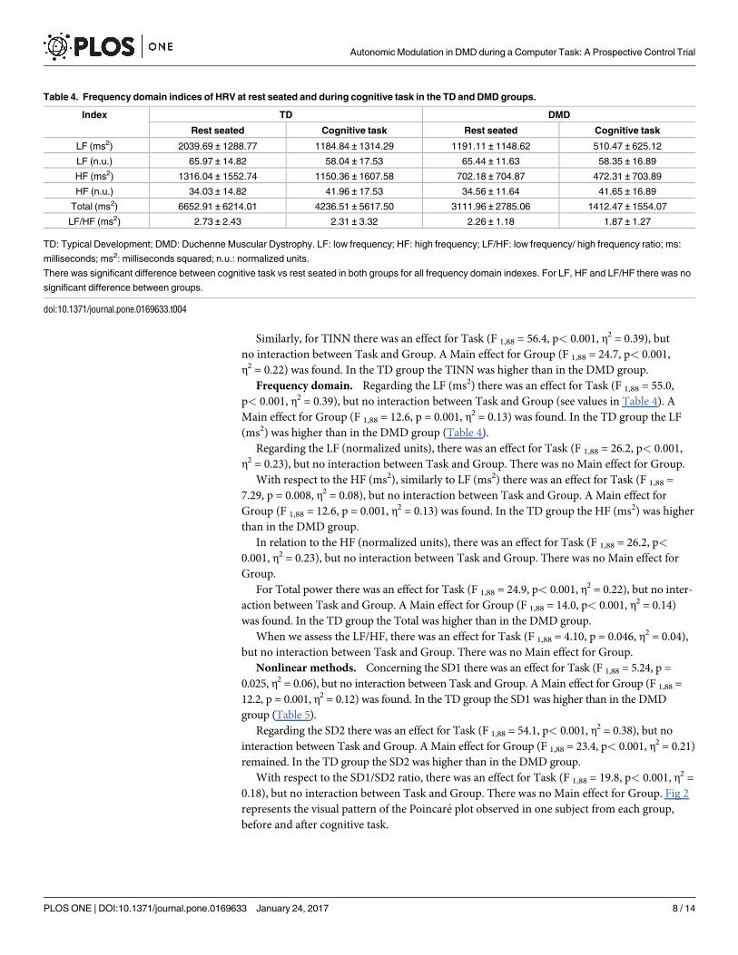
Similarly, for TINN there was an effect for Task (F 1,88 = 56.4, p< 0.001, η2 = 0.39), but
no interaction between Task and Group. A Main effect for Group (F 1,88 = 24.7, p< 0.001,
η2 = 0.22) was found. In the TD group the TINN was higher than in the DMD group.
Frequency domain. Regarding the LF (ms2) there was an effect for Task (F 1,88 = 55.0,
p< 0.001, η2 = 0.39), but no interaction between Task and Group (see values in Table 4). A
Main effect for Group (F 1,88 = 12.6, p = 0.001, η2 = 0.13) was found. In the TD group the LF
(ms2) was higher than in the DMD group (Table 4).
Regarding the LF (normalized units), there was an effect for Task (F 1,88 = 26.2, p< 0.001,
η2 = 0.23), but no interaction between Task and Group. There was no Main effect for Group.
With respect to the HF (ms2), similarly to LF (ms2) there was an effect for Task (F 1,88 =
7.29, p = 0.008, η2 = 0.08), but no interaction between Task and Group. A Main effect for
Group (F 1,88 = 12.6, p = 0.001, η2 = 0.13) was found. In the TD group the HF (ms2) was higher
than in the DMD group.
In relation to the HF (normalized units), there was an effect for Task (F 1,88 = 26.2, p<
0.001, η2 = 0.23), but no interaction between Task and Group. There was no Main effect for
Group.
For Total power there was an effect for Task (F 1,88 = 24.9, p< 0.001, η2 = 0.22), but no inter-
action between Task and Group. A Main effect for Group (F 1,88 = 14.0, p< 0.001, η2 = 0.14)
was found. In the TD group the Total was higher than in the DMD group.
When we assess the LF/HF, there was an effect for Task (F 1,88 = 4.10, p = 0.046, η2 = 0.04),
but no interaction between Task and Group. There was no Main effect for Group.
Nonlinear methods. Concerning the SD1 there was an effect for Task (F 1,88 = 5.24, p =
0.025, η2 = 0.06), but no interaction between Task and Group. A Main effect for Group (F 1,88 =
12.2, p = 0.001, η2 = 0.12) was found. In the TD group the SD1 was higher than in the DMD
group (Table 5).
Regarding the SD2 there was an effect for Task (F 1,88 = 54.1, p< 0.001, η2 = 0.38), but no
interaction between Task and Group. A Main effect for Group (F 1,88 = 23.4, p< 0.001, η2 = 0.21)
remained. In the TD group the SD2 was higher than in the DMD group.
With respect to the SD1/SD2 ratio, there was an effect for Task (F 1,88 = 19.8, p< 0.001, η2 =
0.18), but no interaction between Task and Group. There was no Main effect for Group. Fig 2
represents the visual pattern of the Poincare plot observed in one subject from each group,
before and after cognitive task.
Table 4. Frequency domain indices of HRV at rest seated and during cognitive task in the TD and DMD groups.
Index TD DMD
Rest seated Cognitive task Rest seated Cognitive task
LF (ms2) 2039.69 ± 1288.77 1184.84 ± 1314.29 1191.11 ± 1148.62 510.47 ± 625.12
LF (n.u.) 65.97 ± 14.82 58.04 ± 17.53 65.44 ± 11.63 58.35 ± 16.89
HF (ms2) 1316.04 ± 1552.74 1150.36 ± 1607.58 702.18 ± 704.87 472.31 ± 703.89
HF (n.u.) 34.03 ± 14.82 41.96 ± 17.53 34.56 ± 11.64 41.65 ± 16.89
Total (ms2) 6652.91 ± 6214.01 4236.51 ± 5617.50 3111.96 ± 2785.06 1412.47 ± 1554.07
LF/HF (ms2) 2.73 ± 2.43 2.31 ± 3.32 2.26 ± 1.18 1.87 ± 1.27
TD: Typical Development; DMD: Duchenne Muscular Dystrophy. LF: low frequency; HF: high frequency; LF/HF: low frequency/ high frequency ratio; ms:
milliseconds; ms2: milliseconds squared; n.u.: normalized units.
There was significant difference between cognitive task vs rest seated in both groups for all frequency domain indexes. For LF, HF and LF/HF there was no
significant difference between groups.
doi:10.1371/journal.pone.0169633.t004
Autonomic Modulation in DMD during a Computer Task: A Prospective Control Trial
PLOS ONE | DOI:10.1371/journal.pone.0169633 January 24, 2017 8 / 14
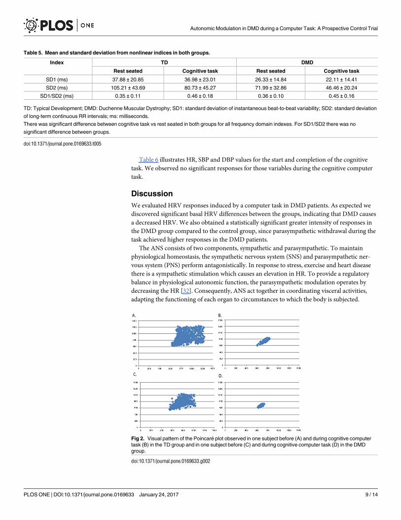
Table 6 illustrates HR, SBP and DBP values for the start and completion of the cognitive
task. We observed no significant responses for those variables during the cognitive computer
task.
Discussion
We evaluated HRV responses induced by a computer task in DMD patients. As expected we
discovered significant basal HRV differences between the groups, indicating that DMD causes
a decreased HRV. We also obtained a statistically significant greater intensity of responses in
the DMD group compared to the control group, since parasympathetic withdrawal during the
task achieved higher responses in the DMD patients.
The ANS consists of two components, sympathetic and parasympathetic. To maintain
physiological homeostasis, the sympathetic nervous system (SNS) and parasympathetic ner-
vous system (PNS) perform antagonistically. In response to stress, exercise and heart disease
there is a sympathetic stimulation which causes an elevation in HR. To provide a regulatory
balance in physiological autonomic function, the parasympathetic modulation operates by
decreasing the HR [32]. Consequently, ANS act together in coordinating visceral activities,
adapting the functioning of each organ to circumstances to which the body is subjected.
Table 5. Mean and standard deviation from nonlinear indices in both groups.
Index TD DMD
Rest seated Cognitive task Rest seated Cognitive task
SD1 (ms) 37.88 ± 20.85 36.98 ± 23.01 26.33 ± 14.84 22.11 ± 14.41
SD2 (ms) 105.21 ± 43.69 80.73 ± 45.27 71.99 ± 32.86 46.46 ± 20.24
SD1/SD2 (ms) 0.35 ± 0.11 0.46 ± 0.18 0.36 ± 0.10 0.45 ± 0.16
TD: Typical Development; DMD: Duchenne Muscular Dystrophy; SD1: standard deviation of instantaneous beat-to-beat variability; SD2: standard deviation
of long-term continuous RR intervals; ms: milliseconds.
There was significant difference between cognitive task vs rest seated in both groups for all frequency domain indexes. For SD1/SD2 there was no
significant difference between groups.
doi:10.1371/journal.pone.0169633.t005
Fig 2. Visual pattern of the Poincare plot observed in one subject before (A) and during cognitive computer
task (B) in the TD group and in one subject before (C) and during cognitive computer task (D) in the DMD
group.
doi:10.1371/journal.pone.0169633.g002
Autonomic Modulation in DMD during a Computer Task: A Prospective Control Trial
PLOS ONE | DOI:10.1371/journal.pone.0169633 January 24, 2017 9 / 14
Considering at rest HRV, this study found that the statistical differences between groups
indicate lower parasympathetic modulation (rMSSD, pNN50, HF, SD1) and overall HRV
(SDNN, LF, RRtri, TINN, SD2) in the DMD group, which reflects a lower ANS adaptive capac-
ity due to pathological impairment.
Our results are supported by Dhargave et al. [33], who evaluated 124 patients with DMD
and compared them with 50 age matched individuals in the supine position, and attained a
reduction in autonomic regulation in the DMD group, with decreased PNS modulation and
increased sympathetic predominance.
The cited studies [34, 35] evaluated patients with DMD and documented an increase in
sympathetic modulation with reduced parasympathetic modulation at various pathological
stages. This suggested that ANS involvement occurs in the early stages of DMD and likely due
to progressive inactivity and neglect of physical fitness. Accordingly, the authors concluded
that with advancing pathology, a secondary autonomic imbalance of cardiopulmonary dys-
function, along with progressive inactivity and lack of conditioning might increase the inher-
ent autonomic abnormalities.
In 2001, Lanza [36] established lower HRV in DMD patients compared with the control
group of healthy individuals. The data of Inoue et al. [37] illustrated that the autonomic abnor-
malities in the patients with DMD are characterized by a significant decrease in parasympa-
thetic modulation and a significant increase in sympathetic modulation.
Whilst performing the computer tasks, this study reports that parasympathetic modulation is
diminished in both groups. However, HRV responses were more intense in the DMD patients,
since the pNN50 index was significant for the DMD group but no significant responses were
found in the control group. Furthermore, the Poincare plot demonstrated decreased HRV while
performing the computer tasks when compared to at rest in both groups. However, the decrease
was greater in the DMD group.
The quantitative analysis of the Poincare plot provides analysis of chaotic behavior of heart
rate dynamics [31]. In this context, we achieve an understanding of nonlinear analysis of
HRV. If only linear methods are applied to RR intervals some information may be lost. Thus,
indicating that the traditional time and frequency domains analysis are mostly insufficient to
characterize the complexity of the heart rate dynamics [38].
Also, nonlinear analysis of HRV does not assess responses associated with the quantifica-
tion of variability. It only provides the quality and correlation properties of the signal [39]. Pre-
vious studies have demonstrated nonlinear methods as clinically important to interpretation
of pathological mechanisms related to HRV. So the nonlinear method provides extra informa-
tion to linear methods alone [40, 41].
The tasks performed in this current study are identical in both groups. Nevertheless, due to
the presence of progressive muscle weakness which is characteristic of the disease, we assert
that muscular effort was greater in the DMD group of patients. This included patients with
various degrees of severity, ranked from 1 to 9 on a scale of Vignos. The greatest effort exerted
Table 6. Mean and standard error for HR (bpm), SBP (mmHg) and DBP (mmHg) initial and final in the cognitive task.
Variable TD DMD
Initial Final p Initial Final p
HR 77.3±2.4 79.1±1.6 0.094 89.7±1.7 89.2±1.9 0.599
SBP 116.2±1.9 122.4±1.8 0.002 104.3±1.9 102.1±1.9 0.191
DBP 72.5±2.0 76.1±1.6 0.029 72.3±1.6 71.5±1.6 0.657
TD: Typical Development; DMD: Duchenne Muscular Dystrophy, HR: Heart rate, SBP: Systolic Blood Pressure, DBP: Diastolic Blood Pressure.
doi:10.1371/journal.pone.0169633.t006
Autonomic Modulation in DMD during a Computer Task: A Prospective Control Trial
PLOS ONE | DOI:10.1371/journal.pone.0169633 January 24, 2017 10 / 14
during the task led to the reduction of parasympathetic modulation, necessary to maintain
homeostasis of the body to the stimulus.
We reflect that the impaired cardiac autonomic modulation in DMD patients hindered
their ANS response to cognitive stimulation, such as the computer task in this study.
Thus, the relationship between HRV and respiratory function in DMD might be explained
by our data. Significant correlation was achieved between forced vital capacity and HRV indi-
ces [36]. Therefore, we hypothesized that the reduced respiratory strength in DMD patients
caused by impaired muscle function is implicit in higher HRV responses to the computer task.
Alternatively, it is important to mention that the authors noted moderate correlation (r = 0.3),
suggestive that other factors are involved in the autonomic change.
The role of dystrophin in cardiac autonomic modulation could be involved in the elevation
of HRV responses to cognitive computer tasks [42]. Dysfunction in the cognitive abilities of
DMD is also suggested to be involved in changes in HRV responses to computer tasks. DMD
is associated with behavioral and cognitive disabilities leading to impaired intellectual disabili-
ties and lowered academic achievement [43].
This study suggests cardiac autonomic modulation data, useful for clinical practice by indi-
cating that the use of computer tasks can support the functional capacities through training
and competence of the ANS. Yet, the computational tasks must be performed under supervi-
sion and care taken to avoid psychological overload and exacerbation to the ANS.
The recent literature has found reduced HRV in DMD [21,22], and it is well known that
HRV in DMD is reduced, possibly leading to cardiac [34–36] or respiratory [36–44] failure. As
the computational task induces sympathetic primacy it may be applied as a tool for improve-
ment of heart rate autonomic modulation.
This study has limitations that should be recognized: (1) Patients were included in the
study that continued to use beta-blockers and angiotensin-converting enzyme (ACE) inhibi-
tors. Despite the interference that medication could cause to autonomic functions. These med-
ications are frequently used and their cessation is not medically feasible. Inoue et al. [37]
reported that by limiting the study without the evaluation of patients taking medication for
congestive heart failure, only individuals with milder form of the disease participated in the
study; (2) The inclusion of patients with varying degrees of pathology (Vignos scale 1–9).
However, the principle of obtaining HRV data at different stages of the disease is vital to better
characterize the population.
Conclusion
DMD patients presented decreased HRV and exhibited greater intensity of cardiac autonomic
responses during computer tasks characterized by vagal withdrawal when compared to the
healthy typically developed control subjects.
Supporting Information
S1 File. Supplementary material about HRV.
(PDF)
Author Contributions
Conceptualization: MPBA TDS VEV RDR CBMM.
Data curation: TDS VEV RDR.
Formal analysis: TDS VEV RDR LCMV.
Autonomic Modulation in DMD during a Computer Task: A Prospective Control Trial
PLOS ONE | DOI:10.1371/journal.pone.0169633 January 24, 2017 11 / 14
Funding acquisition: CBMM.
Investigation: MPBA TDS CBMM.
Methodology: MPBA TDS FMF CBMM.
Project administration: CBMM MPBA.
Resources: TDS FMF CBMM.
Supervision: CBMM VEV.
Validation: MPBA TDS VEV RDR.
Visualization: MPBA FMF.
Writing – original draft: MPBA TDS CBMM.
Writing – review & editing: VEV RDR LCMV DMG.
References1. Lue YJ, Lin RF, Chen SS, Lu YM. Measurement of the functional status of patients with different types
of muscular dystrophy. Kaohsiung J Med Sci. 2009; 25(6):325–33. doi: 10.1016/S1607-551X(09)
70523-6 PMID: 19560997
2. Verhaart IE, Aartsma-Rus A. Gene therapy for duchenne muscular dystrophy. Curr Opin Neurol. 2012;
25(5):588–96. doi: 10.1097/WCO.0b013e328357b0be PMID: 22892952
3. Jung IY, Chae JH, Park SK, Kim JH, Kim JY, Kim SJ, et al. The correlation analysis of functional factors
and age with duchenne muscular dystrophy. Ann Rehabil Med. 2012; 36(1):22–32. doi: 10.5535/arm.
2012.36.1.22 PMID: 22506232
4. Jansen M, Ong M, Coes HM, Eggermont F, Alfen NV, Groot IJM. The assisted 6-minute cycling test to
assess endurance in children with a neuromuscular disorder. Muscle Nerve. 2012; 46:520–530. doi: 10.
1002/mus.23369 PMID: 22987692
5. Aoki Y, Yokota T, Nagata T, Nakamura A, Tanihata J, Saito T, et al. Bodywide skipping of exons 45–55
in dystrophic mdx52 mice by systemic antisense delivery. Proc Natl Acad Sci U S A. 2012; 109
(34):13763–13768. doi: 10.1073/pnas.1204638109 PMID: 22869723
6. Goyenvalle A, Babbs A, Wright J, Wilkins V, Powell D, Garcia L, et al. Rescue of severely affected dys-
trophin/utrophin-deficient mice through scaav-u7snrna-mediated exon skipping. Hum Mol Genet. 2012;
21(11):2559–71. doi: 10.1093/hmg/dds082 PMID: 22388933
7. Athanasopoulos T, Foster H, Foster K, Dickson G. Codon optimization of the microdystrophin gene for
duchenne muscular dystrophy gene therapy. Methods Mol Biol. 2011; 709:21–37. doi: 10.1007/978-1-
61737-982-6_2 PMID: 21194019
8. Li F, Li Y, Cui K, Li C, Chen W, Gao J. Detection of pathogenic mutations and the mechanism of a rare
chromosomal rearrangement in a chinese family with becker muscular dystrophy. Clin Chim Acta.
2012; 414:20–5. doi: 10.1016/j.cca.2012.08.006 PMID: 22910583
9. Adams RD. Diseases of muscle, 3ª ed. Hagerstown, MD: Harper & Row; 1975.p.262–5.
10. Kenneth LT. Origins and early descriptions of duchenne muscular dystrophy. Muscle Nerve. 2003;
28:402–22. doi: 10.1002/mus.10435 PMID: 14506712
11. Cook AM, Hussey SM. Assistive technologies: principles and practices. St. Louis, Missouri. Mosby—
Year Book, Inc., 1995.
12. Neistadt ME, Crepeau EB. Terapia ocupacional. 9˚ ed. Rio de janeiro: Guanabara Koogan, 2002.
P.463.
13. Hashimoto Y, Ushiba J, Kimura A, Liu M, Tomita Y. Change in brain activity through virtual reality based
brain-machine communication in a chronic tetraplegic subject with muscular dystrophy. BMC Neurosci.
2010; 11:117. doi: 10.1186/1471-2202-11-117 PMID: 20846418
14. Lalor EC, Kelly SP, Finucane C, Burke R, Smith R, Reilly RB, et al. Steady-state vep-based brain-com-
puter interface control in an immersive 3 d gaming environment. EURASIP J Appl Signal Processing.
2005; 19:3156–3164.
Autonomic Modulation in DMD during a Computer Task: A Prospective Control Trial
PLOS ONE | DOI:10.1371/journal.pone.0169633 January 24, 2017 12 / 14
15. Leeb R, Lee F, Keinrath C, Scherer R, Bischof H, Pfurtscheller G. Brain computer communication: moti-
vation, aim, and impact of exploring a virtual apartment. IEEE Trans Neural Syst Rehabil Eng. 2007;
15:473–482. doi: 10.1109/TNSRE.2007.906956 PMID: 18198704
16. Mitchell L, Ziviani J, Oftedal S, Boyd R. The effect of virtual reality interventions on physical activity in
children and adolescents with early brain injuries including cerebral palsy. Dev Med Child Neurol. 2012;
54: 667–67. doi: 10.1111/j.1469-8749.2011.04199.x PMID: 22283557
17. Riener R, Dislaki E, Keller U, Koenig A, van Hedel H, Nagle A. Virtual reality aided training of combined
arm and leg movements of children with cp. Stud Health Technol Inform. 2013; 184:349–55. PMID:
23400183
18. Barton GJ, Hawken MB, Foster RJ, Holmes G, Butler PB. The effects of virtual reality game training on
trunk to pelvis coupling in a child with cerebral palsy. J Neuroeng Rehabil. 2013;7; 10:15. doi: 10.1186/
1743-0003-10-15 PMID: 23391156
19. Burdea GC, Cioi D, Kale A, Janes WE, Ross SA, Engsberg JR. Robotics and gaming to improve ankle
strength, motor control, and function in children with cerebral palsy—a case study series. IEEE Trans
Neural Syst Rehabil Eng. 2013; 21(2):165–73. doi: 10.1109/TNSRE.2012.2206055 PMID: 22773059
20. Barzilay O, Wolf A. Adaptive rehabilitation games. J Electromyogr Kinesiol. 2013; 23(1):182–9. doi: 10.
1016/j.jelekin.2012.09.004 PMID: 23141481
21. Thomas TO, Jefferies JL, Lorts A, Anderson JB, Gao Z, Benson DW, et al. Autonomic dysfunction: a
driving force for myocardial fibrosis in young Duchenne muscular dystrophy patients? Pediatr Cardiol.
2015; 36(3):561–8. doi: 10.1007/s00246-014-1050-z PMID: 25399404
22. Dittrich S, Tuerk M, Haaker G, Greim V, Buchholz A, Burkhardt B, et al. Cardiomyopathy in Duchenne
Muscular Dystrophy: Current Value of Clinical, Electrophysiological and Imaging Findings in Children
and Teenagers. Klin Padiatr. 2015; 227(4):225–31. doi: 10.1055/s-0034-1398689 PMID: 26058601
23. Vanderlei LC, Pastre CM, Hoshi RA, Carvalho TD, Godoy MF. Basic notions of heart rate variability and
its clinical applicability. Rev Bras Cir Cardiovasc. 2009; 24(2):205–217. PMID: 19768301
24. Gamelin FX, Berthoin S, Bosquet L. Validity of the polar s810 heart rate monitor to measure r-r intervals
at rest. Med Sci Sports Exerc. 2006; 38(5): 887–93. doi: 10.1249/01.mss.0000218135.79476.9c PMID:
16672842
25. Vignos PJ, Archibald KC. Maintenance of ambulation in childhood muscular dystrophy. J Chronic Dis.
1960; 12(2): 273–89.
26. Souza DE, Franca FR, Campos TF. Teste de labirinto: instrumento de analise na aquisicão de uma
habilidade motora. Rev Bras Fisioter. 2006; 10(3):355–360.
27. Task Force of the European Society of Cardiology (TFESC) & North American Society of Pacing and
Electrophysiology (NASPE). Heart rate variability: standards of measurement, physiological interpreta-
tion and clinical use. Circulation. 1996; 93(5):1043–1065. PMID: 8598068
28. Godoy MF, Takakura LT, Correa PR. Relevancia da analise do comportamento dinamico não linear
(teoria do caos) como elemento prognostico de morbidade e mortalidade em pacientes submetidos àcirurgia de revascularizacão miocardica. Arq Ciênc Saude. 2005; 12(4):167–71.
29. Hoshi RA, Vanderlei LC, de Godoy MF, Bastos FD, Netto J Jr, Pastre CM. Temporal sequence of recov-
ery-related events following maximal exercise assessed by heart rate variability and blood lactate con-
centration. Clin Physiol Funct Imaging. 2016.
30. Brunetto AF, Silva BM, Roseguini BT, Hirai DM, Dartagnan PG. Limiar ventilatorio e variabilidade cardı-
aca em adolescentes. Rev Bras Med Esporte. 2005; 11(1):22–27.
31. Hoshi RA, Pastre CM, Vanderlei LC, Godoy MF. Poincare plot indexes of heart rate variability: relation-
ships with other nonlinear variables. Auton Neurosci. 2013; 177(2):271–4. doi: 10.1016/j.autneu.2013.
05.004 PMID: 23755947
32. Acharya UR, Joseph KP, Kannathal N, Lim CM, Suri JS. Heart rate variability: a review. Med Biol Eng
Comput. 2006; 44:1031–1051. doi: 10.1007/s11517-006-0119-0 PMID: 17111118
33. Dhargave P, Nalini A, Abhishekh HA, Meghana A, Nagarathna R, Raju TR, et al. Assessment of cardiac
autonomic function in patients with Duchenne muscular dystrophy using short term heart rate variability
measures. Eur J Paediatr Neurol. 2014; 18(3):317–20. doi: 10.1016/j.ejpn.2013.12.009 PMID:
24445161
34. Yotsukura M, Sasaki K, Kachi E, Sasaki A, Ishihara T, Ishikawa K. Circadian rhythm and variability of
heart rate in Duchenne-type progressive muscular dystrophy. Am J Cardiol. 1995; 76:947–951. PMID:
7484837
35. Yotsukura M, Fujii K, Katayama A, Tomono Y, Ando H, Sakata K, et al. Nine-year follow-up study of
heart rate variability in patients with Duchenne-type progressive muscular dystrophy. Am Heart J. 1998;
136:289–96. doi: 10.1053/hj.1998.v136.89737 PMID: 9704692
Autonomic Modulation in DMD during a Computer Task: A Prospective Control Trial
PLOS ONE | DOI:10.1371/journal.pone.0169633 January 24, 2017 13 / 14
36. Lanza GA. Impairment of cardiac autonomic function in patients with Duchenne muscular dystrophy:
relationship to myocardial and respiratory function. Am Heart J. 2001; 141:808–12. PMID: 11320370
37. Inoue M, Mori K, Hayabuchi Y, Tatara K, Kagami S. Autonomic function in patients with Duchenne mus-
cular dystrophy. Pediatr Int. 2009; 51(1),33–40. doi: 10.1111/j.1442-200X.2008.02656.x PMID:
19371275
38. Krstacic G, Martinis M, Vargovic E, Krstacic A, Smalcelj A, Jembrek-Gostovic M. Non-linear dynamics
in patients with stable angina pectoris. Comput Cardiol. 2001; 28,45–48.
39. Beckers F, Verheyden B, Aubert AE. Aging and nonlinear heart rate control in a healthy population. Am
J Physiol Heart Circ Physiol. 2006; 290(6):H2560–H70. doi: 10.1152/ajpheart.00903.2005 PMID:
16373585
40. Voss A, Schulz S, Schroeder R, Baumert M, Caminal P. Methods derived from nonlinear dynamics for
analysing heart rate variability. Philos Trans A Math Phys Eng Sci. 2009; 367(1887):277–96. doi: 10.
1098/rsta.2008.0232 PMID: 18977726
41. Buccelletti E, Gilardi E, Scaini E, Galiuto L, Persiani R, Biondi A, et al. Heart rate variability and myocar-
dial infarction: systematic literature review and metanalysis. Eur Rev Med Pharmacol Sci. 2009; 13
(4):299–307. PMID: 19694345
42. Chaussenot R, Edeline JM, Le Bec B, El Massioui N, Laroche S, Vaillend C. Cognitive dysfunction in
the dystrophin-deficient mouse model of Duchenne muscular dystrophy: A reappraisal from sensory to
executive processes. Neurobiol Learn Mem. 2015; 124:111–22. doi: 10.1016/j.nlm.2015.07.006 PMID:
26190833
43. Perronnet C, Vaillend C. Dystrophins, utrophins, and associated scaffolding complexes: role in mam-
malian brain and implications for therapeutic strategies. J Biomed Biotechnol. 2010; 2010:849426. doi:
10.1155/2010/849426 PMID: 20625423
44. Mochizuki H, Okahashi S, Ugawa Y, Tamura T, Suzuki M, Miyatake S, et al. Heart rate variability and
hypercapnia in Duchenne muscular dystrophy. Intern Med. 2008; 47(21):1893–7. PMID: 18981633
Autonomic Modulation in DMD during a Computer Task: A Prospective Control Trial
PLOS ONE | DOI:10.1371/journal.pone.0169633 January 24, 2017 14 / 14