radiation damage to dna : from nucleosides to the cell jean cadet, thierry douki, didier gasparutto...
TRANSCRIPT

RADIATION DAMAGE TO DNA : RADIATION DAMAGE TO DNA : FROM NUCLEOSIDES TO THE CELLFROM NUCLEOSIDES TO THE CELL
Jean Cadet, Thierry Douki, Didier Gasparutto
& Jean-Luc Ravanat
Département de Recherche Fondamentale sur la Matière Condensée, SCIB/Laboratoire “Lésions des Acides
Nucléiques”, CEA/Grenoble, Grenoble, France.

OXIDATIVE DAMAGE TO DNA
PHOTOSENSITIZATION IONIZING RADIATION OXIDATIVE METABOLISM
UV LASER PULSES XENOBIOTICS
- Modified bases- Abasic sites- Single & double strand breaks- DNA-protein cross-links- Aldehyde adducts to aminobases
LETHALITY MUTAGENESIS CARCINOGENESIS AGING
1O2 - e- .OH H2O2 O2-.

Visible
Near-UV
Far-UV
Vacuum UV
Soft X-ray
X-raysGamma rays
Heavy ions
Photosensitization: one-electron oxidation, 1O2
Excitation of the bases: dimerization, adducts
Photoionization: bases, 2-deoxyribose
Atom photoexcitation: Auger effect (P = 2.153 keV)
DNA ionization: direct effect (radical cations)Water radiolysis: indirect effect (.OH radical, e-
aq)
Multiple events: clustered damage
3 eV
4 eV
5.5 eV
25 keV
5 keV
4 MeV
Effects of photons of the electromagnetic range on DNA

Basee-
Base°+
XX
X
XX
X
OH
H
e-aq, H°, HO°
Direct effect: ionization of DNA bases and 2-deoxyribose
Indirect effect:radiolysis of water
ADN
Radiation-induced damage to DNA

10-18 sec
10-12 sec
10-6 sec
100 sec
106 sec
Excitations & ionizationsIntratrack reactions
Indirect effects of water radicals: .OH, e-
aq, H.
Target radical reduction/oxidation
Enzymic removal of O2
.-, H2O2
Enzymic repair of damaged molecules
Cell proliferation
Late tissue effects
Radiation physicsTrack simulation
Pulse radiolysis
Rapid mix techniques
Repair deficient cells
Puck plating techniques
Animal models
Scale of radiation events in the cell
Time Events Experimental approaches
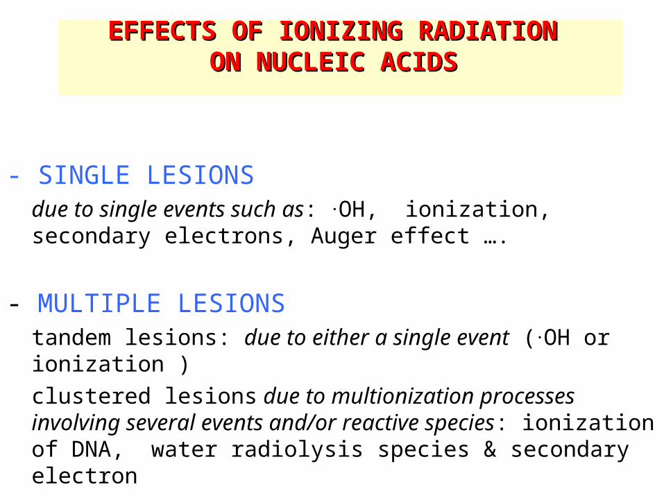
EFFECTS OF IONIZING RADIATION EFFECTS OF IONIZING RADIATION ON NUCLEIC ACIDSON NUCLEIC ACIDS
- SINGLE LESIONS
due to single events such as: .OH, ionization, secondary electrons, Auger effect ….
- MULTIPLE LESIONStandem lesions: due to either a single event (.OH or ionization )clustered lesions due to multionization processes involving several events and/or reactive species: ionization of DNA, water radiolysis species & secondary electron

OXIDATIVE DNA DAMAGEOXIDATIVE DNA DAMAGE
- OLIGONUCLEOTIDE STRAND BREAKS (hydrogen abstraction at 2', 4' and 5' carbons)
- ABASIC SITES * hydrolysis of the N-glycosidic bond (modified bases) * oxidation at C1‘ (2-deoxyribonolactone)
- BASE LESIONS (about 70 modifications identified)
- DNA-PROTEIN CROSSLINKS
- ALDEHYDE ADDUCTS TO AMINOBASES(breakdown products of LOOH and oxidation products of 2-deoxyribose)
- ALKALI-LABILE SITES
(abasic sites and a few oxidized bases including thymine glycols, 5-formyluracil, hydantoins …)

OXIDATION OF NUCLEIC ACIDS (general objectives)
- Model compounds Nucleosides and oligonucleotides for structural and mechanistic studies
- Isolated DNA Search for oxidative lesions, requiring the development of assays based on the chemical and spectroscopic features of the targeted lesions
- DNA in cells and tissues Need of sensitive assays aimed at singling out targeted lesions (at least 1 modification per 106 to 107 normal bases)

DNA lesions(model compounds)
Structural and mechanistic studies
(NMR, MS, X-rays)Theoretical aspects
Damage measurement
Modified oligonucleotides
HumanAnimals
Isolated cellsTissues
Biological fluids DNA
Biomarkers
Biological role(mutagenesis,
translesional synthesis)
Repair
Synthesis
ReactivityHPLC (EC, MS/MS)Comet assayImmunoassays
…………………………………………………………………………….

RADIATION-INDUCED DAMAGE TO ISOLATEDRADIATION-INDUCED DAMAGE TO ISOLATED AND AND
CELLULAR DNACELLULAR DNA (outline)
- Mechanistic studies on isolated DNA and models
- .OH radical degradation of the thymine base
- reactions of the guanine radical cation
- Radiation-induced damage to cellular DNA
- HPLC-MS/MS measurement
- Modified comet assay

OXIDATIVE BASE DAMAGE TO DNA OXIDATIVE BASE DAMAGE TO DNA (current situation)(current situation)
- THYMINE: almost complete available information for isolated DNA and model compounds
- CYTOSINE: comprehensive mechanism for radical oxidation of dCyd. Paucity of information for isolated DNA
- GUANINE: complex reactions with still a strong need of further investigations on both dGuo and isolated DNA
- ADENINE: apparent lack of information (need to be further checked)

N
N
O
O
HCH3
+°OH
60 %35 %
5 %
N
N
O
O
HCH2
°N
N
O
O
HCH3
OHH
°N
N
O
O
HCH3
OH
H°
O2, O2°-, H+
N
N
O
O
HCH3
OHH
OOH
N
N
O
O
HCH3
OOHH
OH
N
N
O
O
HCH2OOH
H
NOH
N
N
O
O
OH
CH3
H
N
N
O
O
HCHON
N
O
O
HCH2OH
1
2 3 4
56 7
8
9 10
12 13
O
OHCH3
H
O
O
N
N
O2, O2°-, H+ O2, O2°-, H+
N
N
O
O
HCH3
OHH
OH
11
Reactions of .OH radical with thymine

OXIDATIVE DAMAGE TO GUANINE OXIDATIVE DAMAGE TO GUANINE
(mechanistic aspects)(mechanistic aspects)
- Single damage: (one-electron oxidation)
- Tandem lesions (.OH radical, one-electron oxidation)
- Charge transfer reaction within DNA
- Modulating effect of 8-oxo-7,8-dihydroguanine (one-electron oxidation)

N N
NHN
O
H2N
HO
H
N N
NN
O
H2N
H+°
N N
NN
O
H2N
H
N N
NN
O
H2N
°
N N
N
HNH2
O
N N
O
H
NH2
NH2
O
H
H2N
O
N N
NN OH
H
°
- e-
H2O
- H+
N N
NN
O
H2N
HO
H
O2, H2O
Reduction
H2O
1 2
3
6
4 5
7
8
Oxidation
Main chemical reactions of the guanine radical cation
Kasai et al, JACS, 1992Cadet et al, JACS 1994Gasparutto et al, JACS 1998Ravanat et al, JACS 2003

- Similar degradation products from 8-hydroxy-7,8-dihydro purinyl radicals: 8-oxo-7,8-dihydro- and Fapy purine derivatives
- 2-Hydroxyadenine: barely detectable with yield much lower than that of FapyAde
- On the overall degradation of guanine 10-fold less efficient than that of adenine in isolated DNA but not in nucleosides.
- Unknown reasons of the apparent lower susceptibility of adenine to both .OH radical and one-electron oxidants.
RADICAL OXIDATION OF RADICAL OXIDATION OF GUANINE AND ADENINE IN ISOLATED DNAGUANINE AND ADENINE IN ISOLATED DNA
((similarities and differences)similarities and differences)

Modulating effect of 8-oxodGuoModulating effect of 8-oxodGuo on one-electron oxidative damage to on one-electron oxidative damage to
DNADNA
- Ionization potential of 8-oxodGuo is lower than that of other DNA nucleosides including dGuo (Prat et al, J. Am. Chem. Soc., 120, 845-846, 1998)
- Rate constant for one-electron oxidation of 8-oxodGuo is about 2 orders of magnitude higher with respect to that of dGuo (Steenken et al, J. Am. Chem. Soc., 122, 2373-2374, 2000)
- 8-OxodGuo could be the ultimate sink of one-electron
oxidation process within DNA

0%
20%
40%
60%
80%
100%
120%
0 1 2 3 4 5
8-oxodGuo
Time (min)
A
dGuo
0 1 2 3 4 5
Time (min)
B
0 5 10 15 20 25 30 35
dGuo
8-oxodGuo
Time (min)
C
Comparative and competitive susceptibility of dGuo and 8-oxodGuo to one-electron oxidant (photoexcited riboflavin)
(Ravanat et al, J. Am. Chem. Soc., 125, 2030, 2003)

°N N
NN
O
H2N
H OH
H
N N
NN
O
H2N
°
N N
NN
O
H2N
H
H2O
N N
NN
O
H2N
HO
H
N N
NN
O
H2N
H+°
-H+
N N
NN
O
H2N
H
-e-
Decompositionproducts
dGuo
8-oxodGuo
Guanine oxidizing radical dGuo
Decompositionproducts
Radical oxidation reactions of isolated 2’-deoxyguanosine

8-OXO-7,8-DIHYDROGUANINE8-OXO-7,8-DIHYDROGUANINE(available information on its formation)(available information on its formation)
- Ubiquitous DNA oxidation product: * singlet oxygen (1O2)
* one-electron oxidation
* .OH radical
* peroxynitrite (ONOO-)
- Present in tandem base modifications that involve initial oxidation reactions of thymine or cytosine
* one-electron oxidation
* .OH radical

Oxidative DNA damage: tandem lesions
(one initial radical hit)
H
NH2
O
NN
N N
ON
H H
HO
OO
POH
O
OHO
O
O
H
HO
OO
POH
O
OHO
O
HH
NO
H
ONN
N N
O
NH2
H
H
NH2
O
NN
N N
O
HO
HO
H
NH2
NN
N N
OHO
HO
H
(5’S)-5',8-cyclodAdo
8-oxodGuo/Fo Fo/8-oxodGuo
(5’R)-5’,8-cyclodGuo
HN
N
O
O
CH3
dRP
dR
N
HN N
NH2N
O
NH
N
O
O
H3C
O
H2N N
NHN
N
dRP
dR
HO
°OONH
N
O
O
H3C
O
H2N N
NHN
N
dRP dR
HO
°
°
HO
dRPdR
N
HN N
NH2N
O
H3C
O
O
N
NHO O
NH
O
H2N N
NHN
N
dRP
dR
OO
H
H
*
°OH O2
O2Fe2+
scissionfragmentation
O O
HO
dRPdR
N
HN N
NH2N
O
H3C
O
O
N
NHNH
N
O
O
H3C
O
H2N N
NHN
N
dR
PdR
HO
°OOH
NH
O
H2N N
NHN
N
dRP
dR
OO
H
HNH
N
O
O
H3C
O
H2N N
NHN
N
dRP dR
°O
HOOH
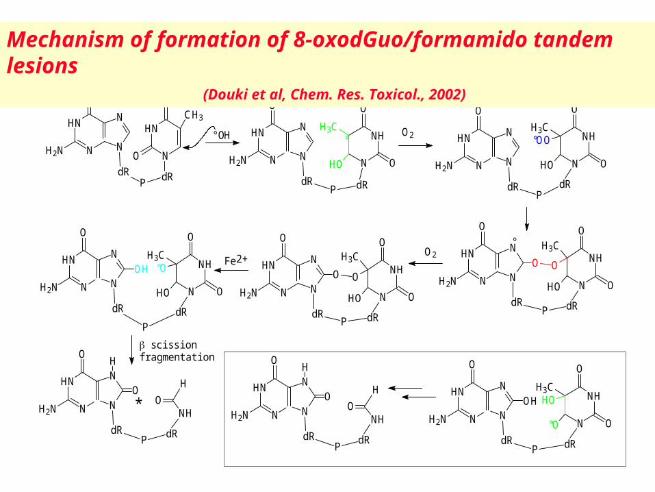
Mechanism of formation of 8-oxodGuo/formamido tandem lesions(Douki et al, Chem. Res. Toxicol., 2002)

CHARGE MIGRATION WITHIN DOUBLE CHARGE MIGRATION WITHIN DOUBLE STRANDED DNASTRANDED DNA
- radical cations may be produced within nucleobases and 2-deoxyribose moieties (ionization)* ionizing radiation
* bi-photonic excitation (high intensity laser pulses)
- positive hole migration was found to occur toward guanine and also adenine (lesser extent) through:
* hopping * phonon-assisted polaron-like hopping * super-exchange

HN
NO ON
NH
O O
dR dRP
HN
NO
N
N
OH
O
O
dR
dRP
T<>T
(6-4) TT (6-4) TC
T<>C
HN
NO
N
N
NH2
O
O
dR
dRP
HN
NO ON
N
O NH2
dR dRP
Biphotonic products:oxidized nucleosides
HN
N
N
NO
OH
H2NdR
N
N
N
NO
NH2 H
dR
HN
NO
O
CH2OH
dR
HN
NO
O
CHO
dR
HN
NO
OCH3
OH
OH
dRThdGly 5-HMdUrd 5-FordUrd
8-oxodGuo 8-oxodAdo
Monophotonic products:pyrimidine dimers
cis-syn trans-syn
S0
S1
T1
Tn
ISC
Ionizationenergy
T+°
h
h
Singlet state Triplet state

0 100 200 3000.0
0.2
0.4
0.6
0.8
1.0
ThdGly5-HMdUrd8-oxodGuo8-oxodAdo
quan
tum
yiel
d x 1
000
intensity (mJ/cm2)
Effects of the intensity on the quantum yield of formation of oxidized nucleosides upon exposure of DNA to UV laser pulses.

Effects of denaturation and addition of polyamine on the quantum yield of formation of modified bases upon exposure of DNA to UV laser pulses
(intensity: 347 mJ/cm2).
0
0.2
0.4
0.6
0.8
1
1.2
1.4
1.6
1.8
2
denatured (heated) control spermidine 0.1 mM
quantu
m y
ield
(x1
00
0)
c,s T<>T t,s T<>TThdGly 8-oxodGuo8-oxodAdo

0 2 4 6 8 100
1
2
3
4
ThdGly5-HMdUrd 5-FordUrd8-oxodGuo8-oxodAdo
quan
tum
yiel
d x 1
000
TRIS concentration (mM)
Effects of the addition of TRIS on the quantum yield of formation of oxidized bases upon exposure of DNA to UV laser pulses (intensity: 347
mJ/cm2).

OXIDATIVE BASE DAMAGE TO CELLULAR OXIDATIVE BASE DAMAGE TO CELLULAR DNADNA
(current situation)(current situation)
* Isolated DNA and model compounds: More than 70 lesions have been identified as oxidative degradation products of thymine, cytosine, adenine, guanine and 5-methylcytosine
* Cellular DNA: only 11 base lesions have been accurately measured: Adenine (2) Cytosine (1) Guanine (2), Thymine (6)

MEASUREMENT OF OXIDATIVE MEASUREMENT OF OXIDATIVE BASE DAMAGE TO DNABASE DAMAGE TO DNA
- As DNA fragments (bases, nucleosides or nucleotides)
- In whole DNA
- In intact cells

MEASUREMENT OF OXIDATIVE BASE DAMAGE MEASUREMENT OF OXIDATIVE BASE DAMAGE TO CELLULAR DNATO CELLULAR DNA
((individual measurementindividual measurement))
- Chromatographic methods: HPLC-MS/MS Optimization of DNA extraction conditions
- Applications: Effects of γ-rays and heavy ions on human monocytes

DNA extraction for chromatographic DNA extraction for chromatographic assaysassays
* Still a critical step
* Various methods have been proposed with or without phenol The background level is within the range of one up to several 8-oxodGuo residue per 106 bp
* Chaotropic method associated with a metal chelator (desferioxamine) The background level is lower than one 8-oxodGuo residue per 106 bp (Helbock et al, PNAS, 95, 288-93, 1998; Ravanat et al, Carcinogenesis, 23, 1911-8, 2002)

OXIDATIVE DNA BASE DAMAGEOXIDATIVE DNA BASE DAMAGEHPLC-MS/MS (electrospray ionization mode) HPLC-MS/MS (electrospray ionization mode)
measurementsmeasurements
- Most recent method (tandem mass spectrometers)
- More sensitive than any other chromatographic methods by about a factor of 10 (this depends on the targeted lesion)
- More straightforward than GC-MS (no derivatization) and versatile than HPLC-ECD (almost all compounds can be detected)
- Extension to more sensitive analytical methods (micro-HPLC, capillary electrophoresis)

Q 1
Parent ion(M-H-)
detector
Q 3
daugther ions(fragments)
N2
collisioncell Q2
HPLCcolumn
dN
dN
dNdN
dN
OxdN
DNA digestion(dN: nucleosidesOxdNs: oxidized
nucleosides)
UV detection(normal bases)
Principle of the HPLC-MS/MS assay for the measurement of
oxidized nucleosides
capillary+ -+
+
-
N2
Tandem mass spectrometry(multiple reaction mode)

Time, min
Intensity, cps29.62
35.7210000
20000
30000
40000
4.15 9.14
ThdGly
13.42
5-OHdUrd
18.35
5-HMdUrd
25.23
5-FordUrd
8-oxodGuo
8-oxodAdo
HPLCMS/MS of oxidized nucleosides(separation - detection – quantitation)
C18 / 2.1mm
Gradient Formate / MeCN
Hydrolyzed DNA sample + Q1 is
M1 F1
Mn Fn
Asa. AisMS/MSIntegration
Ais )Qsa= (AsampQ1 x

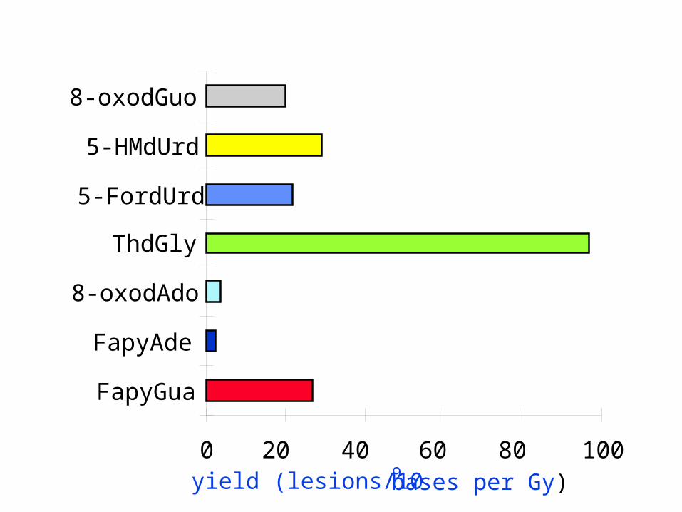
Gamma ray-mediated formation of DNA damage in isolated calf thymus DNA in aerated aqueous
solutions(lesions /106 DNA bases/ Gy)
N
N
O
O
OHHHOH
H N
N
O
O
HCH2OH
N
N
O
O
HCHO
N
N
O
O
HOH
11.4 1.2 5.3 1.4 15.3 0.1 7.6 4.4
N N
NN
O
H2N
HO
HN
NH
CHO
NHN
O
H2N
H N N
NN
O
HNH2
NNH
CHO
NHN
NH2
35.1 0.9 33.0 9.4 3.9 0.5 5.0 0.1
Measurement within the dose rang: 0 100 Gy
0 20 40 60 80 100
FapyGua
FapyAde
8-oxodAdo
ThdGly
5-FordUrd
5-HMdUrd
8-oxodGuo
yield (lesions/109 bases per Gy)

Yields of formation of 11 base modifications in the DNA of monocytes upon exposure to 60Co -rays (LET: 0.2 keV/µm) and 12C6+ ions (LET:
25 keV/µm)
DNA base modifications -rays 12C6+ (per 109 bases and per Gy)
5,6-Dihydroxy-5,6-dihydrothymidine 97 62 (4 diastereomers)
5-(Hydroxymethyl)-2’-deoxyuridine 29 12
5-Formyl-2’-deoxyuridine 22 11
5-Hydroxy-2’-deoxyuridine 0.2 < 0.2
8-Oxo-7,8-dihydro-2’-deoxyadenosine 3 3
Adenine formamidopyrimidine 5 1
8-Oxo-7,8-dihydro-2’-deoxyguanosine 20 10
Guanine formamidopyrimidine 39 22

RADIATION-INDUCED DAMAGE TO RADIATION-INDUCED DAMAGE TO CELLULAR DNACELLULAR DNA
((conclusionsconclusions))
- Thymine is a better substrate than guanine (guanine is a better target than adenine)
- Occurrence of reduction processes for purine base damage
Fapypurines > 8-oxo-7,8-dihydropurine
- Relatively low yields of base damage and strand breaks (with respect to steady-level of oxidative DNA damage)
- Major role played by clustered damage * DNA double strand breaks * base lesion + single strand break (or another base
damage)

MEASUREMENT OF OXIDATIVE BASE DAMAGE MEASUREMENT OF OXIDATIVE BASE DAMAGE TO CELLULAR DNATO CELLULAR DNA
- Isolated cells: Modified comet assay (use of DNA repair enzymes to
convert base damage into strand breaks)
More sensitive but less specific than HPLC-
MS/MS
- Applications: Effects of ionizing radiation

The Comet Assay Cells Agarose gel
Microscope slide
60Co
Fluorescence labeling of DNA prior to analysis
Alkaline cell lysis
Electrophoresis (pH13)
Cells :
modified
untreated
DNA migration
Detection of CSB, CDB & SAL
Untreated cells
Irradiated cell
- +

Fpg
Fpg
Cleavage of DNA by formamidopyrimidine glycosylase (Fpg) at a 8-oxo-7,8-dihydroguanine site
+
Single strand break and 8-oxoGua release

Comet assay associated with DNA Comet assay associated with DNA glycosylases (Fpg)glycosylases (Fpg)
Cells
SSB + DSB + SAL
Alkaline cell lysis
Analysis
electrophoresisTreatment
withFpg
SSB + DSB + SAL+ Fpg sites
electrophoresis
Control cells
8 Gy irradiated cell
Control cells treated wiht Fpg
8 Gy irradiated treated with Fpg

0
10
20
30
40
50
0 0.1 0.2 0.3 0.4 0.5
dose (Gy)
tail
mo
me
nt
(µm
)
SSb+ALS
endo III
FpgSSB: strand breaks
ALS: alkali-labile sites
Endo III sites: oxidized pyrimidines
Fpg sites: oxidized purines
Detection of radiation-induced DNA damage using an optimized version of the
comet assay
Cultured human monocytes exposed to -rays

Formation of 8-oxodGuo, SSB + DSB + ALS and DNA repair glycosylases (Fpg and endo III) sensitive sites in
–irradiated THP-1 tumoral monocytes
SSB + DSB + ALS Fpg sites endo III sites
Background level/109 bases 130 90 90
Background level/cell 910 630 630
Yield /109 bases/Gy 62 22 25
Yield/cell/Gy 430 151 171

Damage in a mammalian cell nucleusDamage in a mammalian cell nucleus (1 Gy of low-LET radiation)(1 Gy of low-LET radiation)
- Initial physical damage Ionizations in cell nucleus ~ 100 000 Ionizations directly in DNA ~ 2 000 Excitations directly in DNA ~
2 000 - - - Selected biochemical damage (Ward 1988) DNA strand breaks DNA ~ 1 000 8-Hydroxyadenine ~ 700 Diol de thymine ~ 200 DNA double-strand breaks ~ 40 DNA-proteins crosslinks ~ 150
Selected cellular effect Lethal events ~ 0.2-0.8
D.T. Goodhead, IJRB, 65, 7-17 1994

RADIATON DAMAGE TO DNARADIATON DAMAGE TO DNA(future work)(future work)
- Clustered damage
- DNA-protein crosslinks
- DNA-aldehyde adducts (LOOH)
- Bystander effects
- Effects of electrons of low energy

OXIDATIVE DAMAGE TO CELLULAR DNAOXIDATIVE DAMAGE TO CELLULAR DNA(conclusion)(conclusion)
- The steady-state level of the main oxidized bases is within the range of 1 lesion per 106 - 107 normal bases.
- The level of radical induced 8-oxoGua is lower than that of FapyGua and thymine glycol.
- The level of 8-oxoAde and FapyAde is about 10-fold lower than that of 8-oxoGua.
- 2-Hydroxyadenine is not detectable.

OXIDATIVE DAMAGE TO CELLULAR DNAOXIDATIVE DAMAGE TO CELLULAR DNA(ESCODD)(ESCODD)
- “European Standards Committee on Oxidative DNA Damage”: set in 1997 with EC funding over the period 2000-2003; it has involved 25 member laboratories in Europe and one in Japan
- Objectives: to standardize and validate procedures for measuring 8-oxodGuo as a biomarker of DNA oxidation
- Levels of 8-oxodGuo (0.5) and Fpg-sensitive sites (0.1) per 106 bases in the DNA of human lymphocytes by HPLC and enzymic methods
- It will be necessary to re-examine anti-oxidant studies that are based on claims of 8-oxodGuo higher values than 1 per 106 bases
(Collins et al, Arch. Biochem. Biophys., 423, 57-65, 2004)