radiation - motec life-uk
TRANSCRIPT

Patti Edwards, Senior Radiographer,
West Herts Hospitals, UK. February 2008.
Radiation Safety

Sub -headings• Background Radiation
• Effects of Radiation
• Safe Levels
• Effective Doses
• ‘ALARA’ Principle
• Radiation Safety within the X-Ray Department
• Radiation Management
• Risk versus Benefit
• Summary
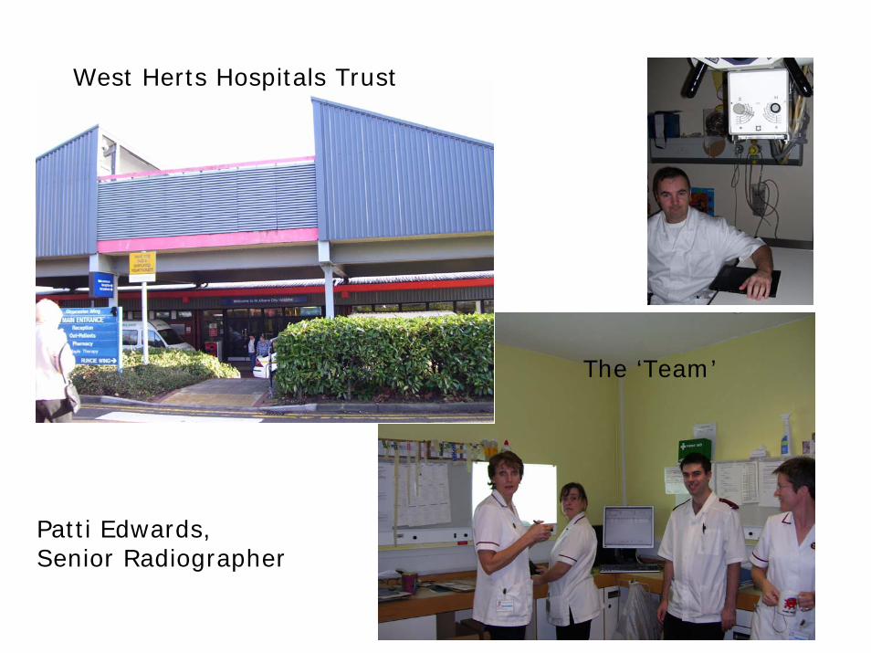
West Herts Hospitals Trust
The ‘Team’
Patti Edwards,Senior Radiographer

Background Radiation
Ionising Radiation

Background RadiationCan be natural or man-made
Natural radiation ~82%
Man-Made ~18%

Background Radiation
•The largest contributor to man-made background radiation is Medical – diagnostics & therapy
•This is why a strict Code of Practice by technicians & referrers is vitally important

Effects of Radiation
•Skin burns•Cataracts•Tumours•Cellular damage (DNA changes)
Recommended UK dose equivalents (in milli Sieverts, mSv):•0.3 mSv for the general public•20 mSv for radiation workers
•Average person in UK receives an annual dose equivalent of ~2.7 mSv

National Radiological Protection Board statutory dose limits
Statutory dose limit 50 mSv/year
NRPB recommended dose limit 20 mSv/year
Air crew from cosmic ray exposure 2 mSv/year
Nuclear industry workers 1 mSv/year
Medical worker 0.1 mSv/year

Sensitive Organs
In order of greatest sensitivity
•Gonads•Breast•Red Bone Marrow•Lung•Thyroid•Bone

Typical effective doses from diagnostic medical exposures in the 1990s (taken from ’Making the best use of a Department of Clinical Radiology –Guidelines for Doctors’. Distributed by The Royal College of Radiologists
Diagnostic procedure Typical effective dose (mSv) Equivalent number of chest xrays Approx equivalent period of natural background radiation
Limb and joint extremity xray Less than 0.01 Less than 0.5 days Less than 1.5 daysChest xray 0.02 1 3 daysSkull xray 0.07 3.5 11 daysHip xray 0.3 15 7 weeksLung ventilation (Xe-133) 0.3 15 7 weeksPelvis xray 0.7 35 4 monthsThoracic spine xray 0.7 35 4 monthsAbdomen xray 1 50 6 monthsLung perfusion (Tc-99m) 1 50 6 monthsKidney (Tc-99m) 1 50 6 monthsThyroid (Tc-99m) 1 50 6 monthsLumbar spine xray 1.3 65 7 monthsBarium Swallow 1.5 75 8 monthsCT Head 2.3 115 1 yearIVU 2.5 125 14 monthsBarium Meal 3 150 16 monthsBarium Followthrough 3 150 16 monthsBone (Tc-99m) 4 200 1.8 yearsPET Head 5 250 2.3 yearsDynamic cardiac (Tc-99m) 6 300 2.7 yearsBarium Enema 7 350 3.2 yearsCT Chest 8 400 3.6 years CT Abdo/Pelvis 10 500 4.5 years
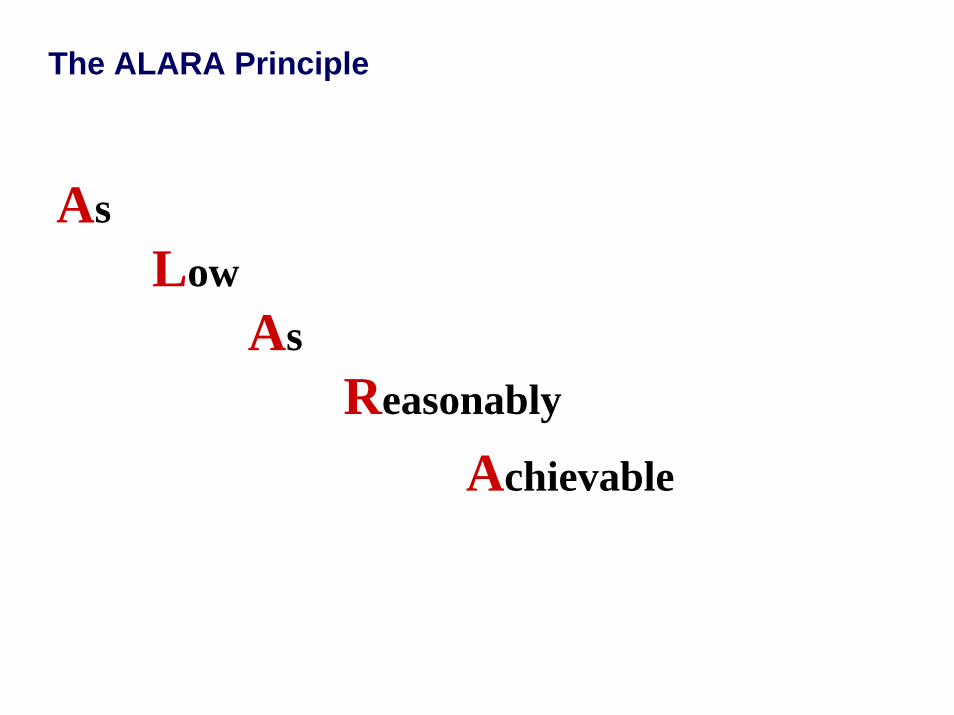
The ALARA Principle
AsLow
AsReasonably
Achievable
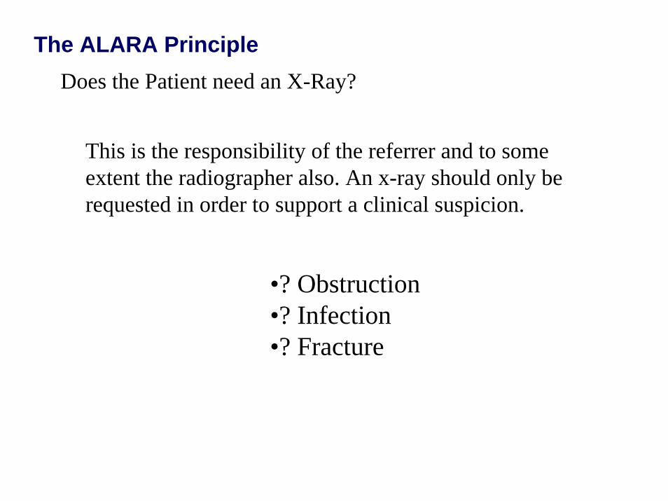
The ALARA PrincipleDoes the Patient need an X-Ray?
This is the responsibility of the referrer and to some extent the radiographer also. An x-ray should only be requested in order to support a clinical suspicion.
•? Obstruction•? Infection•? Fracture


The ALARA Principle
Will the x-ray alter patient management?
- An x-ray cannot be justified if the outcome has no possibility of altering patient management.


Examples of unnecessary X-Rays
Foreign body demonstrations of wood or plastic.

Examples of unnecessary X-Rays

Examples of unnecessary X-Rays
Abdominal x-ray to show a swallowed coin.
A coin below the diaphragm will do no damage and all that is required is frequent inspection of the stools!
Sometime a chest x- ray is indicated to ensure that the coin has in fact passed into the stomach and is not lodged in either the oesophagus or the respiratory tract.Use a high kV technique to visualize behind the heart and include the nasopharynx on a child. (A chest x-ray is roughly one fiftieth the dose of an abdomen).

Radiation Protection within the X-Ray Department
Structural Shielding
•Within the x-ray machine•Within the department structure
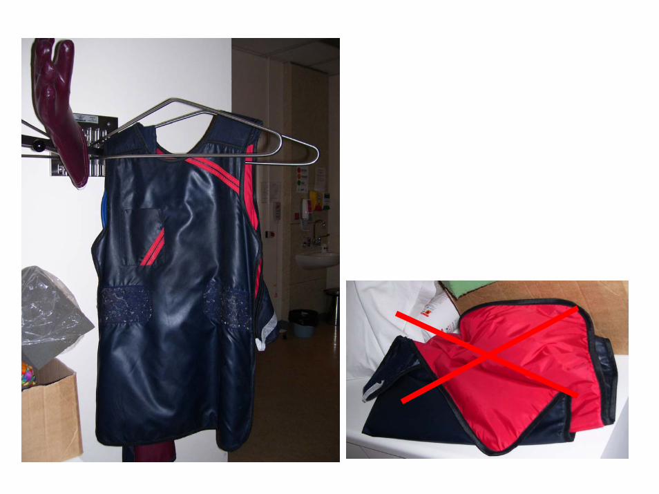
Personal Shielding
•Lead
•Lead-rubber

Inverse Square Law
1m
2m
4m
Source
At 2m, Intensity = 1 / 22 (one quarter)
At 4m, Intensity = 1 / 42 (one sixteenth)
The intensity of the beam over an area is inversely proportionalto its distance from the source

Avoid Repeat Exposures
By:•Correct Positioning•Correct Exposure

Quality Assurance
•Screen Lead Aprons
•Accuracy of Light Beam Diaphragm
•Consistency of Output


Do Not ‘Waste’ Radiation
Use lead shielding (Coning)
Always limit the beam to within the edges of the film (Coning)


General Good Practice
A) Never direct the X-ray beam at a door
B) Shout verbal warnings when X-raying on a ward or theatre
C) Only allow as many people as necessary into the X-ray room
D) Extend the limb away from the body & avoid angling the beam towards the body when X-raying extremities

Risk versus BenefitACTIVITY PROBABILITY OF DYING TIME PERIOD ODDS AGAINST
(per person)
Motorcycling 0.02 Per year
50:1Smoking 0.05 Per year
200:1Air travel 0.003 Per 100 hrs
flying time 330:1Pregnancy 0.00023 Per year 4,350:1
Housekeeping 0.0002 Per year 5,000:1RTA – driver 0.00017 Per year 5,900:1
- passenger 0.00006 16,600:1Jogging 0.00015 Per year 6,700:1
Struck by lightening 0.0000001 Per year 10,000.000:1
Exposure to radiation – At 0.3 mSv per year 0.00001 Per year 100,000:1 At 20 mSv per year 0.001 1,000:1
Taken from ‘Essential Physics for Radiographers’ by John Ball and Adrian D. Moore