radiofrequency surgery in patients with xanthelasma ... · application report radiofrequency...
TRANSCRIPT
APPLICATION REPORT
Radiofrequency Surgery in Patients with Xanthelasma palpebrarumBy Andrei Marinescu, MD, ENT-Practice Winnenden, Germany
The precision of the cut as well the hemostatic qualities of radiosurgery allow a minimal invasive and efficient approach to the delicate structures of the eyelid when resecting Xanthelasma.
Introduction: Xanthelasma is a clearly demarcated yellowish deposit of fat underneath the skin, usually on or around the eyelids. It may be associated with raised cholesterol levels, high-density lipoprotein and triglyceride levels, diabetes, cardio-vascular and thyroidal diseases (1, 2). In the author’s personal experience, a high-incidence of xanthelasma plaques were observed among the patient group excessive smokers with poor sleep-hygiene (including SAS).
Histopathological studies reveal collections of macrophages in the superficial dermis and lipid found in the lesion (3), but the latter was not always present in the resected pieces. Xanthelasmata can be treated with a trichloracetic acid peeling, cauterization or cryotherapy or alternatively removed by conventional surgery, laser or radiofrequency. High-frequency radiosurgery is well known to most ENT and oculoplastic surgeons (3). The author has 14 years of experience and has managed more than 50 cases of xanthelasmata with cold surgery, CO2
laser and radiosurgery. Radiofrequency has proven to be the method of choice: it is easiest to apply and has delivered the best results both in terms of post-operative healing and lack of recurrence.
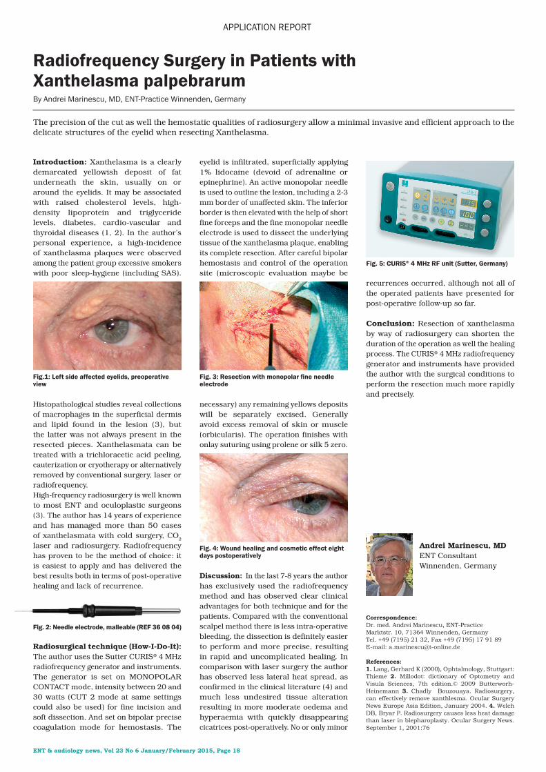
Radiosurgical technique (How-I-Do-It): The author uses the Sutter CURIS® 4 MHz radiofrequency generator and instruments. The generator is set on MONOPOLAR CONTACT mode, intensity between 20 and 30 watts (CUT 2 mode at same settings could also be used) for fine incision and soft dissection. And set on bipolar precise coagulation mode for hemostasis. The
eyelid is infiltrated, superficially applying 1% lidocaine (devoid of adrenaline or epinephrine). An active monopolar needle is used to outline the lesion, including a 2-3 mm border of unaffected skin. The inferior border is then elevated with the help of short fine forceps and the fine monopolar needle electrode is used to dissect the underlying tissue of the xanthelasma plaque, enabling its complete resection. After careful bipolar hemostasis and control of the operation site (microscopic evaluation maybe be
necessary) any remaining yellows deposits will be separately excised. Generally avoid excess removal of skin or muscle (orbicularis). The operation finishes with onlay suturing using prolene or silk 5 zero.
Discussion: In the last 7-8 years the author has exclusively used the radiofrequency method and has observed clear clinical advantages for both technique and for the patients. Compared with the conventional scalpel method there is less intra-operative bleeding, the dissection is definitely easier to perform and more precise, resulting in rapid and uncomplicated healing. In comparison with laser surgery the author has observed less lateral heat spread, as confirmed in the clinical literature (4) and much less undesired tissue alteration resulting in more moderate oedema and hyperaemia with quickly disappearing cicatrices post-operatively. No or only minor
recurrences occurred, although not all of the operated patients have presented for post-operative follow-up so far.
Conclusion: Resection of xanthelasma by way of radiosurgery can shorten the duration of the operation as well the healing process. The CURIS® 4 MHz radiofrequency generator and instruments have provided the author with the surgical conditions to perform the resection much more rapidly and precisely.
Andrei Marinescu, MDENT ConsultantWinnenden, Germany
Fig. 5: CURIS® 4 MHz RF unit (Sutter, Germany)
Fig. 2: Needle electrode, malleable (REF 36 08 04)
Fig.1: Left side affected eyelids, preoperative view
Fig. 3: Resection with monopolar fine needle electrode
Fig. 4: Wound healing and cosmetic effect eight days postoperatively
ENT & audiology news, Vol 23 No 6 January/February 2015, Page 18
Correspondence: Dr. med. Andrei Marinescu, ENT-Practice Marktstr. 10, 71364 Winnenden, Germany Tel. +49 (7195) 21 32, Fax +49 (7195) 17 91 89E-mail: [email protected]
References:1. Lang, Gerhard K (2000), Ophtalmology, Stuttgart: Thieme 2. Millodot: dictionary of Optometry and Visula Sciences, 7th edition.© 2009 Butterworh-Heinemann 3. Chadly Bouzouaya. Radiosurgery, can effectively remove xanthlesma. Ocular Surgery News Europe Asia Edition, January 2004. 4. Welch DB, Bryar P. Radiosurgery causes less heat damage than laser in blepharoplasty. Ocular Surgery News. September 1, 2001:76
Featured Products
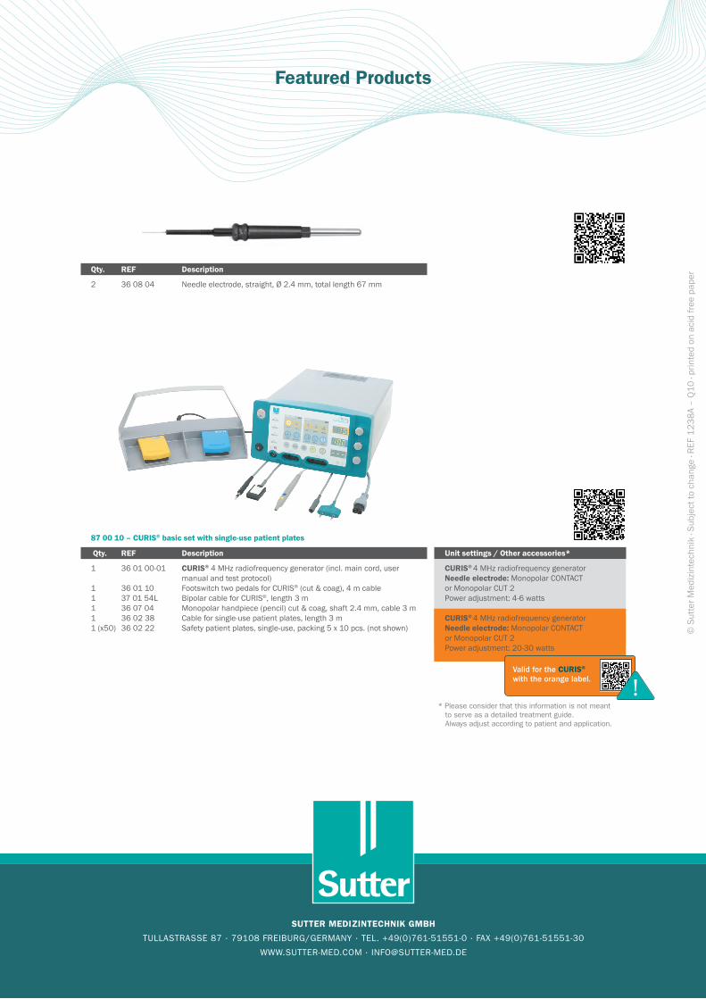
87 00 10 – CURIS® basic set with single-use patient plates
Qty. REF Description
1 36 01 00-01 CURIS® 4 MHz radiofrequency generator (incl. main cord, user manual and test protocol)
1 36 01 10 Footswitch two pedals for CURIS® (cut & coag), 4 m cable1 37 01 54L Bipolar cable for CURIS®, length 3 m1 36 07 04 Monopolar handpiece (pencil) cut & coag, shaft 2.4 mm, cable 3 m1 36 02 38 Cable for single-use patient plates, length 3 m 1 (x50) 36 02 22 Safety patient plates, single-use, packing 5 x 10 pcs. (not shown)
Unit settings / Other accessories*
CURIS® 4 MHz radiofrequency generator Needle electrode: Monopolar CONTACT
or Monopolar CUT 2 Power adjustment: 4-6 watts
CURIS® 4 MHz radiofrequency generatorNeedle electrode: Monopolar CONTACT or Monopolar CUT 2 Power adjustment: 20-30 watts
Valid for the CURIS® with the orange label. !
* Please consider that this information is not meant to serve as a detailed treatment guide. Always adjust according to patient and application.
Qty. REF Description
2 36 08 04 Needle electrode, straight, Ø 2.4 mm, total length 67 mm
SUTTER MEDIZINTECHNIK GMBH
TULLASTRASSE 87 · 79108 FREIBURG/GERMANY · TEL. +49(0)761-51551-0 · FAX +49(0)761-51551-30
WWW.SUTTER-MED.COM · [email protected]
© S
utte
r Med
izin
tech
nik
· Sub
ject
to c
hang
e · R
EF 1
238A
– Q
10 · p
rinte
d on
aci
d fre
e pa
per