radiology chapter’s 2, 3. & 4 electromagnetic spectrum unique abilities some rays visible some...
TRANSCRIPT

Radiology
Chapter’s 2, 3. & 4
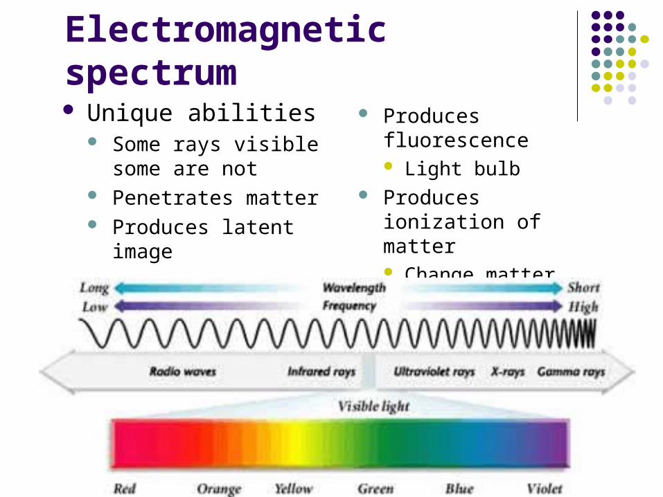
Electromagnetic spectrum Unique abilities
Some rays visible some are not
Penetrates matter Produces latent image
Produces fluorescence Light bulb
Produces ionization of matter Change matter

Matter
Anything that occupies space and has mass
Matter can be altered by energy
Fundamental unit of matter is the Atom
Desk Chair Computer Tissue Muscle Teeth Bone Your Patient
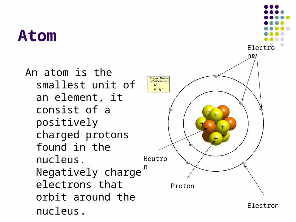
Atom
An atom is the smallest unit of an element, it consist of a positively charged protons found in the nucleus. Negatively charge electrons that orbit around the nucleus.
Neutron
Proton
Electron
Electrons
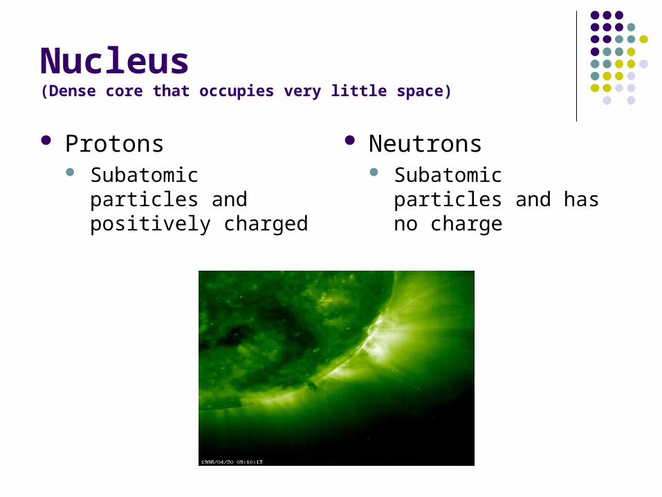
Nucleus(Dense core that occupies very little space)
Protons Subatomic particles and
positively charged
Neutrons Subatomic particles and
has no charge
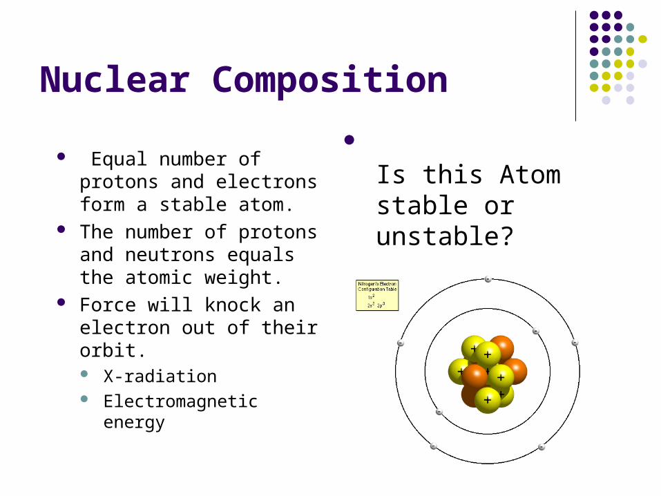
Nuclear Composition
Equal number of protons and electrons form a stable atom.
The number of protons and neutrons equals the atomic weight.
Force will knock an electron out of their orbit. X-radiation Electromagnetic energy
Is this Atom stable or unstable?

Ionization(production of ions)
Converting atoms into ions. To produce ions a force or a collision
such as x-radiation or electromagnetic energy must eject an electron out from its orbit. Thus making the atom unstable.
Spontaneous release excess energy in the form of wave or particles.
ION
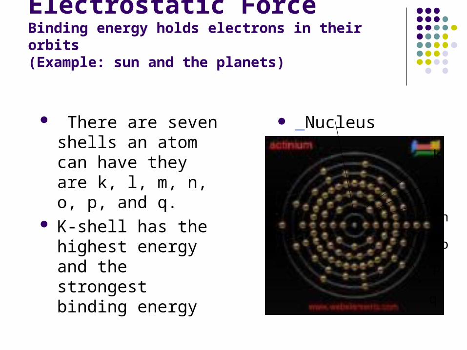
Electrostatic ForceBinding energy holds electrons in their orbits(Example: sun and the planets)
There are seven shells an atom can have they are k, l, m, n, o, p, and q.
K-shell has the highest energy and the strongest binding energy
Nucleus
q
p
o
nm
l
k
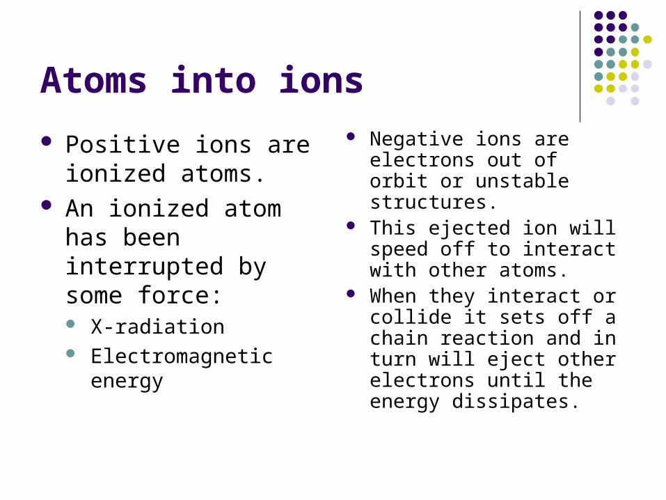
Atoms into ions
Positive ions are ionized atoms.
An ionized atom has been interrupted by some force: X-radiation Electromagnetic energy
Negative ions are electrons out of orbit or unstable structures.
This ejected ion will speed off to interact with other atoms.
When they interact or collide it sets off a chain reaction and in turn will eject other electrons until the energy dissipates.

Elements Elements
Substances made up of only one type of Atom
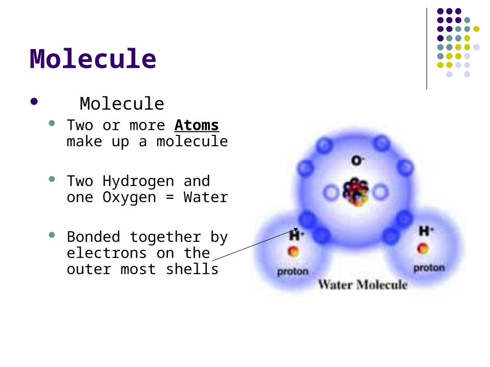
Molecule
Molecule Two or more Atoms
make up a molecule
Two Hydrogen and one Oxygen = Water
Bonded together by electrons on the outer most shells

Ionizing Radiation(all radiation cause biological changes)
Particulate Responsible for
radioactivity
Radioactivity is when atoms spontaneously disintegrate or decay. Power plant Atomic bomb
Electromagnetic Series of wave like
energies with no mass some high energy some low energy
Visible and invisible Man made or natural
occurring

Electromagnetic Energy Made up of both wave and particle that
travel in a straight line X-ray = bundle of energy, is termed, x-ray
photon which has no mass no charge and travels at the speed of light (186,000 MPS)
X-ray photon is what interacts with matter (your patient)
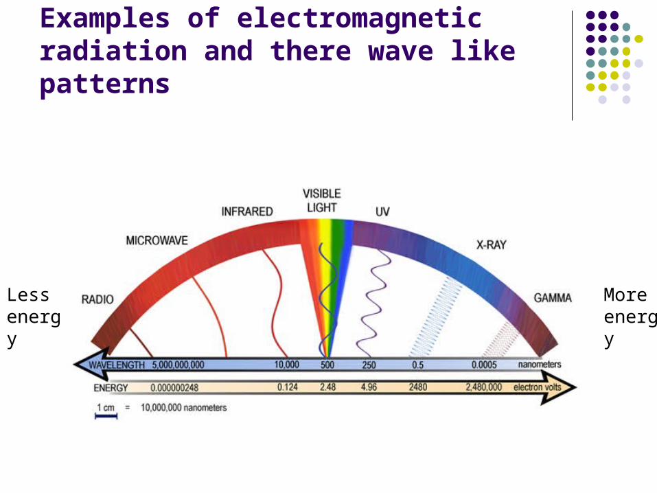
Examples of electromagnetic radiation and there wave like patterns
Less energy
More energy
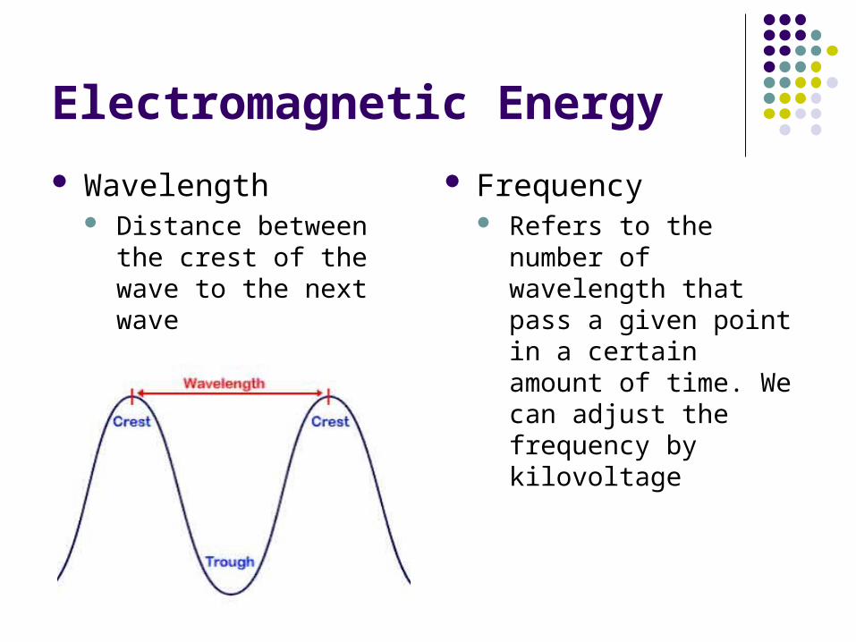
Electromagnetic Energy
Wavelength Distance between the
crest of the wave to the next wave
Frequency Refers to the number of
wavelength that pass a given point in a certain amount of time. We can adjust the frequency by kilovoltage

Voltage
Measurement of electrical force that cause electrons to move from a negative pole to a positive one (Strength)
Dental x-ray units require a high level of electrical potential

Kilo-Voltage Peak(KVp)
Kilovoltage controls the level of penetration Shorter wavelength =
more penetrating High frequency = more penetrating Longer wavelength =
less penetrating Low frequency the =
penetrating
Kilovoltage Kilo = 1000 Volatage = volts 110 or 220 Higher voltage means
greater energies Dental radiographs require
65 to 100 kilovolts Higher KVp should be used
when area is dense or thick Adjust KVp on individual
diagnostic needs Overall QUALITY of
primary beam

Amperage
Measurement of electrons moving through a conductor
Current is measured in amperes

Milliamperes (mA) Milliampere
Milli = 1/1000 Ampere = Electrical
current NOT voltage mA settings
7, 10, and 15 Thermionic emissions
Higher the setting increases temperature resulting in some electrons being ejected out of their orbit.
Ampere allows electrical current to flow thru a filament which results in a cloud of electrons
Depending on mA setting will depend on the QUANTITY of x-rays produced

Milliamperes-Seconds
Exposure time Interval of time in which
photons are being produced (.117 secs)
Longer time = more photons
High mA = more photons
Both mA and exposure time both have a direct influence on the number of electrons produced
If we produce to many photons our dental film will be dark or black
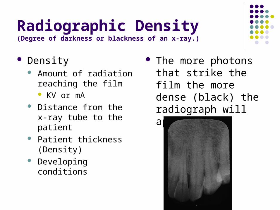
Radiographic Density(Degree of darkness or blackness of an x-ray.)
Density Amount of radiation
reaching the film KV or mA
Distance from the x-ray tube to the patient
Patient thickness (Density)
Developing conditions
The more photons that strike the film the more dense (black) the radiograph will appear
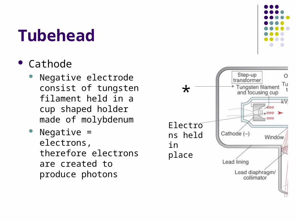
Tubehead
Cathode Negative electrode
consist of tungsten filament held in a cup shaped holder made of molybdenum
Negative = electrons, therefore electrons are created to produce photons
*
Electrons held in place

Tubehead
Anode Positive electrode consist
of a wafer thin tungsten plate embedded in a solid copper rod
Positive = collision = photons
*
Collision produced photons (indicated in red)
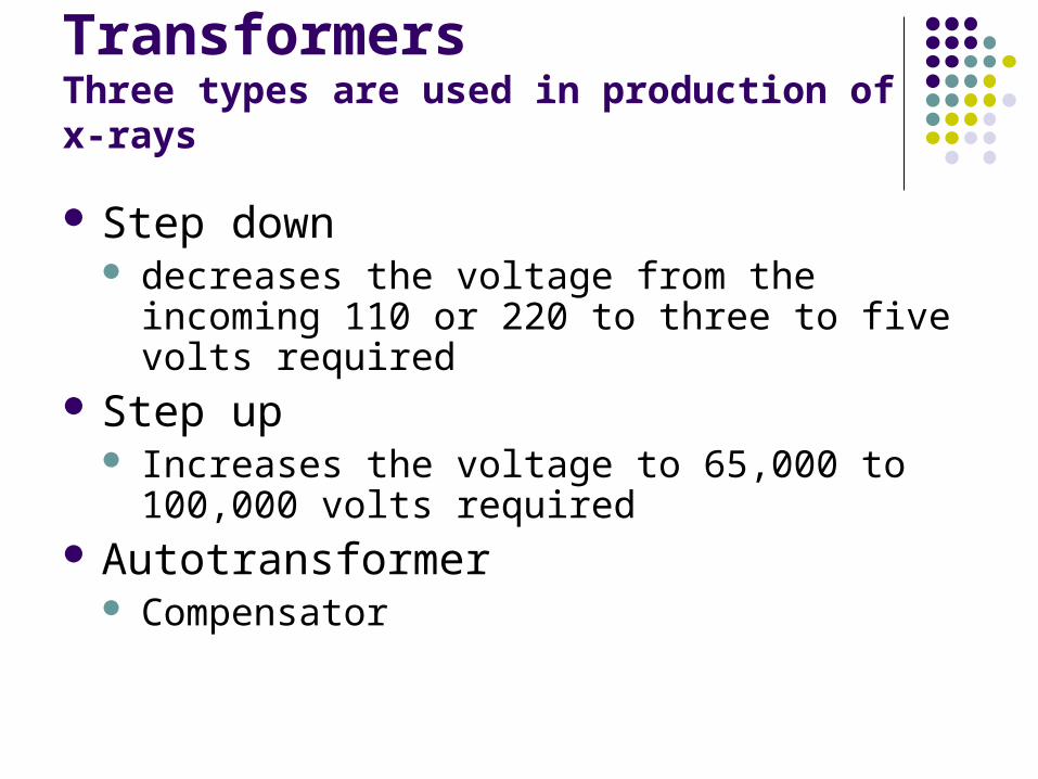
TransformersThree types are used in production of x-rays
Step down decreases the voltage from the incoming 110 or
220 to three to five volts required Step up
Increases the voltage to 65,000 to 100,000 volts required
Autotransformer Compensator

Inside the Tubehead

This is What Happens Electricity excites the filament at 3-5 volts, creating thermionic
emissions, a release of electrons from the tungsten filament when heated, this cloud of electrons stay in place until the exposure button is pushed. The high voltage circuit is activated. The electrons produce are accelerated across the x-ray tube to the anode. The molybdenum cup helps to direct the electrons to the tungsten target. When the electrons strike the tungsten target their energy of motion or kinetic energy is converted to x-ray energy and heat. More heat is created that x-rays and is dissipated through the copper stem and absorbed by the insulating oil. X-rays are produce in all direction only a few will escape through the unleaded portion of the tube. Those x-rays will be directed to the aluminum filter, which will remove the long waves. The collimator will focus the remaining short waves and travel down the lead lined PID and exit the tubehead

Production of radiation(not all produce the same in the tube head)
General radiation AKA braking radiation
An electron passes near the nucleus and is deflected by the positively charge nucleus
Once deflected this kinetic energy is converted into photons
Bremsstrahlung German
(braking radiation)

Production of radiation(not all produce the same in the tube head)
Characteristic radiation
Electron that has been deflected continues to travel ejecting other electrons out of orbit until they loose their kinetic energy Energy in motion
Important Terms
kVp = Kilovoltage peak=quality of beam kVp = density = low contrast = lots of
shades of gray Low kVp = low density = high contrast = lighter
film = black & white film Contrast – varying shades of gray Density – overall blackness or darkness of
film
Important Terms
mA=Milliamperage=quantity of x-rays produced=density To many mA’s=darker film=higher density To little mA’s=light film=lower density # of electrons from cathode to anode # of x-ray photons in beam mA regulates temperature of cathode

Important Terms
Exposure time – refers to interval of time x-rays are produced-measured in impulses Longer time=longer time x-rays emitted=darker
film=greater film density Less time=less time x-rays emitted=lighter films=
less film density Subject Thickness effects quality and
quantity of penetrating power
Important Terms
Kilovoltage peak rule – RHS – when kVp is increased by 15 – exposure time should be decreased by ½
When kVp is decreased by 15 – exposure time should be doubled
Milliampere-seconds (mAs) = combination of milliamperes and exposure time Milliampers X exposure time (seconds) =
milliampere-seconds (increase mAs-decrease time)
Important Terms
Attenuation aka Dose When matter absorbs radiation (only during
secondary) Primary Radiation aka Primary Beam
Penetrating beam – Roentgen Units = R Secondary Radiation
Created when Primary beam hits matter (soft tissues, head , skull, teeth) less penetrating power
Radiation pt receives – RAD = radiation absorbed
Important Terms
Scatter Radiation A type of secondary radiation Has been deflected off of path Travels to all parts of the body & operatory
REM = Roentgen equivalent to man MPD = maximum permissible dose
Dental radiographer = 5.0 REM per year Non health care worker = .1 REM per year .08 RAD’s in FMX

Radiation Biology
Radiation Biology-the study of effects of ionizing radiation on living tissue
Absorption Ionization page 39 – all x-rays harmful to
living tissue When x-rays strike patient tissues ionization
results Free Radical Formation-causes cell damage
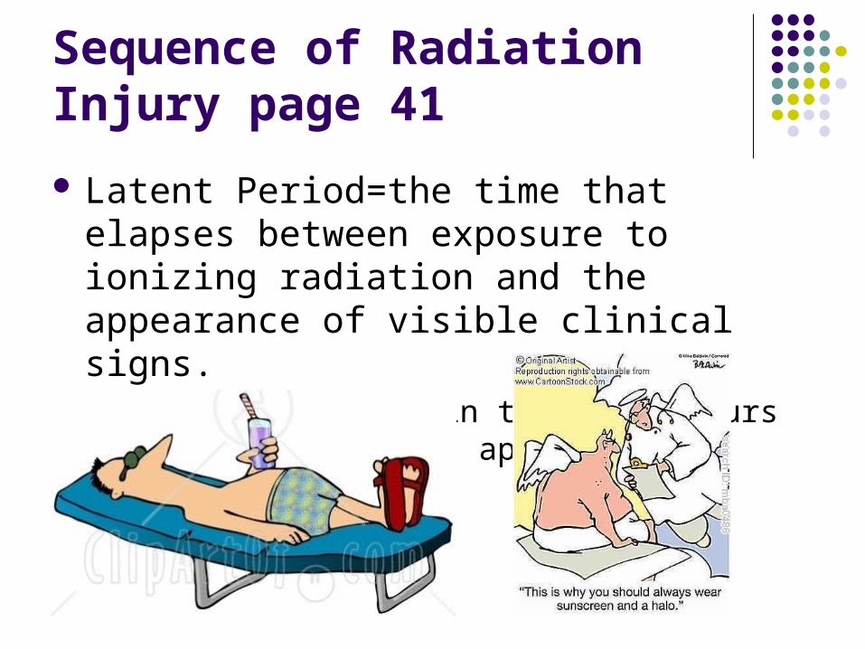
Sequence of Radiation Injury page 41
Latent Period=the time that elapses between exposure to ionizing radiation and the appearance of visible clinical signs. Example: Sitting in the sun – hours later skin
redness appears

Sequence of Radiation Injury
Period of injury After the latent period Cell injury can result as:
cell death changes in cell function – ex: endometriosis breaking or clumping of Chromosomes – ex: (cell)
reproduction problems many more cell specific
Cell injury is the desired result in cancer tx

Sequence of Radiation Injury
Recovery period Not all cell radiation damage is permanent Damage caused by low-level radiation is repaired
within cells of body. Ex: skin was burned – it has repaired itself

Sequence of Radiation Injury
Cumulative effects – overtime Radiation damage accumulates in tissue of entire
body Can lead to:
Poor health Cancer – Thyroid/Skin Cataract formation Birth defects
Dental x-rays do not cause cancer – falls under MPD

Sequence of Radiation injury cont.
Recovery period=Not all cellular radiation injuries are permanent. With each radiation exposure, cellular damage is followed by repair.
Cumulative effects=The effects of radiation exposure are additive, and unrepaired damage accumulates in the tissues, cumulative effects of repeated radiation exposure can lead to health problems.
Determining Factors for Radiation Injury
Total dose – RAD – measurement of attentuation – absorbable dose
Dose rate – rate @ which exposure to radiation occurs & absorption takes place Cells need time to recover
Amount of tissue irradiated – area exposed Cell sensitivity Age
Short-Term and Long-Term Effects
Short-term page 42 Short-term effects are
associated with large mounts of radiation absorbed in a short time. Includes nausea, vomiting, diarrhea, hair loss and hemorrhage.
Long-term page 42 Effects that appear after
years, decades or generations. Long-term effects are associated with small amounts of radiation absorbed repeatedly over a long period.

Somatic and Genetic Effects
Somatic effects Somatic cells are all the
cells except reproductive cells. Major somatic effects of radiation exposure include the induction of cancer, leukemia and cataracts. These are not transmitted to future generations.
Genetic effects Genetic effects are not
seen in the person irradiated but are passed on to future generations. The radiation-induced mutations affect the health of the off-spring. Genetic damage cannot be repaired.

Radiation Measurements MPD, Maximum Permissible dose
The dental radiographer must know radiation measurements to discuss exposure and dose concepts with the dental patient.
Traditional or standard system R-roentgen=measurement of radiation REM (Me)=5.0 year or .01 weekly Pregnant operator=0.1 per year RAD (pt.)=0.1 per year

Risk Estimates
Radiosensitive parts Lymphoid tissue Blood forming tissues Reproductive cells Formative cells Embryo cells
Radioresistant parts Salivary glands Kidney Liver Cells of mature bones Muscle Nerves
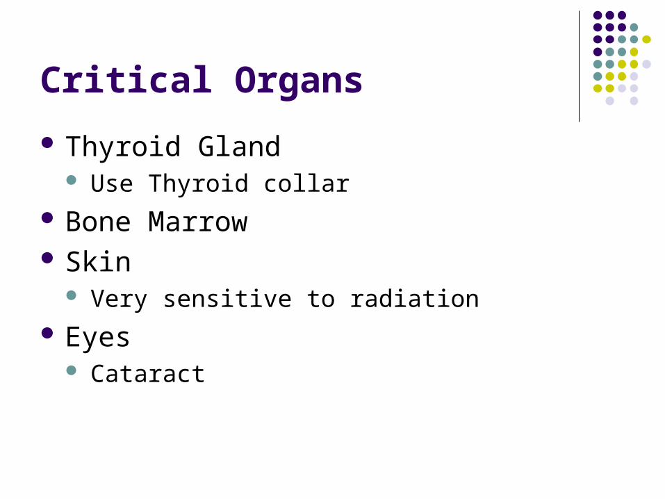
Critical Organs
Thyroid Gland Use Thyroid collar
Bone Marrow Skin
Very sensitive to radiation Eyes
Cataract

Patient Exposure and Dose pg. 46
Film- Using F-speed film instead of D reduces absorbed dose by 60%. Using
F-speed instead of E reduces absorbed dose by an additional 20%
Collimation-Radiation exposure can be limited by using rectangular collimation, reduces absorbed dose by 60%-70% rather the round collimation (PID).
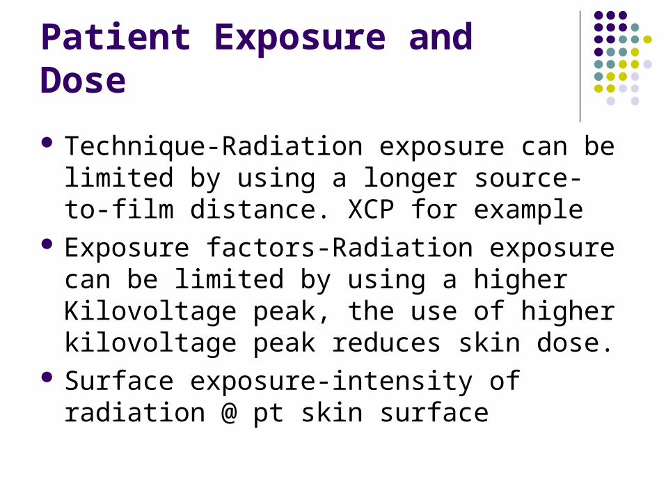
Patient Exposure and Dose
Technique-Radiation exposure can be limited by using a longer source-to-film distance. XCP for example
Exposure factors-Radiation exposure can be limited by using a higher Kilovoltage peak, the use of higher kilovoltage peak reduces skin dose.
Surface exposure-intensity of radiation @ pt skin surface
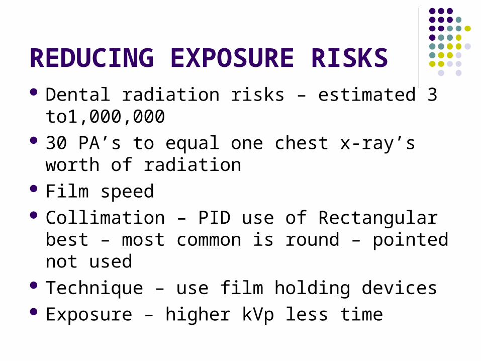
REDUCING EXPOSURE RISKS Dental radiation risks – estimated 3
to1,000,000 30 PA’s to equal one chest x-ray’s worth of
radiation Film speed Collimation – PID use of Rectangular best –
most common is round – pointed not used Technique – use film holding devices Exposure – higher kVp less time

Risk versus Benefit
X-radiation is harmful to living tissues Benefit of disease detection outweighs risk of
biological exposure Properly prescribed & exposed – only when
needed ALARA Principle – as low as reasonably
achievable – minimize risk to pt and operator FMX – full mouth survey – taken no more than
once every 3-5 yrs – 14 PA’s & 4 BW’s