radiology technologist intravenous skills · discuss the medical-legal aspects of ......
TRANSCRIPT

RADIOLOGY
TECHNOLOGIST
INTRAVENOUS
SKILLS
Self-Study Module
CLINICAL EDUCATION

Module Developed by:
University Hospital Clinical Educators
For additional information regarding classes offered by Clinical Education, click on the Education Website, Clinical Education: http://hyper.unm.edu/edcs/educationhome.shtml or
Call 272-6313 for information on Clinical Education classes.
The information contained in this module is maintained and reviewed annually/routinely by educators in the Department of Clinical Education. Efforts were made to present the following content in an accurate, clear, and concise manner while maintaining compliance with acceptable standards of care. Your comments and concerns are important to us. Please do not hesitate to provide feedback or identify errors, omissions or deficiencies in the content or presentation of this important subject matter.
Revised: Jane Potter (July 2014) Phone: 272-6411 Updated and revised: Irene Zamora (June 2010)
© Copyright University of New Mexico
All rights reserved. The reproduction or use of this document, in part or in whole, is
forbidden without the express written permission of UNM.

Please READ!!!!
Directions for Completing this Self-Study Module
This self-study module is followed by an exam to validate learning. The passing grade for the test is
84%. Should you fail the exam, your missed questions will be identified. You may retake your test up
to 3 times to achieve a passing grade.

CONTENTS
I. OVERVIEW OF INTRAVENOUS USE IN THE APPLICABLE RADIOLOGY MODALITIES .......... Error!
Bookmark not defined.
A. INDICATIONS FOR INTRAVENOUS USE ............................................................................................... 1
B. TYPES OF IV ACCESS .....................................................................................................................1
C. POTENTIAL PROBLEMS ................................................................................................................1
II. MEDICAL-LEGAL ASPECTS OF INTRAVENOUS THERAPY............................................................................ 2
A. HISTORY .......................................................................................................................................................... 2
B. TECHNOLOGIST PROCESS ............................................................................................................2
C. CURRENT PRACTICE OF IV THERAPY IN NEW MEXICO ............................................................. 4
D. STANDARD HOSPITAL POLICIES ................................................................................................4
E. RESPONSIBILITIES ASSOCIATED WITH INTRAVENOUS THERAPY ........................................ 4
F. LEGAL IMPLICATIONS ..................................................................................................................6
III. ANATOMY AND PHYSIOLOGY APPLIED TO INTRAVENOUS THERAPY ............................................... 6
A. BASIC VASCULAR SYSTEM ..........................................................................................................6
B. VEINS USED FOR IV THERAPY .....................................................................................................7
C. CONSIDERATIONS IN SELECTION A SUITABLE VEIN .................................................................. 9
IV. INFUSION EQUIPMENT & DELIVERY ........................................................................................................ 11
A. VENOUS ACCESS DEVICES ......................................................................................................... 11
B. DRESSINGS .................................................................................................................................................... 12
C. NORMAL SALINE LOCK .............................................................................................................. 12
V. VENIPUNCTURE & SITE CARE ..................................................................................................................................... 12
A. PREPARATION ............................................................................................................................................. 12
B. INSERTING THE CANNULA ........................................................................................................ 14
C. DOCUMENTATION ..................................................................................................................................... 15
D. IV ASSESSMENT AND SITE CARE .............................................................................................. 15
E. DISCONTINUING THE IV ............................................................................................................. 15
VI. COMPLICATIONS OF INTRAVENOUS THERAPY ................................................................................... 16
A. LOCAL COMPLICATIONS ........................................................................................................... 19
B. SYSTEMIC COMPLICATIONS...................................................................................................... 19
REFERENCES ............................................................................................................................................................ 21
APPENDICES .............................................................................................................................Error! Bookmark not defined.2
A – GLOSSARY OF TERMS .................................................................................................................................................. 22
B – SUMMARY TABLE OF POTENTIAL COMPLICATIONS .............................................................................. 255
C – SUMMARY TABLE OF MEASURES TO PREVENT COMPLICATIONS .................................................. 27
- I-
OBJECTIVES
1. List two advantages and two disadvantages of intravenous therapy.
2. Describe the distinguishing characteristics for the following types of IV access: peripheral,
midline and central.
3. Discuss potential problems with peripheral IVs.
4. Discuss the medical-legal aspects of intravenous therapy.
5. Describe the Technologists Clinical Performance Standards
6. Learn the Seven Rights of Medication Administration.
7. List 4 considerations in choosing a suitable vein for IV therapy.
8. Identify various patient factors that may influence size and condition of veins.
9. Identify the two types of venous access devices used most commonly in Radiology.
10. Describe the steps to perform venipuncture safely.
11. Differentiate between local and systemic complications.
12. Identify signs and symptoms of various local and systemic complications.
13. Describe prompt treatment for each local and systemic complications.
- ii -

I. OVERVIEW OF INTRAVENOUS USE IN THE APPLICABLE RADIOLOGY MODALITIES
A. INDICATIONS FOR INTRAVENOUS USE
Medication Administration. Many radiology exams require the IV administration of medication including exams performed in computerized tomography (CT), fluoroscopy, interventional radiology (IR), magnetic resonance imaging (MRI) and nuclear medicine.
B. TYPES OF IV ACCESS
In Radiology, peripheral IV access is the typical method utilized for medication administration by the imaging specialists. Peripheral IV access refers to use of a butterfly needle or any IV catheter that is inserted only a short distance into a peripheral vein. Peripheral veins are, for the most part, those in the extremities. Peripheral IVs are typically used for short term infusion of medications. Peripheral IV access established in the radiology service area is typically discontinued shortly after the patient’s radiologic procedure. Peripheral IV access that was established by nursing units for inpatients will not be discontinued post-imaging.
Radiologic technologists may use established midline IVs, such as PICC lines, for medication injection but they will never place these types of IVs. A midline IV is an IV line that, while inserted into a peripheral vein (usually the cephalic or basilic), is threaded up so that the tip of the catheter lies just distal to the superior vena cava.
A central IV is an IV line placed such that the catheter tip terminates in either the superior or inferior vena cava. Radiologic technologists may use these lines for medication injection, but will never establish this type of IV access. Either hand or power-injections may be performed through these lines after the hub is wiped for 30 seconds with chlorhexidine and allowed to dry for 30 seconds. Power-injections through central IVs will be performed only through central lines which state on the device hub that they are safety rated for power-injection.
C. POTENTIAL PROBLEMS The administration of IV medications is essential for the successful completion of many radiologic procedures. However, there are inherent, potential problems associated with IV injections.
Some potential problems are:
Extravasation which occurs when an agent infuses into the tissues surrounding the IV site. This is a serious problem if the agent is caustic to tissues. The technologist must be sure that the IV is patent before administering any substance through it.
Phlebitis (including thrombophlebitis) may result when a substance irritates the vein wall.
Nephrogenic systemic fibrosis (NSF) may result in fatal or debilitating systemic fibrosis affecting the skin, muscle and internal organs in those patients with impaired renal function who are given IV gadolinium-based contrast agents.
Other potential acute reactions to IV administered medications include cardiac or respiratory arrest, anaphylactic shock, respiratory distress, and laryngeal edema.
II. MEDICAL-LEGAL ASPECTS OF IV THERAPY
A. HISTORY
During the last fifty years, there has been a great deal of change in intravenous therapy regarding realm of practice. Initially, the radiologist performed the IV insertions and medication administrations for patients. Over time, radiologists began to delegate intravenous tasks to technologists.
The American Society of Radiologic Technologists (ASRT) Practice Standards for Medical Imaging and Radiation Therapy support that, “medication administration, which includes the administration of contrast media, are within radiologic technologists' and radiation therapists' scope of practice when the technologist or therapist is educationally prepared and clinically competent AND where federal or state statues and/or institutional policy permit.”
The University Hospital procedure, ”Medication Orders & Administration” addresses the professional scope of practice of radiologic technologists.
B. TECHNOLOGIST PROCESS
The American Society of Radiologic Technologists has assumed the responsibility for developing standards that apply to the practice of medical imaging regardless of area of professional performance or setting.
The Practice Standards for Medical Imaging and Radiation Therapy consists of standards of Radiography Clinical Performance which include the following topic areas:
Clinical Performance Standards
1. Assessment 2. Analysis/Determination
3. Patient Education
4. Performance
5. Evaluation 6. Implementation 7. Outcomes Measurement
8. Documentation

- 9 -
Clinical Performance Standards Standard 1. Assessment
The technologist asses the patient prior to the procedure. Technologist will evaluate patient’s requisition or exam screening form.
Ask patient pertinent questions regarding medical history including current medication use and previous contrast use, acute and/or chronic renal insufficiency, history of chemotherapy, diabetes, hepatic insufficiency, hypertension, or if patient is over the age of 60.
Determine if patient has current lab work or if they need lab work. If lab work is required, send patient to lab for necessary work (i.e. Creatinine/BUN).
Standard 2. Analysis/Determination
The technologist analyzes the information obtained during the assessment phase and develops an action plan for completing the procedure.
Evaluate lab values prior initiation of contrast-enhanced exam to determine if contrast use is safe for the patient.
Assess if patient has existing IV access that can be safely used for injection. Assess if peripheral IV access can be established if necessary.
Standard 3. Patient Education
The technologist provides information about the procedure and related health issues according to protocol.
Discuss the need for medication use with the patient.
Educate patient regarding contrast use, IV access and what they should expect during and post injection of medication or contrast.
Standard 4. Performance
The technologist performs the action plan established during Analysis/Determination After determining safety of contrast use, establish new peripheral IV or utilize existing IV
access.
Standard 5. Evaluation
The technologist determines whether the goals of the action plan have been achieved Evaluate patient’s response to contrast use both during and after injection. Determine if patient requires any intervention post-contrast injection.
Standard 6. Implementation
The technologist implements the revised action plan if necessary Notify radiologist or rapid response as necessary if patient experiences adverse reaction to
contrast agent.
Standard 7. Outcomes Measurement
The technologist reviews and evaluates the outcome of the procedure Review all diagnostic imaging for completeness and accuracy. Determine whether the actual outcome is within established criteria.
Assess the patient’s physical, emotional and mental status prior to discharge from technologist’s care.
Standard 8. Documentation
The technologist documents information about patient care, the procedure, and the final outcome
Document all data, including contrast or medication administered, in the record in a timely, accurate and comprehensive manner.
Provide appropriate information to authorized individual(s) involved in the patient’s care (nurse and/or attending physician) via either the Ticket to Ride or phone call.
Participate in the billing and coding procedures
"The Practice Standards for Medical Imaging and Radiation Therapy" from the ASRT Web Site, (c) 2007, the American Society of Radiologic Technologists. All rights reserved. Reprinted with permission of the ASRT for educational purposes.

- 10 -
C. CURRENT PRACTICE OF IV THERAPY IN NEW MEXICO
The radiologic technologist’s scope of practice in New Mexico includes IV administration of medication after proper and adequate training is achieved. The radiologic technologists must graduate from an accredited training program, must maintain both their state and federal radiologic technologist’s licensure and attend the UNMH IV Skills Training course, prior to their being allowed to place peripheral IVs.
D. STANDARD HOSPITAL POLICIES
All health care facilities will have policies that define the expected protocol of care for intravenous therapy. The general documents at UNMH, which guide intravenous therapy practice by radiologic technologists, are:
Radiologic Technologist Job Descriptions
Procedure “Medication Orders & Administration”
Procedure “Radiology – Intravenous (IV) Contrast Administration”
In most institutions, radiologic technologists may initiate a peripheral IV. This includes being able to recognize and respond to complications, which may arise as a result of the medication use.
E. RESPONSIBILITIES ASSOCIATED WITH INTRAVENOUS THERAPY
There are several components involved with the safe administration of IV medications. There are several types of IV medications used within the Radiology Service Area and each has potential complications. As with any other medication, the technologist must know the indications, route, dose, timing and potential side effects associated with the medication administered. Any discrepancies related to the physician’s written exam protocol should be called to the radiologist's attention before administration begins.
Following the Seven Rights of Medication Administration is essential for safe practice.
Right Patient. No assumptions should ever be made about a patient’s identity.
The technologist should always ask the patient to state their name and date of birth (when feasible) and/or verify this information on the patient’s armband with the information on the patient’s requisition or the exam work list.
Right Medication. The technologist should always carefully verify the medication prior to administration and compare this with the physician’s written protocol. If any doubt exists about whether the medication is appropriate or not, the technologist should consult a radiologist prior to administration.
- 11 -
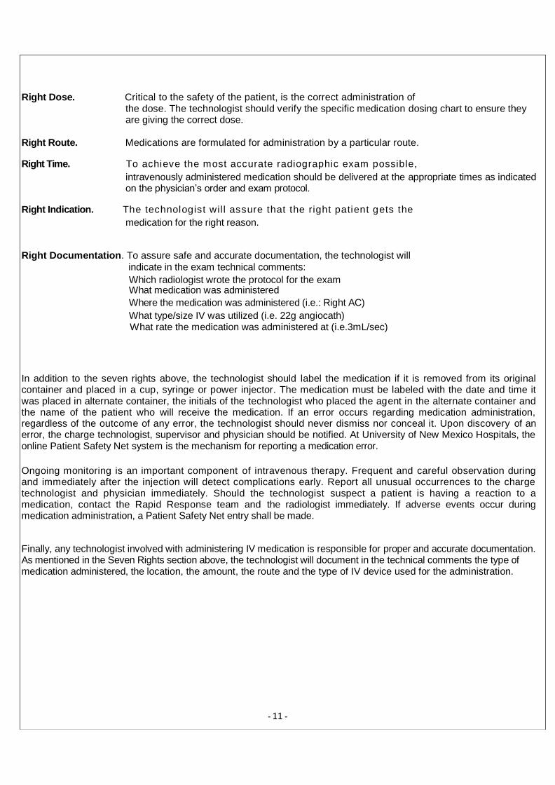
Right Dose. Critical to the safety of the patient, is the correct administration of the dose. The technologist should verify the specific medication dosing chart to ensure they are giving the correct dose.
Right Route. Medications are formulated for administration by a particular route.
Right Time. To achieve the most accurate radiographic exam possible,
intravenously administered medication should be delivered at the appropriate times as indicated on the physician’s order and exam protocol.
Right Indication. The technologist will assure that the right patient gets the
medication for the right reason.
Right Documentation. To assure safe and accurate documentation, the technologist will
indicate in the exam technical comments:
Which radiologist wrote the protocol for the exam What medication was administered
Where the medication was administered (i.e.: Right AC)
What type/size IV was utilized (i.e. 22g angiocath) What rate the medication was administered at (i.e.3mL/sec)
In addition to the seven rights above, the technologist should label the medication if it is removed from its original container and placed in a cup, syringe or power injector. The medication must be labeled with the date and time it was placed in alternate container, the initials of the technologist who placed the agent in the alternate container and the name of the patient who will receive the medication. If an error occurs regarding medication administration, regardless of the outcome of any error, the technologist should never dismiss nor conceal it. Upon discovery of an error, the charge technologist, supervisor and physician should be notified. At University of New Mexico Hospitals, the online Patient Safety Net system is the mechanism for reporting a medication error.
Ongoing monitoring is an important component of intravenous therapy. Frequent and careful observation during and immediately after the injection will detect complications early. Report all unusual occurrences to the charge technologist and physician immediately. Should the technologist suspect a patient is having a reaction to a medication, contact the Rapid Response team and the radiologist immediately. If adverse events occur during medication administration, a Patient Safety Net entry shall be made.
Finally, any technologist involved with administering IV medication is responsible for proper and accurate documentation. As mentioned in the Seven Rights section above, the technologist will document in the technical comments the type of medication administered, the location, the amount, the route and the type of IV device used for the administration.

- 12 -
F. LEGAL IMPLICATIONS
Several of the problems that result from intravenous medication administration may potentially result in legal problems for the technologist and the hospital. With respect to intravenous use, the following patient rights are of particular importance:
The patient has the right to considerate and respectful care; reasonable continuity of care; care in a safe setting; and the right to be free from all forms of abuse or harassment.
Except in emergencies, the patient or the patient's legal guardian for treatment decisions has the right to information necessary to give consent prior to treatment. The patient or legal guardian has the right to refuse treatment. The patient or his or her representative has the right to make informed decisions regarding his or her care. The patient's rights include being informed of his or her health status, being involved in care planning and treatment, and being able to request or refuse treatment. This right must not be construed as a mechanism to demand the provision of treatment or services deemed medically unnecessary or inappropriate.
III. ANATOMY AND PHYSIOLOGY APPLIED TO INTRAVENOUS THERAPY
A. BASIC VASCULAR SYSTEM
The circulatory system is divided into two main systems. The pulmonary system involves the flow of blood from the right ventricle of the heart to the lungs, where it is oxygenated and returned to the left atrium. The systemic system consists of blood flow through the left ventricle, and aorta to the rest of the body and then returned to the right atrium.
Arteries carry blood away from the heart. Veins carry blood towards the heart. Very small arteries are called arterioles, very small veins are called venules. Between arterioles and venules are the capillaries. Arteries and veins consist of three layers. The innermost layer (tunica intima) consists of an inner elastic endothelial lining. This lining allows platelets and blood cells to flow through the vessels without interruption. The middle layer (tunica media) consists of smooth muscle with both elastic and collagenous tissue. The outer layer, (tunica adventitia) consists of connective tissue that surrounds and supports the vessel.
Structurally, arteries differ from veins by the amount of tissue within the walls. Large arteries have very thick walls composed of elastic tissue and some smooth muscle. Arterioles have little elastic tissue but a great deal of smooth muscle, which makes them capable of responding to autonomic nervous control. This al lows arterioles to readily dilate or constrict directly affecting the amount of blood flow each organ and various tissues receive. It is the arterioles that serve as the major control of arterial blood pressure and distribution of blood flow.

- 13 -
Characteristics Differentiating Veins and Arteries.
VEINS... ARTERIES...
have thin walls
tend to collapse
tend to be more superficial
may contain valves to help blood flow against the force of gravity
contain de-oxygenated blood which should appear dark in color
have thick muscular walls
tend not to collapse
tend to be located deep within tissue have no valves
contain oxygen-rich blood which should appear to be bright in color
pulsate
The capillary is a very thin walled vessel made of endothelial cells. There is no elastic or muscle tissue present in the capillary. Capillaries serve as an exchange station. Cellular nutrients and metabolic end products are exchanged through this thin walled vessel.
Venules are small vessels, which serve to collect blood from various capillary beds and move it into the veins. Veins are thin walled vessels that return blood to the heart. Larger veins contain valves to maintain the blood flow toward the heart and to prevent backward flow.
Veins may be classified into one of two categories: deep veins or superficial veins. Deep veins usually run alongside and are enclosed in the same sheath with the artery. Superficial veins are located just beneath the skin. Some superficial veins unite with deep veins especially in the lower extremities. For example, the small saphenous vein eventually runs into the popliteal vein. Veins such as these should be avoided for venipuncture due to a higher incidence of complications.
There are several factors that influence the size and condition of a vein. Age is one such factor. In the elderly, veins may be tortuous, and are very fragile and easily tear with venipuncture. Present and past trauma can cause veins to become sclerosed and hard with increased frequency of blood drawing, IV drug use and/or medication administration. If a patient has a decrease in body temperature, vasoconstriction can occur making venipuncture more difficult. Finally, anxiety may cause vasoconstriction. Talking with the patient prior to IV insertion, may ease anxiety.
B. VEINS USED FOR IV THERAPY
Part of the task of initiating an IV is choosing a vein suitable for venipuncture. There are four basic things to consider when selecting a vein for venipuncture: location, condition of the vein, purpose of the infusion and duration of the IV placement.
The Center for Disease Control (CDC) states that in all patients, the upper extremity should be used in preference to lower extremities for intravenous therapy. This is because peripheral IVs placed in the lower extremities are associated with stagnant blood flow, which increases risk for thrombus, and infection. Additionally, venipuncture of veins in the lower extremity tends to be more difficult due to the condition of the veins.
- 14 -
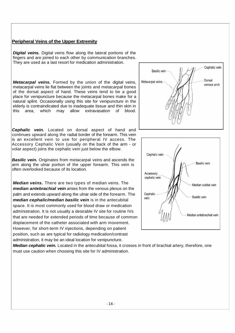
Cephalic vein. Located on dorsal aspect of hand and continues upward along the radial border of the forearm. This vein is an excellent vein to use for peripheral IV access. The Accessory Cephalic Vein (usually on the back of the arm - or volar aspect) joins the cephalic vein just below the elbow.
Basilic vein. Originates from metacarpal veins and ascends the arm along the ulnar portion of the upper forearm. This vein is often overlooked because of its location.
Peripheral Veins of the Upper Extremity
Digital veins. Digital veins flow along the lateral portions of the fingers and are joined to each other by communication branches. They are used as a last resort for medication administration.
Metacarpal veins. Formed by the union of the digital veins, metacarpal veins lie flat between the joints and metacarpal bones of the dorsal aspect of hand. These veins tend to be a good place for venipuncture because the metacarpal bones make for a natural splint. Occasionally using this site for venipuncture in the elderly is contraindicated due to inadequate tissue and thin skin in this area, which may allow extravasation of blood.
Median veins. There are two types of median veins. The
median antebrachial vein arises from the venous plexus on the
palm and extends upward along the ulnar side of the forearm. The
median cephalic/median basilic vein is in the antecubital
space. It is most commonly used for blood draw or medication
administration. It is not usually a desirable IV site for routine IVs
that are needed for extended periods of time because of common
displacement of the catheter associated with arm movement.
However, for short-term IV injections, depending on patient
position, such as are typical for radiology medication/contrast
administration, it may be an ideal location for venipuncture.
Median cephalic vein. Located in the antecubital fossa, it crosses in front of brachial artery, therefore, one
must use caution when choosing this site for IV administration.

- 15 -
Median basilic vein. Located on the outside of antecubital fossa on the ulnar curve of the arm and is least desirable for venipuncture for long-term use as hematoma may occur if patient flexes elbow, but may be used for short-term use such as with radiology medication administration.
Median antebrachial. Arises from the venous plexus on the palm of the hand and extends along the ulnar side of the front lower forearm.
Young Children and Infants
Sites for children include hand and arms as described above. For newborns, the LIP or RN will place the IV.
C. CONSIDERATIONS IN SELECTION OF A SUITABLE VEIN
The following variables must be considered when selecting a vein for medication injection purposes when several alternatives are available:
patient factors and vein condition
the anticipated length of use of the IV
catheter (or cannula) size needed
Patient Factors and Vein Condition
Patient's General Condition. An assessment of the patient’s general physical condition may yield some information about the type of IV to be used (angiocath vs. butterfly).
Patient's Current Health Problem and Disease History. It is important to evaluate the patient’s current health problem and their disease history prior to placement of the intravenous line.
Age of Patient. The anatomy of the venous system, like many other physical attributes, has age-specific characteristics that must be addressed during the vein selection process. The elderly tend to have very fragile and delicate veins. A looser tourniquet should be used when preparing for venipuncture on an elderly patient. Infants, too, have age-specific venipuncture challenges. Neonates and infants do not have as many accessible veins as adults do. For this reason, hands and antecubital area are usually the venipuncture sites of choice for children in radiology.
Condition of Skin. The condition of the skin is assessed when selecting a site for venipuncture because the condition of the skin may indicate some alteration of circulation or venous pathology below the skin. It is, therefore, recommended that you avoid areas that are reddened, sore or painful to the touch, tender, blanched, black and blue/discolored, cool, swollen, or with evidence of a hematoma.
Limb Mobility. A brief assessment of the patient’s upper extremity mobility is also done before an intravenous line is inserted.
- 16 -
Fluid Volume Status. The patient’s fluid volume and hydration status should also be determined and evaluated prior to the initiation of intravenous therapy. Venous dilation challenges are often encountered when patients are dehydrated or they have a decreased circulating volume.
Patient Activity and Personal Preference. Consider the patient’s preference when choosing a venipuncture site. Whenever possible ask the patient which arm or hand is preferable to him or her.
Other Patient Specific Conditions. Placement of an intravenous line on the same extremity as any of the following condition is contraindicated:
mastectomy •arteriovenous shunt or fistula • hemodialysis shunt •serious burns •skin graft •paralysis
Additionally, areas to be avoided are those that have encountered recent complications related to IV therapy, such as phlebitic sites, sclerosed areas, thrombosed areas; or edematous areas.
Anticipated Length of Use of the IV
The selection of suitable veins for intravenous therapy is far greater when the anticipated duration of therapy is short lived as is the case for the majority of administrations of radiology medications. On occasion, an IV may be placed in one modality and then left in place so that it may then be used again on the same day in another modality (such as in CT and then MRI). This dual use of the same IV is done to prevent the patient from having multiple intravenous injection sites in the course of their visit to Radiology. The technologists in the multiple areas where the IV is needed will communicate to ensure that the patient is seen in both modalities in a timely fashion. The IV site will be flushed with 10 mL of normal saline after the initial use of the site, prior to the secondary use. If there is a time lapse of more than 30 minutes between procedures, the IV will be discontinued and another IV placed at the time of the second procedure.
However, numerous venipunctures and multiple sites are often necessary. In such cases, the choice of veins becomes progressively more limited because the need to preserve veins (for later use) must be taken into account. In order to ensure adequate veins for future use, arms should be alternated with the most distally suitable vein accessed. For example, if a patient had an intravenous line in the right arm that must now be replaced, use a left hand or left lower forearm vein for the next venipuncture. If the other arm cannot be accessed for whatever reason, a more proximal vein on the same arm should be used.
Catheter (or Cannula) Size
Peripheral venous catheters range in variety from straight steel and winged needles to catheters made of silicon, PVC, or other materials. They vary in length and gauge to meet the needs of a wide variety of patients, ranging from the premature neonate to the elderly. IV catheters can range from a small 27 gauge 1/4 inch in length to a 14 gauge 3 inches in length. Due to the viscosity of most radiologic medications, a catheter size in the range of 18 to 22 gauge is preferable.

Dealing With Difficult Veins
Some of the problems that can be encountered during the venipuncture process include a lack of venous distention, rolling veins; and fragile veins. Venipuncture and catheter insertion cannot be successful unless the vein is adequately distended to permit the entry and threading of the catheter. If the vein does not dilate enough with the tourniquet, remove the tourniquet and:
apply heat or a warm compress to create vasodilation;
lower the limb into a dependent position to facilitate a greater volume of fluid in the vein;
gently pat the vein;
apply a small amount of pressure just above the intended site of insertion; or
rub the vein gently in one continuous motion from the proximal to the distal.
Rolling veins are also a challenge during the venipuncture process. If the desired vein starts to move or roll out of position after you have pierced the skin, perform another gentle thrust of the needle parallel to the skin while holding the area taunt. Fragile veins, on the other hand, may not respond well to a tourniquet. On some rare occasions, such as when a fragile vein is encountered, it is necessary to avoid the use of a tourniquet. Tourniquets are also not necessary when a vein is adequately dilated, visible, and palpable without it.
IV. INFUSION EQUIPMENT & DELIVERY
A. VENOUS ACCESS DEVICES
Most intravenous therapy is del ivered peripherally using a short catheter made of Teflon, silicon or polyurethane. A 20 or 22 gauge catheter is used in most situations for adults, whereas a 22 or 24 gauge catheter can be used for children and older adults or for patients with small or fragile veins. In cases where rapid infusion such as power injection is necessary an 18 gauge may be necessary.
The vein is usually accessed using an over-the-needle (ONC) catheter. Once the vein is successfully penetrated by the introducer needle, blood may be seen in the flash back chamber, at which point, the catheter is threaded into the vein. A safety mechanism may then be activated to retract the needle into the needle guard thereby preventing a subsequent needle stick injury.
Winged needles (also known as butterflies) are sometimes used for short-term infusions of 24 hours or less. The wings are used to direct the needle into the vein as well as secure the device by being taped against the skin.
- 11 -

- 12 -
B. DRESSINGS
Dressings may be either standard sterile gauze and tape dressings or the more popular sterile, transparent, semi-permeable dressing (which allow for continuous visual inspection of the catheter site). If the patient is diaphoretic, or if the site is bleeding or oozing, gauze dressing is preferable to a transparent semi-permeable dressing. Dressings should always be applied or removed using aseptic technique.
C. NORMAL SALINE LOCK (MEDLOCK)
For In-patients: When intravenous use consists only of brief medication administration and no continuous infusion,
there will be no IV bag or regulating apparatus to maintain. In such cases, a saline lock (also known as a medlock or heplock) will be used to cap the catheter. The SAS method is routinely used with medication administration via this method and involves flushing the line with normal saline (S), administration of the medication (A), followed by another flush with saline (S).
For Out-patients: If the IV site was accessed immediately (within 15 minutes) prior to use, it is not necessary to flush it with saline either prior to or after use as the IV device will normally be removed after it is used for out-patient procedures.
V. VENIPUNCTURE & SITE CARE
A. PREPARATION
Always check and verify the doctor’s order. Venipuncture and medication administration, require an order in the form of a written exam protocol.
Gather necessary supplies and equipment. Minimally, you will need disposable gloves, a tourniquet, chlorhexidine skin wipe, tape, a transparent, occlusive IV dressing or another sterile dressing, saline solution for injection in a pre-filled sterile syringe or power-injector, catheters of varying sizes and the ordered intravenous medication in either a labeled syringe or power-injector. Always wipe the top of the medication bottle with chlorhexidine and allow to dry for 30 seconds prior to puncturing the bottle. If the medication bottle has been spiked, as in multi-use bottles, wipe the top of the clave on the spike tubing and allow to dry for 30 seconds prior to attaching the syringe to the clave and drawing up the medication.
Before drawing up any IV fluids or medications, examine them for expiration date and clarity. Report and discard all fluids and medications that have exceeded their expiration date. Expired substances should not be accessible for possible use by others. Report and discard all substances that are cloudy or with particles. Cloudiness and particulate matter are indications that the infusate is not as it should be. Sometimes an entire lot, or batch, is bad so it is necessary to report this finding and the lot number of the product to your supervisor.

- 13 -
Prepare your supplies and equipment so they are ready for use. Use sterile technique to prepare sterile supplies like the dressing, catheter, and tubing. The medication must be prepared in a clean area. Use of a newly cleaned mayo stand with a clean “chuck” on it for each injection is a must. After the injection, the chuck is to be thrown away, the stand wiped clean and a new chuck placed on the stand.
Images A and B below show the proper set up for the mayo stand.
Image A. Image B.
IVs and venipuncture are sterile procedures. If the person is to have a continuous intravenous flow of medication via a power-injection, clear the IV tubing by attaching it to the saline solution and letting the fluid fill the tubing to the tip.
Identify the patient. Prior to any radiologic procedure, check the person’s identification band and ask his or her name and date of birth. Starting an IV on the wrong patient is considered a medical error.
Explain the procedure and the purpose of the procedure to the patient. Patients have a right to receive full information about any treatments. They also have the right to refuse. If the person is not competent or is a minor, the procedure and the purpose of the IV should be explained to the family member, guardian, healthcare surrogate, health proxy, etc., as indicated. Explaining procedures and treatments to patients rel ieves anxiety and increases their cooperation.
The patient should be positioned in a manner that allows for optimum conditions for venous access. The preferable position is fowler’s with the patient’s upper body upright. However, cannulation in radiology is typically performed with the patient in a flat, supine position.
Perform proper hand hygiene. Hand-washing is the single most important thing that you can do to prevent the spread of nosocomial or hospital acquired infections. Hand-washing must be done for at least 15 seconds, using ample friction and soap. Hand-washing must be done before and after every direct patient contact and should be performed before donning gloves and immediately after the removal of gloves.

- 14 -
CDC, OSHA, all healthcare facilities and other bodies mandate the use of gloves during venipuncture and any other procedure when contact with blood or another bodily fluid is possible in order to prevent the spread of blood-borne pathogens such as HIV/AIDS and hepatitis. Donning gloves and then removing the finger tip of the glove before performing venipuncture is an unacceptable practice and is not OSHA and hospital policy compliant.
B. INSERTING THE CANNULA
Select the vein. Place the tourniquet 3 to 6 inches above the site that was selected. The tourniquet will distend the vein. The tourniquet should be tight enough to distend the vein but not too tight as to cut off arterial circulation. Check the radial pulse to ensure that the arterial circulation is not impaired. The selected vein must be palpable and visible before venipuncture is attempted. If the vein is not distended enough, additional measures may be necessary. For example, placing the limb in a dependent position may create satisfactory distention of the vein. Do not leave the tourniquet in place if venipuncture cannot be successfully completed within 1 – 2 minutes. If additional time is needed, release the tourniquet and reapply it a few minutes later to continue the procedure.
Ask a responsive patient to clench and unclench their f ist several times on the side selected for venipuncture. This fist clenching will bring greater blood flow to the area and provides a greater degree of distention.
Using the chlorhexidine wipe, cleanse the skin in back and forth manner. Allow chlorhexidine to air-dry for 30 seconds before insertion. Note that chlorhexidine is considered too harsh an antiseptic for infants younger than 2 months of age. For this patient population betadine is the antiseptic of choice.
Remove the needle guard. Grasp the extremity with your thumb and gently pull the skin taut distally to the intended insertion site. This stabilizes the vein and also makes entry into the vein easier. With the bevel up, insert the needle at a 10 to 30 degree angle directly into or along-side the vein from an angle of entry with a gentle, slow, and steady movement. Holding the bevel up allows the sharpest and smallest part of the needle to enter the vein. Slow and steady movement ensures that you can see blood return and/or feel a decreased resistance before you inadvertently pass through the tunica intima (inside of the vein) and pass through the other side.
Look for blood return and feel for decreased resistance in the vein as you enter (both are indications that the vein has been successfully entered). Blood return indicates that you have successfully entered the vein’s tunica intima. A feeling of decreased resistance also indicates that you have entered the vein’s hollow interior. Lower the angle of the catheter against the skin. This lowering prevents piercing all the way through the opposite side of the vein. Advance the catheter into the vein about 1/4 inch. This advancement ensures placement in the vein.
The following step may be performed using a one handed or two handed technique as shown in the images. Pull back on the needle so it separates from the catheter and advance the catheter another 1/4 inch while applying a small amount of pressure just above the placed catheter tip and release the

- 15 -
tourniquet. Removing the needle prevents an accidental piercing of the vein while advancing the catheter into place. Applying light pressure at the catheter tip prevents excessive blood flashback as the needle is being removed and the IV tubing is being connected.
INITIATION OF VENIPUNCTURE SHOULD BE LIMITED TO TWO ATTEMPTS PER PERSON.
Attach the cleared and primed IV tubing for power injections or the pre-filled saline syringe and microclave to the hub of the catheter.
C. DOCUMENTATION
Document the procedure in technical comments in the Radiology Information System (RIS), including who started the IV, location of the IV catheter, gauge of the catheter, hand vs. power injection and, if power injected, the flow rate per second.
D. IV ASSESSMENT AND SITE CARE
Routine IV assessment should be carried out by the technologist during the medication administration. Check to see that the IV is infusing correctly and that the medication has not extravasated or infiltrated. Ascertain from the patient if they are having any pain during the course of the administration.
The IV site is considered a wound and therefore should be treated like one. It should always be covered with a sterile dressing. The CDC recommends that either a sterile gauze dressing or a bio-occlusive dressing be placed over the site.
E. DISCONTINUING THE IV
The majority of radiology IV placements are removed immediately after the medication is injected. The exception is for inpatients who arrive in radiology with venous access in place. For inpatients, their IV will be left in place when they return to the hospital unit. For outpatients, when their injection(s) are complete, the procedure for discontinuing a peripheral venous catheter is:
1. Gather necessary supplies: gloves, sterile gauze and tape.
2. Stop the infusion by discontinuing the power injection if in fact an infusion is running.
3. Wash your hands.
4. Put on gloves.
5. Remove the dressing.
6. Hold the sterile gauze against the insertion site.
7. Gently pull the catheter out parallel to the skin surface.
8. Look at the catheter to insure that it is still intact and unbroken.
9. Discard the catheter and old dressing in the proper manner for biohazardous waste.
10. Apply pressure to the site for about two minutes. Apply pressure for about 5 to 10 minutes if the person
is taking anticoagulation medications.
11. Apply a sterile dressing to the site. 12. Secure the sterile dressing with tape. 13. Instruct the patient to not remove sterile dressing for approximately one hour.

- 16 -
VI. COMPLICATIONS OF INTRAVENOUS THERAPY
One of the most important responsibilities technologists have with respect to intravenous therapy is the monitoring for the development of complications. The need to recognize complications and take appropriate steps when they occur is obvious. Complications are usually described as being either local or systemic.
A. LOCAL COMPLICATIONS
Local complications of IV therapy are associated with adverse reactions or trauma to the venipuncture site or surrounding tissue. Local complications are rarely life-threatening and are easily recognized early with frequent assessment of the IV site.
Hematoma
A hematoma (i.e., bruise) is a localized collection of blood, usually clotted, within tissue. The formation of a hematoma during intravenous therapy is related to the flow of blood into surrounding tissue at the venipuncture site and is most apt to form when initiating or discontinuing an IV. Specific actions attributable to the formation of hematomas include:
pushing the needle through the vein during an unsuccessful venipuncture attempt
discontinuing an IV without applying adequate pressure to the venipuncture site
application of a tourniquet to an extremity immediately after a venipuncture attempt
Measures that help prevent formation of hematomas include:
ensuring that the catheter and tubing are securely anchored
checking the site often
good venipuncture technique and no more than two unsuccessful attempts at any one site
applying pressure to the site after an IV catheter is removed
Symptoms of the hematoma include ecchymotic discoloration (i.e., purplish) and/or swelling around the venipuncture site. Treatment of a hematoma consists of application of pressure with a dry sterile gauze over the site (after the catheter is removed) to minimize further bleeding and elevation of the extremity to reduce swelling.
Infiltration
An infiltration is the seepage or infusion of non-caustic IV solution (e.g., normal saline) into the surrounding tissue.
Typically the causes of infiltration include:
dislodgement of the catheter out of the vein
phlebitis (inflammation may narrow the vein which can no longer support the infusion rate therefore causing fluid to leak at the site of catheter insertion
- 17 -
Measures that help prevent infiltration include:
securing the catheter and tubing well with tape
use of an arm board, if necessary to prevent dislodgement of the catheter, and
checking the site and tubing often
Symptoms of infiltration include dependent edema, cool, blanched and/or taut skin, slow or sluggish IV infusion, and patient may complain of pain or pressure. Treatment measures include discontinuing the IV, elevating the extremity and apply warm compresses to the site. The warm packs assist in reabsorption of the fluid (warm packs, however, are not used on neonates).
Extravasation
Extravasation is a form of infiltration in which the fluid is caustic to the tissue and can cause permanent damage. These agents are termed vesicant fluids. If extravasation does occur, the technologist should stop the infusion immediately and notify the radiologist. The radiologist will direct the technologist on necessary treatment for the extravasation.
Thrombosis
A thrombosis (i.e., blood clot) is a common complication during intravenous therapy and can occur during intravenous therapy in a number of ways:
Injury to the vein wall during venipuncture (which activates the clotting process)
Irritation within the vein wall by the catheter after placement (an area of vein flexion is most susceptible, as in central lines that curve within the subclavian vein)
Blood stasis (e.g., brought about by letting an infusion run dry and allowing blood to back up into the lumen)
Measures that help prevent the formation of a thrombosis include:
Good venipuncture technique
Ensuring the catheter is securely anchored
Dilution of irritating medications
Checking the IV bag frequently
Symptoms associated with a thrombosis include a decreased flow of the IV solution, or difficulty in flushing the line. The patient may also complain of some aching in the area. Since the thrombosis occurs within the vein or lumen of the catheter, it may be present even though the venipuncture site appears healthy. Treatment measures for a thrombosis are to discontinue the IV and restart in a new vein. It is important that the technologist does not attempt to irrigate the catheter as it can propel the clot into circulation where it becomes an embolism.
Phlebitis
Phlebitis is an inflammation of the vein. It is one of the most common of the local complications. The vein can become inflamed in a number of ways. Some of the causes include:
Poor catheter insertion technique.

- 18 -
Mechanical irritation of the vein from the catheter (e.g., cannula too large for the size of the vein and rubs against the vein wall, or the catheter is not anchored and constant movement irritates the vein wall)
Chemical irritation from medications.
Measures that help prevent phlebitis include:
Good venipuncture technique
Choosing an appropriately sized vein for the infusing catheter
Symptoms associated with phlebitis include redness, area warm to touch, local swelling, pain along the course of the vein, and possibly an elevated body temperature. Treatment measures when phlebitis is assessed include immediately discontinuing the infusion and restarting the IV in a different extremity; elevate the extremity and application of warm moist packs to the affected area.
Thrombophlebitis
Thrombophlebitis is a twofold injury caused by the presence of both a thrombosis and phlebitis. Typically the presence of either one induces the formation of the other (e.g., the blood clot causes inflammation of the vein).
Symptoms associated with thrombophlebitis included a sluggish IV flow rate; the extremity becomes swollen; the venipuncture site is warm to touch; the vein becomes tender and cordlike and redness may follow up the arm along the course of the vein. Treatment measures include immediate discontinuation of the IV at that site and notifying the physician. If possible, the IV should be moved to another extremity. Warm compresses may be applied (except on the neonate) to the affected area.

-
19 -
CDC Guidelines to Decrease
Infection Related to IV Therapy
Use an antiseptic to cleanse the skin at the intended site of venipuncture; do not palpate
the site immediately afterwards
Use transparent or sterile gauze dressing to
cover the insertion site
Replace IV tubing, including any piggyback tubing and stopcocks, no more frequently than at 72-hour intervals unless clinically indicated
Replace tubing used to administer blood, blood products, or lipid emulsions within 24 hours of
initiating infusion
Replace the dressing over peripheral sites whenever catheter is replaced or when
dressing becomes damp, loosened, or soiled
Palpate the insertion site for tenderness daily
Clean injection ports with antiseptic agents
before accessing the system
Replace short, peripheral venous catheters and rotate sites every 96 hours or immediately when complications appear
Perform hand hygiene before and after palpating, inserting, replacing or dressing any
IV device
Local Infection
Local infections are a common complication associated with IV therapy. The infection is g e n e r a l l y c a u s e d b y t w o s o u r c e s o f contamination: the cannula or the fluid being infused.
Contamination of the cannula is the most common source of local infections. It can occur during venipuncture or anytime during therapy from unclean skin at the insertion site. Infections are preventable by maintaining aseptic technique and following guidelines established by the CDC. Good hand-washing technique and inspection of IV fluid and containers before using are all basic preventative measures that, if followed, greatly reduce the incidence of local infection.
The symptoms associated with local infect ion inc lude redness, sw el l ing and tenderness at the site, possible exudate (oozing); and possible elevated temperature. If a local infection does occur the physician should be notified. Treatment includes discontinuing the IV and changing the IV site.
B. SYSTEMIC COMPLICATIONS
Although systemic complications are not common, when they occur, they are much more serious and can be life-threatening.
Embolism
There are three different types of embolism, which can occur during intravenous therapy: thrombus, air embolism and catheter embolism. Thrombi as discussed previously can develop on the tip of a catheter. If they break loose, the clot will migrate to the pulmonary bed where it can cause a pulmonary embolus.
Air embolism is more commonly seen with central venous catheters but can be associated with peripheral
catheters. They are caused by:
air infused into the patient
loose connections in the IV tubing into which air is drawn
poor technique with dressing, tubing or cap changes
Initial symptoms of an air embolus include anxiety, respiratory distress, hypoxia, and hypotension. If untreated, the patient will have a reduction in mental alertness and may eventually fall into a coma, then cardiac arrest. Treatment should be immediate as an air embolism is considered an emergent situation. The IV should be discontinued and the patient should be placed in a left sided Trendelenburg position (to allow air to enter the right atrium and be disperse via the pulmonary artery). Administer oxygen and monitor vital signs.
- 20 -
Catheter embolism is an uncommon complication that occurs when part of the catheter breaks off and becomes free floating. The object could migrate and lodge in the right ventricle or pulmonary artery.
Symptoms include pain and discomfort along the length of the vein, hypotension, and a weak, thready and rapid pulse. If the embolus reaches the pulmonary artery, cyanosis and loss of consciousness may occur. Treatment should commence immediately and involve discontinuing the IV and inspecting the catheter for rough and broken areas (do not discard the catheter). Most sources suggest placing a tourniquet above the elbow to prevent the catheter from migrating (unless symptoms otherwise strongly suggest it has already migrated centrally). If a catheter embolus is suspected, the physician should be contacted and the patient will most likely have an x-ray to determine the location of the catheter.

- 21 -
References
Campbell, L.S., Jackson, K. (1991). Starting Intravenous Lines in Children: Tips for Success. Journal of Emergency Nursing 17(3), 77-178.
Feldstein, A. (1986). Detect phlebitis and infiltration before they harm your patient. Nursing 86 16(1), 44-46. Hankins, Judith. (2001). Infusion
therapy in clinical practice. W. B. Saunders. St. Louis.
Johnson, M. et.al. (2001). Nursing Diagnosis, Outcomes & Interventions. Mosby, Inc. St. Louis. Lenox, A.C. (1990). I.V. therapy
reducing the risk of infection. Nursing 90 20(3), 60-61
Maki, D. G. (1986). Skin as a source of nosocomial infection: Direction for future research. Infection Control. 7(2), 113- 115.
Malseed, R.T. (1985). Pharmacology: drug therapy and nursing considerations (2nd ed.). Philadelphia: J. B. Lippincott Co. Meares, C. (1992). PICC & M.L.C.
Lines. Nursing 92. 22(10), 52-55.
Millam, D.A. (1992). Starting IVs - How to Develop Your Venipuncture Expertise. Nursing 92 22(9), 22-48
Perry, A.G, Potter, PA. (2010). Clinical nursing skills & techniques (7th ed.) Mosby/Elsevier. St. Louis. pp 740-770 Phillips, L.D. (1993). Manual of IV
Therapeutics. Philadelphia: F.A. Davis Company.
Plumer, Ada Lawrence. (1987). Principles and practice of Intravenous Therapy. (4th edition). Boston: Little, Brown and Company.
New Mexico Nursing Practice Act. Chapter 61 Article 3
The American Society of Radiologic Technologists. All rights reserved. Reprinted with permission of the ASRT for educational purposes. (2007). The Practice Standards for Medical Imaging and Radiation Therapy" from the ASRT Web Site, (c).
University of New Mexico Hospital. (revised 2009). Intravenous Therapy Guideline. University of New Mexico. Radiologic Technologist Scope of Practice and
Guideline.
Weinstein, Sharon (ed.). (2001). Plumer's principles and practice of intravenous therapy. Lippincott Williams & Wilkins. Philadelphia.

- 22 -
®
Cannula ............................................................................................. A specialized polymer plastic, Teflon ®
, or metal hollow tube that
APPENDIX A GLOSSARY OF TERMS
Access injection cap ...................................................................... A hard device with a resealable cap that is used to cap off a female-type
opening on an IV catheter, an extension tube, or an intravenous administration tubing.
Adverse drug event (ADE) ............................................................. Unexpected or undesirable adverse event that requires a medication to
be discontinued, a dosage to be modified, a prolongation of hospi-
talization, or additional supportive measures.
Adverse drug reaction (ADR) ........................................................ Undesirable and often serious side effect of a medication that occurs
when the medication is used in an approved manner. However, some pharmacology resources refer to all side effects as adverse reactions
Air embolus ................................................................................................... A clot or plug that may occur where there is an inadvertent opening of the catheter system without clamping the catheter; air embolus can occur with
the accidental disconnection of the tubing or damage to the catheter that results in the introduction of air.
Aseptic technique ............................................................................. An infection control technique that ensures either sterility or the cleanest
possible completion of a procedure when sterile technique is not possible.
For example, venipuncture is done using medical asepsis, rather than surgical or sterile asepsis, because the skin punctured during venipuncture cannot be sterilized.
Bevel ................................................................................................. The slanted part of the needle or cannula that has the opening.
is used to access the vascular system. These devices can remain in place for three days to several weeks depending upon the type of cannula used, thereby, reducing the number of times the patient needs to have
this needle changed. ® ®
Catheters Specialized polymer plastic, Teflon , Vialon , or metal hollow tubes that are used to access the vascular system. These devices can remain in
place for three days to several weeks depending upon the type of cannula used, reducing the number of times the patient needs to have this needle changed.
Central line .................................................................................................... A long-term intravenous access device that is placed into a large central
vein with the tip in the superior vena cava. This line can be used to infuse chemotherapy, total parenteral nutrition (TPN), blood components, antibiotics, cardiac medications (inotropic agents), investigational drugs, and other fluids.
Central venous catheters (CVCs) ................................................... A flexible catheter that is inserted into the tip of the superior vena cava.
CVCs can be used to administer TPN. Purcutaneously-inserted catheters are inserted directly into the subclavian vein or through the internal
jugular. CVCs can remain in the patient for an extended time period. They have longer lumen diameters than a PICC or midline catheter.
Contraindication ............................................................................... A condition or circumstance that makes the medication not appropriate for
use. Some contraindications are absolute, that is, the medication is never given in a particular circumstance or with a particular disease state even if
certain cautions and precautions are followed. Other contraindications are precautionary.
Distal ............................................................................................................... A point away from a given point of reference; throughout this course, distal denotes the equipment point that is farthest from the patient’s heart.
Endothelial lining ............................................................................ The delicate, thin layer of cells that lines the heart and the blood vessels
of the circulatory system.
Exit site infection .............................................................................. An infection that results from bacterial contamination at the exit site of a
catheter. This complication may be related to inadequate site care or the patient’s immunocompromised status.
External tunneled catheter .............................................................These catheters have the end of the catheter protruding outward from the
body and are used for long-term vascular access and for patients who lack suitable sites for peripheral vascular access.
Extravasation ................................................................................................ A discharge or escape of blood, medication, or other substance from a
vessel into the surrounding tissue.

- 23 -
Fascia .............................................................................................. Fascia lies below the dermis. It covers the muscles of the body and the blood vessels.
Fractionated plasma products ....................................................... These blood products include colloid solutions such as albumin, plasma protein fraction, and albumin in combination with plasma protein fraction; immune serum globulins that contain large amounts of gamma globulin
in an aqueous solution; intravenous immunoglobulins; Factor VIII concentrate; and Factor IX concentrates.
Groshong ............................................................................... This special valve on the tip of a catheter is used on PICCs, external
tunneled catheters, and ports. Developed by Dr. Leroy Groshong in 1978, the closed rounded tip allows fluids to flow in or out, but the
pressure sensitive valve remains closed when not in use.
Hematoma ..................................................................................................... a bruise caused by the collection of blood, usually clotted, within tissue
Heparin lock .................................................................................................. A venous access device that was used for intermittent intravenous
therapy or to keep the vein accessible should a need for access arise. It was kept patent, or open, using a heparin flush. Now, heparin is no longer
used in the flush so the device is now called a saline lock.
Implanted port ............................................................................................... A small central venous catheter with a small reservoir or portal attached
that is surgically placed completely under the skin surface in the subcutaneous tissue. This type of catheter is used for long-term IV therapy when an external catheter is not appropriate.
Implanted vascular access devices (IVADs) .............................A vascular access device that is an implanted port device attached to a
small catheter for IV therapy.
Medical asepsis ........................................................................................... Medical asepsis is the type of asepsis that eliminates bacterial pathogens.
It is sometimes referred to as clean technique. Venipuncture is performed using medical asepsis.
Medlocks ............................................................................... Also referred to as saline locks, provide continuous venous access without the need for continuous infusions of fluid.
Midline catheter ................................................................................ An intravenous catheter with a tip that extends approximately midway between the antecubital space and the head of the clavicle. It is longer in
length than a peripheral line but shorter that a central catheter line. A longer lasting IV catheter than a peripheral catheter, it can be used for up to four weeks of IV therapy. It is particularly useful to those patients who
may have poor veins.
Multi-lumen catheter ....................................................................... A catheter that has multiple lumens that permit the simultaneous infusion of different solutions or medications.
Negligence ..................................................................................................... A departure from the conduct (breach of duty) expected of a reasonably
prudent person under similar circumstances. When a healthcare provider
fails to act or whose conduct falls below the accepted standard of care established by law, they may be held accountable for injuries and losses incurred by the patient as a result of such omission or substandard
conduct.
Noncoring needle ............................................................................ A specialized needle used to enter or access the port of an external
catheter that has a deflected point to help prevent coring and damage to
the port’s septum.
Nontunneled central venous catheter lines (CVCs) A central venous catheter that is placed without tunneling into the
subclavian, internal jugular vein, or external jugular. After placement, it is
then sutured for security.
Occlusion .......................................................................................................... The blocking of a vessel that causes an obstruction of flow.
Osmolarity .......................................................................................................The concentration of a solution.
Osmosis .......................................................................................... Osmosis is the fundamental bodily process in which water passes from the side of a semi-permeable membrane that has the lower solute concentration to the side that has the higher solute concentration.
®
PASport .................................................................................. PASport is a peripheral port that is smaller than a regular port. It can, therefore, be inserted into the anterior forearm.
Patent or Patency ............................................................................. The unobstructed, free flow of fluids.
Peripherally-inserted central catheter (PICC) ....................... A PICC is a long (14 to 28 inches) access device made of silicone or
polymer. It is inserted into one of the superficial veins of the peripheral
vascular system and advanced upward toward the central venous system.

- 24 -
Peripheral IV ..................................................................................................A peripheral IV is the most common IV access. Intravenous therapy with a ®
peripheral IV is delivered with a short catheter, made of Teflon or another
synthetic material that is inserted into a peripheral vein—usually in the forearm area—and used for the delivery of many fluids and medications.
Phlebitis ............................................................................................................. Phlebitis is an inflammation of the vein.
Potential adverse drug event .........................................................A reaction to a medication that has the potential to cause injury.
Precautions ...................................................................................... Warnings about a medication that should be heeded and addressed, if in fact the medication is to be used.
Portal ............................................................................................................... A component of an implantable port system that consists of a small chamber sealed at the top with a septum made of self-sealing silicone attached to a thin, flexible catheter.
Proximal ............................................................................................A point closest to a given point of reference. Throughout this course, proximal denotes the inserted end nearest the patient’s heart.
Saline lock ...................................................................................................... A venous access device that is used for intermittent intravenous therapy or to keep the vein accessible should the need for access arise. It is kept patent, or open, using a saline flush. In the past, this same device was
flushed with heparin and, therefore, called a heparin lock.
SAS ..................................................................................................... An IV line flush procedure that entails using a saline flush, followed by the administration of the ordered medication or solution, and then followed by
another saline flush in order to keep it patent, or open.
Side Effects .................................................................................................... ALL effects of a medication, other than the main effect(s).
Surgical asepsis .............................................................................. The type of asepsis that eliminates all microorganisms, viruses, spores,
etc. Surgical asepsis is sterile technique.
Trendelenberg position ................................................................... Position where the body is placed so that the head is low and the body
and legs are on an inclined plane.
Tunica adventitia (outer layer) ....................................................... One of the three layers of a vein. It is the outer coat of the vein. It is
comprised of connective tissue.
Tunica intima (innermost layer) ..................................................... One of the three layers of a vein. It is the innermost part of the vein, the
endothelial lining of the vein’s lumen. Under normal circumstances, the
tunica intima is smooth and continuous. Clotting occurs when this lining is not smooth and intact.
Tunica media (middle layer) .......................................................... Another of the three layers of a vein. It is between the tunica intima and
the tunica adventitia. It is comprised of elastic and muscle tissue. This layer contracts and dilates.
Vasovagal reaction ..........................................................................A reflex of the involuntary nervous system that causes bradycardia; as a
result the heart puts out less blood, blood pressure drops, circulating blood tends to go into the legs; the brain is deprived of oxygen and a fainting episode occurs.

- 25 -
APPENDIX B POTENTIAL COMPLICATIONS OF IV THERAPY
COMPLICATION SIGNS & SYMPTOMS INTERVENTIONS
Infiltration: The infusion of a
Coolness, blanching, skin tautness, and
Discontinue the IV fluid and start another
nonvesicant fluid into the
extravascular tissue as a result of catheter dislodgement.
swelling around the site.
Pain or tenderness at the site.
one.
Elevate the extremity and apply warm or cold compresses depending on your
Slowed or stopped IV flow. facility's procedure.
No blood flashback in the tubing or
catheter.
Extravasation: The infusion of a vesicant fluid, usually chemotherapy, into the extravascular tissue as a
Coolness, skin tautness, tissue necrosis, and swelling around the site.
Immediately stop the infusion.
Begin your facility's extravasation
result of catheter dislodgement.
Pain or tenderness at the site.
Slowed or stopped IV flow.
procedure, usually consisting of aspiration of the remaining contents of the catheter, injection antidote into the IV tubing, after No blood flashback in the tubing or
catheter. which it is immediately removed, and
subcutaneous multiple antidote injections into surrounding tissue for several hours.
Notify the physician.
Phlebitis thrombophlebitis: Vein inflammation that is caused by some
Redness, heat, soreness, and pain around the site.
Discontinue the IV.
mechanical, chemical irritant and
injury or a clot. A red streak above the site.
Apply cold and/or warm compresses,
according to your facility's procedure.
Slow or ceased IV flow. Thrombosis: The vein becomes occluded with clot or thrombus.
Slow or ceased IV flow.
Discontinue the IV and immediately notify the physician.
(occurs most often with central lines). Swelling and pain at site and proximal to the site. Sometimes the following are ordered:
Heat; Elevation of the affected extremity;
Inability to draw blood from the central
line. Anticoagulation therapy or antibiotics.
Catheter embolus: A floating piece
Pain and discomfort along the length of
Discontinue the IV and inspect the
of the IV catheter in the venous system. the vein. catheter for rough and broken areas. Do
not discard the catheter.
Hypotension Apply a tourniquet above the site and take
Weak, thready and rapid pulse. emergency medical measures for any
Cyanosis and loss of consciousness hypotension, etc.
Notify the physician.
Sometimes an x-ray is taken to visualize
the presence of catheter fragments followed by surgical removal of
particles.
- 26 -
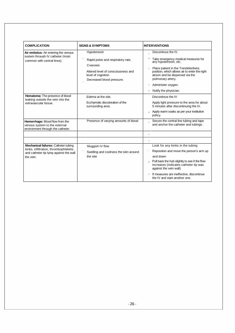
COMPLICATION SIGNS & SYMPTOMS INTERVENTIONS
Air embolus: Air entering the venous
system through IV catheter (most
Hypotension
Discontinue the IV.
common with central lines).
Rapid pulse and respiratory rate. Take emergency medical measures for
any hypotension, etc.
Cyanosis Place patient in the Trendelenberg
Altered level of consciousness and level of cognition
position, which allows air to enter the right atrium and be dispersed via the
Decreased blood pressure. pulmonary artery.
Administer oxygen.
Notify the physician.
Hematoma: The presence of blood
leaking outside the vein into the extravascular tissue.
Edema at the site.
Ecchymotic discoloration of the
surrounding area
Discontinue the IV
Apply light pressure to the area for about
5 minutes after discontinuing the IV.
Apply warm soaks as per your institution
policy.
Hemorrhage: Blood flow from the
venous system to the external environment through the catheter.
Presence of varying amounts of blood
Secure the central line tubing and tape
and anchor the catheter and tubings.
Mechanical failures: Catheter tubing
kinks, infiltration, thrombophlebitis, and catheter tip lying against the wall
Sluggish IV flow
Swelling and coolness the skin around
Look for any kinks in the tubing
Reposition and move the person's arm up
the vein. the site and down
Pull back the hub slightly to see if the flow increases (indicates catheter tip was against the vein wall)
If measures are ineffective, discontinue
the IV and start another one.

- 27 -
APPENDIX C
MEASURES TO PREVENT COMPLICATIONS COMPLICATION PREVENTIVE MEASURES
Infiltration Secure the catheter and tubing well with tape.
Use an arm board, if necessary, to prevent dislodgement of the catheter.
Check the site and tubing often.
Extravasation Make sure that the catheter is secured properly to decrease the chance irritation.
Change the peripheral IV site every 48 to 96 hours, as dictated by facility policy.
Ensure that irritating medications are diluted properly, run at the proper slow rate, and are instilled into large enough veins to prevent chemical irritation.
Phlebitis Make sure that the catheter is secured properly to decrease the chance irritation.
Change the peripheral IV site eve, 48 to 96 hours, as dictated by facility policy.
Ensure that irritating medications are diluted properly, run at the proper slow rate, and are instilled into large enough veins to prevent chemical irritation.
Thrombosis Ensure that irritating medications are diluted properly, run at the proper slow rate, and are instilled into preferably the superior vena cava to prevent chemical irritation.
Catheter embolus Make sure that the needle is never threaded into the catheter once withdrawn.
Air embolus Ensure that air is never permitted to enter into a central venous catheter or any tubing (peripheral, midline, and central venous).
Hematoma Check the site often.
Ensure that the catheter and tubing are securely anchored.
Hemorrhage Connect central line tubing to an adapter cap and not directly to the central line.
Tape and secure all catheters and tubings.
Keep pressure on the site of a discontinued IV for at least 2 minutes and for at least 5 to 10 minutes if the patient is taking an anticoagulant.
Infection and bacteremia Change the IV site every 48 to 96 hours, according to the facility's policy to prevent phlebitis, a major risk factor, which leads to infection.
Use aseptic technique when starting and dressing the IV.
Maintain the integrity of the IV system. It is a closed sterile system.
Date all IV fluids and tubing.
The maximum number of hours for IV supplies is as follows IV tubing: 48 to 72 hours; IV fluids: 24 hours; IV dressings: 48 to 96 hours
Mechanical failures Secure the catheter and tubing.
Use an arm board when necessary.
Check the IV frequently for kinks and patency.
Immediately respond to IV controller and pump alarms.
- 28 -
Please READ!!!!
Directions for Completing this Self-Study Module
1. Read this Self-Study Module.
2. Complete the post-test in Learning Central.
3. A score of 84% or better is needed for passing.
4. You may take three attempts to pass. If you do not pass during this time, the post-test will need to be reset by your Educator for you re-take the test.

- 29 -
POST-TEST Revision 4-19-16
1. Peripheral IV access refers to use of a butterfly needle or any IV catheter that is inserted only a short distance into a peripheral vein.
A . T r u e
B . F a l s e
2. During which Clinical Performance Standard, would a technologist ask the patient if they have had a reaction to IV contrast in the past?
A. Assessm ent B. Analysis/Determination
C. Performance D. Implementation
3. Which of the following is not part of the Seven Rights of Medication Administration?
A. Right Medication B. Right Dose C. Right Rate D. Right Documentation
4. Except in emergencies, the patient or their legal guardian has the right to information necessary to give consent prior to treatment.
A . T r u e
B . F a l s e
5. Which of these factors may affect your ability to perform venipuncture?
A. Patient’s age B. Present and past trauma to vein C. Body tem perature
D. Al l of the above
6. Which factor(s) may influence your choice of lower extremity venipuncture?
A. Possibility of development of thrombus
B. Possibility of development of infection C. A a n d B
D. A o n l y
7. Venous dilation challenges are rarely encountered when patients are dehydrated or they have a decreased circulating volume.
A . T r u e
B . F a l s e

- 30 -
8. Your patient has had right-sided paralysis for 10 years as a result of a stroke. His left lower forearm peripheral IV has just infiltrated. What is the appropriate action to take with respect to restarting the IV?
A. Restart the IV at the same place in the right arm regardless of the paralysis
B. Restart the IV in a foot because both the right and left arms can no longer be used C. Restart the IV in the left upper forearm D. Restart the IV in any vein on the left hand
9. Which of the following is not a way to deal with difficult veins? A. Apply heat or a warm compress to create vasodilation B. Lower the limb into a dependent position to facilitate a greater volume of fluid in the vein C. Repeatedly strike the patient’s vein
D. Rub the vein gently in one continuous motion from the proximal to the distal
10. If you suspect infiltration of IV fluids, you should:
A. Administer oxygen B. Place a tourniquet above site C. Place the patient in Trendelenberg position
D. Discontinue the IV and start another one
11. When you are using chlorhexidine for IV site preparation, you should:
A. Wipe off the excess to prevent skin irritation. B. Blow it dry so it does not become sticky. C. Allow it to air dry.
D. Know that it is a skin sanitizer.
12. Proper tourniquet application includes:
A. Placing the tourniquet 2 - 6 inches above the site. B. Leaving it on arm for 3 minutes. C. Cutting off arterial blood flow. D. Placing the tourniquet 1 inch above the site.
13. Preventing intravenous therapy complications requires:
A. Daily monitoring by the physician. B. The technologist’s ongoing monitoring and care.
C. Little care and monitoring. D. Tech assistants monitor the site and flow rate.
14. Complications that may arise from a poor venipuncture technique include:
A. Hematoma B. Phlebitis C. Thrombophlebitis D. All of the above

- 31 -
15. Which vein is ideal for an adult peripheral IV placement?
A. Antecubital B. Cephalic C. Digital D. Scalp
16. The _____________ vein is found along the ulnar side of the inner lower forearm:
A. Metacarpal B. Median antebrachial C. Basilic D. Digital
17. One of the most common complications associated with IV therapy is:
A. Air or catheter embolism
B. Hematoma
C. Phlebitis D. Septicemia
18. Air embolism associated with IV therapy is most commonly caused by:
A. Contamination of the cannula
B. Loose connections in the IV tubing C. Rapid infusion of fluid D. Pushing the needle through the vein
19. Systemic complications from IV therapy are usually related to the following causes:
A. Poor aseptic technique
B. Contaminated equipment C. Outdated equipment D. All the above
20. If a patient experiences difficulty during or after an IV injection of isotope or contrast agent, you should contact the radiologist immediately.
A. True B. False

- 32 -
COMPETENCY VALIDATION Peripheral IV Insertion
Name:__________________________________________________________
Preceptor:_______________________________________________________
Date of 1st competency:____________ Date of 2
nd competency:____________ Date of 3
rd competency:____________
Competency Statement: Performs peripheral IV insertion while demonstrating correct technique.
Performance Criteria: Participant must complete three successful peripheral IV insertions. A passing score of 60 or more is required for each individual IV insertion.
Preceptor will rate each step individually using a 1 – 4 scale as follows:
Not Competent:
1 = Is unable to state reason for task and needs instructions to perform task.
2 = Understands reason for task but needs instructions to perform task.
Competent:
3 = Understands reason for task and is able to perform task but needs to increase speed.
4 = Understands reason for task and is able to perform task proficiently.
1 2 3
1. ——— ——— ——— Review physician order.
2. ——— ——— ——— Identify patient using at least two patient identifiers.
3. ——— ——— ——— Introduce self to patient and/or family and provide for privacy.
4. ——— ——— ——— Explain the proposed procedures in terms the patient can understand. Assess patient’s previous or perceived experience with IV therapy and arm placement preference.
5. ——— ——— ——— Elevate the bed or stretcher to a level of comfort for starting an IV.
6. ——— ——— ——— Protect patient’s clothing and bedding with an under pad or towel.
7. ——— ——— ——— Wash hands.
8. ——— ——— ——— Set up the necessary supplies on the bedside mayo stand.
9. ——— ——— ——— Don disposable gloves. NOTE: GLOVES CAN BE LEFT OFF TO LOCATE VEIN, BUT MUST BE
APPLIED PRIOR TO PREPARING SITE.
10. ——— ——— ——— Identify accessible vein for IV placement. Apply tourniquet 3 to 6 inches (10-15 cm)
above the proposed insertion site.
11. ——— ——— ——— Select the vein.
12. ——— ——— ——— Release tourniquet. Clip arm hair with scissors (if necessary). DO NOT SHAVE AREA.
13. ——— ——— ——— Cleanse insertion site using firm, up and down motion 2 to 3 inches from insertion site with Chlorhexidine.
14. ——— ——— ——— Reapply tourniquet without touching cleaned area.
15. ——— ——— ——— Perform venipuncture. NOTE: Reserve the catheter packaging to place used needle in.

- 33 -
16. ——— ——— ——— Stabilize the catheter or butterfly. For catheters, apply gentle, but firm, pressure with
the index finger of non-dominant hand 11/4 inches (3 cm) above the insertion site. Release
tourniquet with dominant hand and retract needle. Slide the catheter off the needle and place in
catheter packaging.
17. ——— ——— ——— Quickly connect micro-clave, syringe or primed IV tubing to the hub of the catheter. Ensure good connection.
18. ——— ——— ——— Tape or secure catheter or butterfly.
19. ——— ——— ——— Dispose of used needles in appropriate sharps container. Discard supplies. Remove
gloves and perform hand hygiene.
20. ——— ——— ——— Document in the technical comments for exam in RIS: size of angiocatheter or butterfly, site of venipuncture, amount of medication administered, if medication was hand injected or power-injected. If power-injected, what flow rate was used (mL/sec).
——— ——— ——— Total Scores
——— ——— ——— Initials of Preceptor