radiolysis studies of kappa carrageenan for bio base
TRANSCRIPT
Radiolysis studies of kappa carrageenan
for biobased materials development
(生体由来材料開発のためのカッパ・カラギーナンの
放射線分解に関する研究)
by
Lucille V. Abad(ルシル ベレス アバド)
Department of Nuclear Engineering and Management
Graduate School of Engineering
The University of Tokyo

What t
D
the heart
Dedicated
This one
Tabong,I’ve g
t has once
to Mama
e is for you
breakfast wgot this one
e known, i
a and Tab
Ma.
will no longee ready for y
it shall ne
bong
er be readyyou.
ver forge
but …
et.

This work was done under the sponsorship of the
Japan Society for the Promotion of Science
RONPAKU (PhD Dissertation) Program
in cooperation with the
Department of Science and Technology
Philippines
Through the support of the following institutes:
Philippine Nuclear Research Institute
Nuclear Research Engineering Laboratory
Department of Nuclear Engineering and Management
The Univeristy of Tokyo
Takasaki Advanced Radiation Research Institute
Japan Atomic Energy Agency
Neutron Science Laboratory
Institute for Solid State Physics
The University of Tokyo
“Something that has always puzzled me all my life is why, when I am in
special need of help, the good deed is usually done by somebody on whom I
have no claim.”
William Feather
To my Advisers…
Dr. Hisaaki Kudo and Dr. Alumanda M. De la Rosa
Thank you for guiding me all the way and making sure that I finish this successfully.
To my Japanese Supervisors…
Prof. Mitsuhiro Shibayama, Dr. Masao Tamada, and Prof. Yosuke Katsumura
Thank you for giving me the rare opportunity to work with you in your laboratory.
To my Panelists
Prof. Yosuke Katsumura, Prof. Takayuki Terai, Prof. Mitsuhiro Shibayama, Dr. Hisaaki Kudo, and Dr. Masao Tamada
To my Japanese Mentors
Saiki-san and Nagasawa-san
Thanks for all the technical discussions, the planning and implementation of my experimental design, and most of all for teaching me the Japanese ways of doing things.
To my Japanese friends and acquaintances…
Lin-san, Muroya-san, Ira, Satoshi-san, Fu-san, Hiroki-san, Hasegawa-san, Tokoro-san, Asahi-san, Okamoto-san, Makuuchi-san, Yoshii-san, Kume-san, Takigami-san,
Amada-san, Imura-san, my gaijin tomodachis, and Japanese gakuseis…
A million thanks for putting up with all my “ “
お願いします

To my PNRI family…
Alum, Boss Elvies, Lorna, Chat, Biboy, Jay, Andrew, Ryan, Adel, Angie, Simon, Rina, Aileen, Sol, Sony, Mon, Jo Mike, Fe “Ima”, Yen-yen, Maricel, Boss Gin, my “ka-
bonding”, and the rest of the clan.
Thanks for all your support –technical or non-technical, for the spices of light moments that you provide!
To my OJT students and chemists…
Jaimee, Yam, Eco, Ron-ron, Julienne, Day, Arlyn
Thank you for your eagerness to learn and patience to repeatedly do accurately the analyses.
To my friends in Japan…
Agnes & Gigi with their two “chikitings”, Anabel, Adelfa, Michelle, Mida, Teks, Ely & Kuya Roger, Joji & Jun, Cora, Ascen, Mila, Goie, Kimie-san & Seigi-san and TA Japan
Thanks for the happy days in Japan over a glass of “biru” with matching spaghetti and “magic sing”.
To my Teresian Family…
Eufro, Bebing, Queenie, Amyting, Baleleng, Julie (+), Ester, Melds, Roli, Cena, Thelma, Remy, Mimi, Marj, Margie, Sally, Chuchi, Novie and all my special friends.
Thanks for your encouragement and raining heaven with your prayers.
To the best of my Kin… Teddy & Yvonne, Gemma & Raul, Danny & Pining, Justine and Joe, their siblings…
Thanks for your gestures of countless love and support.
And above all…
Thank you God for whispering in my ear – Yes You Can!
ABSTRACT
Kappa (-) carrageenan oligomers are known to have several biological
activities such as anti-HIV, anti-herpes, antitumor and antioxidant properties. Recent
progress in the development of radiation modified -carrageenan has resulted in new
applications such as plant growth promoter, radiation dose indicator and hydrogels for
wound dressing. This study would investigate on the changes in chemical structure,
gelation and conformational transition behavior and molecular size of -carrageenan
at doses from 0 to 200 kGy and would be correlated to these functions for the
development of bio-based materials.
Pulse radiolysis studies on -carrageenan was carried out to determine what
transient species directly affects the degradation rate of -carrageenan in aqueous
solution. The results reveal that there is no seeming reaction of the hydrated electron
with -carrageenan. OH• reacts with -carrageenan at a fast rate of approximately 1.2
x 109 M-1s-1. This value was influenced by conformational change from helix to coil
by the addition of the metal ion Na+, reduction of molecular weight by hydrolysis
reaction and reduction of reactive sites by sonolysis or irradiation.
Most applications from the radiation degradation of polysaccharides started
with the use of the “hit and miss” process where polysaccharides were irradiated at a
certain dose range and finding out which dose is suitable for a specific function.
Measurement of the radiation degradation yield (Gd) at different conditions can give
an approximation of the Mw at an absorbed dose. This will allow the production of
oligomers with a specified Mw. With the use of the Gd both in solid and in aqueous
solution, one can also make a rough calculation whether it is more economical to
irradiate -carrageen in solid or in aqueous solution. Results of this experiment reveal

ii
that the radiation degradation yields (Gd) of -carrageenan in solid and in aqueous
(1%) were as follows: 2.5, 1.7 and 1.2 x 10⁻⁷ mol J-1 for solid in atmosphere, solid in
vacuum and at 1% aqueous solution, respectively. The presence of N2O gas in
aqueous -carrageenan solution increased its Gd whereas a decrease in Gd was
observed in the presence of N2 gas. The Gd of -carrageenan was also affected by the
conformational change from helix to coil in the presence of Na+ ion.
This study would also investigate on the chemical structure of the radiolytic
products of -carrageenan at doses from 0 to 200 kGy at different irradiation
conditions. The results would determine the extent of destruction of its basic structure
[(13)-4 sulfate--D galactose (14)-3, 6 anhydro-"-D-galactose)] with absorbed
dose. The new functional groupings produced by radiation will be determined. The
findings would be related to some possible uses of the -carrageenan oligomers e.g.
plant growth promoter, anti-viral, anti-tumor, antioxidants, stimuli responsive gels,
hydrogels and as radiation dose indicators. Chemical and spectral analyses were
carried out using UV-Vis spectroscopy, FT-IR spetroscopy, NMR spectroscopy,
reducing sugar analysis, free sulfate and carboxylic acid analysis. The chemical and
spectral analyses of the radiolytic products indicated increasing reducing sugars,
carbonyl, carboxylic acids, and sulfates with increasing doses which reach a
maximum level at a certain dose depending on the irradiation condition. Values were
very much lower in solid irradiation (in vacuum and in air) as compared to aqueous
irradiation. NMR data also revealed an intact structure of the oligomer irradiated at
100 kGy in the specific fraction that contains an Mw = (3-10) kDa.
In addition to changes in chemical structure with absorbed dose, it would also
be important to determine the changes in dynamic behavior and structure form of -

iii
carrageenan with absorbed dose. This would indicate changes in gelation behavior
and conformational transition temperatures of -carrageenan or the destruction of its
polymer chain. The results would be important in determining the appropriate
molecular size needed for its biological activities. Dynamic light scattering (DLS) and
small angle neutron scattering (SANS) were carried out for this purpose. DLS
experiments reveal that at a dose of up to 50 kGy, sol-gelation transition was still
observed. Beyond 50 kGy, no gelation took place, instead appearance of fast
relaxation mode in characteristic decay time function was observed at doses of (75–
150) kGy. Optimum peak intensity was found at 100 kGy (Mol wt .5 – 10 kDa)
which coincides with the optimum plant growth promoter effect in -carrageenan. At
a dose beyond 150 kGy, the conformational transition temperature from coil to helix
was no longer observed. In addition, SANS experiment indicated a unique structure
form at 100 kGy wherein a power law behavior with a fractal dimension -1.84 was
observed.
The correlation of all the results gave us a wide spectrum of the possible uses
of -carrageenan for bio-based material development.
iv
TABLE OF CONTENTS
Pages
CHAPTER 1: INTRODUCTION……………………………………………... 1
Biological Activity of Carrageenan………………………………………... 5
Carrageenan-based radiation dose indicator……………………………….. 8
Polyvinyl pyrrolidone – - Carrageenan……………………………………. 10
Hydrogels for Burn / Wound Dressing
Objectives……………………………………………………………….…. 11
CHAPTER 2: RATE CONSTANTS OF REACTIONS
OF -CARRAGEENAN WITH HYDRATED
ELECTRON AND HYDROXYL RADICAL…………………. 18
2.1. INTRODUCTION…………………………………………………..... 18
2.2. MATERIALS AND METHODS………………..………………….... 19
2.2.1. Materials…………………………………………………….….. 19
2.2.2. Sample Preparation…………………………………………..…. 20
2.2.3. Laser Photolysis of -carrageenan………………………..……. 20
2.2.4. Pulse Radiolysis of -carrageenan…………………………..…. 20
2.2.5. Irradiation of -carrageenan………………………………..…... 22
2.2.6. Sonication of -carrageenan………………………………..…... 22
2.2.7. Molecular weight Determination of -carrageenan…………..… 22
2.2.8. UV-Vis Analysis……………………………………………...… 23
2.2.9 Viscosity Measurement……………………………………...….. 23
2.3. RESULTS AND DISCUSSION…………………………….……….. 23
2.3.1 Reaction of hydrated electrons with -carrageenan…………….. 23
2.3.2. Rate constant of hydroxyl radicals reactions
with -carrageenan……………………………………………. 25
2.3.3. pH Dependence…………………………………………………. 27
2.3.4. Pulse radiolysis studies of sonicated and irradiated
-carrageenan…………………………………………………... 29

v
Pages
2.3.5. The effect of sodium ion on the rate constant
of reaction of OH radical with - carrageenan…………………. 33
2.4. CONCLUSION…………………………………………………………. 37
CHAPTER 3: RADIATION DEGRADATION
YIELD OF -CARRAGEENAN…………………………….. 41
3.1. INTRODUCTION……………………………………………………… 41
3.2 METHODOLOGY……………………………………………………… 45
3.2.1. Sample Preparation……………………………………………….. 45
3.2.2 Gamma Irradiation of -carrageenan……………………………… 46
3.2.3. GPC Analyses of -carrageenan………………………………… 46
3.2.4. UV Vis Spectroscopy……………………………………………... 46
3.3. RESULTS AND DISCUSSION………………………………………... 46
3.3.1. Radiation Degradation Yield of -Carrageenan………………….. 46
3.3.2. The Effect of N2 and N2O gas on the
Radiation Degradation Yield of -Carrageenan………………… 51
3.3.3. The Effect of Na+ on the Gd of -carrageenan…………………… 54
3.4. CONCLUSION…………………………………………………………. 56
CHAPTER 4: CHEMICAL AND SPECTRAL CHARACTERIZATION
OF THE RADIOLYTIC PRODUCTS OF
-CARRAGEENAN …………………………………………… 61
4.1. INTRODUCTION……………………………………………………… 61
4.2. METHODOLOGY……………………………………………………... 64
4.2.1. Gamma Irradiation of -carrageenan……………………………... 64
4.2.2. UV Vis Spectroscopy……………………………………………... 64
4.2.3. FT-IR Spectroscopy……………………………………………… 64
4.2.4. Chemical Analyses………………………………………………... 64
4.2.5. Fractionation of Irradiated -carrageenan………………………... 65
4.2.6. NMR of Irradiated -carrageenan………………………………… 65
4.3. RESULTS AND DISCUSSION………………………………………... 66
4.3.1. UV-Vis Spectrum………………………………………………….66
vi
Pages
4.3.2. FT-IR Spectrum…………………………………………………... 69
4.3.3. Reducing Sugars………………………………………………….. 71
4.3.4. Acidity……………………………………………………………..73
4.3.5. Fractionation of Irradiated -carrageenan………………………... 77
4.3.6. NMR of Fractionated Irradiated -carrageenan …………………. 80
4.3.6.1. NMR of carrabiose standards……………………………… 81
4.3.6.2. NMR of oligomers from irradiated -carrageenan……….. 87
4.4. CONCLUSION…………………………………………………………. 95
4.4.1. For 1% Aqueous Solution Irradiation …………………………… 95
4.4.2. For Solid Irradiation …………………………………………. 96
CHAPTER 5: STRUCTURAL AND DYNAMIC BEHAVIOR OF
IRRADIATED - CARRAGEENAN…………………………. 103
5.1. INTRODUCTION……………………………………………………… 103
5.2. METHODOLOGY……………………………………………………... 104
5.2.1. Dynamic Light Scattering………………………………………… 104
5.2.2. Small Angle Neutron Scattering………………………………….. 105
5.3. RESULTS AND DISCUSSION……………………………………….. 107
5.3.1. Dynamic Light Scattering Studies of Irradiated -Carrageenan….. 107
5.3.1.1. Molecular weight dependence and characteristic
relaxation time dependence of -carrageenan
with absorbed dose………………………………………. 108
5.3.1.2. Coil-helical transition of irradiated -carrageenan……….. 110
5.3.1.3. Gelation temperatures of -carrageenan as a
function of dose…………………………………………… 116
5.3.1.4. Fast relaxation mode peaks corresponding to
optimum biological activity of -carrageenan………………119
5.3.2. Small-angle Neutron Scattering Study on Irradiated
-Carrageenan……………………………………………………. 121
5.3.2.1. Physical Properties of irradiated -carrageenan gels……… 121
5.3.2.2. SANS profile of -carrageenan gels………………………. 123
5.4. CONCLUSION………………………..……………………………….. 127
vii
Pages
CHAPTER 6: RADIOLYSIS REACTIONS OF -CARRAGEENAN……….. 132
6.1. INTRODUCTION………………………………………………………132
6.2. METHODOLOGY……………………………………………………... 133
6.3. RESULTS AND DISCUSSION……………………………………….. 133
6.3.1. Electron Spin Resonance of -carrageenan………………….. 133
6.3.2. Proposed Mechanism for the Radiolysis Reaction
of -carrageenan…………………………………………….. 137
CHAPTER 7: SUMMARY AND CONLUSIONS……………………………... 148

viii
LIST OF FIGURES
Pages
Figure 1.1. Idealized structure of -, - and -carrageenan……………………... 2
Figure 1.2. Plant growth promnotion effect of -carrageenan in rice…………... 7
Figure 1.3. Different formulations of -carrageenan-based
radiation dose indicator………………………………………………………. 9
Figure 1.4. PVP- -carrageenan hydrogel for wound/burn dressing…………….. 11
Figure 1.5. Over-all view of the different chapters………………………...……. 14
Figure 2.1. The schematic diagram of experimental apparatus of a pulse
radiolysis or laser photolysis combined with photo-spectroscopic method….. 21
Figure 2.2. Decay profiles of the absorbance at 720 nm after
pulse radiolysis of -carrageenan at varying concentrations………………... 24
Figure 2.3. Determination of the rate constant reaction of ·OH with
-carrageenan by competition method………………………………………. 26
Figure 2.4. Determination of the rate constant reaction of ·OH with
-carrageenan by competition method at varying pH (laser photolysis
method)………………………………….…………………………………... 28
Figure 2.5. Determination of the rate constant reaction of ·OH with
-carrageenan by competition method after neutralization at pH =2
(laser photolysis method)……………………………………………...…….. 28
Figure 2.6. Determination of the rate constant reaction of OH radical
with sonicated -carrageenan…………………………………………..…… 30
Figure 2.7. Determination of the rate constant reaction of OH radical
with irradiated -carrageenan…………………………………..…………… 30
Figure 2.8. UV-Vis spectra of sonicated -carrageenan………………………... 33
Figure 2.9. The effect of Na+ on the rate constant of OH· reaction
with -carrageenan………………………………………..…………………. 34
Figure 2.10. Two models of conformational transition in -carrageenan………. 36
Figure 3.1. Main mechanism for acid hydrolysis of -carrageenan……………... 44
Figure 3.2. GPC Chromatogram of a) irradiated solid
-carrageenan and b) irradiated aqueous -carrageenan……………………. 47

ix
Pages
Figure 3.3. Molecular Weight of -carrageenan in air, in vacuum and
in aqueous solution at various doses……………………………………….... 48
Figure 3.4. Reciprocals of Mw of solid -carrageenan in air and in vacuum
at varying doses…………………………………………………….………... 49
Figure 3.5. Reciprocal of Mw of 1% aqueous -carrageenan at varying doses… 50
Figure 3.6. Reciprocal of Mw of 1% aqueous -carrageenan at varying doses
purged with different gases……………………………………………..…... 52
Figure 3.7. UV absorbance at 260nm of -carrageenan (0.25%) irradiated at
different conditions…………………………………………………………. 53
Figure 3.8. The Effect of Na+ on the reciprocals of Mw of -carrageenan……... 55
Figure 4.1. Structure of Neocarrabiose Standards………………………………. 66
Figure 4.2. UV-Vis spectra of -carrageenan irradiated as
a) solid in atmosphere b) solid in vacuum and c) 1% aqueous
solution at varying doses…………………………………………………….. 67
Figure 4.3. UV absorbance (260nm) of irradiated -carrageenan
at different conditions……………………………………………………… 68
Figure 4.4. FT-IR spectra of -carrageenan irradiated in air, in vacuum
and in 1% aqueous solution at different doses……………………..……….. 70
Figure 4.5. Percent reducing sugar of -carrageenan irradiated at increasing
doses………………………..………………………………………………... 72
Figure 4.6. Percent Acidity of -carrageenan as a function of dose at
different conditions………………………………………………………….. 74
Figure 4.7. Fractional yield of irradiated -carrageenan………………………. 79
Figure 4.8. Proton NMR of neocarrabiose standards…………………………... 83
Figure 4.9. 13C NMR of neocarrabiose standards……………………………... 84
Figure 4.10. 1H NMR of irradiated -carrageenan oligomers…….……………....91
Figure 4.11. 13C NMR of irradiated -carrageenan oligomers…………………. 92
Figure 4.12. 1H and 13C NMR of -carrageenan polymer……………………... 93
Figure 4.13. Summary of chemical and spectral analyses of
-carrageenan irradiated in 1% aqueosus solution at varying doses………….97

x
Pages
Figure 4.14. Summary of chemical and spectral analyses of
solid -carrageenan irradiated in vacuum at varying doses………….………. 98
Figure 4.15. Summary of chemical and spectral analyses of
solid -carrageenan irradiated in air at varying doses……………………….. 98
Figure 5.1. The DLS Set-up………………………………………………..…….. 104
Figure 5.2. Schematic Diagram of the SANS-U Set-up………………..……….. 106
Figure 5.3. (a) Intensity correlation function and (b) decay time
distribution of 0.5% irradiated -carrageenan (in 0.05M KCl) at 80oC…….. 109
Figure 5.4. Relationship of the molecular weight of -carrageenan
(0.5% in 0.05M KCl) and 1/ (T=80oC and a scattering angle of 90o)
with absorbed dose………………..………………………………………..… 110
Figure 5.5. Decay time distribution function of 0.5% irradiated
-carrageenan (in 0.05M KCl) at varying temperatures at doses
of (a) 0 kGy (b) 150 kGy (c) 300 kGy……………………………………… 111
Figure 5.6. Conformational transition temperature as a function
of absorbed dose……………..……………………………………………… 112
Figure 5.7. Effect of gamma radiation on the structure of -carrageenan………. 115
Figure 5.8. Conformational transition models of -carragenan…………………. 117
Figure 5.9. ICF 0.5% irradiated -carrageenan (in 0.05M KCl) at varying
temperatures at doses of (a) 0 kGy (b) 100 kGy and (c) 200 kGy……...……. 118
Figure 5.10. Decay time distribution function of 0.5% irradiated
-carrageenan (in 0.05M KCl) at varying temperatures at doses
of (a) 0 kGy (b) 50 kGy (c) 75 kGy (d) 100 kGy (e) 150 kGy
and (f) 200 kGy………………………………………………………………. 120
Figure 5.11. -carrageenan gels in 0.05M KCl at 20oC………………………….. 122
Figure 5.12. Gelation temperatures of 5% -carrageenan………………………. 123
Figure 5.13. SANS intensity curves of -carrageenan
(in 0.0M KCl at 20oC) at varying doses……………………………………... 124
Figure 5.14. Power law behavior of -carrageenan
(5% in 0.05M KCl at 20oC) irradiated at 100 kGy………………………….. 126
Figure 5.15. Distribution of -carrageenan polymer chains
with dose…………………………………………………………………... 126

xi
Pages
Figure 5.16. Changes in gelation and conformational transition
behavior of irradiated -carrageenan at varying doses………………………..129
Figure 6.1. ESR of -carrageenan irradiated at varying doses in air…………......134
Figure 6.2. ESR of -carrageenan at varying temperatures
irradiated in vacuum…………………………………………………………. 136
Figure 6.3. ESR of -carrageenan irradiated in vacuum and in air
at 5 and 50 kGy…………………………………………………………….. 136
Figure 7.1. Effective dose range of irradiated - carrageenan
for bio-based materials development…………………………………………153
Figure 7.2. Over-all view of the current research and some future works………..154
xii
LIST OF TABLES
Pages
Table 1.1. Summary of the biological functions of -carrageenan oligomer….…8
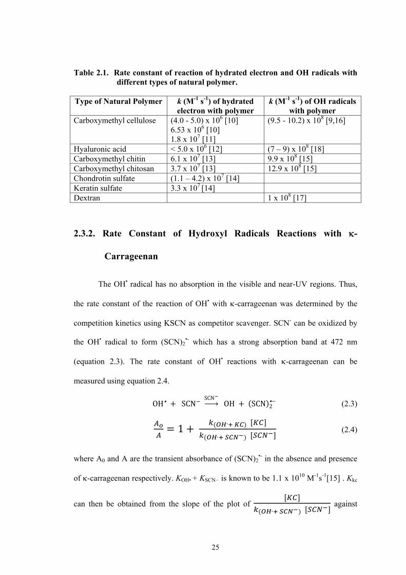
Table 2.1. Rate constant of reaction of hydrated electron and
OH radicals with different types of natural polymer………………………... 25
Table 2.2. Rate constant of reaction of OH radicals with
-carrageenan at different conditions…………………………………….…..36
Table. 3.1 Radiation degradation yield of different polysaccharides
irradiated in solid and aqueous solution……………………………………. 51
Table 4.1a. Acidic composition of gamma irradiated solid
-carrageenan (in air) with absorbed dose………………………………….. 76
Table 4.1b. Acidic composition of gamma irradiated solid
-carrageenan (in vacuum) with absorbed dose…………………………….. 76
Table 4.1c. Acidic composition of gamma irradiated -carrageenan
(1% aqueous solution) with absorbed dose…………………………………. 76
Table 4.2. Molecular weight of the fractionated samples of
irradiated -carrageenan…………………………………………………….. 78
Table 4.3. UV-Vis ( = 260nm) of fractionated irradiated
-carrageenan (0.025%)……………………………………………………… 78
Table 4.4. Chemical shifts of the 1H NMR of neocarrabiose standards………… 85
Table 4.5. Chemical Shifts of the 13C NMR of neocarrabiose standards………. 86
Table 4.6. Chemical Shifts of the proton NMR of irradiated
-carrageenan oligomers……………………………………………………... 94
Table 4.7. Chemical shifts of the C-13 NMR of irradiated
-carrageenan oligomers……………………………………………………... 94
Table 5.1. Molecular weight (Mw), conformational transition
temperature (CTT) and gelation temperature (GT) of
-carrageenan at varying doses…………………………………...…………. 119
CHAPTER 1
INTRODUCTION
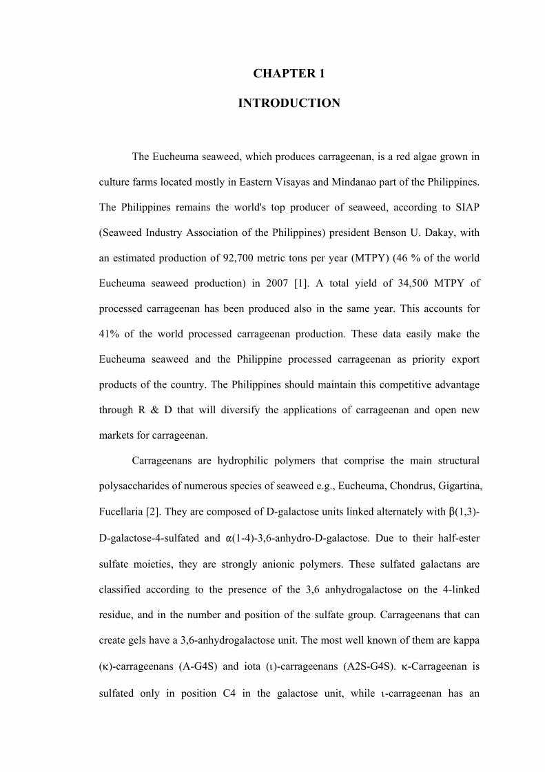
The Eucheuma seaweed, which produces carrageenan, is a red algae grown in
culture farms located mostly in Eastern Visayas and Mindanao part of the Philippines.
The Philippines remains the world's top producer of seaweed, according to SIAP
(Seaweed Industry Association of the Philippines) president Benson U. Dakay, with
an estimated production of 92,700 metric tons per year (MTPY) (46 % of the world
Eucheuma seaweed production) in 2007 [1]. A total yield of 34,500 MTPY of
processed carrageenan has been produced also in the same year. This accounts for
41% of the world processed carrageenan production. These data easily make the
Eucheuma seaweed and the Philippine processed carrageenan as priority export
products of the country. The Philippines should maintain this competitive advantage
through R & D that will diversify the applications of carrageenan and open new
markets for carrageenan.
Carrageenans are hydrophilic polymers that comprise the main structural
polysaccharides of numerous species of seaweed e.g., Eucheuma, Chondrus, Gigartina,
Fucellaria [2]. They are composed of D-galactose units linked alternately with $(1,3)-
D-galactose-4-sulfated and "(1-4)-3,6-anhydro-D-galactose. Due to their half-ester
sulfate moieties, they are strongly anionic polymers. These sulfated galactans are
classified according to the presence of the 3,6 anhydrogalactose on the 4-linked
residue, and in the number and position of the sulfate group. Carrageenans that can
create gels have a 3,6-anhydrogalactose unit. The most well known of them are kappa
()-carrageenans (A-G4S) and iota ()-carrageenans (A2S-G4S). -Carrageenan is
sulfated only in position C4 in the galactose unit, while -carrageenan has an

2
additional sulfate unit in position C2 of the 3,6-anhydrogalactose unit. Both the
number and the position of sulfate [3] and the 3,6-anhydrogalactose [4], have a
dramatic effect on the tertiary structure and the possible interactions of the different
types of carrageenans. -carrageenan forms gels that are hard, strong and brittle,
whereas -carrageenan forms soft and weak gels [5]. Lambda ()-carrageenan differs
from - and -carrageenan by having a disulfated-D-galactose residue and no 4-sulfate
in the -D-galactose residue. Instead of 4-sulfate ester groups, -carrageenan contains
variable amounts of 2-sulfate ester groups (G2,6S-G2S). Lambda carrageenan does
not gel at all. The repeating units -, -, and -carrageenans are shown in Figure 1.1.
Figure 1.1. Idealized structure of -, - and -carrageenan
O
O
CH2OH
HO
O
-O3SO
OCH2OSO3
-
-O3SO
OSO3-
OCH2OH
-O3SO
OH
O
H2CO
O
iota-carrageenan
OH
OCH2OH
-O3SO
OH
O
H2CO
O
kappa-carrageenan
O
O
O
O
CH2OH
HO
O
-O3SO
OCH2OSO3
-
-O3SO
O
O
CH2OH
HO
O
-O3SO
OCH2OSO3
-
-O3SO
OSO3-
OCH2OH
-O3SO
OH
O
H2CO
O
iota-carrageenan
OH
OCH2OH
-O3SO
OH
O
H2CO
O
kappa-carrageenan
O
O
lambda-carrageenan
3
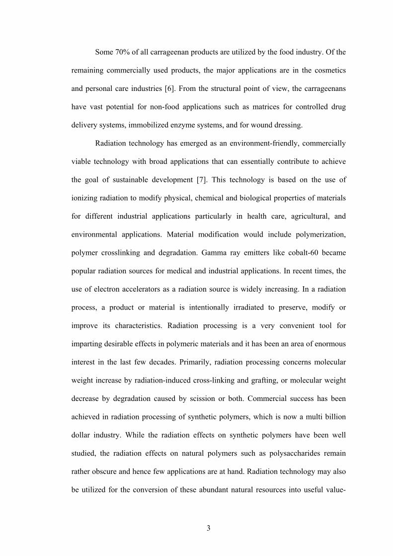
Some 70% of all carrageenan products are utilized by the food industry. Of the
remaining commercially used products, the major applications are in the cosmetics
and personal care industries [6]. From the structural point of view, the carrageenans
have vast potential for non-food applications such as matrices for controlled drug
delivery systems, immobilized enzyme systems, and for wound dressing.
Radiation technology has emerged as an environment-friendly, commercially
viable technology with broad applications that can essentially contribute to achieve
the goal of sustainable development [7]. This technology is based on the use of
ionizing radiation to modify physical, chemical and biological properties of materials
for different industrial applications particularly in health care, agricultural, and
environmental applications. Material modification would include polymerization,
polymer crosslinking and degradation. Gamma ray emitters like cobalt-60 became
popular radiation sources for medical and industrial applications. In recent times, the
use of electron accelerators as a radiation source is widely increasing. In a radiation
process, a product or material is intentionally irradiated to preserve, modify or
improve its characteristics. Radiation processing is a very convenient tool for
imparting desirable effects in polymeric materials and it has been an area of enormous
interest in the last few decades. Primarily, radiation processing concerns molecular
weight increase by radiation-induced cross-linking and grafting, or molecular weight
decrease by degradation caused by scission or both. Commercial success has been
achieved in radiation processing of synthetic polymers, which is now a multi billion
dollar industry. While the radiation effects on synthetic polymers have been well
studied, the radiation effects on natural polymers such as polysaccharides remain
rather obscure and hence few applications are at hand. Radiation technology may also
be utilized for the conversion of these abundant natural resources into useful value-

4
added products. In recent years, natural polymers are being looked at with interest
because of their unique characteristics like inherent biocompatibility, biodegradability
and easy availability. Among the most abundant natural polymers, cellulose, chitin,
carrageenan, and alginates have been found to be the most promising candidates for
radiation processing [8]. Many new areas of use of radiation modified natural
polymers are being explored by researchers, such as pharmaceuticals, nanotechnology,
biomaterials, biofuels and biochemicals [9].
The irradiation of natural polymeric materials with ionizing radiation (gamma
rays, X-rays, or accelerated electrons) leads to the formation of very reactive
intermediates and free radicals. These intermediates can follow several reaction paths
that result in disproportion, hydrogen abstraction, arrangements and/or the formation
of new bonds. The degree of these transformations depends on the structure of the
polymer and the conditions of treatment before, during and after irradiation. Thorough
control of all of these factors facilitates the modification of polymers by radiation
processing. Radiation modification of natural polymers covers cross-linking (in the
presence of water soluble synthetic polymers), grafting, curing (for natural oils) and
degradation. With the end in view of developing non-food applications of
carrageenans, several works have been done on the radiation modification of
carrageenan to produce lower molecular weight fragments or crosslinked hydrogels.
Its applications cover the following: a) radiation processed carrageenan oligomers as
plant growth promoter; b) -carrageenan / polyethylene oxide (PEO) hydrogels as
radiochemical dosimeters; and c) Polyvinyl pyrolidone (PVP)-carrageenan hydrogels
as wound/burn dressing.

5
Biological Activity of Carrageenan
Carrageenans are known to have valuable biological functions. Due to the
superior gelling and high viscosity properties of the native carragenans, their
utilization for biological applications is in most cases in the form of their oligomers.
Oligo--carrageenans induce secretion of laminarinase from Rubus cells and
protoplast [10]. Degraded λ-carrageenan is reported to have tumor inhibiting activities
[11, 12]. Oligomers from carrageenans suggest promising antiherpetic and anti-HIV
(human immunodeficiency virus) activities [13 - 16]. Oligomers of carrageenan can
easily be prepared through depolymerisation either by chemical or enzymatic
hydrolysis.
Recently, degradation by radiation processing of polysaccahrides has gained
much attention due to its technological effectiveness in producing low molecular
weight oligomers. Radiation degraded polysaccharides such as chitin, chitosan,
carrageenan, alginates can induce various kinds of bioactivities such as growth
promotion of plants, suppression of heavy metal stress on plants and anti
microbiological activities [17]. Irradiated alginate shows a strong effect on the
growth-promotion of rice and peanut. Degraded alginate (MW ca. 7000) in 4%
alginate solution irradiated at 100 kGy or from powder irradiated at 500 kGy has a
remarkable effect on growth promotion of rice [18]. Oligochitosan with Mw of 2,000
inhibited the growth of fungi and that with Mw 800 enhances the growth of the same
typical fungi [19]. Depolymerized chitosan with an Mw of 47,000 shows growth
promoting effect in spring rape seeds [20]. Anti-bacterial activity of irradiated
chitosan has been tested against Escherichia coli B/r [21]. Irradiated pectin induces
Phytoalexin elicitor activity to prevent infection of plants by several fungi [22-27].
Irradiated chitosan has anti-fungal activity when used as coats for mango [28].
6
Irradiated lignocllulosic materials suppresses the heavy metal and salt stress on barley
plants [28]. Oligoalginate prepared by irradiation with an Mw of approximately
14,000 is found to be effective for in vitro propagation of flower plants [29].
Oligogalacturonide fragments are found to have root-inhibiting and flower-
stimulating effects [30- 31].
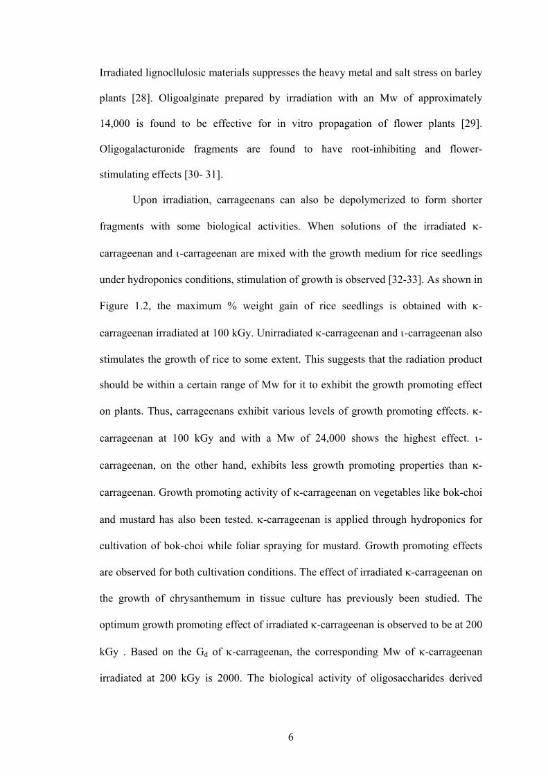
Upon irradiation, carrageenans can also be depolymerized to form shorter
fragments with some biological activities. When solutions of the irradiated -
carrageenan and -carrageenan are mixed with the growth medium for rice seedlings
under hydroponics conditions, stimulation of growth is observed [32-33]. As shown in
Figure 1.2, the maximum % weight gain of rice seedlings is obtained with -
carrageenan irradiated at 100 kGy. Unirradiated -carrageenan and -carrageenan also
stimulates the growth of rice to some extent. This suggests that the radiation product
should be within a certain range of Mw for it to exhibit the growth promoting effect
on plants. Thus, carrageenans exhibit various levels of growth promoting effects. -
carrageenan at 100 kGy and with a Mw of 24,000 shows the highest effect. -
carrageenan, on the other hand, exhibits less growth promoting properties than -
carrageenan. Growth promoting activity of -carrageenan on vegetables like bok-choi
and mustard has also been tested. -carrageenan is applied through hydroponics for
cultivation of bok-choi while foliar spraying for mustard. Growth promoting effects
are observed for both cultivation conditions. The effect of irradiated -carrageenan on
the growth of chrysanthemum in tissue culture has previously been studied. The
optimum growth promoting effect of irradiated -carrageenan is observed to be at 200
kGy . Based on the Gd of -carrageenan, the corresponding Mw of -carrageenan
irradiated at 200 kGy is 2000. The biological activity of oligosaccharides derived

7
from irradiated -carrageenan at different doses in potato tissue culture bioassay has
also been tested. Unirradiated carrageenan inhibits the growth of potato in tissue
culture while carrageenan irradiated at 30 kGy (Mw = 10,000) shows the highest
growth promoting effect. Compared to the control, the fresh biomass and shoot height
increases by 35% and 15%, respectively, when supplemented with oligosaccharide
with an Mw of 10,000 [34].
While much progress has been done in studies related to the application of
carrageenans as plant growth promoter, very few fundamental researches on the
radiation chemistry of carrageenan and radiolytic products formed have been made.
Studies on the effect of radiation on carrageenan and the characterization (chemical
structure and molecular size) of the radiolytic products will provide a better insight on
the preparation of these biomaterials as plant growth promoter. Furthermore, the
degradation response of carrageenan to the absorbed dose at different radiation
conditions will optimize conditions for the preparation of such materials which is an
important factor if economics have to be considered.
Figure 1.2. Plant growth promotion effect of -carrageenan in rice
8
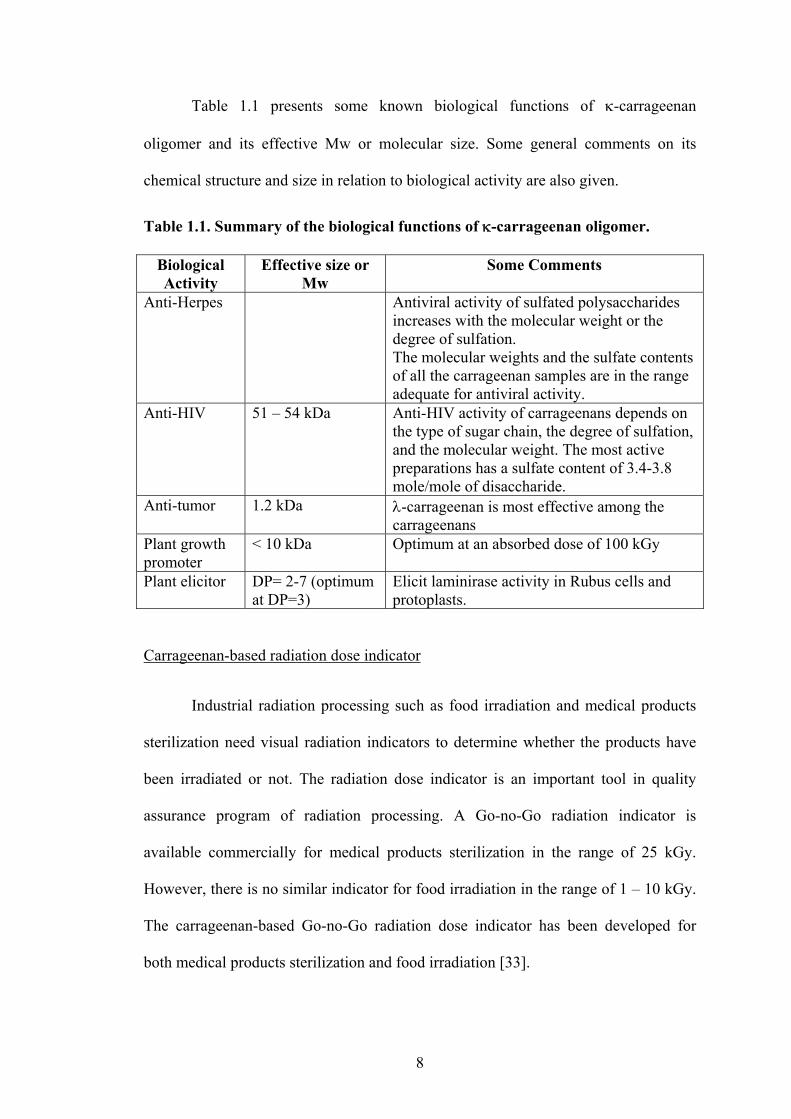
Table 1.1 presents some known biological functions of -carrageenan
oligomer and its effective Mw or molecular size. Some general comments on its
chemical structure and size in relation to biological activity are also given.
Table 1.1. Summary of the biological functions of -carrageenan oligomer.
Biological Activity
Effective size or Mw
Some Comments
Anti-Herpes Antiviral activity of sulfated polysaccharides increases with the molecular weight or the degree of sulfation. The molecular weights and the sulfate contents of all the carrageenan samples are in the range adequate for antiviral activity.
Anti-HIV 51 – 54 kDa Anti-HIV activity of carrageenans depends on the type of sugar chain, the degree of sulfation, and the molecular weight. The most active preparations has a sulfate content of 3.4-3.8 mole/mole of disaccharide.
Anti-tumor 1.2 kDa -carrageenan is most effective among the carrageenans
Plant growth promoter
< 10 kDa Optimum at an absorbed dose of 100 kGy
Plant elicitor DP= 2-7 (optimum at DP=3)
Elicit laminirase activity in Rubus cells and protoplasts.
Carrageenan-based radiation dose indicator
Industrial radiation processing such as food irradiation and medical products
sterilization need visual radiation indicators to determine whether the products have
been irradiated or not. The radiation dose indicator is an important tool in quality
assurance program of radiation processing. A Go-no-Go radiation indicator is
available commercially for medical products sterilization in the range of 25 kGy.
However, there is no similar indicator for food irradiation in the range of 1 – 10 kGy.
The carrageenan-based Go-no-Go radiation dose indicator has been developed for
both medical products sterilization and food irradiation [33].

9
It consists of phenol red, an acid-sensitive dye mixed homogeneously with a
polymer base such as carrageenan or the carrageenan-based hydrogels (KC/MPEO
(medium molecular weight polyethylene oxide) hydrogel). The indicator is being
developed based on the observation that carrageenan releases the sulfate group during
irradiation thereby changing the pH around the carrageenan environment. A decrease
in pH of -carrageenan from pH 6.7 to pH 3.4 with dose (5 to 30 kGy) is observed.
This change in pH will elicit a reaction from the acid-sensitive dye resulting in a
change in color as the specific pH is reached. The radiation indicator being developed
is in gel or film form which can be affixed to the product packaging (Figure 1.3).
This product developed is simply based from the practical observation that
carrageenan would decrease its pH with dose. A study on the acidic components
produced from the irradiation of -carrageenan and the mechanism involved in their
formation would give additional information to further develop this application.
Figure 1.3. Different formulations (a-d) of -carrageenan-based radiation dose indicator
10
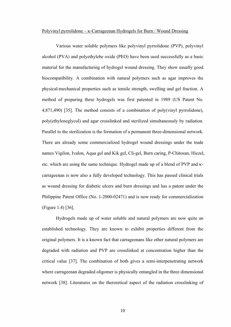
Polyvinyl pyrrolidone – -Carrageenan Hydrogels for Burn / Wound Dressing
Various water soluble polymers like polyvinyl pyrrolidone (PVP), polyvinyl
alcohol (PVA) and polyethylebe oxide (PEO) have been used successfully as a basic
material for the manufacturing of hydrogel wound dressing. They show usually good
biocompatibility. A combination with natural polymers such as agar improves the
physical-mechanical properties such as tensile strength, swelling and gel fraction. A
method of preparing these hydrogels was first patented in 1989 (US Patent No.
4,871,490) [35]. The method consists of a combination of poly(vinyl pyrrolidone),
poly(ethyleneglycol) and agar crosslinked and sterilized simultaneously by radiation.
Parallel to the sterilization is the formation of a permanent three-dimensional network.
There are already some commercialized hydrogel wound dressings under the trade
names Vigilon, Ivalon, Aqua gel and Kik gel, Cli-gel, Burn caring, P-Chitosan, Hiezel,
etc. which are using the same technique. Hydrogel made up of a blend of PVP and -
carrageenan is now also a fully developed technology. This has passed clinical trials
as wound dressing for diabetic ulcers and burn dressings and has a patent under the
Philippine Patent Office (No. 1-2000-02471) and is now ready for commercialization
(Figure 1.4) [36].
Hydrogels made up of water soluble and natural polymers are now quite an
established technology. They are known to exhibit properties different from the
original polymers. It is a known fact that carrageenans like other natural polymers are
degraded with radiation and PVP are crosslinked at concentration higher than the
critical value [37]. The combination of both gives a semi-interpenetrating network
where carrageenan degraded oligomer is physically entangled in the three dimensional
network [38]. Literatures on the theroretical aspect of the radiation crosslinking of

11
PVP hydrogels are quite extensive but very few are available on the radiation
degradation of carrageenan oligomers.
Figure 1.4. PVP- -carrageenan hydrogel for wound/burn dressing
Objectives:
From the introduction, several works have already been done to exploit the use
of carrageenan for agricultural, biomedical and industrial application. While the
application of radiation processed carrageenan oligomers has been thoroughly studied,
knowledge on the radiation-induced changes both in aqueous and in solid media and
the kinetics of radiation induced reactions in aqueous carrageenan are quite few. This
information would be quite essential in understanding the processes involved and the
radiolytic products formed (chemical structure, functional groups and molecular size)
that may be useful in the development of carrageenan-containing biomaterials for
functional material development. Furthermore, the factors (absorbed dose, in solid or
aqueous state, in air or in vacuum) affecting the radiation degradation of -

12
carrageenan for modification to useful low molecular weight oligomers would
optimize the radiation processing conditions for specific applications. The degradation
yield obtained at different conditions would determine how much dose would be
needed to attain an oligomer with specific Mw. Choices can then be made whether it
is more practical or economical to irradiate in solid or in aqueous solution. It is
expected that the yield of degradation of -carrageenan irradiated in vacuum would be
lower than irradiation in air. It may offer some advantages though in terms of keeping
the chemical structure of the oligomer intact in the absence of O2. Oxygen promotes
the formation of oxidizing species such as peroxy radicals.
This study will focus only on -carrageenan. It specifically aims to do the
following:
To study the kinetics of the reactions of the intermediate products of water
radiolysis with -carrageenan.
To determine the radiation yields of scission (Gd) of -carrageenan irradiated in
solid and aqueous state and some factors affecting the Gd.
To characterize the chemical structure of the radiolytic products formed with
absorbed dose.
To determine structure and dynamic behavior of -carrageenan as a function of
absorbed dose.
To propose mechanism for the radiolysis reaction of -carrageenan in solid and in
aqueous solutions.
Chapter two would study the rate constants for the reactions of carrageenans with
hydrated electron and hydroxyl radical as investigated by electron beam pulse
13
radiolysis and excimer flash laser photolysis. The influence of changes in pH on the
rate constant of hydroxyl radical with carrageenan is also studied. The rate constants
of irradiated and sonicated -carrageenan (at decreasing molecular weights) will also
be examined. The study will determine the effect of conformational changes (addition
of ions e.g. Na+ ) on the rate constant reaction of OH with -carrageenan.
Chapter three will study on the radiation yields of scission of -carrageenan -
irradiated in solid state and aqueous state at different conditions (solid in air and in
vacuum, aqueous in the presence of N2O or N2). The effect of conformational changes
(presence of Na+) on the Gd will also be investigated.
Chapter four will determine the radiolytic products formed at different doses.
The chemical structure of irradiated -carrageenan as analyzed by spectroscopic
methods and some chemical analyses will be discussed.
Chapter five will study the structure and dynamic behavior of irradiated -
carrageenan by dynamic light scattering and small angle neutron scattering. Changes
on the conformational transition temperature and gelation behavior with absorbed
dose will be discussed.
Chapter six will propose some possible mechanism for the radiolysis reaction
of -carrageenan.
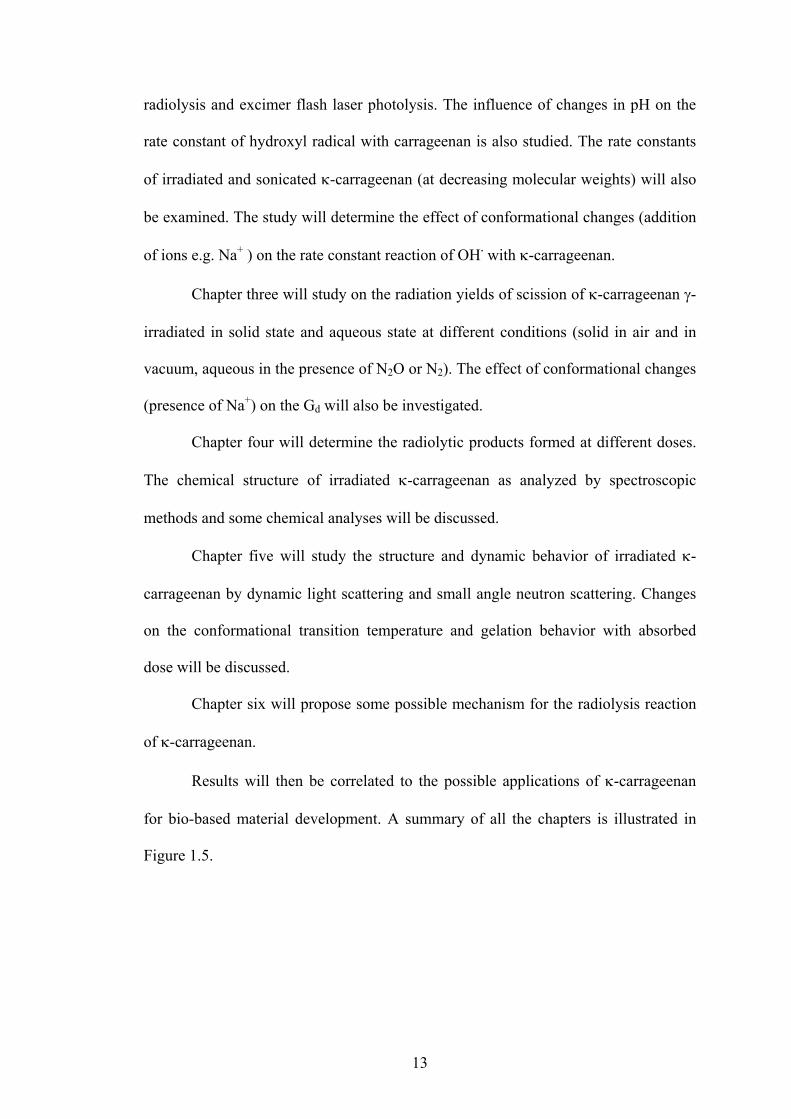
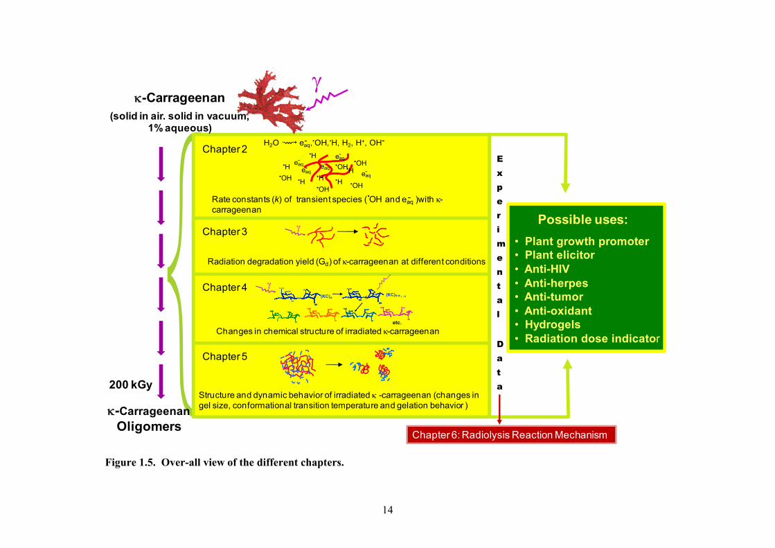
Results will then be correlated to the possible applications of -carrageenan
for bio-based material development. A summary of all the chapters is illustrated in
Figure 1.5.
14
Figure 1.5. Over-all view of the different chapters.
Rate constants (k) of transient species ( OH and eaq )with -carrageenan
-.
H2O eaq, OH, H, H2, H+, OH-- . .
H. OH.eaq-
H. H.
H.
H.
H.
eaq-
eaq-
eaq-
eaq-
OH.
OH.
OH.
OH.
Chapter 2
200 kGy
-CarrageenanOligomers
-Carrageenan(solid in air. solid in vacuum,
1% aqueous)
Chapter 6: Radiolysis Reaction Mechanism
Possible uses:
• Plant growth promoter• Plant elicitor• Anti-HIV• Anti-herpes• Anti-tumor• Anti-oxidant• Hydrogels• Radiation dose indicator
Radiation degradation yield (Gd) of -carrageenan at different conditions
Chapter 3
etc.
O
O
OH
O
O
OH
(KC)nO
O
OH
O
O
OH
-O3 SO
CH2OH
O
OH
O
O
H2C
O
O
O
O
OH
-O3SO-O3SO
CH2OHCH2OH
O
O
OO
O
O
OH
CH2OHCH2OH
O
O
O
O
OH
-O3SO-O3SO-O3SO-O3SO
CH2OHCH2OHCH2OHCH2OH
O
O
OOOO
O
O
OH
CH2OHCH2OHCH2OHCH2OH
O
O
O
O
O
OH
-O3SO-O3SO
CH2OHCH2OH
O
O
H2CH2C
O
O
O
OHOO
O
O
O
O
O
OH
-O3SO-O3SO-O3SO-O3SO
CH2OHCH2OHCH2OHCH2OH
O
O
H2CH2CH2CH2C
O
O
O
OHOO
-O3SO
O
O
O
O
OH
-O3SO
CH2OHCH2OH
O
O
OO
O
O
OH
CH2OHCH2OH
OO
-O3SO
O
O
O
O
OH
-O3SO
CH2OHCH2OHCH2OHCH2OH
O
O
OOOO
O
O
OH
CH2OHCH2OHCH2OHCH2OH
OOOO
O
O O
-O3SO-O3SO
CH2OHCH2OH
O
O
OO
O
OHOH
CHCH2OH
OOO
O O
-O3SO-O3SO-O3SO-O3SO
CH2OHCH2OHCH2OHCH2OH
O
O
OOOO
O
OHOHOHOH
CHCH2OHCHCH2OH
OOOO
CH2 OHO
O
OH
O
O
O
OH
CH2 OHO
O
OH
O
O
O
OH
(KC)n-xCH2 OH
O
O
OH
O
O
O
OH
CH2 OHO
O
OH
H2CH2CH2CH2CO
O
O
OH
-O3 SO
i ii→
Changes in chemical structure of irradiated -carrageenan
Chapter 4
Structure and dynamic behavior of irradiated -carrageenan (changes in gel size, conformational transition temperature and gelation behavior )
Chapter 5
Experimental
Data

15
REFERENCES:
1. B. Dakay, “Developing Partnership between the Philippines and Indonesia in the Seaweed Industry”, 1st Indonesia Seaweed Forum, South Sulawesi, Indonesia, 27-30 October 2008.
2. G. Therkelsen, 1993. Industrial Gums, Polysaccharides and their Derivatives. R.L. Whistler and BeMiller, eds. Academic Press, San Diego.
3. M. Roberts, B. Quemener, Trends Food Sci. Technol. 10 (1999) 169. In A.
Antonopoulo, B. Herbreteau, M. Lafosse, W. Helbert, J. Chromatogr. A, 1023 (2004) 231.
4. J. Le Questel, S. Cros, W. Mackie, S. Pérez, Int. J. Biol. Macromol. 17 (1999) 161.
In: A. Antonopoulo, B. Herbreteau, M. Lafosse W. Helbert, J. Chromatogr. A. 1023 (2004) 231.
5. F. Van de Velde, H. Peppelman, H. Rollema, R. Tromp, Carbohydr. Res. 331
(2001) 271.
6. R. Tye, Carbohydr. Polym. 10(1998) 259.
7. IAEA-TECDOC-1386, Emerging Applications of Radiation Processing, January 2004.
8. IAEA- TECDOC-1422, Radiation Processing of Polysaccharides, November 2004.
9. M. Haji-Saeid, M. Sampa, N. Ramamoorthy, O. Güven, A. Chmielewski, Nucl.
Instr. and Meth. in Phys. Res. B. 265 (2007) 51.
10. P. Patier, P. Potin, C. Rochas, B. Kloareg, J. Yvin, Y. Lienard, Plant Sci. 110 (1995) 27.
11. G. Zhou, Y. Sun, H. Xin, Z. Zhang, Z. Xu, Pharm. Res. 50 (2004). 47.
12. G. Zhou, H. Xin, W. Sheng, S. Yueping, Z. Li, Z. Xu. Pharm. Res. 51 (2005)153.
13. M. Carlucci, C. Pujol, M. Ciancia, M. Noseda, M. Matulewicz, E. Damonte, A.
Cerezo, Int. J. Biol. Macromol. 20 (1997) 97.
14. T. Yamada, A. Ogamo, T. Saito, J. Watanabe, H. Uchiyama, Y. Nakagawa, Carbohydr. Polym. 32 (1997) 51.
15. T. Yamada, A. Ogamo, T. Saito, H. Uchiyama, Y. Nakagawa, Carbohydr. Polym.,
41 (2000) 115.
16. P. Cáceres, M. Carlucci, E. Damonte, B. Matsuhiro, E. Zúñiga, Phytochem., 53 (2000) 81.

16
17. T. Kume, In: Processing of Agro Waste by using Radiation Technology, IAEA, RCA National Executive Management Seminars (NEMS), Islamabad, (2000) 17.
18. N. Hien, N. Nagasawa, L. Tham, F. Yoshii, V. Dang, H. Mitomo, K. Makuuchi,
T. Kume, Radiat. Phys. Chem. 59 (2000) 97.
19. L. Hai, T. Diep, N. Nagasawa, F. Yoshii, T. Kume, Nucl. Instr. and Meth. in Phys. Res. B. 208 (2003) 466.
20. A. Chmielewski, W. Migdal, J. Swietoslawski, U. Jakubaszek, T. Tarnowski,
Radiat. Phys.Chem. 76 (2007) 1840.
21. S. Matsuhashi, T. Kume, J. Sci. Food Agric. 73 (1997) 237.
22. G. Darvill, P. Albersheim, Ann. Rev. Plant Physiol. 35 (1984) 243.
23. A. Ryan, Biochemistry 27 (1988) 8879.
24. W. Schnabel, Polymer Degradation: Principles and Practical Applications, Hanser Verlag, 1981.
25. Y. Shigemasa, K. Saito, H. Sashiwa, H. Saimoto, Int. J. Biol. Macromol. 16
(1994) 43.
26. E. Muraki, F. Yaku, H. Kojima, Carbohydr. Res. 239 (1993) 227.
27. A. Andrady, A. Torikai, T. Kobatake, J. Appl. Polym. Sci. 62 (1996) 1465.
28. T. Kume, N. Nagasawa, F. Yoshii Radiat. Phys. Chem. 63 (2002) 625.
29. L. Luan, N. Hien, N. Nagasawa T. Kume, F. Yoshii, M. Nakanishi, Biotechnol. Appl. Biochem. 38 (2003) 283.
30. D. Bellincampi, G. Salvi, G. Lorenzo, F. Cervone, V. Marfà, S. Eberhard, A.
Darvill, P. Albersheim, Plant J. 4 (1993) 207.
31. V. Marfa`, D.J. Gollin, S. Eberhard, D. Mohnen, A. Darvill, P. Albersheim P, Plant J. 1 (1991) 217.
32. L. Relleve, L. Abad, C. Aranilla, A. Aliganga, A. de la Rosa, F. Yoshii, T. Kume,
N. Nagasawa, “ Biological Activities of Radiation-Degraded Carrageenan” Proceedings of the Symposium on Radiation Technology in Emerging Industrial Applications, Beijing, People’s Republic of China, 6-10, November 2000.
33. A. de la Rosa, L. Abad, L. Relleve, C. Aranilla, “ Radiation-Modified
Carrageenan For Agricultural And Health Care Applications”. IAEA Report, International Atomic Energy Agency (IAEA) Project Coordination Meeting on Radiation Processing of Chitin/Chitosan, Bangkok, Thailand, 18-20 March 2002

17
34. L. Relleve, N. Nagasawa, L.Q. Luan, T. Yagi, C. Aranilla, L. Abad, T. Kume, F. Yoshii, A. dela Rosa, Polym. Degrad. Stabil. 87 (2005) 403.
35. J. Rosiak, A. Rucihska-Rybus and W. Pekala (1989). US Patent no. 4,871,490.
36. L. Abad, A. de la Rosa (2008). Philippine Patent no. 1-2000-02471.
37. J. Rosiak, J. Olejniczak, W. Pecala, Radiat. Phys. Chem. 36 (1990) 747.
38. L. Abad, L. Relleve, C. Aranilla, A. de la Rosa, Radiat. Phys. Chem. 68 (2003)
901.
18
CHAPTER 2
RATE CONSTANTS OF REACTIONS OF -CARRAGEENAN
WITH HYDRATED ELECTRON AND HYDROXYL RADICAL
2.1. INTRODUCTION
In dilute aqueous polymeric solutions, the energy of radiation is absorbed
mainly by water. Direct effect of radiation on the polymer itself is quite minimal. This
leads to the formation of OH-radicals, solvated electrons and H-atoms as reactive
intermediates (equation 2.1). Radiation chemical yield of these transients is similar for
gamma and electron beam irradiation, i.e. G(eaq) = G(OH) = 2.8 x 10⁻⁷ mol J-1, G(H) =
0.6 x 10⁻⁷ mol J-1 and it is constant in the wide range of pH [1]. The hydrated electron,
eaq, is the simplest reducing specie which may react with some transient species of the
water molecule, with surrounding water molecules or with the polymer itself.
Hydrated electrons react with N2O almost quantitatively giving the corresponding
amount of .OH with the rate constant k = 9.8 x 10⁹ M-1 s-1 as shown in equation 2.2.
[2, 3]. The .OH is a reactive oxidizing species, which can abstract hydrogen within the
polymer. The H radicals react with polymers at a rate constant one order lower than
the OH radicals whose value is often times negligible [1].
Pulse radiolysis is well known as an excellent method for direct observations
of short-lived transient species and their reactions. A method of nanoseconds /
(2.1)
(2.2)eaq + N2O + H2O .OH + N2 + OHeaq + N2O + H2O .OH + N2 + OH
H2O eaq, OH, H, H2, H+, OH-- . .
19
picoseconds electron beam pulse radiolysis or flash laser photolysis combined with
absorption spectroscopy has been used for the investigation of these radiation-induced
intermediates. The electron beam pulse radiolysis technique involves the use of high-
energy electrons (from a linear electron accelerator) that are delivered to a sample at
times < 1 s [4]. Laser photolysis technique involves a technique where sample is
firstly excited by a strong pulse of light from a laser of < 1 s pulse width. This first
strong pulse of both techniques starts a chemical reaction. Typically the absorption of
light by the sample is recorded within short time intervals to monitor relaxation or
reaction processes initiated by the pulse by photo spectroscopic method such as UV-
spectroscopy. Using both methods, this chapter will investigate the rate constants (k)
of the reaction of -carrageenan and its oligomers with the primary water radicals
using both techniques. It will also investigate the effect of conformational changes in
-carrageenan on its k.
2.2. MATERIALS AND METHODS
2.2.1. Materials
Refined -carrageenan was obtained from Shemberg Corporation, Philippines.
This was further purified. -carrageenan was dissolved in distilled water and
precipitated with isopropyl alcohol. The precipitated carrageenan was dissolved in a
buffer solution containing 0.1N NaCl and 0.005M ethylenediaminetetraacetic acid
(EDTA). The sample was dialyzed (Mol. wt. cut-off = 12,000-14,000) against a
NaH2PO4/ Na2HPO4 buffer solution for 72 hrs. The dialyzed solution was then
reprecipitated with isopropyl alcohol and freeze dried [5, 6].

20
2.2.2. Sample Preparation
The purified samples of refined -carrageenan were dissolved in Millipore
water to make a 50 mM concentration (based on Mw of repeating unit). This was
stirred overnight to insure complete dissolution. The -carrageenan samples were then
diluted to the desired concentrations for both the Laser Photolysis and Electron-beam
radiolysis experiments. The pH of the solutions were adjusted either by the addition of
HClO4 or NaOH. Perclorate anion does not react with the .OH. In order to investigate
the reaction of OH radical with the polymer, the solution was first saturated with N2O
to convert the eaq, into .OH (equation 2.2). On the other hand, the solution was
saturated with Ar when reaction of eaq with the polymer was being studied. This
eliminates the dissolved atmospheric oxygen from scavenging the hydrated electrons.
For analysis using the laser photolysis, polymer solutions were added with 5mM H2O2
to generate .OH.
2. 2.3. Laser Photolysis of -carrageenan
A 248 nm (KrF) excimer laser was used with the maximum energy of 330 mJ
per 20 ns pulse. The analyzing light was perpendicular with the laser beam, and the
optical length was 15 mm. Fresh sample solutions were placed in the optical cell by
batch.
2.2.4. Pulse Radiolysis of -carrageenan
Pulse radiolysis experiments were performed using an electron beam (10ns) of
35 MeV delivered from a linear accelearator. The preparation and analysis of the
samples were the same as that in laser photolysis, except that the optical path length

21
was 18mm. Figure 2.1 shows the schematic diagram for the laser photolysis and pulse
radiolysis set-up with all the essential parts. In this figure, LINAC is a linear
accelerator. The pulsed lamp is a Xe lamp which provides high-intensity probe light
from UV to near IR region. This probe light is being optimized by the presence of
lenses which passes through the monochromator for proper selection of wavelength.
The pulse generator produces a series of control signals to LINAC, Excimer, pulse Xe
lamp and oscilloscope et al. The photodiode detects the change in intensity of the
probing light where the signals are being amplified by the amplifier. The digital
oscilloscope then collects and records these data which is transmitted to the computer
for storage and processing purposes. Control for the pulse generator, oscilloscope and
monochromator is done through a GPIB interface using a homemade software based
NI LabVIEW (National InstrumentsTM).
Figure 2.1. The schematic diagram of experimental apparatus of a pulse radiolysis or laser photolysis combined with photo-spectroscopic method.
Lenses Cell
Beam
LINAC orExcimer
Mono-chromator Photodiode
Pulse duration: 10 or 50 nsDose per pulse: 20 or 50Gy
PulseGenerator
Computer
Oscilloscope
Amplifier
Lamp
22
2.2.5. Irradiation of -carrageenan
Irradiation of the purified -carrageenan was carried out using the Co-60
facility of the Takasaki Advanced Radiation Research Institute, Japan Atomic Energy
Agency in atmospheric condition, at a dose rate of 10 kGy/h with absorbed doses
ranging from 1 to 100 kGy.
2.2.6. Sonication of -carrageenan
Sonochemical degradation of -carrageenan was performed using the Cole
Parmer 4710 Series, Ultrasonic Homogenizer, with a frequency of 80 kHz. Solutions
with concentration of 5%, 2.5 % and 1% were sonicated for 30mins.
2.2.7. Molecular weight Determination of -carrageenan
The molecular weight of -carrageenan was determined by Gel Permeation
Chromatography. GPC analyses were performed on a Tosoh chromatograph equipped
with DP-8020 pump, CO-8020 column oven, RI-8020 refractive index detector and
four TSK gel PWXL columns in series (G6000 PWXL, G4000 PWXL, G3000 PWXL
and G2500 PWXL. Elution was carried out using 0.1M NaNO3 (to suppress
electrostatic effects [7, 8] as the mobile phase at a flow rate of 0.5 ml/min. The
temperatures of the column and detector were both maintained at 40oC. A calibration
curve was constructed using polyethylene oxide as standards. All molecular masses
reported in this work are based on PEO standards and are not absolute.

23
2.2.8. UV-Vis Analysis
UV-visible spectroscopy of carrageenan solutions was performed using a Shimadzu
spectrophotometer UV-265 FW (wavelength range = 200 - 600 nm) at ambient
temperature and at 0.025% (w/v) concentration.
2.2.9. Viscosity Measurement
Viscosity measurement of carrageenan solutions (1%) was done using a
Tokimec Viscometer TV-20L with a THM-11 spindle at 25oC and a rotor speed of 6
rpm.
2.3. RESULTS AND DISCUSSION
2.3.1. Reaction of Hydrated Electrons with -Carrageenan
The rate constant of the reaction of hydrated electron with -carrageenan can
be estimated by measuring the rate of disappearance of eaq- at 720 nm. The first order
rate constants for the reaction of eaq- with -carrageenan can be obtained from the
linear plot of log (absorbance) against time after pulse. From the slope of the straight
line of the plot of the first order rate constant with polymer concentration, the second-
order rate constant can then be estimated.
The plot of absorbance with time at different concentrations of -carrageenan
is shown in Figure 2.2. The figure indicates no seeming reaction of the eaq- with -
carrageenan. The rate of decay decreased with increasing polymer concentration. This
trend of decreasing rate constants may simply be a result of the slow diffusion of eaq-
into the polymer due to increasing viscosity. Even at very low concentrations of

24
-carrageenan where viscosity approximates that of water (0.2-0.5 mM), this
decreasing trend of rate constants is still observed. Thus it can be deduced that the
reaction with hydrated electron with -carrageenan is negligible. The reactivity of eaq-
towards the carbohydrates is generally low (typically < 107 M-1 s-1) [9]. Investigations
done on the reaction rates of hydrated electron with carboxymethyl cellulose (with
different degrees of substitution), carboxymethyl chitin, carboxymethyl chitosan,
chondrotin sulfate and keratin sulfate are shown in Table 2.1.
water
10mM
water
10mM
Figure 2.2. Decay profiles of the absorbance at 720 nm after pulse radiolysis of -carrageenan at varying concentrations.
25
Table 2.1. Rate constant of reaction of hydrated electron and OH radicals with different types of natural polymer.
Type of Natural Polymer k (M-1 s-1) of hydrated
electron with polymer k (M-1 s-1) of OH radicals
with polymer Carboxymethyl cellulose (4.0 - 5.0) x 106 [10]
6.53 x 10⁶ [10] 1.8 x 10⁷ [11]
(9.5 - 10.2) x 108 [9,16]
Hyaluronic acid < 5.0 x 10⁶ [12] (7 – 9) x 108 [18] Carboxymethyl chitin 6.1 x 10⁷ [13] 9.9 x 10⁸ [15] Carboxymethyl chitosan 3.7 x 10⁷ [13] 12.9 x 10⁸ [15] Chondrotin sulfate (1.1 – 4.2) x 107 [14] Keratin sulfate 3.3 x 10⁷ [14] Dextran 1 x 10⁸ [17]
2.3.2. Rate Constant of Hydroxyl Radicals Reactions with -
Carrageenan
The OH• radical has no absorption in the visible and near-UV regions. Thus,
the rate constant of the reaction of OH• with -carrageenan was determined by the
competition kinetics using KSCN as competitor scavenger. SCN- can be oxidized by
the OH• radical to form (SCN)2•- which has a strong absorption band at 472 nm
(equation 2.3). The rate constant of OH• reactions with -carrageenan can be
measured using equation 2.4.
OHC SCN SCN
OH SCN 2C (2.3)
1 .
. (2.4)
where A0 and A are the transient absorbance of (SCN)2•- in the absence and presence
of -carrageenan respectively. KOH• + KSCN! is known to be 1.1 x 10¹⁰ M-1s-1[15] . Kkc
can then be obtained from the slope of the plot of .
against

26
A/A0 – 1 where either the KSCN or -carrageenan concentration can be fixed. In the
current procedure, KSCN concentration was fixed at 2mM. Figure 2.3 shows the plot
of the curves using laser photolysis and e-beam radiolysis. The values of Kkc obtained
for both methodologies were quite high and were in close agreement with each other,
1.14 x 10⁹ for pulse radiolysis and 1.21 x 10⁹ M-1 s-1 for laser photolysis. This value is
quite close to the reported values for CM-cellulose, CM-chitosan and CM-chitin
shown in Table 2.1. The rate constants for the reaction of OH• with dextran and
hyaluronic acid are lower.
0.0 4.0e-10 8.0e-10 1.2e-9 1.6e-9 2.0e-90.0
0.4
0.8
1.2
1.6
2.0
Pulse radiolysisPhotolysis
A0/
A -
1
[KC] / KOH + SCN- [SCN-]
K = 1.14 x10⁹
K = 1.21 x10⁹
Figure 2.3. Determination of the rate constant reaction of ·OH with -carrageenan by competition method.
27
2.3.3. pH Dependence
The rate constant of reaction of the OH radical with a charged polymer can be
pH or ionic strength dependent. This phenomenon can be influenced by the changes in
conformational structure of the polymer due to these variations. CM-cellulose has
higher rate constant at a basic pH and in the presence of a salt (NaCl). This is due to
its extended conformation in these prevailing conditions. As the pH is increased,
ionization, through its repulsive interactions, causes extension of macromolecule
chains. Ions of NaCl reduce the mutual interaction of charges on macromolecules,
thus the stiffness of chains reduces, resulting in coiling [17] . Similar results are also
observed in CM-chitin and CM-chitosan where both exhibit pH dependence. The rate
constant is lowest at pH = 4.4, near the isoelectric point of CM-chitin [16]. On the
other hand, CM-chitosan exhibits maximum k at a pH range of 2.0 – 7.3 [16]. At this
pH range, both polymers tend to have the coiled conformation. -carrageenan is also a
negatively charged polymer with sulfate groups attached to it. The rates of reaction of
OH• with -carrageenan may also exhibit pH dependence. Figure 2.4 shows the
influence of pH (2 - 10) on the k reaction of OH• with -carrageenan. At pH higher
than 10, OH• dissociates into O• as shown in equation 2.5. The pKa of OH• is 11.5.
OH OH•
O• H2O (2.5)
Rate constant was faster at an acidic pH = 2. When the acidic solution of -
carrageenan was neutralized back to pH 7.7, the rate constant did not revert back to
the original value of 1.21 x 10⁹ M-1 s-1 (Figure 2.5). This indicates that the increase in
rate constant at pH =2 was not due to any conformational change in the -carrageenan
but to some structural changes. At an acidic pH, it is possible that hydrolysis reaction

28
Figure 2.4. Determination of the rate constant reaction of ·OH with -carrageenan by competition method at varying pH (laser photolysis method)
Figure 2.5. Determination of the rate constant reaction of ·OH with -carrageenan by
competition method after neutralization at pH =2 (laser photolysis method)
0 4.0e‐10 8.0e‐10 1.2e‐9 1.6e‐9 2.0e‐90.0
0.5
1.0
1.5
2.0
2.5
3.0
A0
/A -
1
[KC] / KOH + SCN- [SCN-]
pH 7.7 (control) pH 2 pH 4 pH 6 pH 10
4.0e-10 6.0e-10 8.0e-10 1.0e-9 1.2e-9
0.0
0.4
0.8
1.2
1.6
2.0
pH 7.7pH2 pH 2 after neutralization
A0
/A -
1
[KC] / KOH + SCN- [SCN-]
29
may have occurred resulting in the fragmentation of -carrageenan [20]. Thus, OH•
can diffuse faster into the polymer resulting in a higher rate constant. Measurement of
the viscosity of - carrageenan at pH 2 and at neutral pH indicated values of 61 and
85 mPa·s, respectively. These results would indicate further that at pH 2, hydrolysis
reaction took place which resulted in the formation of lower molecular fragments that
consequently decreased the viscosity of -carrageenan. In cellulose, glycosidic
linkages are stable to alkali but are readily hydrolyzed by dilute acid solutions [21].
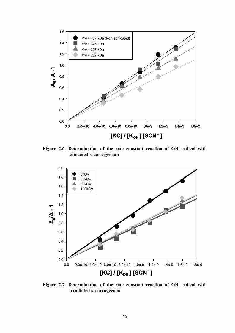
2.3.4. Pulse radiolysis studies of sonicated and irradiated -
carrageenan
The molecular weight of the sonicated -carrageenan solutions was analyzed
by GPC. Mw was determined to be as follows: 376,000; 267,000; and 202,000. The
rate constant of OH• reaction with -carrageenan of these samples was measured by e-
beam pulse radiolysis. A decrease in molecular weight of -carrageenan would
signify a decrease in viscosity, which expectedly would increase the diffusion rate of
OH radicals and consequently increase the rate constant. The results, however,
indicated a reverse trend than what was expected. Figure 2.6 shows that the rate
constants decreased from k = 9.9 x 10⁸ (unsonicated -carrageenan) to k = 9.4 x 10⁸,
8.3 x 10⁸ and 6.8 x 10⁸ M-1s-1 with decreasing molecular weights. Similarly, the rate
constants of irradiated carrageenan at 25, 50 and 100 kGy (computed Mw = 188,000;
105,000; and 59,000 respectively) decreased (from k = 1.1 x 10⁹ to an average of k =
8.2 x 10⁸ M-1s-1) but did not vary with increasing doses (Figure 2.7). Two factors may
affect the reaction rate of a polymer. First, a reduction of viscosity or molecular size
leads to higher diffusion of OH• in the polymer resulting in increased rate constant.
Second, rate constant is directly proportional to the number of reactive
30
0.0 2.0e-10 4.0e-10 6.0e-10 8.0e-10 1.0e-9 1.2e-9 1.4e-9 1.6e-9 1.8e-9
0.0
0.2
0.4
0.6
0.8
1.0
1.2
1.4
1.6
1.8
2.0
0kGy25kGy50kGy100kGy
A0/
A -
1
[KC] / [KOH.] [SCN- ][KC] / [KOH.] [SCN- ]
Figure 2.6. Determination of the rate constant reaction of OH radical with
sonicated -carrageenan
Figure 2.7. Determination of the rate constant reaction of OH radical with irradiated -carrageenan
0.0 2.0e-10 4.0e-10 6.0e-10 8.0e-10 1.0e-9 1.2e-9 1.4e-9 1.6e-9
0.0
0.2
0.4
0.6
0.8
1.0
1.2
1.4
1.6
Mw = 436,915 (Non -sonicated)
Mw = 376,139
Mw = 267,467
Mw = 202,244
[KC] / [KOH.] [SCN- ]
0.0 2.0e-10 4.0e-10 6.0e-10 8.0e-10 1.0e-9 1.2e-9 1.4e-9 1.6e-9
0.0
0.2
0.4
0.6
0.8
1.0
1.2
1.4
1.6
Mw = 437 kDa (Non-sonicated)
Mw = 376 kDa
Mw = 267 kDa
Mw = 202 kDa
[KC] / [KOH.] [SCN- ]
A0
/ A
-1

31
sites for the OH• interaction. Based on the results, the latter factor predominates in the
sonication or irradiation of -carrageenan. Cleavage of glycosidic linkage in -
carrageenan is the most likely effect in the sonication or irradiation of -carrageenan
as evidenced by a rapid decrease in molecular weight by either these two processes.
But cleavage of glycosidic linkage alone by hydrolysis reaction would have increased
the rate constant of OH• reaction similar to the one observed in -carrageenan at pH
= 2. Experiments on chitosan indicate that the rate constant of OH• reaction with
chitosan is increased with decreasing chain length [22]. Most likely, sonication or
irradiation of -carrageenan could have generated products that have reduced the
reactive sites for OH• reaction with -carrageenan. Thus, decreasing rate constant of
OH• interaction with decreasing molecular weight was observed both in sonicated and
irradiated -carrageenan. The degradation mechanism of macromolecules by
ultrasound is frequently attributed to cavitation (mechanical) effects and partially to
the stress concentration on the segment of macromolecules [23]. At lower
frequencies, 20–50 kHz range (the ‘low’ frequency domain reaches to 100 kHz), these
effects are observed [24]. When ultrasound of a frequency >500 kHz is applied, an
additional factor (radical reactions similar to radiolysis effect) may become more
pronounced [25]. Since the frequency used for the sonication of -carrageenan was
only 80 kHz, it was expected that cleavage of glycosidic linkage could be caused only
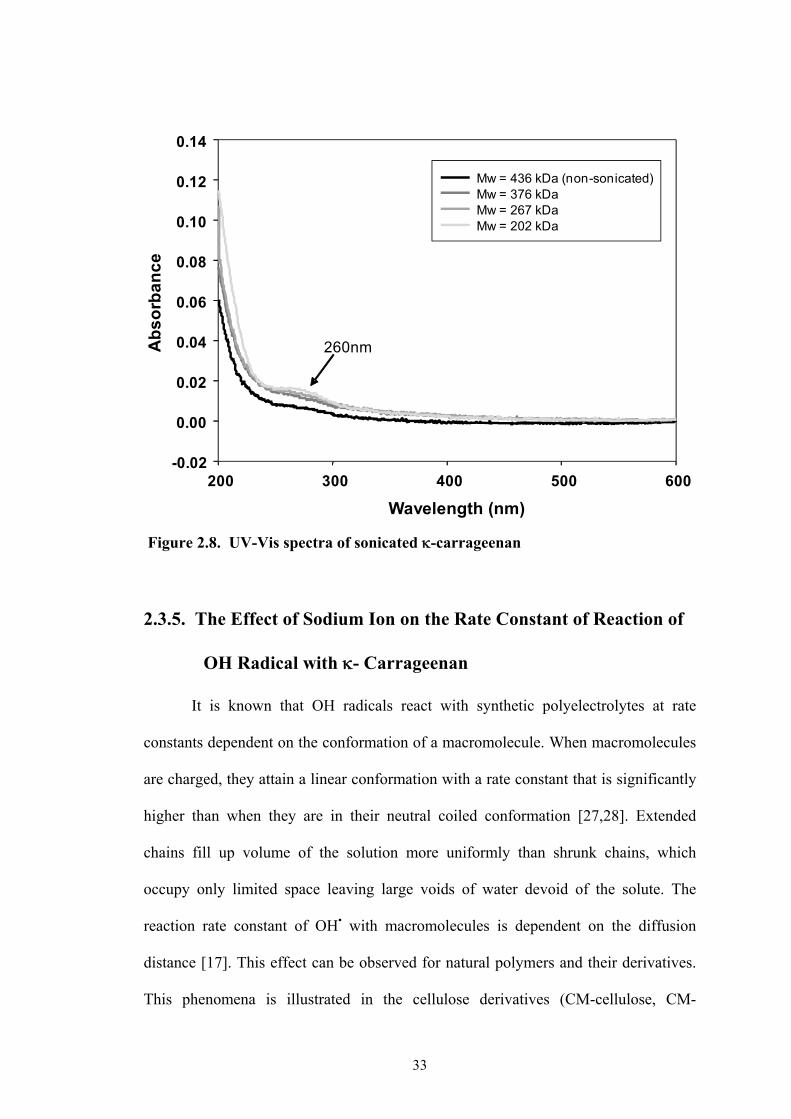
by vibrational effect. However, UV-Vis spectra of sonicated -carrageenan solutions,
revealed otherwise as seen in Figure 2.8. The figure shows slightly increasing UV
absorbance at 260nm. Similar experiments done previously on sonicated chitosan
(360kHz) show two absorption bands (at 265 and at 297 nm), absent in the starting
material, in the UV–Vis spectroscopy. These are some transformations of chitosan-
derived radicals that lead to the formation of carbonyl groups [22]. Ultrasound-

32
induced degradation of chitosan in Ar-saturated solutions, both caused by OH• or
vibrational effects, is accompanied by side reactions. One source of such processes is
a terminal radical formed as a result of glycosidic bond breakage. In the case of OH•-
mediated process, there are also non-terminal radicals located along the chain, which
may not be capable of causing chain breakage, but may undergo other reactions [25,
26]. The effect of sonochemical degradation of -carrageenan may follow the same
scheme as that of chitosan. Since the frequency is not so high, the formed carbonyl
may simply be a terminal radical formed as a result of glycosidic bond breakage.
Thus, only a slight increase in carbonyl bonds (UV-Vis spectra in Figure 2.8) was
observed with increasing doses. As a consequence, diminishing reactive site for OH
radical interaction was also observed as indicated by decreasing rate constant k with
sonication time (decreasing molecular weight). In the case of the reaction of ionizing
radiation with -carrageenan, reactions of OH• and other radicals can produce
carbonyl groups in several sites not only in the terminal groups. Reactions can be
more severe than the sonication process especially under air condition where some
peroxy radicals are generated. Oxidation reactions may take place with the formation
of carbonyl groups/carboxyl groups and which can eventually lead to fragmentation
patterns that may result in ring opening of the galacto-pyranose ring. This phenomena
may then lead to a drastic reduction of reactive sites for OH• interaction at all levels of
absorbed doses (decreasing molecular weight). Thus, the k for OH• reaction with -
carrageenan levels off. Two events - decrease in viscosity (increase in k) and decrease
in reactive sites (decrease in k) with increasing radiation may have occurred
simultaneously producing an over-all effect of a consistently uniform rate constant for
all doses.
33
2.3.5. The Effect of Sodium Ion on the Rate Constant of Reaction of
OH Radical with - Carrageenan
It is known that OH radicals react with synthetic polyelectrolytes at rate
constants dependent on the conformation of a macromolecule. When macromolecules
are charged, they attain a linear conformation with a rate constant that is significantly
higher than when they are in their neutral coiled conformation [27,28]. Extended
chains fill up volume of the solution more uniformly than shrunk chains, which
occupy only limited space leaving large voids of water devoid of the solute. The
reaction rate constant of OH• with macromolecules is dependent on the diffusion
distance [17]. This effect can be observed for natural polymers and their derivatives.
This phenomena is illustrated in the cellulose derivatives (CM-cellulose, CM-
Figure 2.8. UV-Vis spectra of sonicated -carrageenan
Mw = 436,195 (non-sonicated)Mw = 376,139Mw = 267,467Mw = 202,244
Ab
so
rba
nc
eMw = 436 kDa (non-sonicated)Mw = 376 kDaMw = 267 kDaMw = 202 kDa
260nm
200 300 400 500 600-0.02
0.00
0.02
0.04
0.06
0.08
0.10
0.12
0.14
Wavelength (nm)

34
0 1x10-12 2x10-12 3x10-12 4x10-12 5x10-12
0.0
0.1
0.2
0.3
0.4
0.5Kappa Carrageenan
Kappa Carrageenan added with 0.08M NaClO4
[KC] / [KOH.] [SCN- ] / M-1s-1
A0/
A -
1 /
a.u
.
0 1x10-12 2x10-12 3x10-12 4x10-12 5x10-12
0.0
0.1
0.2
0.3
0.4
0.5Kappa Carrageenan
Kappa Carrageenan added with 0.08M NaClO4
[KC] / [KOH.] [SCN- ] / M-1s-1
A0/
A -
1 /
a.u
.
8.8 x 10⁸ M-1s-1
4.5 x 10⁸ M-1s-1
- Carrageenan-Carrageenan with NaClO4
0 1x10-12 2x10-12 3x10-12 4x10-12 5x10-12
0.0
0.1
0.2
0.3
0.4
0.5Kappa Carrageenan
Kappa Carrageenan added with 0.08M NaClO4
[KC] / [KOH.] [SCN- ] / M-1s-1
A0/
A -
1 /
a.u
.
0 1x10-12 2x10-12 3x10-12 4x10-12 5x10-12
0.0
0.1
0.2
0.3
0.4
0.5Kappa Carrageenan
Kappa Carrageenan added with 0.08M NaClO4
[KC] / [KOH.] [SCN- ] / M-1s-1
A0/
A -
1 /
a.u
.
8.8 x 10⁸ M-1s-1
4.5 x 10⁸ M-1s-1
0 1x10-12 2x10-12 3x10-12 4x10-12 5x10-12
0.0
0.1
0.2
0.3
0.4
0.5Kappa Carrageenan
Kappa Carrageenan added with 0.08M NaClO4
[KC] / [KOH.] [SCN- ] / M-1s-1
A0/
A -
1 /
a.u
.
0 1x10-12 2x10-12 3x10-12 4x10-12 5x10-12
0.0
0.1
0.2
0.3
0.4
0.5Kappa Carrageenan
Kappa Carrageenan added with 0.08M NaClO4
[KC] / [KOH.] [SCN- ] / M-1s-1
A0/
A -
1 /
a.u
.
8.8 x 10⁸ M-1s-1
4.5 x 10⁸ M-1s-1
- Carrageenan-Carrageenan with NaClO4
chitosan, and CM-chitin) as discussed earlier under Chapter 2.3.3. It is also a known
fact that carrageenans undergo thermoreversible ion-induced conformational
transition from a disordered (coil) to an ordered (helix) form. The ability of some
cations to promote helical formation follows this order: Li+, Na+, NR4+ <<NH4
+ <K+,
Cs+ <Rb+. These transitions are both temperature and concentration dependent [30].
At temperatures below its transition temperature, -carrageenan assumes a helical
conformation. Transition temperatures differ with the type of carrageenan, 22oC and
57oC (in 0.2M NaCl) for - and - respectively [31]. The effect of Na+ (NaClO4) on
the rate constant of reaction of OH• with -carrageenan was investigated. The effect
of conformational change on the radiolysis of carrageenan is shown in Figure 2.9. The
rate constant of OH• reaction with - carrageenan with and without the addition of
0.08M NaClO4 was investigated by electron pulse radiolysis. Results show a dramatic
decrease of rate constant from 8.8 x 10⁸ M-1s-1 to 4.5 x 10⁸ M-1s-1.
Figure 2.9. The effect of Na+ on the rate constant of OH• reaction with -carrageenan
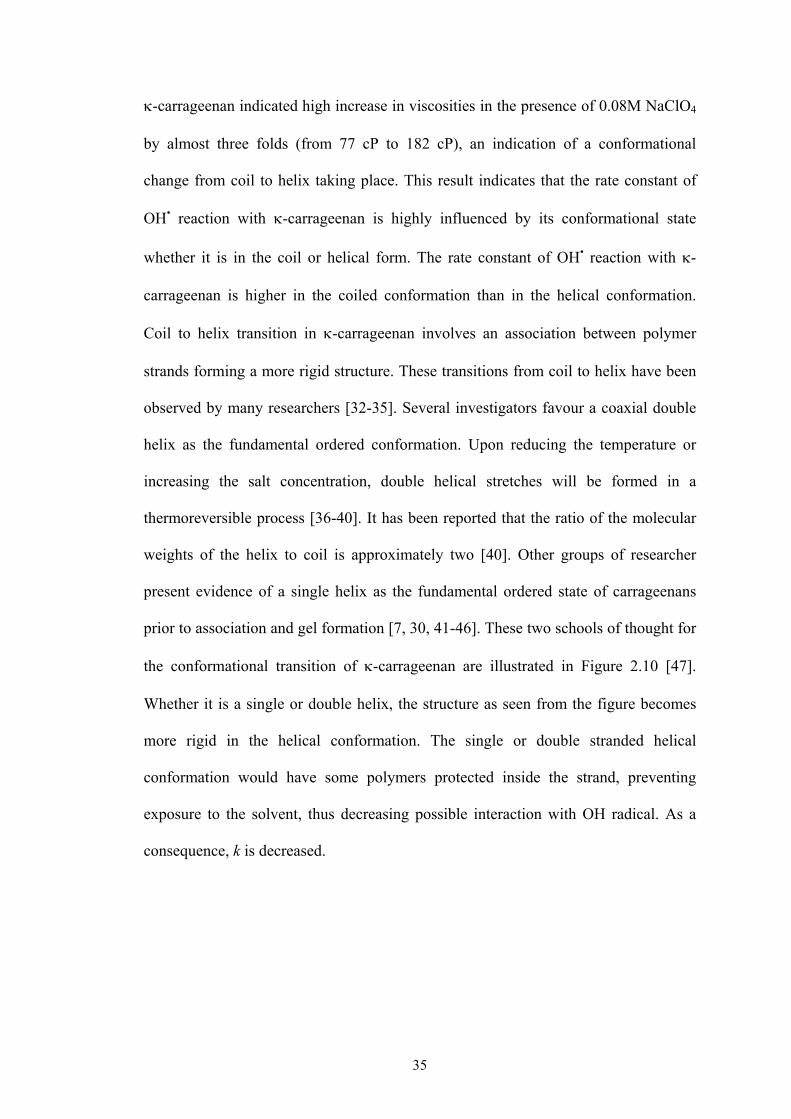
35
-carrageenan indicated high increase in viscosities in the presence of 0.08M NaClO4
by almost three folds (from 77 cP to 182 cP), an indication of a conformational
change from coil to helix taking place. This result indicates that the rate constant of
OH• reaction with -carrageenan is highly influenced by its conformational state
whether it is in the coil or helical form. The rate constant of OH• reaction with -
carrageenan is higher in the coiled conformation than in the helical conformation.
Coil to helix transition in -carrageenan involves an association between polymer
strands forming a more rigid structure. These transitions from coil to helix have been
observed by many researchers [32-35]. Several investigators favour a coaxial double
helix as the fundamental ordered conformation. Upon reducing the temperature or
increasing the salt concentration, double helical stretches will be formed in a
thermoreversible process [36-40]. It has been reported that the ratio of the molecular
weights of the helix to coil is approximately two [40]. Other groups of researcher
present evidence of a single helix as the fundamental ordered state of carrageenans
prior to association and gel formation [7, 30, 41-46]. These two schools of thought for
the conformational transition of -carrageenan are illustrated in Figure 2.10 [47].
Whether it is a single or double helix, the structure as seen from the figure becomes
more rigid in the helical conformation. The single or double stranded helical
conformation would have some polymers protected inside the strand, preventing
exposure to the solvent, thus decreasing possible interaction with OH radical. As a
consequence, k is decreased.
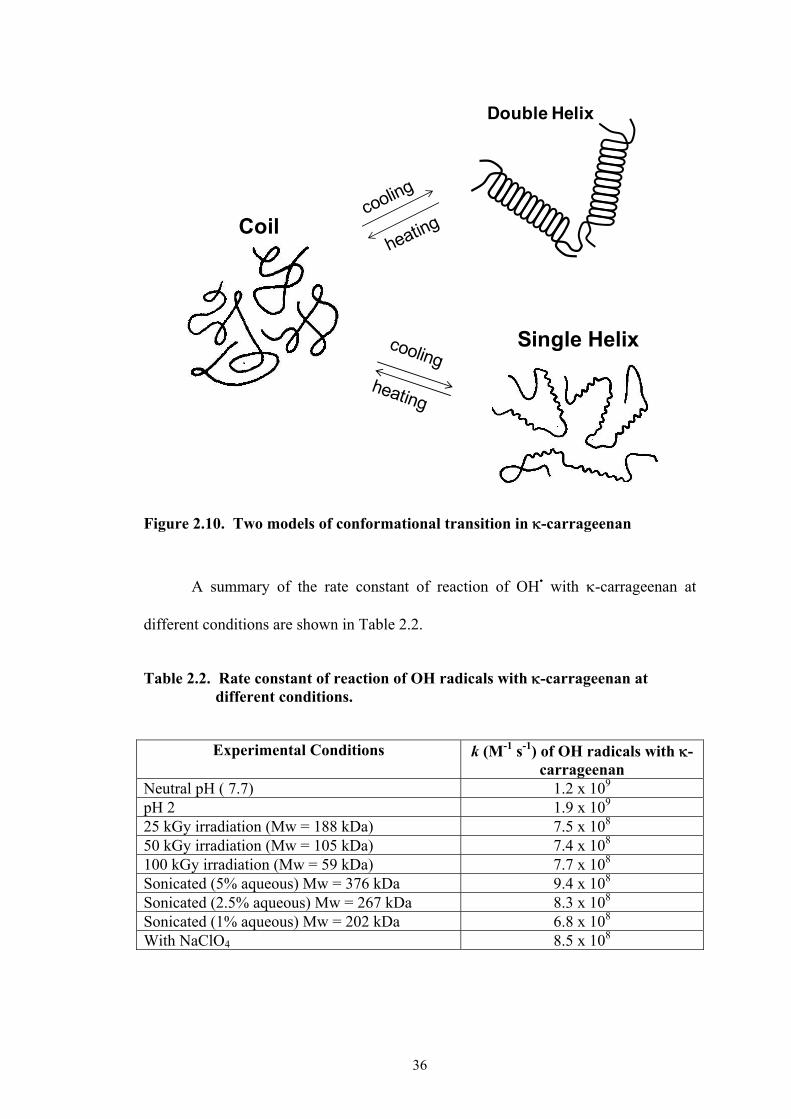
36
Figure 2.10. Two models of conformational transition in -carrageenan
A summary of the rate constant of reaction of OH• with -carrageenan at
different conditions are shown in Table 2.2.
Table 2.2. Rate constant of reaction of OH radicals with -carrageenan at different conditions.
Experimental Conditions k (M-1 s-1) of OH radicals with -
carrageenan Neutral pH ( 7.7) 1.2 x 10⁹ pH 2 1.9 x 10⁹ 25 kGy irradiation (Mw = 188 kDa) 7.5 x 10⁸ 50 kGy irradiation (Mw = 105 kDa) 7.4 x 10⁸ 100 kGy irradiation (Mw = 59 kDa) 7.7 x 10⁸
Sonicated (5% aqueous) Mw = 376 kDa 9.4 x 10⁸
Sonicated (2.5% aqueous) Mw = 267 kDa 8.3 x 10⁸
Sonicated (1% aqueous) Mw = 202 kDa 6.8 x 10⁸
With NaClO4 8.5 x 10⁸
Double Helix
Coil
Single Helix
37
2.4. CONCLUSION
Pulse radiolysis experiments indicate no seeming reaction of the hydrated
electron with -carrageenan. The rate constant for the reaction of OH• with -
carrageenan was approximately 1.2 x 10⁹ M-1s-1. It was faster at an acidic pH of 2
which is probably due to fragmentation from the hydrolysis of -carrageenan.
The rate constant of OH• interaction with sonicated -carrageenan decreased
with decreasing molecular weight. On the other hand, the OH• interaction with
irradiated -carrageenan decreased but did not vary significantly with decreasing
molecular weight. Metal ion (Na+) induced conformational transition into helical form
decreased the rate constant of OH• reaction with -carrageenan.
The radiation depolymerization (decrease in Mw) of aqueous solution of -
carrageenan is caused primarily by the indirect effect of OH•. Obviously, greater
depolymerization is expected in aqueous solution than in solid due to this OH•. Like
most other natural polymers, decrease in molecular weight at higher doses is lower
and reaches a point where it forms a plateau. This phenomenon can thus be explained
by the fact that more carbonyl groups are formed with increasing dose which
decreases reactive sites and consequently lowers rate constant of OH• with -
carrageenan.
Most commercially available -carrageenans are of the Na+ or K+ form. It is
then expected that in solution these would assume a helical conformation which
would decrease k of OH• with -carrageenan. Theoretically speaking,
deploymerization of aqueous commercial carrageenans would be lower than its
purified form. However, for practical reasons, it is more convenient to irradiate in its
commercial form without further purification for bio-based material development.

38
REFERENCES:
1. R. Wach, H. Kudoh, M. Zhai, N. Nagasawa, Y. Muroya, F. Yoshii, Y. Katsumura, Polymer 45 (2004) 8165.
2. C. von Sonntag, E. Bothe, P. Ulanski, J. Deeble, Radiat. Phys. Chem. 46 (1995) 527.
3. C. von Sonntag and HP. Schuchmann, Angew. Chem. Int. Ed. Engl. 30 (1991)
1229.
4. C. von Sonntag, Int. J. Radiat. Biol. 65 (1994) 19.
5. F. van de Velde, A. Antipova, H. Rollema, T. Burova, N. Grinberg, L. Pereira, P. Gilsenan, R. Tromp, B. Rudolphe, V. Grinberg, Carbohydr. Res. 340 (2005) 1113.
6. J. Aguilan, 2005. Structural Analysis of kappa and iota carrageenan from
Philippine Seaweeds, PhD Dissertation, Ateneo de Manila University Graduate School, Philippines.
7. D. Slootmaekers, J. van Dijk, F. Varkevisser, C. van Treslong, H. Reynaers,
Biophy. Chem. 41 (1991) 51.
8. S. Singh, S. Jacobsson, Carbohvdr. Polym. 23 (1994) 89.
9. R. Wach, H. Kudoh, M. Zhai, Y. Muroya, Y. Katsumura, Journal of Polymer Science: Part A: Polym. Chem. 43 (2005) 505.
10. G. Phillips, D. Wedlock, I. Micic, B. Milosorljevic. Radiat. Phys. Chem. 15
(1980) 187.
11. E. Balazs, J. Davies, G. Phillips, D. Scheufele. J. Chem. Soc. (C). 1968;1420.
12. P. Myint, D. Deeble, P. Baumont, S. Balke, G. Phillips, Biochim. Biophys. Acta. 925 (1987) 194.
13. M. Zhai, H. Kudoh, G. Wu, R. Wach, Y. Muroya,Y. Katsumura, N. Nagasawa,
L. Zhao, F. Yoshii, Biomac. 5 (2004) 453.
14. J. Moore, G. Phillips, Carbohy. Res. 12 (1970) 253.
15. G. Buxton, C. Greenstock, W. Helman, A. Ross, J. Phys Chem Ref Data 17 (1988) 513.

39
16. R. Wach, H. Kudoh, M. Zhai, N. Nagasawa, Y. Muroya, F. Yoshii, Y. Katsumura, Polymer 45 (2004) 8165.
17. M. Zhai, H. Kudoh, G. Wu, R. Wach, Y. Muroya, Y. Katsumura, N. Nagasawa,
L. Zhao, F. Yoshii, Biomac. 5 (2004) 458.
18. G. Buxton, C. Greenstock, W. Helman, A. Ross, J. Phys. Chem, Ref Data, 17 (1988) 513.
19. D. Deeble, E. Bothe, H. Schuchman, B. Parson, G. Phillips, C. von Sonntag,
Naturforsch Teil C 45 (1990) 1031–43.
20. Y. Jiang, X. Guo, X. Tian, Carbohyd. Polym. 61 (2005) 399.
21. A. Chapiro. Radiation Chemistry of Polymeric Systems: High Polymers Vol. 15. New York, Interscience Publishers (1962) p. 533.
22. R. Czechowska-Biskup, B. Rokita, S. Lotfy, P. Ulanski, J. Rosiak, Carbohyd.
Polym. 60 (2005) 175.
23. T. Mason, J. Lorimer, (1988). Sonochemistry: theory, applications and uses of ultrasound in chemistry, Chichester, UK: Ellis Horwood pp. 99–138: In C. Lii, C. Chen, A. Yeh, V. Laic, Food Hydrocolloids 13 (1999) 477.
24. N. Kardos, J. Luche, Carbohyd. Res. 332 (2001) 115.
25. G. Portenlänger, H. Heusinger, Ultrasonics Sonochemistry, 4 (1997) 127: In C.
Lii, C. Chen, A. Yeh, V. Laic, Food Hydrocolloids 13 (1999) 477.
26. P. Ulanski, C. Von Sonntag, Journal of the Chemical Society, Perkin Transactions 2 (2000) 2022.
27. C. von Sonntag, C. (1987). The chemical basis of radiation biology. London:
Taylor and Francis.
28. P. Ulanski, J. Rosiak, J. Radioanal. Nucl. Chem. Lett. 186 (1994) 315.
29. P. Ulanski, E. Bothe, C. von Sonntag, Radiat. Phys. Chem. 56 (1999) 467.
30. M. Ciancia, M. Milas, M. Rinaudo, Int. J. Biol. Macromol. 20 (1997) 35.
31. Y. Yuguchi, T. Thuy, H. Urakawa, K. Kajiwara, Food Hydrocolloids. 16 (2002) 515.
32. F. van de Velde, A. Antipova, H. Rollema, B. Tatiana, N. Grinberg, L. Pereira, P.
Gilsenan, R. Tromp, B. Rudolphe, V. Grinberg, Carbohyd. Res. 340 (2005) 1113.
33. T. Hjerde, O. Smidsrød, B. Christenesen, Carbohydr. Res. 288 (1996) 175.
40
34. M. Ciszkowska, J. Osteryoung, J. Am. Chem. Soc. (Commun) 121 (1999) 1617.
35. S. Cara, C. Tamerler, H. Bermek, O. Pekcan, Int. J. Biol. Macromol. 31 (2003)
177.
36. M. Ciszkowska, I. Kotlyar, Anal. Chem.71 (1999) 5013.
37. D. Reid, T. Bryce, A. Clark, D. Rees, Faraday Discuss. Chem. Soc. 57 (1974) 230.
38. T. Hjerde, O. Smidsrod, B. Christensen, Biopolymers, 49 (1999) 71.
39. R. Jones, E. Staples, A. Penman, Chem. Soc. Perkin Trans., II (1973) 1608.
40. B. Wittgren, J. Borgström, L. Piculell, K. Wahlund, Biopolymers. 45 (1998) 85.
41. C. Viebke, J. Borgström, L. Piculell, Carbohyd. Polym. 27 (1995) 145.
42. S. Bailey, Biochim. Biophys. Acta. 17 (1955) 194.
43. Smidsrød, in 27th Int. Congress of Pure and Appl.Chem., Varmavuori A. Ed.,
Pergamon, Oxford (1980) p 315-327.
44. O. Smidsrød, I. Andresen, H. Grasdalen, B. Larsen, T. Painter, Carbohydr. Res., 80 (1980) c11.
45. D. Slootmaekers, M. Mandel, H. Reynaers, Int. J. Biol. Macromol. 13 (1991) 17.
46. K. Ueda, M. Itoh, Y. Matsuzaki, H. Ochiai, A. Imamura, Macromolecules 31
(1998) 675.
47. H. Reynaers, Fibres Text. East Eur. 11(2003) 88.
41
CHAPTER 3
RADIATION DEGRADATION YIELD OF -CARRAGEENAN
3.1. INTRODUCTION
The irradiation of polymeric materials with ionizing radiation (gamma rays, X
rays, accelerated electrons, ion beams) leads to the formation of very reactive
intermediates. These chemically active species as free radicals or radical ions react
with other radicals or molecules to form stable products. It can follow several reaction
paths, which result in rearrangements and/or formation of new bonds [1-7]. These
reactive intermediates are formed either from direct action of radiation on the polymer
chains or from indirect effect, i.e. reaction of the intermediates generated in water
with polymer molecules in aqueous solutions [8]. The ultimate effects of these
reactions can be the formation of oxidized products, grafts, scission of main chains
(degradation) or cross-linking. The degree of these transformations depends on the
structure of the polymer and the conditions of treatment before, during and after
irradiation [9]. The yields of radiation –induced reactions are expressed as G-values,
where G is the number of molecules changed (formed or destroyed) per 100 eV of
energy.
The most important reactions occurring during radiolysis of polymers are
those that lead to permanent changes in their molecular weight. The reactions leading
to either increases or decreases in molecular weight are referred to as cross-linking
and chain scission, respectively. Radicals would also be expected to
dimerize/polymerize: abstraction of a hydrogen atom from a polymer radical followed
by addition of this polymer radical to another polymer radical that would yield a

42
molecule of increased molecular weight. Carbon-carbon scission following loss of a
hydrogen atom from a polymer radical could account for the formation of fragments
as in the case of most polysaccharides. This results in the decrease of molecular
weight. In general, cross-linking and scission processes can occur simultaneously in
any irradiated material; however, it is often observed that one tends to dominate over
the other, and thus polymers can be broadly placed into the categories cross-linking or
degrading. Over the years a solid understanding of the relationship between polymer
structure and the relative yields of cross-linking and chain scission has been acquired
[10].
In the early 1950s, Charlesby and co-workers were considering theoretical
approaches to the description of the changes in molecular weight of polymers during
irradiation. As early as 1954 they reported that the molecular weight of PMMA,
measured by solution viscometry, was inversely proportional to the absorbed dose
[11,12]. This has then been the basis for the theoretical expression of the theory of
degradation. Since that time a number of refinements to these initial observation and
theoretical expression have been made [13-17]. In summary, the number- and weight-
average molecular weights change is described below:
(3.1)
(3.2)
where Gd (mol/J) expresses the degradation susceptibility of the polymer during
radiation, where Mn / Mw is the number / weight-average molecular weight at
absorption dose; Mn0 / Mw0 is the initial number / weight- average molecular weight;
D is the absorbed dose in kGy.
In polysaccharides, glycosidic linkages between constituent units may be
broken by ionizing radiation, resulting in the shortening of polysaccharide chains.
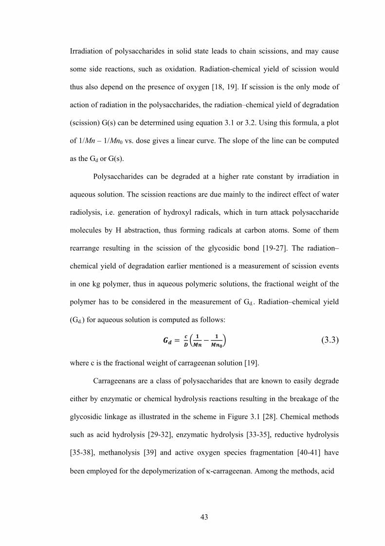
43
Irradiation of polysaccharides in solid state leads to chain scissions, and may cause
some side reactions, such as oxidation. Radiation-chemical yield of scission would
thus also depend on the presence of oxygen [18, 19]. If scission is the only mode of
action of radiation in the polysaccharides, the radiation–chemical yield of degradation
(scission) G(s) can be determined using equation 3.1 or 3.2. Using this formula, a plot
of 1/Mn – 1/Mn0 vs. dose gives a linear curve. The slope of the line can be computed
as the Gd or G(s).
Polysaccharides can be degraded at a higher rate constant by irradiation in
aqueous solution. The scission reactions are due mainly to the indirect effect of water
radiolysis, i.e. generation of hydroxyl radicals, which in turn attack polysaccharide
molecules by H abstraction, thus forming radicals at carbon atoms. Some of them
rearrange resulting in the scission of the glycosidic bond [19-27]. The radiation–
chemical yield of degradation earlier mentioned is a measurement of scission events
in one kg polymer, thus in aqueous polymeric solutions, the fractional weight of the
polymer has to be considered in the measurement of Gd.. Radiation–chemical yield
(Gd.) for aqueous solution is computed as follows:
(3.3)
where c is the fractional weight of carrageenan solution [19].
Carrageenans are a class of polysaccharides that are known to easily degrade
either by enzymatic or chemical hydrolysis reactions resulting in the breakage of the
glycosidic linkage as illustrated in the scheme in Figure 3.1 [28]. Chemical methods
such as acid hydrolysis [29-32], enzymatic hydrolysis [33-35], reductive hydrolysis
[35-38], methanolysis [39] and active oxygen species fragmentation [40-41] have
been employed for the depolymerization of -carrageenan. Among the methods, acid
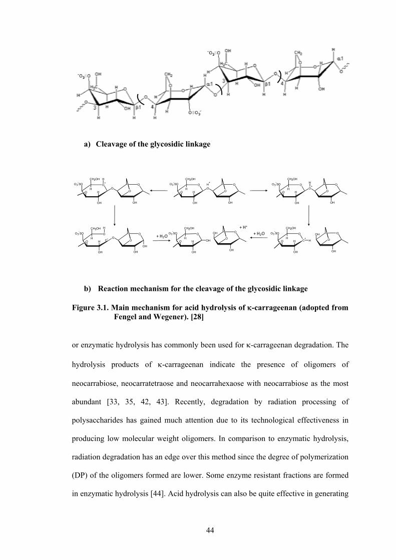
44
a) Cleavage of the glycosidic linkage
b) Reaction mechanism for the cleavage of the glycosidic linkage Figure 3.1. Main mechanism for acid hydrolysis of -carrageenan (adopted from
Fengel and Wegener). [28]
or enzymatic hydrolysis has commonly been used for -carrageenan degradation. The
hydrolysis products of -carrageenan indicate the presence of oligomers of
neocarrabiose, neocarratetraose and neocarrahexaose with neocarrabiose as the most
abundant [33, 35, 42, 43]. Recently, degradation by radiation processing of
polysaccharides has gained much attention due to its technological effectiveness in
producing low molecular weight oligomers. In comparison to enzymatic hydrolysis,
radiation degradation has an edge over this method since the degree of polymerization
(DP) of the oligomers formed are lower. Some enzyme resistant fractions are formed
in enzymatic hydrolysis [44]. Acid hydrolysis can also be quite effective in generating
+ H2O+ H2O
O+
HH
O3-SO
O
OH
CH2OH
O
H
O
O
OH
H+
O
HH
O3-SO
O
OH
CH2OH
OO
O
OH
O
HH
O3-SO
O
OH
CH2OH
O+
O
O
OH
H
O
HH
O3-SO
O
OH
CH2OH
O
H
O
OH
O
OH
C+
O
O
OH
OHO
C+
HH
O3-SO
O
OH
CH2OH
H
O
HH
O3-SO
O
OH
CH2OH
OH
O
O
OH
OH
+ H+

45
low DP oligomers but this method would need further purification step to remove all
the mineral acids. Several studies have already been done to determine the kinetics /
rate constant of reactions by acid hydrolysis for the reduction of molecular weight of
-carrageenan [45-47]. While both common methods of degrading -carrageenan
have extensively been studied, this chapter will study the degradation yield of -
carrageenan with the use of gamma irradiation at different experimental conditions.
3.2. METHODOLOGY
3.2.1. Sample Preparation
For Solid Irradiation:
Purified solid -carrageenan were placed in small polyethylene bags and sealed.
Another set of solid -carrageenan were placed in glass tubes. These tubes were
connected to a vacuum line aided with methanol ice and liquid nitrogen traps. The
tubes were then evacuated while initially tapping and heating with a blower the
portion of tubes containing the polymer to facilitate the escape of gases. Once fully
evacuated, the tubes were then sealed directly from the vacuum line.
For Aqueous Irradiation:
Purified -carrageenan was dissolved in water at 1% concentration (w/v) and was
divided into three different sets while placing them in separates tubes (5 tubes per
set). The different sets of tubes were then treated in the following manner:
a) Tubes were sealed with parafilm.
b) Tubes were purged with N2O gas for 30 mins. and heat sealed while still
purging the gas at the mouth of the tubes.
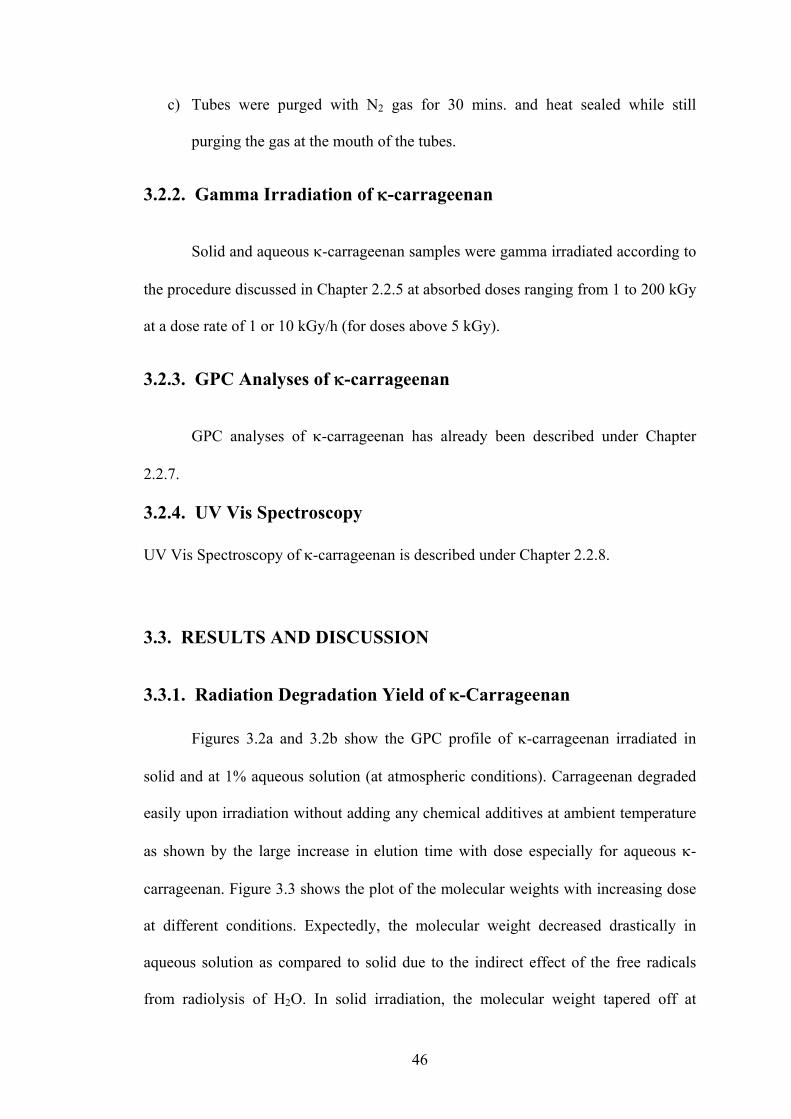
46
c) Tubes were purged with N2 gas for 30 mins. and heat sealed while still
purging the gas at the mouth of the tubes.
3.2.2. Gamma Irradiation of -carrageenan
Solid and aqueous -carrageenan samples were gamma irradiated according to
the procedure discussed in Chapter 2.2.5 at absorbed doses ranging from 1 to 200 kGy
at a dose rate of 1 or 10 kGy/h (for doses above 5 kGy).
3.2.3. GPC Analyses of -carrageenan
GPC analyses of -carrageenan has already been described under Chapter
2.2.7.
3.2.4. UV Vis Spectroscopy
UV Vis Spectroscopy of -carrageenan is described under Chapter 2.2.8.
3.3. RESULTS AND DISCUSSION
3.3.1. Radiation Degradation Yield of -Carrageenan
Figures 3.2a and 3.2b show the GPC profile of -carrageenan irradiated in
solid and at 1% aqueous solution (at atmospheric conditions). Carrageenan degraded
easily upon irradiation without adding any chemical additives at ambient temperature
as shown by the large increase in elution time with dose especially for aqueous -
carrageenan. Figure 3.3 shows the plot of the molecular weights with increasing dose
at different conditions. Expectedly, the molecular weight decreased drastically in
aqueous solution as compared to solid due to the indirect effect of the free radicals
from radiolysis of H2O. In solid irradiation, the molecular weight tapered off at
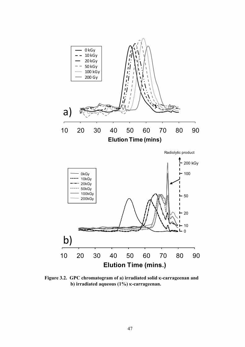
47
Figure 3.2. GPC chromatogram of a) irradiated solid -carrageenan and
b) irradiated aqueous (1%) -carrageenan.
Elution Time (mins)
10 20 30 40 50 60 70 80 90
0 kGy10 kGy20 kGy50 kGy100 kGy200 Gy
a)
10 20 30 40 50 60 70 80 90
0kGy 10kGy20kGy50kGy100kGy200kGy
Radiolytic product
Elution Time (mins.)
0kGy 10kGy20kGy50kGy100kGy200kGy
0kGy 10kGy20kGy50kGy100kGy200kGy
b)0
10
20
50
100
200 kGy
48
Figure 3.3. Mw of -carrageenan in air, in vacuum and in aqueous solution at
various doses.
100 kGy for both air and vacuum. On the other hand, in aqueous, no further decrease
in Mw (3,000) was observed beyond 50 kGy. Instead, a marked increase in radiolytic
product (Elution time = 73.7 mins.) appeared with increasing dose as shown in the
GPC profile in Figure 3.2b. Approximately, the computed minimum DP obtained
from the gamma irradiation of aqueous -carrageenan at these high doses is 7. The
decrease in molecular weight of -carrageenan irradiated in vacuum was slightly
lower than that in air as shown in Fig. 3.4. This result is expected as irradiation in the
presence of oxygen (in air) can result in the formation of some peroxy radicals as
shown in the following equation [48]:
R. + O2 ROO. (3.4)
This could indirectly increase the yield of scission products [49]. On the other hand,
decrease in molecular weight with dose was obviously higher in 1% aqueous solution
Dose (kGy)
0 50 100 150 200 250
Molecular Weight
0
1e+5
2e+5
3e+5
4e+5
5e+5
Solid ‐ carrageenan in atmosphere
Soild ‐carrageenan in vacuumAqueous ‐carrageenan (1%)

49
Figure 3.4. Reciprocals of Mw of solid -carrageenan in air and in vacuum at varying doses.
as shown by the sharp drop in Mw (at <10 kGy). This is due to the indirect effect of
water radiolysis products. Computing the Gd (equation 3.3) of 1% -
carrageenan solution at high doses (above 10 kGy) did not give the typical linear
relationship of 1/Mn – 1/Mn0 vs. absorbed dose, thus Gd was obtained by measuring
the molecular weights at doses of as low as 1 to 10 kGy. Figure 3.5 shows the graph
of the relationship of 1/Mn – 1/Mn0 vs. absorbed dose in 1% aqueous solution. Gd of
- carrageenan (equations 3.1 to 3.3) at different conditions were computed as
follows: 2.5, 1.7 and 1.2 x 10⁻⁷ mol J-1 for solid in atmosphere, solid in vacuum and at
1% aqueous solution, respectively. The Gd for -carrageenan in aqueous solution (1.2)
was almost half the value of the Gd in solid irradiation. Considering that the scission
events contributed by the direct effect of radiation on -carrageenan accounts only 1%
0 50 100 150 200 2500
1
2
3
4
5
6
In AirIn Vauum
Dose (kGy)
(Mw
‐1–Mw0‐1)* 106
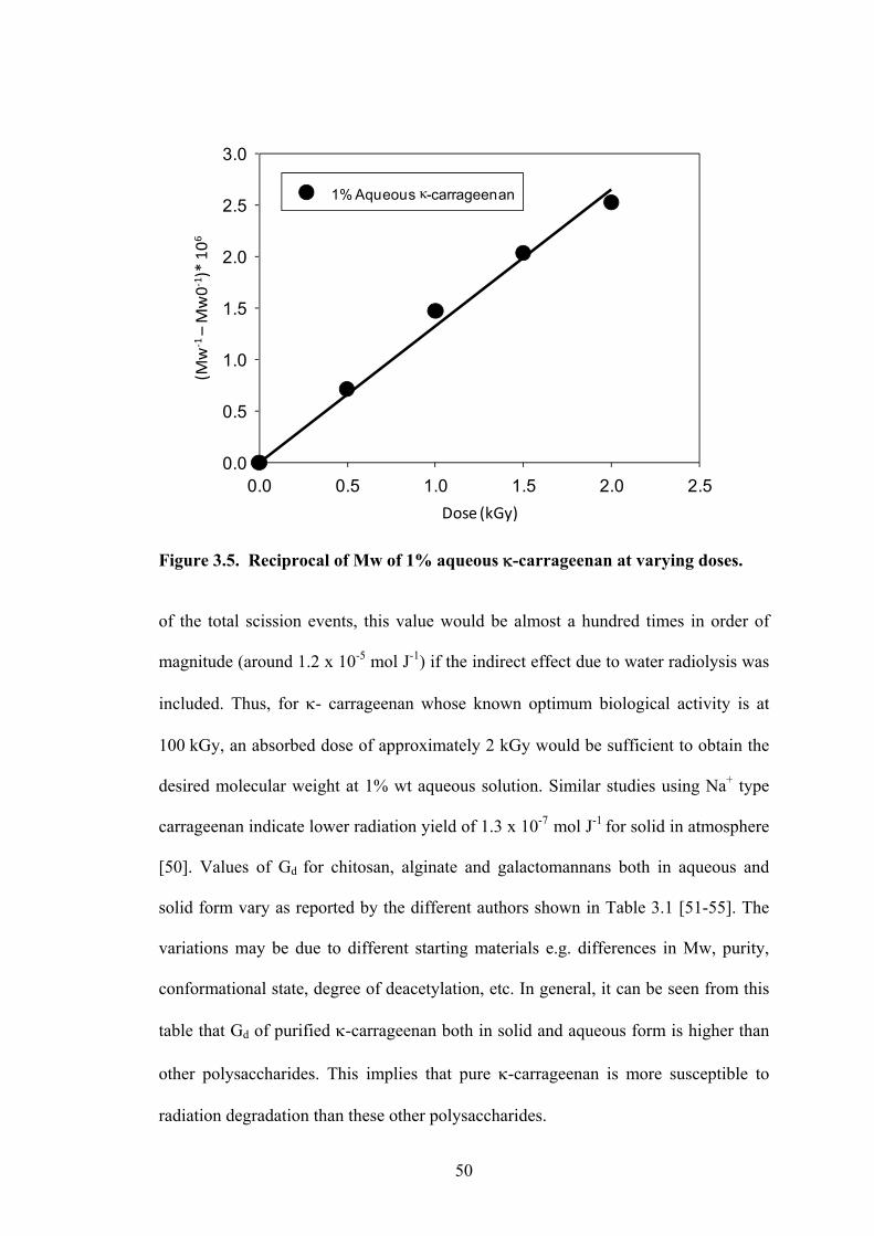
50
Figure 3.5. Reciprocal of Mw of 1% aqueous -carrageenan at varying doses.
of the total scission events, this value would be almost a hundred times in order of
magnitude (around 1.2 x 10⁻⁵ mol J-1) if the indirect effect due to water radiolysis was
included. Thus, for - carrageenan whose known optimum biological activity is at
100 kGy, an absorbed dose of approximately 2 kGy would be sufficient to obtain the
desired molecular weight at 1% wt aqueous solution. Similar studies using Na+ type
carrageenan indicate lower radiation yield of 1.3 x 10⁻⁷ mol J-1 for solid in atmosphere
[50]. Values of Gd for chitosan, alginate and galactomannans both in aqueous and
solid form vary as reported by the different authors shown in Table 3.1 [51-55]. The
variations may be due to different starting materials e.g. differences in Mw, purity,
conformational state, degree of deacetylation, etc. In general, it can be seen from this
table that Gd of purified -carrageenan both in solid and aqueous form is higher than
other polysaccharides. This implies that pure -carrageenan is more susceptible to
radiation degradation than these other polysaccharides.
0.0 0.5 1.0 1.5 2.0 2.50.0
0.5
1.0
1.5
2.0
2.5
3.0
1% Aqueous -carrageenan(M
w‐1–Mw0‐1)* 106
Dose (kGy)

51
Table. 3.1 Radiation degradation yield of different polysaccharides irradiated in solid and aqueous (1%) solution.
Gd (x 10-7 moles/joule)
Solid on air 4% aqueous 1% aqueous-carrageenan 1.3 [50]
2.5 0.3 [50] 1.2
chitosan 6.0 [51]1.8 [52]
0.9 [52]
3.53 [54]
galactomannans 0.9-1.1 [53] alginate 1.9 [27] 0.7 [27]
0.1 [54] 0.6 [27] 0.8 [54]
3.3.2. The Effect of N2 and N2O gas on the Radiation Degradation
Yield of -Carrageenan
The Gd of 1% aqueous solution of -carrageenan irradiated in N2O, N2 and in
air was determined to find out effects of these gases on the degradation of -
carrageenan. From equation 2.2 of chapter 2, N2O reacts with solvated electrons to
produce more OH radicals. It was discussed previously that OH• radicals react at a
very fast rate with -carrageenan (k . 1.2 x 10⁹ M-1 s-1). The effect of air on the
degradation yield of solid -carrageenan has also been discussed. Thus, it would be
interesting to know to what extent is the Gd of -carrageenan affected by the presence
of N2O and by purging N2 gas to remove air in the solution. Figure 3.6 shows the plot
of 1/Mn – 1/Mn0 against absorbed dose in the presence of these gases. From the
graph, it is quite obvious that in the presence of N2O, Gd was increased substantially.
Expectedly, the Gd of -carrageenan in the presence of N2 was lowest when compared
to N2O and in air. There was only a slight difference in the slope of the curves
between N2 and air. The computed Gd values were as follows: 1.7, 1.3 and 1.1 x 10⁻⁷
mol J-1 for N2O, air and N2 respectively. Studies on alginate do not show any

52
Figure 3.6. Reciprocal of Mw of 1% aqueous -carrageenan at varying doses
purged with different gases.
difference in the Gd of 1% solution in air and in N2 (Gd = 0.55 x 10⁻⁷ mol J-1) [27]. In
some cases, it has been reported that the radiation-chemical yield of scission has been
reduced in oxygen containing polymeric solution. The radiation-chemical yields of
chain scission for a 0.01 M chitosan are Gd = 3.4 × 10⁻⁷ mol J-1 in N2O-saturated and
G = 2.1 × 10⁻⁷ mol J-1 in N2O–O2-saturated solutions [19]. Similar trends have been
observed in pectic substances [23]. Interestingly, the starting molecular weight of -
carrageenan for this set of experiments is quite low (Mw = 32 kDa) as compared to
previous experiments in Section 2.3.1. (Mw = 450 kDa). Yet, results of the Gd for 1%
-carrageenan in air for both types of -carrageenan did not vary significantly (1.2
and 1.3 x 10⁻⁷ mol J-1). The Gd remained constant for large or small polymer chains of
-carrageenan.
0 1 2 3 4 5 6 70
2
4
6
8
10
12
14
in Airin N2
in N2
O
(Mw
‐1–Mw0‐1)* 106
Dose (kGy)

53
Figure 3.7 shows the UV absorbance at 260nm for aqueous -carrageenan in
the presence of N2O, N2 and in air with increasing doses. As previously discussed in
Chapter 2.3.4, this 260nm peak is attributed to the formation of carbonyl bond in the
pyranose ring. A slight increase in intensity of this peak was seen with increasing
doses. The UV absorbance trend follows the same order as that of their Gd values
where N2O > Air > N2. Thus, the greater is the radiation degradation yield, the greater
are the number of carbonyl groups formed. Free carbonyl groups are formed in two
ways, first, by radiolytically induced hydrolytical cleavage of glycosidic bonds, and
second, as the result of oxidation of carbohydrate radicals generated inside the galacto
pyranose ring [23].
Figure 3.7. UV absorbance at 260nm of -carrageenan (0.25%) irradiated at
different conditions.
Dose (kGy)
0 1 2 3 4 5 6 7
0.00
0.01
0.02
0.03
0.04
0.05
N2O
Air
N2
UV Absorbance (= 260nm)
54
3.3.3. The Effect of Na+ on the Gd of -carrageenan
It has been discussed in Chapter 2.3.5 that the presence of cations such as Na+
in -carrageenan promotes the formation of helical structures. The helical
conformation of - carrageenan also mentioned in the same chapter is currently still a
subject of debate. Some authors propose monomolecular helices (single strand) [55-
57] while other experimental results suggest double-stranded bimolecular structure
(double helix) of the carrageenan [58-62]. The subject on weight-average molecular
weight in the helical conformation has also been studied. Some authors show an
approximate doubling of the weight-average molecular weight for both - and -
carrageenan helices [63]. Ultrasonically degraded and - carrageenans also indicate
a higher weight-average molecular weight in the helix state [64].
The conformational or physical state of polysaccharides may have a strong
influence on the stability towards degradation [65]. Crystalline cellulose shows strong
resistance towards acid hydrolysis as compared to amorphous cellulose [66]. The
formation of multiple-stranded ordered structures stabilized by non-covalent inter-
chain bonds may increase the stability as they can tolerate cleavages of glycosidic
linkages without any pronounced changes in the physical structure [65].
In the initial stage of radiolysis reaction, Chapter 2.3.5 has demonstrated that
the presence of Na+ lowers the rate constant of reaction of OH• with -carrageenan. It
would be interesting to know if the yield of degradation would also be affected by
conformational transitions with higher structure rigidity e.g. helical conformation.
Figure 3.8 shows the effect of the addition of Na+ on the relationship of 1/Mn – 1/Mn0
with absorbed dose. The Gd had substantially been decreased by the presence of Na+
from a value of 1.2 to 0.7 x 10⁻⁷ mol J-1. This result clearly suggests that
conformational change from coil to helix in the presence of metal ions (Na+) affects

55
Figure 3.8. The effect of Na+ on the reciprocals of Mw of -carrageenan.
the degradation yield of -carrageenan. Since the transition to helical state of -
carrageenan forms rigidity of structure that promotes closer interaction of polymer
chains, there is the possibility of crosslinking simultaneously taking place with
degradation. Thus, Gd became lower in the helical state. It is also possible that these
results may be due to the higher stability in glycosidic linkage brought about by non-
covalent inter-chain bonds as explained in the previous paragraph. In a similar study,
the influence of the conformational state of - and - on the rate of acid hydrolysis has
been investigated. Results indicate an increase by a factor of 200 and 10 for - and -
carrageenan respectively when passing above the conformational transition
temperature [67].
Dose (kGy)
0.0 0.5 1.0 1.5 2.00
0.5
1.0
1.5
2.0
2.5
3.0
- Carrageenan
-Carrageenan with NaClO4
(1/M
n –
1/M
n0
) x 1
06

56
3.4. CONCLUSION
The radiation degradation yields of -carrageenan in solid and in aqueous
(1%) form at different conditions were found to be as follows: 2.5, 1.7 and 1.2 x 10⁻⁷
mol J-1 for solid in atmosphere, solid in vacuum and at 1% aqueous solution,
respectively. These values are higher than other known polysaccharides which make
-carrageenan more susceptible to radiation degradation attack. The Gd of solution
saturated with N2O solution was expectedly much higher with a value of 1.7 x 10⁻⁷
mol J-1. On the other hand, a lower Gd of 1.1 x 10⁻⁷ mol J-1 was obtained from -
carrageenan solution saturated with N2. The Gd in aqueous form was affected by the
conformational state of -carrageenan. The helical conformation gave a lower Gd (0.7
x 10-7 mol J-1) than the coiled conformation (Gd = 1.2 x 10⁻⁷ mol J-1).
Gd in solid which show higher value than in aqueous solution would indicate
higher efficiency. Note that the Gd in aqueous solution (1.2 x 10⁻⁷ mol J-1) constitutes
only 1% of the total depolymerization effect. If the radiation effect due to the indirect
action of transient OH• (in Chapter 2), then the degradation yield would be almost 100
times higher than its actual Gd value.
Information on the Gd will give the economic benefits of choosing the proper
condition for irradiating -carrageenan – whether in solid or in aqueous solution, solid
in air or in vacuum, aqueous in N2O, in N2 or in air. Ultimately, the users will have to
determine what would be best fitting for them considering their available resources
and for what specific purpose they want to depolymerize -carrageenan. For some
whose available energy Co-60 source is quite low, irradiation by aqueous solution
might be more beneficial where a dose of only 2 kGy (1% aqueous) is needed to
produce an equivalent of 100 kGy dose in solid irradiation. Where larger volumes of

57
-carrageenan are needed, solid irradiation may be more practical. Concentration of
aqueous -carrageenan can still be adjusted to higher concentration to maximize
benefits of aqueous irradiation. While the data obtained from this chapter gives us
outright the necessary absorbed dose at different conditions to obtain a specific
molecular weight -carrageenan, this would not be a sufficient basis for determining
the optimum dose needed for bio-based material development. Molecular weight/size
coupled with the chemical structure, functional groups formed, gelation and
conformational change behavior with absorbed dose need to be determined to harness
the full benefits of -carrageenan oligomer produced by radation processing.
REFERENCES:
1. A. Charlesby, Atomic Radiation and Polym. Vol. 1, Permagon Press, Oxford, 1960.
2. A. Chapiro, ed. Radiation Chemistry of Polymeric Systems, Vol. XV, Interscience, New York, 1962.
3. F. Williams, Radiat. Chem. Macromol. 1 (1972) 7.
4. J. H.O’Donnell and D. F. Sangster, Principles of Radiation Chemistry, Edward
Arnold, London, 1970.
5. J.W. T. Spinks and R. J.Woods, An Introduction to Radiation Chemistry, 3rd ed., John Wiley & Sons, Inc., New York, 1990.
6. C. D. Jonah, B. S. Madhava Rao, Radiation chemistry: present status and future
trends, Elsevier, 2001.
7. C. von Sontagg, Int. J. Radiat. Biol. 65 (1994) 19.
8. A. O. Allen, J. Phys. Chem. 52 (1948), 479.
9. IAEA-TECDOC-1420, Advances in radiation chemistry of polymers, Proceedings of a technical meeting held in Notre Dame, Indiana, USA, 13–17 September 2003
10. D. J. T. Hill, and A. K. Whittaker, "Radiation Chemistry of Polymers",
Encyclopedia of Polymer Science and Technology, 2004, on-line edition.
58
11. A. Charlesby, Proc. R. Soc. London, Ser. A 224 (1954) 120.
12. P. Alexander, A. Charlesby, M. Ross, Proc. R. Soc. London, Ser. A 223 (1954) 392.
13. M. Inokuchi, M. Dole, J. Chem. Phys. 38 (1963) 3006.
14. O. Saito, H. Y. Kang, M. Dole, J. Chem. Phys. 46 (1967) 3607.
15. H. Y. Kang, O. Saito, M. Dole, J. Polym. Sci., Polym. Symp. 25 (1968) 123.
16. O. Saito, Radiat. Chem. Macromol. 1 (1972) 223.
17. M. Tsuda, S. Oikawa, J. Polym. Sci., Polym. Chem. Ed. 17 (1979) 3759.
18. W. Dziedziela, D. Kotynska, J.Radiat. Phys. Chem. 23 (1984) 723.
19. P. Ulanski C. Von Sonntag, J. Chem. Soc., Perkin Trans. 2 (2000) 2022.
20. P. Ulanski, J. Rosiak, Radiat. Phys. Chem. 39, (1992) 53.
21. I. Idimecheva, R. Kisel, O. Shadyro, K. Kazem, H. Murase, T. Kagiya. J. Radiat.
Res. 46 (2005) 319.
22. G. O. Phillips and G. J. Moody, Int. J. Appl. Radiat. Isot. 6 (1959) 78.
23. H. Zegota, Food Hydrocolloids 13 (1999) 51.
24. T. Bucknall, H. Edwards, K. Kemsley, J. Moore, G. Phillips, Carbohydr. Res. 62 (1978) 49.
25. L. Huang, M. Zhai, J. Peng, J.Li, G.Wei, Carbohydr. Polym. 67 (2007) 305.
26. P. Baugh, J. Goodall, G. Phillips, C. Von Sonntag, M. Dizdaroglu, Carbohydr.
Res. 49 (1976) 315
27. N. Nagasawa, H. Mitomo, F. Yoshii, T. Kume, Polym. Degrad. Stab. 69 (2000) 279.
28. Fengel, D., Wegener, G. 1984. Wood: Chemistry, Ultrastructure, Reactions.
Berlin: Walter de Gruyter.
29. Y. Jiang, X. Guo, Carbohydr. Polym. 61 (2005) 441.
30. T. Hjerde, O. Smidsrød, B. Christensen, Carbohyd. Res. 288 (1996) 175.
31. A. Karlsson, S. Singh, Carbohydr. Polym. 38 (1999) 7.
32. G. Yu, H. Guan, A. Ioanoviciu, S. Sikkander, C. Thanawiroon, J. Tobacman, T. Toida, R. Linhardt, Carbohydr. Res. 337 (2002) 433.

59
33. S. Knutsen, H. Grasdalen Carbohydr. Polym. 19 (1992) 199.
34. S. Knutsen, M. Sletmoen, T. Kristensen, T. Barbeyron, B. Kloareg, P. Potin,
Carbohydr. Res. 331 (2001) 101.
35. M. Guibet, N. Kervarec, S. Génicot, Y. Chevolota, W. Helbert, Carbohydr. Res. 341 (2006) 1859.
36. R. Falshaw, R. Furneaux, H. Wong, Carbohydr. Res. 338 (2003) 1403.
37. T. Stevenson, R. Furneaux, Carbohydr. Res. 210 (1991) 277.
38. A. Usov, Food Hydrocolloids. 12 (1998) 301.
39. S. Knutsen, H. Grasdalen, Carbohydr. Res. 229 (1992) 233.
40. T. Yamada, A. Ogamo, T. Saito, H. Uchiyama, Y. Nakagawa, Carbohydr. Polym.
41 (2000) 115.
41. T. Yamada, A. Ogamo, T. Saito, J. Watanabe, H. Uchiyama, Y. Nakagawa, Carbohydr. Polym.. 32 (1997) 51.
42. C. Rochas, M. Rinaudo, M. Vincendon, Int. J. Biol. Macromol. 5 (1983) 111.
43. G. Yu, H. Guan, A. Ioanoviciu, S. Sikkander, C. Thanawiroon, J. Tobacman, T.
Toida, R. Linhardt, Carbohydr. Res. 337 (2002) 433.
44. C. Rochas, A. Heyraud, Polym. Bull.5 (1981) 81.
45. T. Hjerde, O. Smidsrød, B. Stokke, E. Christensen, Carbohydr. Res. 288 (1996) 175.
46. T. Hjerde, T. Kristiansen, B. Stokke, O. Smidsrød, B. Christensen, Carbohydr.
Polvmers 24 (1994) 265.
47. S. Singh, S. Jacobsson, Carbohydr. Polym. 23 (1994) 89.
48. C. von Sonntag, H-P. Schuchmann, Angeuz. Chem. Int. Ed. Engl. 30 (1991) 1229-1253.
49. C. von Sonntag, E. Bothe, P. Ulanski, and D. J. Deeble, Radiat. Phys. Chem.
46(1995) 527.
50. L. Relleve, N. Nagasawa, L. Luan, T. Yagi, C. Aranilla, L. Abad, T. Kume, F. Yoshii, A. De la Rosa, Polym Degrad Stab. 87 (2005) 403.
51. R. Czechowska-Biskup, B. Rokita, B., P. Ulanski, J. Rosiak, J. Nucl. Instr. and
Meth. in Phys. Res. B. 236 (2005) 383.
60
52. L. Hai, TB. Diep, N. Nagasawa, F. Yoshii, T. Kume, Nucl. Instr. and Meth. in Phys. Res. B., 208 (2003) 466.
53. M. Sen, B. Yolacan, O. Guven, Nucl. Instr. and Meth. in Phys. Res. B., 265
(2007) 429.
54. J. Wasikiewicza, F. Yoshii, N. Nagasawa, R. Wach, H. Mitomo, Radiat. Phy. Chem., 73 (2005) 287.
55. S. Paoletti, O. Smidsrød, H. Grasdalen, Biopolymers. 23 (1984) 177l.
56. C. Rochas, S. Landry, Carbohydr. Polym. 7 (1987) 435.
57. M. Bosco, A. Segre, S. Miertus, A. Cesàro, S. Paoletti, Carbohydr. Res. 340
(2005) 943.
58. S. Nilsson, L. Piculell,. Macromolecules. 24 (1991) 3804.
59. V. Grinberg, N. Grinberg, A. Usov, N. Shusharina, A. Khokhlov, K. de Kruif, Biomacromolecules. 2 (2001) 864.
60. Y. Yuguchi, T. Thuy, H. Urakawa, K. Kajiwara, Food Hydrocolloids. 16 (2002)
515.
61. S. Janaswamy, R. Chandrasekaran, Carbohydr. Polym. 60 (2005) 499.
62. M. Mangione, D. Giacomazzaa, D. Bulonea, V. Martoranaa, P. San Biagioa, Biophys. Chem. 104 (2003) 95.
63. C. Viebke, J. Borgström L. Piculell, Carbohydr. Polym. 27 (1995) 145.
64. V. Meunier, T. Nicolai, D. Durand, Int. J. Biol. Macromol. 28 (2001) 157.
65. T. Hjerde, T. Kristiansen, B. Stokke, O. Smidsrød, B. Christensen, Carbohvdr.
Polym. 24 (1994) 265.
66. A. Sharples, Trans. Faraday Soc. 53 (1957) 1003.
67. T. Hjerde, O. Smidsrød, B. Christensen, Carbohydr. Res. 288 (1996) 175.

61
CHAPTER 4
CHEMICAL AND SPECTRAL CHARACTERIZATION OF THE
RADIOLYTIC PRODUCTS OF -CARRAGEENAN
4.1. INTRODUCTION
The radiation chemistry of carbohydrates involves the abstraction of hydrogen
atoms from positions which are -to a hydroxyl group. In the case of aqueous
solutions, abstraction is achieved through the hydroxyl radicals (from radiolysis of
water). Hydrated electrons react more slowly, if at all. Hydrogen atoms react more
slowly than hydroxyl group but would be expected to abstract similarly. The
formation of the carbohydrate radicals (e.g. glucose radicals) may be represented as
follows : [1-3]
(4.1)
(4.2)
(4.3)
In the absence of oxygen, the gluconic acid and glucosone (2-oxo-D-
arabinoaldohexose) can be formed by abstraction of hydrogen atoms from the radicals
O
OH
HH
H
OH
OH
H OH
H
CH2OH
O
C
OH
HH
H
OH
OH
H OH
CH2OH
O
OH
HH
OH
OH
H OH
H
CH2OH
.
O
OH
HH
H
OH
OH
H OH
H
CHOH.
O
C
OH
HH
H
OH
OH
H OH
CH2OH
OH
COOHHH
H
OH
OH
H OH
CH2OH
O
OH
HH
OH
OH
H
H
CH2OH
O
O
C
OH
HH
H
OH
OH
H OH
CH2OH
(-H + H2O)
(-H)

62
R-H R.(C1 – C6) + H.
R-H + H. R.(C1 – C6) + H2
R.(C1, C4) F1 + F2 (scission)
F. R(C=O)
.
formed in equations 4.2 and 4.3. The formation of glucuronic acid on irradiation in
the presence of oxygen may be due to the reaction of oxygen with the radicals formed
by reaction 4.3 yielding an unstable aldehyde which becomes oxidized to glucuronic
acid. The absence of glucosone may be accounted for by the addition of oxygen to the
radical formed in 4.2, followed by a chain of reactions including C-C scission, giving
rise to the formation of arabinose and formaldehyde.
The chemical changes occurring in polysaccharides are similar to those with
simple carbohydrates. Reducing groups (carbonyl) and acid groups appear and
carbon-carbon scission occurs. Radiation degradation of several natural
polysaccharides e.g. cellulose [4], chitosan [5-8], carboxymethyl chitosan [9],
alginates [10-11], galactomanans [12], xyloglucan [13], cycloamylose [14] and
pectins [15] have already been studied. Degradation schemes have been proposed for
the cleavage of the glycosidic linkage leading to the degradation of the polymeric
chain e.g. chitosan, pectin, dextrans, etc. with C(1) 6 C(4) linkages. These schemes
can be summarized as follows:
Solid Irradiation:
(4.4)
(4.5)
(4.6)
(4.7)
63
H2O .OH + H. + eaq
N2O + eaq + H+ .OH + N2
.OH(H.) + RH R.(C1 – C6) + H2O(H2)
F1 + F2 (hydrolysis)R.(C1, C4, C5)
F3 + F4 (scission)
F1(F3) R(C=O) or R(COOH)
2R. (F.) 2R (F) (disproportionation)
.
-
.
. .
-
R.(C1 – C6) + O2 ROO. R(C=O)
Aqueous Irradiation:
(4.8)
(4.9)
(4.10)
(4.11)
(4.12)
(4.13)
Aqueous or Solid Irradiation:
(4.14)
From these schemes, the radiolytic products yield not only the formation of low
molecular weight oligomers but also some carbonyl bonds (carboxyl and aldo-keto
groups) and some functional groupings that are cleaved off e.g. NH3 in chitosan.
Depolymerization of carrageenan is easily achieved by chemical means as
already discussed in Chapter 3.1. These hydrolysis reactions are accompanied by
increase in reducing sugars [16-17] and release of sulfate [18-20]. Some other
methods make use of ultrasonic [21-22] and microwave degradation [23].
Mechanochemistry and radical reactions similar to the radiation chemistry of
carbohydrates are expected to contribute to chain scission using the ultrasonic
degradation processes [24-25]. While the scission reactions of the chemical
degradation process have already been carefully studied, this chapter will study the

64
chemistry of the radiolytic products of -carrageenan processed by gamma radiation
as well as the possible mechanisms involved.
4.2. METHODOLOGY
4.2.1. Gamma Irradiation of -carrageenan
Solid and aqueous -carrageenan samples were gamma irradiated (Chapter
2.2.5) with absorbed doses ranging from 1 to 200 kGy at a dose rate of 1 or 10 kGy/h
(for doses above 5 kGy).
4.2.2. UV Vis Spectroscopy
UV Vis Spectroscopy of -carrageenan is already described under Chapter
2.2.8.
4.2.3. FT-IR Spectroscopy
FT-IR spectra of samples in KBr pellets (1mg / 100mg KBr) were measured
using an FT-IR Nicolet Magna 550 at ambient temperature in the region of 4000-
400cm-1.
4.2.4. Chemical Analyses
The reducing group of the carrageenans was determined using the Nelson-
Somogyi method of analysis with galactose as the standard [26].
The total acidity of the carrageenans was determined by acid–base titration
method. Carrageenan solutions were titrated against standardized NaOH using a
phenolphthalein indicator to determine end point. The acidity was reported as %
H2SO4 in carrageenan.

65
Samples of -carrageenan were washed three times in ethanol (95% purity) to
remove the free sulfates. The samples were then freeze dried. Percent carboxylic acid
was determined by acid-base titration with standardized NaOH of these washed
samples. The % free sulfates was then computed as follows:
% Free Sulfates = % Total acidity– % Acidity due to COOH
The percent cleaved sulfate was computed as follows:
% Desulfation = % free sulfates / 20.98 x 100
where the value 20.98 is the theoretical percent sulfate (as HSO3-) present in -
carrageenan per mole of the repeating unit.
4.2.5. Fractionation of Irradiated -carrageenan
Irradiated -carrageenan samples (solid-100 kGy in air, solid - 100 kGy in
vacuum and 1% aqueous - 2 kGy) were fractionated using an Amicon Pressure
Filtration set up pressurized with N2O gas (not exceeding 1.5 kgf/cm2) using
Millipore filters with molecular weight cut offs of >30 kDa, 10-30 kDa, and 3-10
kDa.
4.2.6. NMR of Irradiated -Carrageenan
Fractionated samples with molecular weight cut off of 3–10 kDa were
dissolved in D2O at concentrations of 30-40 mg/ml. Nuclear Magnetic Resonance
(NMR) analyses were carried out using a Bruker 300 Ultra Shield equipment with
300MHz frequency at room temperature (25oC). The 1H spectra were recorded using 8
scans. 13C spectra were obtained using 2400 scans with 3-(Trimethylsilyl)-1-
propanesulfonic acid (DDS) as internal standard. Neocarrabiose-4-O-Sulphate (DP=1,

66
mol.wt = 449) and Neocarrahexaose-4 1,3,5-tri-O-suphate (DP=3, mol. wt = 1265)
were also analyzed as reference materials (Figure 4.1).
Figure 4.1. Structure of Neocarrabiose Standards
4.3. RESULTS AND DISCUSSION
4.3.1. UV-Vis Spectrum
It is already known that radiation degradation of polysaccharides leads to
some chemical changes such as the formation of carbonyl, carboxyl or double bonds
[6, 10, 15]. These changes are seen in the UV spectra as a new absorbance peak at
around 265 and 297nm for chitosan. Likewise, it has also been reported that UV
spectra of all types of irradiated carrageenan show absorbance peak at around 260nm.
Its intensity increases with increasing dose [27]. Figures 4.2a, b, and c show a new
absorbance peak at 260nm for -carrageenan irradiated in solid (in air and in vacuum)
and at 1% aqueous solution. The peak intensities increased with increasing doses.
Absorbance of aqueous carrageenan solutions was very much higher than in solid in
O
OH
O3SO
OOH
OH
O
OH
OOH
Na
O
OH
O3SO
O
OH
O
OH
OO
OH
O3SO
O
OH
O
OH
OOO
OH
O3SO
OOH
OH
O
OH
OO OH
Na Na Na
Neocarrabiose‐4‐O‐Sulphate
Neocarrahexaose‐4 1,3,5‐tri‐O‐suphate
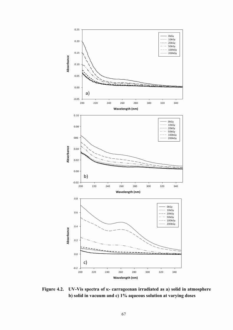
67
Wavelength (nm)
Absorban
ce
200 220 240 260 280 300 320 340
‐0.05
0.00
0.05
0.10
0.15
0.20
0.25
0kGy
10kGy
20kGy
50kGy
100kGy
200kGy
a)
Wavelength (nm)
Absorbance
200 220 240 260 280 300 320 340
‐0.02
0.00
0.02
0.04
006
0.08
0.10
0kGy10kGy20kGy50kGy100kGy200kGy
b)
Wavelength (nm)
Absorban
ce
200 220 240 260 280 300 320 340
‐0.2
0.0
0.2
0.4
0.6
0.8
0kGy
10kGy
20kGy
50kGy
100kGy
200kGy
c)
Figure 4.2. UV-Vis spectra of - carrageenan irradiated as a) solid in atmosphereb) solid in vacuum and c) 1% aqueous solution at varying doses
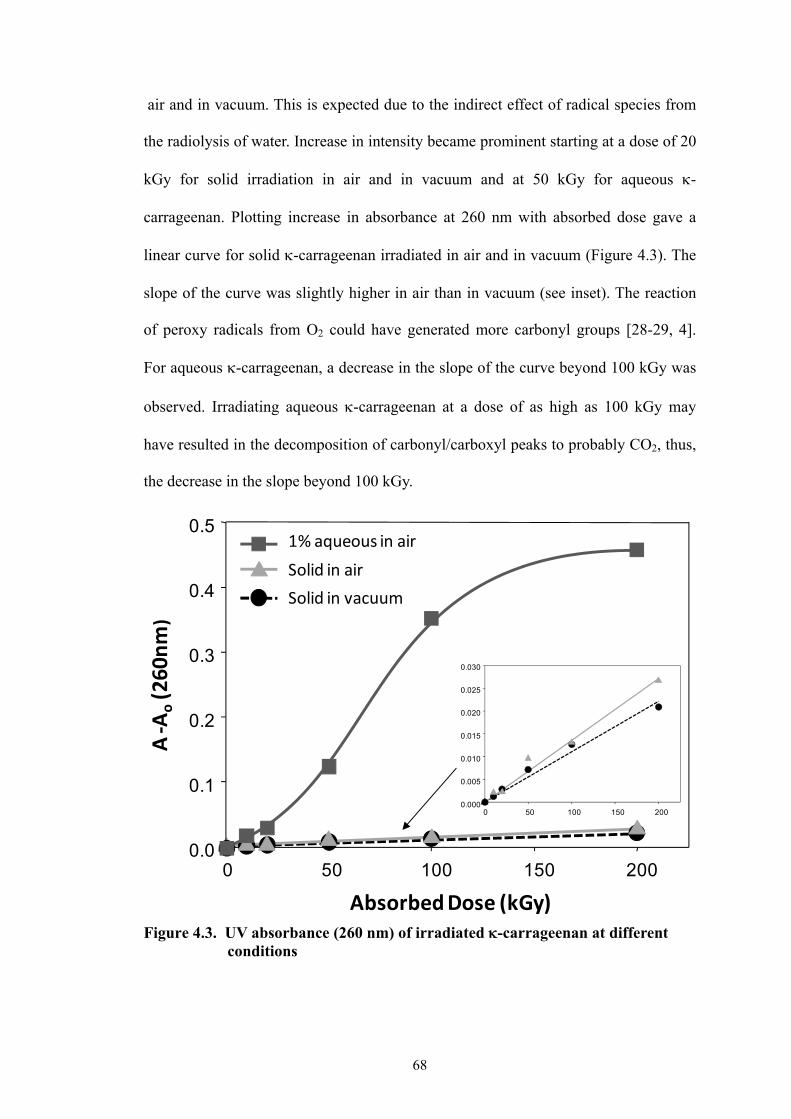
68
air and in vacuum. This is expected due to the indirect effect of radical species from
the radiolysis of water. Increase in intensity became prominent starting at a dose of 20
kGy for solid irradiation in air and in vacuum and at 50 kGy for aqueous -
carrageenan. Plotting increase in absorbance at 260 nm with absorbed dose gave a
linear curve for solid -carrageenan irradiated in air and in vacuum (Figure 4.3). The
slope of the curve was slightly higher in air than in vacuum (see inset). The reaction
of peroxy radicals from O2 could have generated more carbonyl groups [28-29, 4].
For aqueous -carrageenan, a decrease in the slope of the curve beyond 100 kGy was
observed. Irradiating aqueous -carrageenan at a dose of as high as 100 kGy may
have resulted in the decomposition of carbonyl/carboxyl peaks to probably CO2, thus,
the decrease in the slope beyond 100 kGy.
Figure 4.3. UV absorbance (260 nm) of irradiated -carrageenan at different conditions
0 50 100 150 2000.0
0.1
0.2
0.3
0.4
0.5
A‐A
o(260nm)
Absorbed Dose (kGy)
0 50 100 150 2000.000
0.005
0.010
0.015
0.020
0.025
0.030
1% aqueous in air
Solid in air
Solid in vacuum
69
4.3.2. FT-IR Spectrum
Several references are available for the FT-IR of -carrageenan [30-35]. FT-IR
spectra in Figure 4.4 show the finger print functional groupings of the unirradiated -
carrageenan [36] and some changes with irradiation. Similar spectrum has been
obtained with low molecular weight -carrageenan processed by acid hydrolysis [37–
38]. The spectra of irradiated - carrageenan in solid form in air indicated that the
functional groupings were entirely kept intact even at a high dose of 200 kGy. The
only change observed was the appearance of a new absorption peak at 1728 cm-1
corresponding to the carbonyl grouping and decrease in the intensity of polymer
bound water peak at 200 kGy. For vacuum irradiation, no changes were alsoobserved
up to a dose of 100 kGy. However, distortion of peak corresponding to the
galactopyranose ring / glycosidic linkage became evident at 200 kGy. Loss of peaks
corresponding to covalent sulfate, 3,6 anhydro bridge and D-galactose-4-sulfate were
also observed. There was a decrease in the intensity of polymer bound water peak.
The change in the polymer bound water may signify that water radiolysis may have
also occurred in the system. It could not be discounted that changes in vacuum at 200
kGy may also be caused by post-irradiation effects after exposure of the sample in air.
In 1% aqueous solution -carrageenan, increasing carbonyl peaks was observed with
increasing doses. The FT-IR data showing the appearance of carbonyl peaks which
increased with absorbed dose further supports data obtained in the UV-Vis spectra
which also indicated the same trend of increasing carbonyl peaks. At 2 kGy, the finger
print functional groups of 1% aqueous -carrageenan solution were still intact.
Changes in the functional groupings were observed starting at a dose of 10 kGy. The
spectra of aqueous -carrageenan started to become distorted at this dose with a

70
Figure 4.4. FT-IR spectra of -carrageenan irradiated in air, in vacuum and in
1% aqueous solution at different doses.
Absorption (cm-1)
1640-1645
1370-1375
1210-1260
1010-1080
928-933
840-850
Functional Groups
polymer bound water
methylene group
covalent sulfate
glycosidic linkage
3,6 anhydro-D-galactose
D-galactose-4-sulfate
0kGy
200kGy
2 kGy
10 kGy
100 kGy
200kGy
100kGy
100kGy
600800100012001400160018002000
Wavenumber (cm‐1)
Relative Absorban
ceSolid Powder in air 1% aq. soln. Solid Powder in vacuum
Carbonyl
Polymer Bound
Water
MethyleneGroup
CO, COH
Covalent
Sulfate
Galactose
Pyeanose
ring
Glycosidic
linkage
3,6 anhydro
bridge
D‐galactose
4‐sulfate
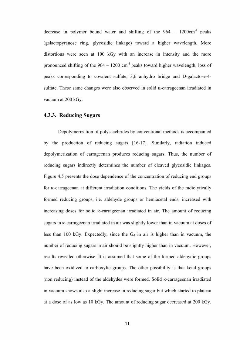
71
decrease in polymer bound water and shifting of the 964 – 1200cm-1 peaks
(galactopyranose ring, glycosidic linkage) toward a higher wavelength. More
distortions were seen at 100 kGy with an increase in intensity and the more
pronounced shifting of the 964 – 1200 cm-1 peaks toward higher wavelength, loss of
peaks corresponding to covalent sulfate, 3,6 anhydro bridge and D-galactose-4-
sulfate. These same changes were also observed in solid -carrageenan irradiated in
vacuum at 200 kGy.
4.3.3. Reducing Sugars
Depolymerization of polysaachrides by conventional methods is accompanied
by the production of reducing sugars [16-17]. Similarly, radiation induced
depolymerization of carrageenan produces reducing sugars. Thus, the number of
reducing sugars indirectly determines the number of cleaved glycosidic linkages.
Figure 4.5 presents the dose dependence of the concentration of reducing end groups
for -carrageenan at different irradiation conditions. The yields of the radiolytically
formed reducing groups, i.e. aldehyde groups or hemiacetal ends, increased with
increasing doses for solid -carrageenan irradiated in air. The amount of reducing
sugars in -carrageenan irradiated in air was slightly lower than in vacuum at doses of
less than 100 kGy. Expectedly, since the Gd in air is higher than in vacuum, the
number of reducing sugars in air should be slightly higher than in vacuum. However,
results revealed otherwise. It is assumed that some of the formed aldehydic groups
have been oxidized to carboxylic groups. The other possibility is that ketal groups
(non reducing) instead of the aldehydes were formed. Solid -carrageenan irradiated
in vacuum shows also a slight increase in reducing sugar but which started to plateau
at a dose of as low as 10 kGy. The amount of reducing sugar decreased at 200 kGy.

72
Figure 4.5. Percent reducing sugar of -carrageenan irradiated at increasing
doses.
Free carbonyl groups in glucose yields carrageenan macromolecule which are formed
in two ways, first, by radiolytically induced cleavage of glycosidic bonds, and second,
as the result of oxidation of carbohydrate radicals generated inside the carrageenan
residue. These radiolytically formed free carbonyl groups can be generated via the
different schemes as illustrated in the last part of this chapter. Radiolysis reaction in
glucose yields glucosone (a ketone) in the absence of oxygen (eq. 4.3). Results on the
reduction of the amount of reducing sugar at 200 kGy coincided with the FT-IR
results of vacuumed -carrageenan irradiated at 200 kGy where the absorbance peak
corresponding to galactose pyranose ring and glycosidic linkage has been destroyed at
this dose. Aqueous -carrageenan, obviously, yielded more reducing end groups as
more radiolytically formed reducing group are expected from the indirect effect of
Dose (kGy)
0 50 100 150 200 250
Pe
rce
nt
Re
du
cin
g S
ug
ar
0
2
4
6
8
10
12
14 Solid iin air
Solid in vacuum
1% aq. soln.

73
OH radicals promoting scission and hydrolysis reactions. Radiation effects on the
polysaccharide pectin solutions also yield reducing groups which increase rapidly
with increasing doses. The presence of oxygen during irradiation of pectin solutions
influences to some extent the concentration of these groups [15]. Gamma irradiation
of aqueous solutions of D-glucose in oxygen yields the reducing sugars erythrose,
glyoxal, formaldehyde, arabinose and xylose [2]. Maximum amount of reducing sugar
in aqueous solution was obtained at 50 kGy. This result coincided with the weight
average molecular weight obtained at 50 kGy (Chapter 3.3.1). No further decrease in
Mw was observed beyond a dose of 20 kGy which would indicate no further cleavage
in glycosdic linkage and therefore no further increase in reducing groups. Beyond 50
kGy, these reducing groups may have been oxidized further to carboxylic groups or
other decomposition products which resulted in the decrease of reducing sugar
beyond this dose.
4.3.4. Acidity
Sulfated galactans like carrageenans are known to release their sulfates after
depolymerization [18-20] rendering them acidic. Acid hydrolysis of -carrageenan
and chondroitin sulfate has been observed to undergo significant desulfation [39].
Figure 4.6 shows a slight increase in acidity (as H2SO4) with solid carrageenans
irradiated in vacuum and in air. Increase in acidity of the vacuumed samples was
almost zero. Irradiation in aqueous solution, on the other hand, showed a sharp
increase in acidity up to 50 kGy and starts to plateau beyond this dose. A high percent
acidity of around 16% percent (as H2SO4) was observed at a dose of 200 kGy. Since
the source of acidity of irradiated carrageenans does not originate only from the
cleaved sulfates but also from the carboxylic groups formed (from oxidation of
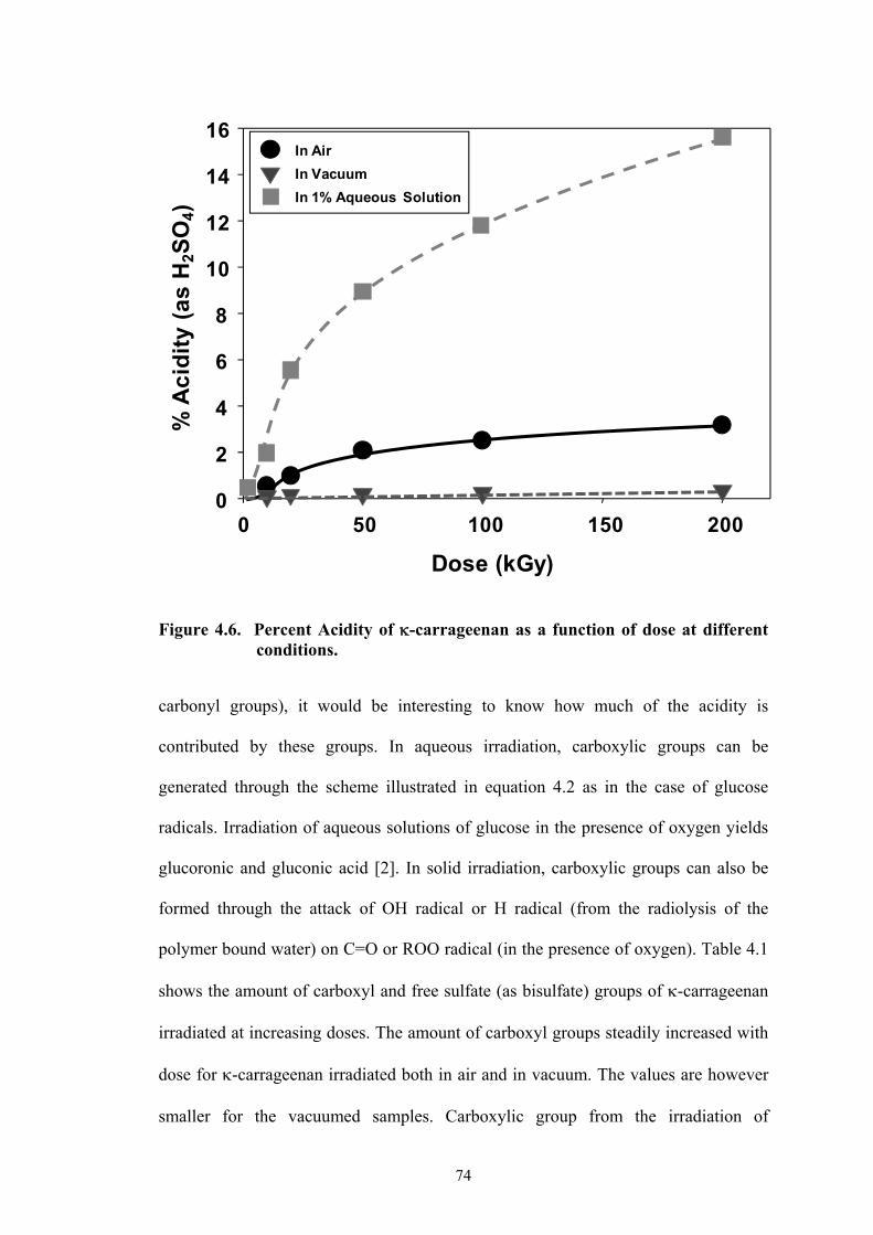
74
carbonyl groups), it would be interesting to know how much of the acidity is
contributed by these groups. In aqueous irradiation, carboxylic groups can be
generated through the scheme illustrated in equation 4.2 as in the case of glucose
radicals. Irradiation of aqueous solutions of glucose in the presence of oxygen yields
glucoronic and gluconic acid [2]. In solid irradiation, carboxylic groups can also be
formed through the attack of OH radical or H radical (from the radiolysis of the
polymer bound water) on C=O or ROO radical (in the presence of oxygen). Table 4.1
shows the amount of carboxyl and free sulfate (as bisulfate) groups of -carrageenan
irradiated at increasing doses. The amount of carboxyl groups steadily increased with
dose for -carrageenan irradiated both in air and in vacuum. The values are however
smaller for the vacuumed samples. Carboxylic group from the irradiation of
Figure 4.6. Percent Acidity of -carrageenan as a function of dose at different
conditions.
0 50 100 150 2000
2
4
6
8
10
12
14
16In Air
In Vacuum
In 1% Aqueous Solution
Dose (kGy)
% A
cid
ity
(as
H2S
O4)
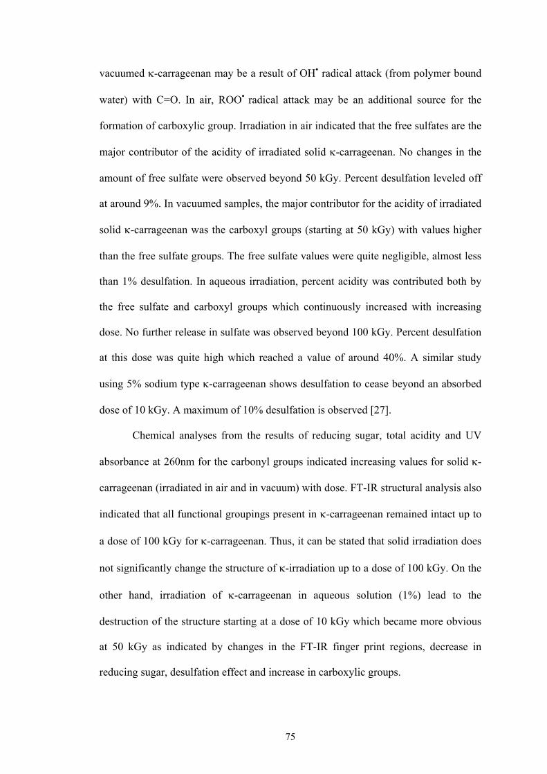
75
vacuumed -carrageenan may be a result of OHC radical attack (from polymer bound
water) with C=O. In air, ROOC radical attack may be an additional source for the
formation of carboxylic group. Irradiation in air indicated that the free sulfates are the
major contributor of the acidity of irradiated solid -carrageenan. No changes in the
amount of free sulfate were observed beyond 50 kGy. Percent desulfation leveled off
at around 9%. In vacuumed samples, the major contributor for the acidity of irradiated
solid -carrageenan was the carboxyl groups (starting at 50 kGy) with values higher
than the free sulfate groups. The free sulfate values were quite negligible, almost less
than 1% desulfation. In aqueous irradiation, percent acidity was contributed both by
the free sulfate and carboxyl groups which continuously increased with increasing
dose. No further release in sulfate was observed beyond 100 kGy. Percent desulfation
at this dose was quite high which reached a value of around 40%. A similar study
using 5% sodium type -carrageenan shows desulfation to cease beyond an absorbed
dose of 10 kGy. A maximum of 10% desulfation is observed [27].
Chemical analyses from the results of reducing sugar, total acidity and UV
absorbance at 260nm for the carbonyl groups indicated increasing values for solid -
carrageenan (irradiated in air and in vacuum) with dose. FT-IR structural analysis also
indicated that all functional groupings present in -carrageenan remained intact up to
a dose of 100 kGy for -carrageenan. Thus, it can be stated that solid irradiation does
not significantly change the structure of -irradiation up to a dose of 100 kGy. On the
other hand, irradiation of -carrageenan in aqueous solution (1%) lead to the
destruction of the structure starting at a dose of 10 kGy which became more obvious
at 50 kGy as indicated by changes in the FT-IR finger print regions, decrease in
reducing sugar, desulfation effect and increase in carboxylic groups.
76
Table 4.1a. Acidic composition of gamma irradiated solid -carrageenan (in air)
with absorbed dose.
Table 4.1b. Acidic composition of gamma irradiated solid -carrageenan (in vacuum) with absorbed dose.
Table 4.1c. Acidic composition of gamma irradiated -carrageenan (1% aqueous solution) with absorbed dose.
Absorbed
Dose (kGy)
% Acidity (as
H2SO4) %COOH
Free Sulfates
(as % HSO3-)
% Desulfation
10 0.52 0.00 0.48 2.29
20 0.97 0.04 0.85 0.19
50 2.07 0.08 1.81 8.63
100 2.49 0.17 2.11 10.06
200 3.15 1.01 1.88 8.96
Absorbed
Dose (kGy)
% Acidity (as
H2SO4) %COOH
Free Sulfates
% HSO3-
% Desulfation
10 0.07 0.02 0.05 0.24
20 0.14 0.06 0.07 0.33
50 0.17 0.15 0.01 0.05
100 0.21 0.19 0.01 0.05
200 0.33 0.27 0.04 0.19
Absorbed
Dose (kGy)
% Acidity (as
H2SO4) %COOH
Free Sulfates
% HSO3-
% Desulfation
2 0.46 0.01 0.41 1.95
10 1.96 0.40 1.39 6.62
20 5.52 3.35 1.72 8.20
50 8.94 4.44 3.77 17.97
100 11.78 4.67 8.23 39.22
200 15.59 5.67 8.64 41.18

77
4.3.5. Fractionation of Irradiated -carrageenan
From the introduction, the effective Mw of oligosaacharides as plant growth
promoter is < 10 kDa. The optimum biological activity of -carrageenan is attained at
100 kGy (in solid) as discussed also in the introduction. Previous result in Chapter
3.3.1 has demonstrated that the Mw obtained at 100 kGy can likewise be attained by
irradiating 1% aqueous -carrageenan solution at around 2 kGy. Chapter 5.3.1.4 will
discuss on some DLS experiments demonstrating the presence of new fast mode
peaks in the ICF at 100 kGy with an Mw ca. 5,000 - 10,000 which is believed to be
related to the biological activity in plants. Thus, solid and aqueous -carrageenans
were irradiated at these doses and were fractionated for structural elucidation. Several
literatures are available for the separation of the different oligomers of -carrageenan
[40-45]. Most of these procedures utilize some appropriate columns that make the
process very tedious and give very small yield. Fractionation was done with the use of
Millipore filters with appropriate molecular weight cut-offs. This procedure, however,
gives a wide range of molecular weight distribution of the fraction. Table 4.2 shows
the weight average molecular weight obtained by GPC of the different fractions of -
carrageenan irradiated in solid state in air and in vacumm at 100 kGy and 1% aqueous
solution at 2 kGy. Fractionation of solid -carrageenan irradiated in air (100 kGy) and
1% aqueous -carrageenan (2 kGy) was well achieved as seen from the Mw of the
different fractions. Fractionation of the irradiated vacuumed sample (100 kGy), on the
other hand, was not well achieved. The fractionated -carrageenan containing < 3 kDa
had an additional radiolytic product peak with an Mw of around 900. This fraction
also contained higher carbonyl and/or double bonds as indicated by the UV-Vis
absorbance at 260nm in Table 4.3. Thus, only the fraction of oligomers with an Mw of

78
Table 4.2. Molecular weight of the fractionated samples of irradiated -carrageenan
Table 4.3. UV-Vis ( = 260nm) of fractionated irradiated -carrageenan
(0.025%)
3- 10 kDa was considered to probably contain the biological active components for
plant growth promoter. From the results in Figure 4.7, the fraction containing an Mw
of 3- 10 kDa constitutes only 11% and 25% for air and aqueous solution respectively.
This implies that the active component for the biological activity of -carrageenan
comprises only a small fraction of the irradiated -carrageenan per unit weight.
Higher amount of the biological active component can be obtained in irradiated 1%
Mol. Wt. Cut-off (kDa)
Mw Peak 1 Mw Peak 2
100 kGy in Air < 3,000 9,000 910
3,000-10,000 15,000 -
10,000-30,000 26,000 -
> 30,000 38,000 -
2 kGy
1% Aqueous
<3,000 7,000 930
3,000-10,000 13,000 -
10,000-30,000 32,000 -
> 30,000 30,000 -
100 kGy in Vacuum
< 3,000 10,000 890
3,000-10,000 29,000 -
10,000-30,000 26,000 -
> 30,000 - -
Mol. Wt. Cut-off Fraction
Solid (100 kGy) 1% Aqueous (2 kGy)
Vacuum (100 kGy) 0.1%
> 30,000 - - -
10,000-30,000 0.005 0.006 0.047
3,000-10,000 0.004 0.028 0.051
<3,000 0.025 0.023 0.118
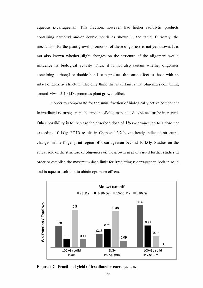
79
aqueous -carrageenan. This fraction, however, had higher radiolytic products
containing carbonyl and/or double bonds as shown in the table. Currently, the
mechanism for the plant growth promotion of these oligomers is not yet known. It is
not also known whether slight changes on the structure of the oligomers would
influence its biological activity. Thus, it is not also certain whether oligomers
containing carbonyl or double bonds can produce the same effect as those with an
intact oligomeric structure. The only thing that is certain is that oligomers containing
around Mw = 5-10 kDa promotes plant growth effect.
In order to compensate for the small fraction of biologically active component
in irradiated -carrageenan, the amount of oligomers added to plants can be increased.
Other possibility is to increase the absorbed dose of 1% -carrageenan to a dose not
exceeding 10 kGy. FT-IR results in Chapter 4.3.2 have already indicated structural
changes in the finger print region of -carrageenan beyond 10 kGy. Studies on the
actual role of the structure of oligomers on the growth in plants need further studies in
order to establish the maximum dose limit for irradiating -carrageenan both in solid
and in aqueous solution to obtain optimum effects.
Figure 4.7. Fractional yield of irradiated -carrageenan.
0.28
0.18
0.56
0.11
0.250.29
0.5 0.48
0.150.11 0.09
0
100kGy solidIn air
2kGy1% aq. soln.
100kGy solidIn vacuum
Mol.wt cut ‐off
<3kDa 3‐10kDa 10‐30kDa >30kDa
Wt. fraction / Total w
t.
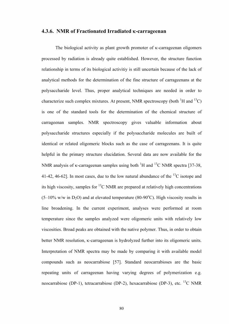
80
4.3.6. NMR of Fractionated Irradiated -carrageenan
The biological activity as plant growth promoter of -carrageenan oligomers
processed by radiation is already quite established. However, the structure function
relationship in terms of its biological activitiy is still uncertain because of the lack of
analytical methods for the determination of the fine structure of carrageenans at the
polysaccharide level. Thus, proper analytical techniques are needed in order to
characterize such complex mixtures. At present, NMR spectroscopy (both 1H and 13C)
is one of the standard tools for the determination of the chemical structure of
carrageenan samples. NMR spectroscopy gives valuable information about
polysaccharide structures especially if the polysaccharide molecules are built of
identical or related oligomeric blocks such as the case of carrageenans. It is quite
helpful in the primary structure elucidation. Several data are now available for the
NMR analysis of -carrageenan samples using both 1H and 13C NMR spectra [37-38,
41-42, 46-62]. In most cases, due to the low natural abundance of the 13C isotope and
its high viscosity, samples for 13C NMR are prepared at relatively high concentrations
(5–10% w/w in D2O) and at elevated temperature (80-90oC). High viscosity results in
line broadening. In the current experiment, analyses were performed at room
temperature since the samples analyzed were oligomeric units with relatively low
viscosities. Broad peaks are obtained with the native polymer. Thus, in order to obtain
better NMR resolution, -carrageenan is hydrolyzed further into its oligomeric units.
Interpretation of NMR spectra may be made by comparing it with available model
compounds such as neocarrabiose [57]. Standard neocarrabioses are the basic
repeating units of carrageenan having varying degrees of polymerization e.g.
neocarrabiose (DP-1), tetracarrabiose (DP-2), hexacarrabiose (DP-3), etc. 13C NMR

81
of -carragenan would give useful information on the type of Carbons present. It
would determine whether new bonded Carbon atoms are formed aside from the
original galacto pyranose ring e.g. new fragmentation patterns, formation of carbonyl
or carboxyl groups. 1H NMR would give information on the removal of sulfate
groups, formation of reducing ends, glycosidic bond cleavage, destruction of anhydro
ring, or other bond formations. For nomenclature purposes, the following
abbreviations are made throughout the discussion:
G4S – galactopyranose 4-sulfate unit A – anhydro galactose unit
nr – non reducing unit r – reducing unit
4.3.6.1. NMR of carrabiose standards
The 1H-NMR spectra of two oligosaccharides of neocarrabiose 4-sulphate
(neocarrabiose-4-O-sulphate (A-G4S) and neocarrahexaose-4 1,3,5-tri-O-suphate [(A-
G4S)3] are shown in Figure 4.8 The spectra of both neocarrabiose indicated well
resolved peaks which resembled those already presented in literatures. The main
difference between G4S and (A-G4S)3 is that non reducing and reducing groups are
present in (A-G4S)3 whereas the disaccharide G4S contains only a reducing end.
Thus, 1H-NMR of Ar of the disaccharide differs from that of the higher
oligosaccharides in the sense that it has the characteristics of both a unit next to a
reducing end (Ar) and a unit occupying the non-reducing end (Anr). The spectra of
(A-G4S)3 looked more complicated showing more peaks. The most upfield resonance
was found in G-2 (-3.5 ppm) whereas the most downfield was found in G4S-1
(-5.2ppm). Unresolved low-intensity signals were found at chemical shifts ()
between 3.8 and 4.2. 1H-NMR of the different neocarrabiose has already been
extensively studied [55, 57-58]. In the current experiment, was assigned to the

82
different protons in neocarrabiose by comparing the peaks obtained from the spectra
with those in literatures. The chemical shift data are given in Table 4.4. The chemical
shifts were similar but slightly lower than those found in literatures as shown in the
table. The spectra showed some overlapping peaks in (A-G4S)3 especially for the
anomeric types and hydrogen attached to the same carbon. For practical purposes,
the same were assigned to these protons.
13C NMR spectra of A-G4S and (A-G4S)3 are shown in Figure 4.9. A-G4S
show a total of 20 different Carbons. (A-G4S)3 on the other hand had a total of 32
Carbons with the additional carbons due to the presence of non-reducing groups. The
same peaks were also found in literatures [57]. The chemical shift data are found in
Table 4.5. The observed values were quite close to those given in literature. The
assigned indicated 12 different carbons for the galactose unit of A-G4S (each carbon
in the ring containing and units) and eight different carbons for the
anhydrogalactose unit. (A-G4S)3 had 16 different carbons for the galactose unit and
14 carbons for the anhydrogalactose unit. The most upfield resonance was found in
G-6 (-61.9ppm and 61.9 for A-G4S and (A-G4S)3 respectively). The most downfield
resonance on the other hand was found in found in A-1 (-95ppm) and Gnr-1
(-102.9ppm) for A-G4S and (A-G4S)3 respectively.

83
neocarrabiose‐4‐O‐sulphate
neocarrahexaose‐4 1,3,5‐tri‐O‐suphate
Figure 4.8. Proton NMR of neocarrabiose standards
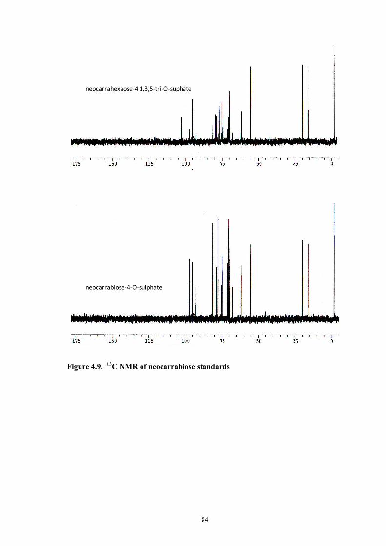
84
neocarrahexaose‐4 1,3,5‐tri‐O‐suphate
neocarrabiose‐4‐O‐sulphate
Figure 4.9. 13C NMR of neocarrabiose standards

85
Table 4.4. Chemical shifts of the 1H NMR of neocarrabiose standards.
A-G4S A-G4S Ref [57]
(A-G4S)3 (A-G4S)3 Ref [57]
ArH-1 5.003 5.102 5.026 5.120 ArH-1 4.984 5.084 5.008 5.103 AnrH-1 4.989 5.084 AH-1 4.989 5.103 ArH-2 4.005 4.106 4.044 4.142 ArH-2 4.005 4.106 4.044 4.142 AnrH-2 4.017 4.102 AH-2 4.044 4.138 ArH-3 4.255 4.357 4.443 4.535 ArH-3 4.255 4.357 4.443 4.535 AnrH-3 4.255 4.352 AH-3 4.443 4.529 ArH-4 4.374 4.478 4.511 4.607 ArH-4 4.374 4.478 4.511 4.607 AnrH-4 4.385 4.479 AH-4 4.511 4.607 ArH-5 4.308 4.408 4.567 4.650 ArH-5 4.308 4.408 4.567 4.650 AnrH-5 4.317 4.409 AH-5 4.567 4.650 ArH-6a 3.920 4.020 3.985 4.061 ArH-6a 3.920 4.020 3.985 4.061 AnrH-6a 3.942 4.020 AH-6a 3.985 4.061 ArH-6b 4.129 4.218 4.148 4.234 ArH-6b 4.095 4.202 4.126 4.224 AnrH-6b 4.117 4.203 AH-6b 4.126 4.224 G4S-H-1 5.217 5.320 5.224 5.320 G4S-H-1 4.536 4.650 4.553 4.654 G4Snr-H-1 4.572 4.658 G4S-H-1 4.572 4.660 G4SrH-2 3.814 3.919 3.823 3.917 G4SrH-2 3.489 3.594 3.501 3.593 G4Snr-H-2 3.501 3.596 G4S-H-2 3.501 3.599 G4Sr-H-3 4.068 4.165 4.090 4.165 G4Sr-H-3 3.869 3.967 3.900 3.983 G4Snr-H-3 3.920 4.003 G4S-H-3 3.920 4.013 G4Sr-H-4 4.832 4.895 4.827 4.901 G4Sr-H-4 4.793 4.835 4.802 4.835 G4Snr-H-4 4.802 4.855

86
Cont’d
Table 4.5. Chemical Shifts of the 13C NMR of neocarrabiose standards
A-G4S A-G4S Ref [57]
(A-G4S)3 (A-G4S)3 Ref [57]
G4S-H-4 4.802 4.855 G4Sr-H-5 4.054 4.190 4.090 4.186 G4Sr-H-5 3.678 3.774 3.689 3.774 G4Snr-H-5 3.689 3.817 G4S-H-5 3.689 3.817 G4Sr-H-6a 3.718 3.793 3.757 3.790 G4Sr-H-6a 3.678 3.78 3.689 3.783 G4Snr-H-6a 3.689 3.800 G4S-H-6a 3.689 3.800 G4Sr-H-6b 3.624 3.731 3.639 3.731 G4Sr-H-6b 3.678 3.783 3.689 3.783 G4Snr-H-6b 3.689 3.800 G4S-H-6b 3.689 3.800 G4S-H-1 5.217 5.320 5.224 5.320
A-G4S (A-G4S)3 (A-G4S)2 Ref [57]
Anr C-1 95.08 95.00 Anr C-2 69.82 69.89 Anr C-3 81.31 81.28 Anr C-4 70.48 70.44 Anr C-5 77.84 77.79 Anr C-6 69.48 69.45 Ar-C1 95.06 95.08 95.00 Ar-C2 69.94 69.95 69.96 Ar-C1 94.99 95.00 94.94 Ar-C2 70.05 70.03 70.10 Ar-C3 81.31 79.61 79.56 Ar-C4 78.82 78.85 78.83 Ar-C5 77.83 77.07 77.02 Ar-C6 69.47 69.82 69.78 G4S C-1 92.80 92.87 92.77 G4S C-2 67.69 67.67 67.60 G4S C-3 75.71 75.53 75.58 G4S C-4 75.39 75.19 75.28 G4S C-5 70.50 70.48 70.49 G4S C-6 61.98 61.88 61.95 G4S C-1 96.99 96.99 96.94 G4S C-2 71.04 70.48 70.97

87
Cont’d
4.3.6.2. NMR of oligomers from irradiated -carrageenan
1H-NMR and 13C spectra of the fractionated oligomers from irradiated -
carrageenan are shown in Figures 4.10 and 4.11. Similar spectra are observed in the
native -carrageenan polymer (Figure 4.12). The 1H-NMR indicated a simple spectra
with 12 different protons for the different types of irradiated -carrageenan oligomers
(-carragenan irradiated in solid in air, solid in vacuum and at 1% aqueous solution).
This may correspond to the protons of the 12 different carbons of the dimer unit of -
carrageenan. The peaks in the spectra were rather broad which probably contains
some overlapping unresolved peaks. This clearly indicates that the oligomers obtained
were still large enough for the details of the proton to be seen by NMR like those
obtained from the neocarrabiose standards. The spectra of these oligomers resemble
more the spectra of a -carrageenan polymer (shown in Figure 4.12) rather than that
of the neocarrabiose oligosaccharides. Based from the obtained MW from GPC
experiments, these oligomers would have approximately a DP of 39, 36 and 75 for -
carrageenan irradiated in solid in air, at 1% aqueous solution, and solid in vacuum
Cont A-G4S (A-G4S)3 (A-G4S)2 Ref [57]
G4S C-3 78.82 78.71 78.65 G4S C-5 75.00 74.21 74.97 G4S C-6 61.87 61.88 61.84 G4Snr C-1 102.91 102.84 G4Snr C-2 69.53 69.78 G4Snr C-3 78.61 78.52 G4Snr C-4 74.13 74.15 G4Snr C-5 75.01 75.15 G4Snr C-6 61.68 61.64

88
respectively. From the 1H-NMR spectra, the degree of polymerization can also be
estimated based on the ratio between integrated areas of the resonances from all H-1
protons of 3,6-anhydrogalactose divided by the average area of the H-3 and H-5 of the
non-reducing end 3,6 anhydrogalactose and multiplying by 2 [57]. However, since the
peaks for the non-reducing ends were not resolved, this method could not be done.
The proton NMR chemical shift data are shown in Table 4.6. did not vary
significantly with the three different fractions of oligomers. Their values were quite
close to each other for the same type of proton. The given denotes only the protons
attached to specific carbon and does not distinguish the different types whether it is
-, -, reducing or non reducing protons. The only well resolved peaks seen in the
spectra were those protons attached to A-6 with chemical shifts ranging from 3.98 to
4.13 ppm for A-6a, A-6b and A-6a nr. The values were quite close to reference
values taken from a polymer [60] and a low molecular weight (LMW) -carragenan
from an acid hydrolyzed sample [37] as seen in the table. Other reference values for
the polymeric and oligomeric -carrageenan are also available [52, 55]. From the 1H-
NMR spectra, the G-4 proton could not be seen in -carrageenan irradiated in vacuum
and in air. This proton was seen only as a slight bump in aqueous -carrageenan at =
4.829 ppm. For the neocarrabiose standard, G-4 proton does not exist as a singlet but
consists of two separate peaks, 4.802 ppm and 4.827 ppm for G4S-4 and G4S-
respectively. One possibility for the disappearance of the G-4 peak is the removal of
the 4-sulphate groups at C-4. Proton NMR of -carrageenan, a desulfated -
carragenan with OH attached to G-6, does not show any peak at .4.8 ppm. The
chemical shift data of -carrageenan resembles that of -carrageenan except for the
G-4 which appears upfield at 4.14 / 4.36 ppm [61-62]. Removal of the 4-sulphate

89
group also shifts slightly upfield the resonances of R-GH-1 and R-GH-1nr [57]. A
closer look at the 1H-NMR spectra of all the irradiated -carrageenan oligomers
would reveal a slight bump at .5.19, indicative of the presence of the reducing end
R-GH-1. This appears a little bit upfield from the neocarrabiose standards ( = 5.224
ppm). Resonances from end residues of carrageenan molecules of a higher molecular
weight are decreased in intensities as they have lesser reducing end groups. Other
unidentified small peaks ( .4.9) were also seen in the spectra of -carrageenan
oligomers irradiated in solid in air and at .3.2 for all types of irradiated -
carrageenan oligomers. Possibly these are protons coming from a different type of
Carbon other than those from the intact dimer A-G4S of -carrageenan. These
probably could be some of the radiolytic products present in small quantities in the
sample.
13C NMR spectra of radiation processed -carrageenan oligomers (irradiated
in solid and in air and as aqueous solution) are shown in Figure 4.11. The spectra
indicated only 11 different types of carbons. In comparison with the 13C NMR profile
of standard neocarrabiose where the non-reducing, reducing ends, and types for
the same carbon atom indicate distinctly different peaks, the 13C NMR spectra of
radiation processed -carrageenan oligomers showed only one peak for each Carbon
atom in the galactopyranose ring dimer. The data are seen in Table 4.7. Chemical
shift values of the three different types of -carrageenan oligomers were almost the
same for the different types of carbon atoms. The values were quite close to reference
values from the polymer and oligomer (DP4) [38]. Other references give also similar
results [49, 54-57, 59]. The reference values indicate that A-2 and A-6 have almost
the same chemical shifts. Thus, carbons from A-2 and A-6 of irradiated -carragenan

90
oligomers were assigned with the same (70.01). This would account for the 12
different carbons present in the galacto pyranose ring. Other reference values are also
quite close. No evidence of carbon atom from carbonyl bonds ( > 160 ppm) was
seen. FT-IR results of solid -carrageenan at 100 kGy (in air and in vacuum) and 1%
aqueous -carrageenan at 2 kGy indicated the presence of carbonyl bonds (Chapter
4.3.2). Carbonyl carbons do not have hydrogen attached to them so they often appear
less intense than other carbons in the molecule. It is also possible that the carbonyl
groups present in this fraction are quite minimal which could not easily be detected by
13C NMR. Most likely, a large fraction of the -carrageenan oligomer containing the
carbonyl groups may have been present in the separated fraction with the molecular
weight cut-off of less than 3,000 Da.
Based on 13C and 1H NMR spectra, the -carrageenan oligomers obtained
from the polymer irradiated in solid in air and in vacuum and at 1% aqueous solution
indicated still an intact repeating unit of the dimer with no changes in the chemical
structure of the galacto-pyranose ring. This result could present further explanation on
the optimum plant growth promoter effect found at 100 kGy for solid irradiation.

91
Figure 4.10. 1H NMR of irradiated -carrageenan oligomers.
solid ‐carrageenanirradiated in vacuum
G4S‐1A‐5
A‐4
G4S‐2
G4S‐3
G4S‐5 & 6
A‐1 A‐2A‐3A‐6a
A‐6a*A‐6b
D2O
aqueous (1%) ‐carrageenanirradiated in airG4S‐4
H2O
G4S‐1
solid ‐carrageenanirradiated in air
? ?

92
Figure 4.11. 13C NMR of irradiated -carrageenan oligomer.
solid ‐carrageenanirradiated in vacuum
solid ‐carrageenanirradiated in air
aqueous (1%) ‐carrageenanirradiated in air
G4S‐1
A‐1
A‐3
A‐4
G4S‐3
A‐5
G4S‐4
A‐2 & A‐6
G4S‐5
?( 71.45)
G4S‐2
G4S‐6
Internal Standard (DDS)
G4S‐1
A‐1
A‐3
A‐4
G4S‐3
A‐5
G4S‐4
A‐2 & A‐6
G4S‐5
G4S‐2
G4S‐6
G4S‐1
A‐1
A‐3
A‐4
G4S‐3
A‐5
G4S‐4
A‐2 & A‐6
G4S‐5
G4S‐2
G4S‐6
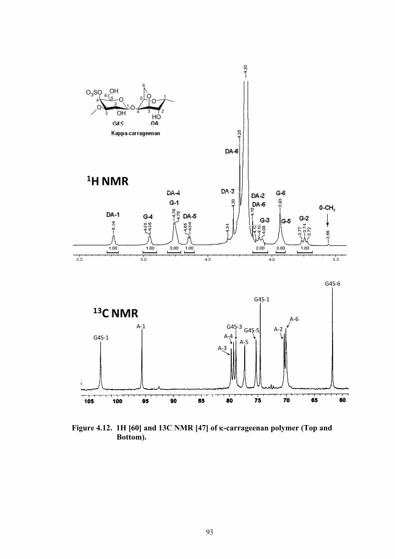
93
Figure 4.12. 1H [60] and 13C NMR [47] of -carrageenan polymer (Top and Bottom).
G4S‐1
A‐1
A‐3
A‐4
G4S‐3
A‐5
G4S‐5
A‐6
A‐2
G4S‐1
G4S‐6
1H NMR
13C NMR
12
3
4 56 1
234
5
6
O
OH
O3SO
OO
OH
O
OH
O
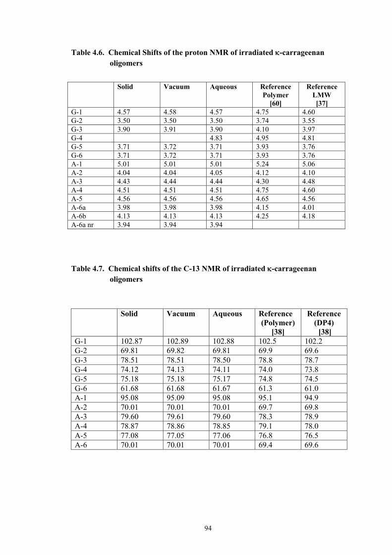
94
Table 4.6. Chemical Shifts of the proton NMR of irradiated -carrageenan oligomers
Table 4.7. Chemical shifts of the C-13 NMR of irradiated -carrageenan oligomers
Solid Vacuum Aqueous Reference (Polymer)
[38]
Reference (DP4) [38]
G-1 102.87 102.89 102.88 102.5 102.2 G-2 69.81 69.82 69.81 69.9 69.6 G-3 78.51 78.51 78.50 78.8 78.7 G-4 74.12 74.13 74.11 74.0 73.8 G-5 75.18 75.18 75.17 74.8 74.5 G-6 61.68 61.68 61.67 61.3 61.0 A-1 95.08 95.09 95.08 95.1 94.9 A-2 70.01 70.01 70.01 69.7 69.8 A-3 79.60 79.61 79.60 78.3 78.9 A-4 78.87 78.86 78.85 79.1 78.0 A-5 77.08 77.05 77.06 76.8 76.5 A-6 70.01 70.01 70.01 69.4 69.6
Solid Vacuum Aqueous Reference Polymer
[60]
Reference LMW [37]
G-1 4.57 4.58 4.57 4.75 4.60 G-2 3.50 3.50 3.50 3.74 3.55 G-3 3.90 3.91 3.90 4.10 3.97 G-4 4.83 4.95 4.81 G-5 3.71 3.72 3.71 3.93 3.76 G-6 3.71 3.72 3.71 3.93 3.76 A-1 5.01 5.01 5.01 5.24 5.06 A-2 4.04 4.04 4.05 4.12 4.10 A-3 4.43 4.44 4.44 4.30 4.48 A-4 4.51 4.51 4.51 4.75 4.60 A-5 4.56 4.56 4.56 4.65 4.56 A-6a 3.98 3.98 3.98 4.15 4.01 A-6b 4.13 4.13 4.13 4.25 4.18 A-6a nr 3.94 3.94 3.94
95
4.4. CONCLUSION
Chemical structure of irradiated -carrageenan at different doses (1% aqueous
and solid in air and in vacuum) was investigated using several methods (UV-Vis / FT-
IR / NMR spectroscopy / reducing sugar analysis / sulfate and carboxylic acid
analysis). The implications of all these chemical analyses in relation to bio-based
material development can be described as follows:
4.4.1. For 1% Aqueous Solution Irradiation
Figure 4.13 summarizes the radiolytic products of 1% aqueous -carrageenan
with dose. Values of reducing sugar, carbonyl groups, free sulfates and carboxylic
groups are relatively high especially at doses over 50 kGy. It is at these doses where
there is either a reduction or a plateau of radiolytic products formed. FT-IR at 100
kGy further suggests destruction of peaks corresponding to covalent sulfate, 3,6
anhydro bridge, D-galactose-4-sulfate, polymer bound water and the galacto-pyranose
ring. Note that Chapter 3 discussed that at dose beyond 50 kGy, there is no longer a
decrease in Mw but increase in radiolytic product peak (Mw . 1,000) is observed
instead. Thus, the region beyond 50 kGy can be considered the decomposition region
and therefore will not be useful for any application
On the other hand, doses between 10 to 50 kGy suggest a continuous build-up
of radiolytic products. FT-IR indicates that at a starting dose of 10 kGy, destruction of
peaks corresponding to galacto-pyranose ring and polymer bound water takes place.
This region may no longer be useful for applications related to the biological activity
of -carrageenan due to the destruction of its chemical structure. However, the acidic
components, reducing sugars and carbonyl groups can still be useful for other

96
applications e.g. radiation dose indicators or as antioxidants. Carbonyl groups in
aldehydic (reducing sugars) and ketone forms are considered antioxidants.
There is a narrow window (< 10 kGy) for the -carrageenan oligomer
containing an intact chemical structure. It is only in this region that applications
related to the biological function of -carrageenan can be considered.
4.4.2. For Solid Irradiation
The radiolytic products of -carrageenan irradiated in vacuum and in air are
shown in Figures 4.14 and 4.15. Unlike the values of 1% aqueous -carrageenan, the
formation of carbonyl groups, free sulfates and carboxylic groups are very minimal
both irradiated solid in air and in vacuum. These are good indicators of a well
preserved chemical structure. For vacuum irradiation, at a high dose of 200 kGy, FT-
IR spectra show that there is already a destruction of the chemical structure of -
carrageenan. However, a 200 kGy dose is rather too large for its biological activity
function considering that the optimum dose for plant growth promotion effect is
known only at 100 kGy. From these results on chemical structure, -carrageenan
irradiated both in vacuum and in air are expected to have good biological activity up
to a dose of 100 kGy. This dose extends further to 200 kGy for solid -carrageenan
irradiated in air. The expected benefits would be higher in vacuumed -carrageenan
due to its very low radiolytic product formation. The carbonyl, carboxylic and free
sulfates groups are lower than in air. In the use of -carrageenan oligomers as anti-
HIV, the main factors that influence the anti-HIV activity of carrageenans are the type
of sugar chain, the degree of sulfation, and the molecular weight [63]. The sulfated
oligosaccharide with the special structure of (13)-4 sulfate--D galactose (14)-3,
6 anhydro-"-D-galactose have antitumor and immunostimulating activity [64]. Thus,
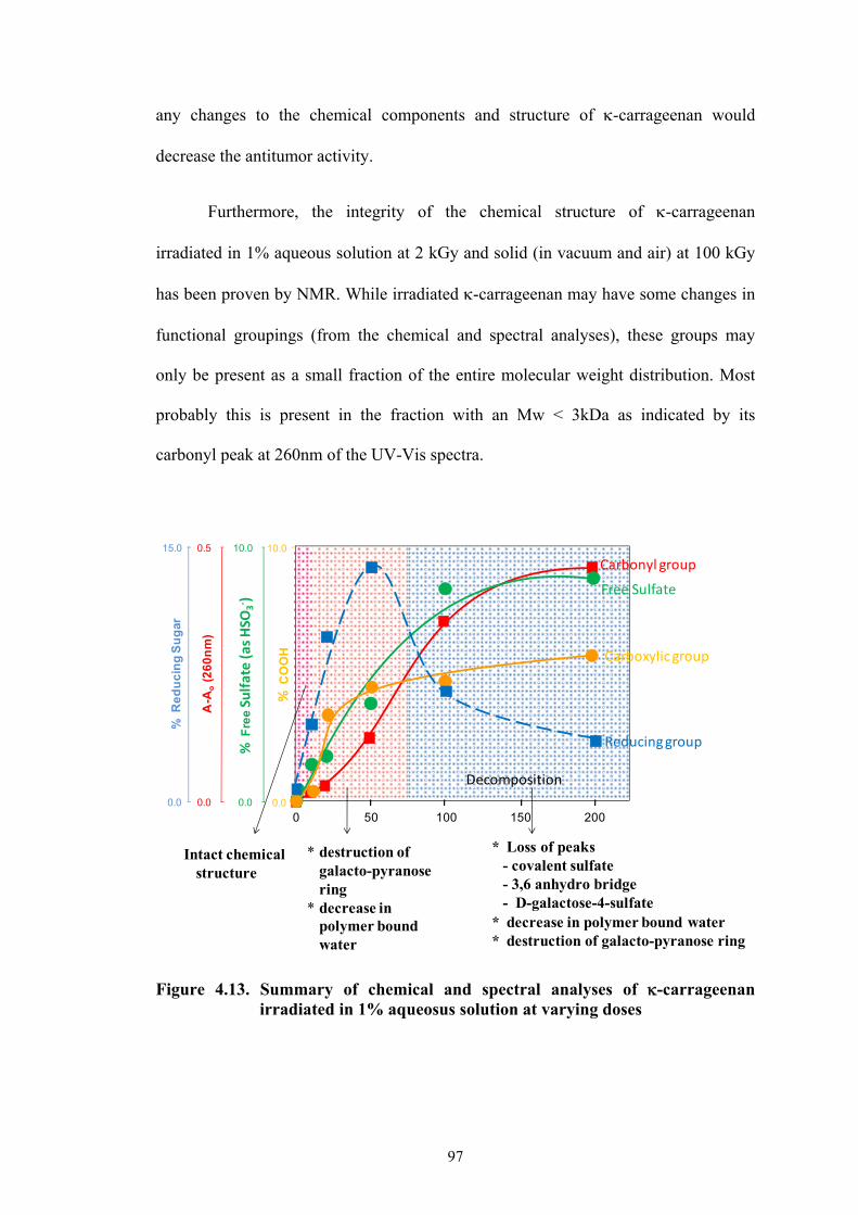
97
any changes to the chemical components and structure of -carrageenan would
decrease the antitumor activity.
Furthermore, the integrity of the chemical structure of -carrageenan
irradiated in 1% aqueous solution at 2 kGy and solid (in vacuum and air) at 100 kGy
has been proven by NMR. While irradiated -carrageenan may have some changes in
functional groupings (from the chemical and spectral analyses), these groups may
only be present as a small fraction of the entire molecular weight distribution. Most
probably this is present in the fraction with an Mw < 3kDa as indicated by its
carbonyl peak at 260nm of the UV-Vis spectra.
Figure 4.13. Summary of chemical and spectral analyses of -carrageenan irradiated in 1% aqueosus solution at varying doses
* destruction of galacto-pyranosering
* decrease in polymer bound water
Decomposition
* Loss of peaks - covalent sulfate - 3,6 anhydro bridge - D-galactose-4-sulfate
* decrease in polymer bound water* destruction of galacto-pyranose ring
50 100 150 200
Intact chemical structure
Carbonyl group
00.0
15.0
% R
ed
uc
ing
Su
gar
0.0
0.5
A-A
o(2
60
nm
)
0.0
10.0
% F
reeSulfate (as HSO
3‐ )
0.0
10.0
% C
OO
H
Free Sulfate
Carboxylic group
Reducing group

98
Figure 4.14. Summary of chemical and spectral analyses of solid -carrageenan
irradiated in vacuum at varying doses.
Figure 4.15. Summary of chemical and spectral analyses of solid -carrageenan
irradiated in air at varying doses.
0.0
15.0
% R
ed
uc
ing
Su
gar
0.0
0.5
A-A
o(2
60
nm
)
0.0
10.0
% F
reeSulfate (as HSO
3‐ )
0.0
10.0
% C
OO
H
0 50 100 150 200
Absorbed Dose (kGy)
Carbonyl group
Reducing Sugar
Carboxylic AcidFree Sulfate
* Loss of peaks - covalent sulfate - 3,6 anhydro bridge - D-galactose-4-sulfate
* decrease in polymer bound water* destruction of galacto-pyranose ring
Intact chemical structure
Decomposition
0 50 100 150 200
Absorbed Dose (kGy)
Carbonyl Group
Reducing Sugar
Free Sulfate
Carboxylic Acid
Intact chemical structure
0.0
15.0
% R
ed
uc
ing
Su
gar
0.0
0.5
A-A
o(2
60
nm
)
0.0
10.0
% F
reeSulfate (as HSO
3‐ )
0.0
10.0
% C
OO
H

99
REFERENCES:
1. A. Swallow. Radiation Chemistry: An Introduction. New York. Wiley. 1973. pp. 231-234.
2. G. Phillips, G. Moody, Int. J. Appl. Radiat. Is. 6 (1959) 78. 3. I. Idimecheva, R. Kisel, O. Shadyro, K. Kazem, H. Murase, T. Kagiya. J. Radiat.
Res. 46 (2005) 319. 4. W. Dziędziela, D. Kotyńska, Radiat. Phys. Chem. 23 (1984) 723. 5. L. Hai, T. Diep, N. Nagasawa, F. Yoshii, K. Tamikazu, Nucl. Instr. and Meth. in
Phys. Res. B. 208 (2003) 466. 6. P. Ulanski, J. Rosiak, Radiat. Phys. Chem. 39, (1992) 53. 7. U. Gryczka, D. Dondi, A. Chmielewski, W. Migdal, A. Buttafava, A. Faucitano,
Radiat. Phys. Chem. 78 (2009) 543. 8. P. Ulanski C. Von Sonntag, J. Chem. Soc., Perkin Trans. 2 (2000) 2022. 9. L. Huang, M. Zhai, J. Peng, J. Li, G. Wei, Carbohydr. Polym. 67 (2007) 305. 10. N. Nagasawa, H. Mitomo, F. Yoshii, T. Kume, Polym. Degrad. Stab. 69 (2000)
279. 11. J. Scott, M. Tigwell, Carbohydr. Res. 47 (1976) 105. 12. M. Şen, B. Yolaçan, O. Güven, Nucl. Instr. and Meth. in Phys. Res. B. 265
(2007) 429. 13. M. Vodeničarová, G. Dřímalová, Z. Hromádková, A. Malovíková, A.
Ebringerová Ultrason. Sonochem. 13 (2006) 157. 14. P. Baugh, J. Goodall, G. Phillips, C. von Sonntag, M. Dizdaroglu, Carbohydr.
Res. 49 (1976) 315. 15. H. Zegota, Food Hydrocolloids 13 (1999) 51. 16. T. Hjerde, O. Smidsrød, B. Christenesen, Carbohydr Res. 288 (1996) 175. 17. C. Rigouin, C. Ladrat, C. Sinquin, S. Colliec-Jouault, M. Dion, Carbohy. Polym.
76 (2009) 279. 18. A. Karlsson, S. Singh, Carbohydr. Polym. 38 (1999) 7. 19. D. Navarro, M. Flores, C. Stortz, Carbohydr. Polym. 69 (2007) 742.
100
20. I. Miller, J. Blunt, Carbohydr. Res. 309 (1998) 39. 21. C. Lii, C. Chen, A. Yeh, V. Lai, Food Hydrocolloids 13 (1999) 477. 22. V. Meunier, T. Nicolai, D. Durand, Int. J. Biol. Macromol. 28 (2001) 157. 23. G. Zhou, W. Yao, C. Wang, Carbohydr. Polym. 64 (2006) 73. 24. R. Czechowska-Biskup, B. Rokita, P. Ulanski, J. Rosiak, Nucl. Instr. and Meth.
in Phys. Res. B. 236 (2005) 383. 25. R. Czechowska-Biskup, B. Rokita, S. Lotfy, P. Ulanski, J. Rosiak, Carbohyd.
Polym. 60 (2005) 175. 26. J. Hodge, B. Hofreiter, B.T. (1962) In Whistler, R.L. & Wolfrom, M.L.(Eds.)
Methods in Carbohydrate Chemistry (Vol. 1, pp 380-394). Academic Press: New York.
27. L. Relleve, N. Nagasawa, L. Luan, T. Yagi, C. Aranilla, L. Abad, T. Kume, F.
Yoshii, A. De la Rosa, Polym Degrad Stab. 87 (2005) 403. 28. M. Akhlaq, S. Baghdadi, C. von Sonntag, Carbohydr. Res.164 (1987) 71. 29. C. von Sonntag, E. Bothe, P. Ulanski, J. Deeble, Radiat. Phys. Chem. 46 (1995)
527. 30. N. Anderson, T. Dolan, A. Penman, D. Rees, G. Mueller, D. Stancioff. N.
Stanley, J. Chem. Soc. C (1968) 602. 31. S. Peats, The infrared spectra of carrageenans extracted from various algae. In:
Proc. of the Xth Interrnational Seaweed Symposium (1981), Walter de Gruyter, Berlin, pp. 495-501.
32. E. Lacerna, R. Veroy, A. Luistro, G. Cajipe, Extracts from some red and brown
seaweeds of the Philippines. In: Proc. of the Xth Interrnational Seaweed Symposium (1981), Walter de Gruyter, Berlin, pp. 4441-448.
33. B. Matsuhiro, Hydrobiologia 326-327 (1996) 481. 34. G. Santos, Aqua. Bot. 36 (1989) 55. 35. I. Fournet, E. Ar Gall, E. Deslandes, J. Huvenne, B. Sombret, J. Floćh, Bot.
Mar. 40 (1997) 45. 36. J. Prado-Fernández, J.A. Rodríguez-Vázquez, E. Tojo, J.M. Andrade, Anal.
Chim. Acta. 480 (2003) 23.Y. Jiang, X. Guo, X. Tian, Carbohydr. Polym. 61 (2005) 399.

101
37. Y. Jiang, X. Guo, X. Tian, Carbohydr. Polym. 61 (2005) 399. 38. C. Rochas, M. Rinaudo, M. Vincendon, Int. J. Biol. Macromol. 5 (1983) 111. 39. N. Volpi, A. Mucci, L. Shenetii, Carbohydr. Res. 315 (1999) 345. 40. T. Malfait, F. van Cauwelaert, J. Chromatogr. A, 504 (1990) 369. 41. A. Kolender, M. Matulewicz, Carbohydr. Res. 337 (2002) 57. 42. S. Knutsen, M. Sletmoen, T. Kristensen, T. Barbeyron, B. Kloareg, P. Potin,
Carbohydr. Res. 331 (2001) 101. 43. N. Caram-Lelham, R. Cleland, L. Sundelöf, Int. J. Biol. Macromol. 16 (1994)
71. 44. N. Caram-Lelham, L. Sundelöf, T. Andersson, Carbohydr. Res. 273 (1995) 71. 45. A. Antonopoulos, B. Herbreteau, M. Lafosse, W. Helbert, J. Chromatogr. A
1023 (2004) 231. 46. Y. Jiang, X. Guo, Carbohydr. Polym. 61 (2005) 441. 47. F. Velde, H. Peppelman, H. Rollema, R. Tromp, Carbohydr. Res. 331 (2001)
271. 48. A. Usov, Food Hydrocolloid. 12 (1998) 301. 49. A. Usov, S. Yarotsky, S. A. Shaskov, Biopolymers. 19 (1980) 977. 50. C. Stortz, B. Bacon, R. Cherniak, A. Cerezo, Carbohydr. Res. 261 (1994) 317. 51. E. Murano, R. Toffanin, E. Cecere, R. Rizzo, S. Knutsen, Mar. Chem. 58 (1997)
319. 52. E. Tojo, J. Prado, Carbohydr. Polym. 53 (2003) 325. 53. F. Van de Velde, A. Antipova, H. Rollema, B. Tatiana, N. Grinberg, L. Pereira,
P. Gilsenan, R. Tromp, B. Rudolphe, V. Grinberg, Carbohyd. Res. 340 (2005) 1113.
54. F. van de Velde, L. Pereira, H. Rollema, Carbohydr. Res. 339 (2004) 2309. 55. F. van de Velde, S. Knutsen, A. Usov, H. Rollemay, A. Cerezo, Trends Food.
Sci. Tech. 13 (2002) 73.

102
56. M. Ciancia, M. Matulewicz, P. Finch, A. Cerezo, Carbohydrate Research, 238 (1993) 241.
57. S. Knutsen, H. Grasdalen, Carbohydr. Res., 229 (1992) 233. 58. S. Knutsen, H. Grasdalen, Carbohydr. Polym. 19 (1992) 199. 59. T. Turquois, S. Acquistapace, F. Arce Vera, D Welti. Carhohydr. Polym. 31
(1996) 269. 60. V. Campo, D. Kawano, D. da Silva Jr., I. Carvalho, Carbohydr. Polym. 77
(2009) 167. 61. A. Kolender, M. Matulewicz, Carbohydr. Res. 339 (2004) 1619. 62. R. Falshaw, R. Furneaux, H. Wong, Carbohydr. Res. 338 (2003) 1403.
63. T. Yamada, A. Ogamo, T. Saito, J. Watanabe, H. Uchiyama, Y. Nakagawa,
Carbohydr. Polym. 32 (1997) 51.
64. H. Yuan, J. Song, X. Li, N. Li, J. Dai, Cancer Letters, 243 (2006) 228.

103
CHAPTER 5
STRUCTURAL AND DYNAMIC BEHAVIOR OF IRRADIATED
-CARRAGEENAN
5.1. INTRODUCTION
Most of the available data on the analysis of the radiolytic products of natural
polysaccharides are those related to their chemical structure. The dynamic behavior
and structural heterogeneity of irradiated polysaccharides have not been studied. The
various structural elements of a macromolecule e.g. -carrageenan, such as
polydispersity, overall molecular dimensions, hydrodynamic behavior and internal
mobility would have an influence in its properties in solution. The reduction in
molecular size and changes in chemical structure of -carrageenan as an effect of
radiolytic degradation may have an influence on these structural elements.
It is well known that -carrageenan easily undergoes association in the
presence of ions (Chapter 2.3.5) and changes in temperature. Thermoreversible
conformational changes from coil to helix to gel are expected with this association.
With radiation, the conformational characteristic and gelation behavior of -
carrageenan may be altered. Dynamic light scattering (DLS) can be a convenient tool
to observe such changes. Likewise, the different diffusion modes of the oligomers
produced from radiation may also be observed.
The availability of structural heterogeneity makes it possible to perform
experimental procedures based on scattering of the incident radiation. The small-angle
neutron scattering technique (SANS) is a convenient and efficient tool for the
determination of structural characteristics in a scale from one up to hundreds of
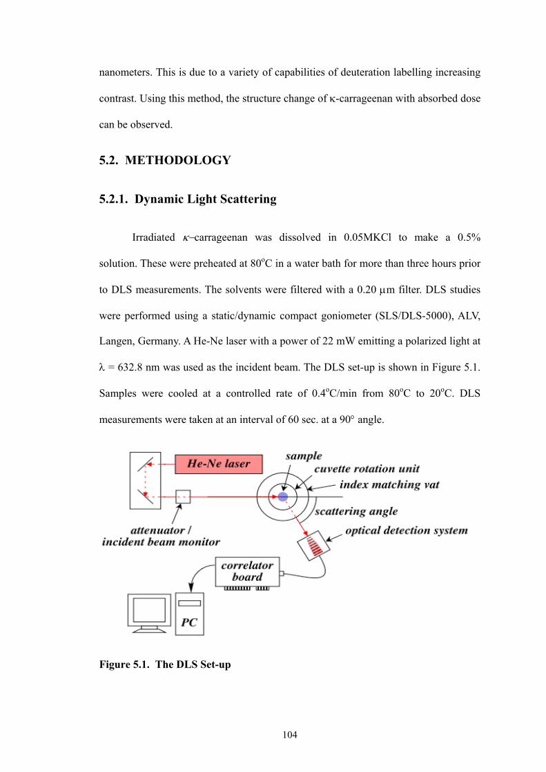
104
nanometers. This is due to a variety of capabilities of deuteration labelling increasing
contrast. Using this method, the structure change of -carrageenan with absorbed dose
can be observed.
5.2. METHODOLOGY
5.2.1. Dynamic Light Scattering
Irradiated carrageenan was dissolved in 0.05MKCl to make a 0.5%
solution. These were preheated at 80oC in a water bath for more than three hours prior
to DLS measurements. The solvents were filtered with a 0.20 m filter. DLS studies
were performed using a static/dynamic compact goniometer (SLS/DLS-5000), ALV,
Langen, Germany. A He-Ne laser with a power of 22 mW emitting a polarized light at
= 632.8 nm was used as the incident beam. The DLS set-up is shown in Figure 5.1.
Samples were cooled at a controlled rate of 0.4oC/min from 80oC to 20oC. DLS
measurements were taken at an interval of 60 sec. at a 90 angle.
Figure 5.1. The DLS Set-up

105
The laser (He-Ne) passes through a collimator lens and then hits the cell with
the solution. The light is scattered and detected by a photomultiplier that transform a
variation of intensity into a variation of voltage. After the photomultiplier, the signal
is immediately preamplified and then sent to the computer for data processing. A
time-dependent fluctuations in the scattered intensity will be observed.
5.2.2. Small Angle Neutron Scattering
Non-irradiated and irradiated -carrageenan samples were dissolved in D2O
and in 0.05M KCl/D2O solutions at elevated temperatures at a fixed concentration of
5% by weight. The samples were then placed in drum cells with a sample thickness of
0.21 mm. SANS experiments were carried out with the small-angle neutron scattering
instrument, SANS-U, at the Institute for Solid State Physics, The University of Tokyo,
located at the Japan Atomic Energy Agency, Tokai, Japan. A flux of cold neutrons
with a wavelength of 0.7 nm was irradiated to the sample and the scattered intensity
profile was collected with an area detector of 128 x 128 pixels. The sample to detector
distances of 2.00 m and 8.00 m were used which covered the accessible q range of
0.06 to 1.5 nm-1, where q is the scattering vector. The sample drum cells with quartz
windows were placed in a brass chamber and the chamber was thermo-regulated with
a circulating water bath. SANS experiments were initially done at 20oC and the
temperature was raised to 60oC. The obtained intensities were corrected for the
detector inhomogeneities, cell scattering, fast neutrons, transmission, and incoherent
scatterings, and then were scaled to the absolute intensity with an intensity calibration
standard sample (Lupolen). Figure 5.2. shows the schematic diagram of the SANS
set-up.

106
This instrument is composed of (a) a mechanical neutron velocity selector
(NVS), (b) a pre-sample flight path, (c) a multi purpose sample stage, (d) an
evacuated post-sample flight path, (e) an area detector, (f) a point detector for
transmission measurements, (g) a beam stop made of B4C, and (h) a data acquisition
system. The incident neutron beam from the cold source (white beam flux » 2 × 10⁸
cm−2s−1 with the peak wavelength of 0.4 nm) is monochromatized by a NVS with
helical slits. Cold neutron beam from the reactor is guided to the NVS. Just behind
the NVS is located the pre-sample flight path, which consists of (j) pinhole tubes
coated with B4C inside and (k) alternate neutron guide tubes coated with Ni inside.
By replacing the pinholes and the guide tubes in or out of the beam, users can change
the effective source-to-sample distance (= collimation length, CL) to 1, 2, 4, 8, 12, or
16 m in order to vary the divergence and flux of the incident beam at the sample
position. The sample to-detector distance (SDD) can be continuously varied from 1
to 16 m, which covers the Q-range from 0.03 to 2.7 nm-1. Normally, a symmetric
optical arrangement, i.e., CL = SDD, is recommended to optimize the flux and
divergence of the beam. The collimator apertures are 20 mmø each and the sample
aperture can be chosen from 1, 3, 5, 7, 8, 10 to 16 mmø. The most-commonly used
Figure 5.2. Schematic Diagram of the SANS-U Set-up
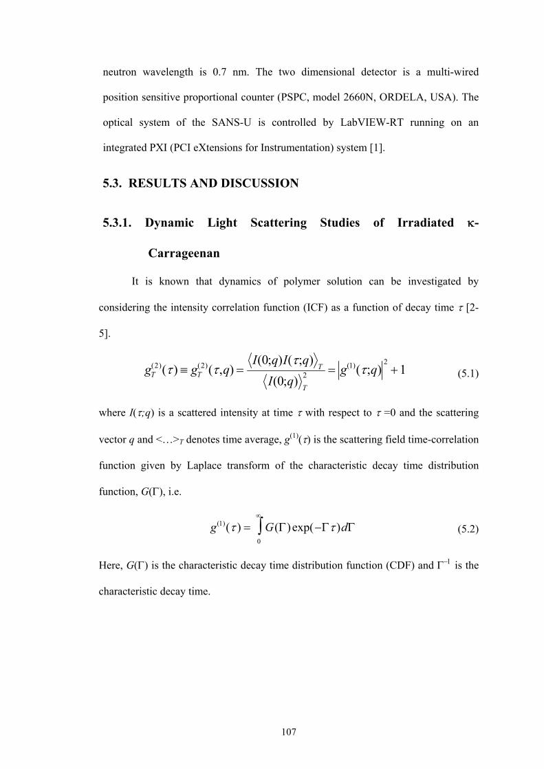
107
neutron wavelength is 0.7 nm. The two dimensional detector is a multi-wired
position sensitive proportional counter (PSPC, model 2660N, ORDELA, USA). The
optical system of the SANS-U is controlled by LabVIEW-RT running on an
integrated PXI (PCI eXtensions for Instrumentation) system [1].
5.3. RESULTS AND DISCUSSION
5.3.1. Dynamic Light Scattering Studies of Irradiated -
Carrageenan
It is known that dynamics of polymer solution can be investigated by
considering the intensity correlation function (ICF) as a function of decay time [2-
5].
gT(2)() gT
(2)(,q) I(0;q)I(;q)
T
I(0;q)T
2 g(1)(;q)21 (5.1)
where I(;q) is a scattered intensity at time with respect to =0 and the scattering
vector q and <…>T denotes time average, g(1)() is the scattering field time-correlation
function given by Laplace transform of the characteristic decay time distribution
function, G(), i.e.
g(1)( ) G()exp( )d0
(5.2)
Here, G() is the characteristic decay time distribution function (CDF) and is the
characteristic decay time.

108
5.3.1.1. Molecular weight dependence and characteristic relaxation time
dependence of -carrageenan with absorbed dose
Figures 5.3a and b show the ICFs and the CDFs respectively for -
carrageenan. The ICFs of 0.5% irradiated -carrageenan solutions in the presence of
0.05M KCl were obtained at varying doses. The shoulder of the ICF shifted to the
region of the smaller relaxation times with increasing dose. This indicates that smaller
fragments of -carrageenan produced by gamma irradiation tend to move faster than
non-irradiated -carrageenan. This trend becomes more obvious using the decay time
distribution of the inverse Laplace transformation of ICF (Figure 5.3b). G() revealed
the presence of two peaks. The fast mode peaks were in the range of 0.1 to 10 ms.
Non-irradiated -carrageenan showed sharper peaks in the range of 4 to10 ms. With
irradiation, peaks shifted towards faster relaxation time and started to broaden. At 300
kGy, the peak became very broad ranging from 0.1 to 4ms. Polydispersity of the
molecular size distribution may have resulted in the broadening of peaks. Molecular
weight distribution is expected to become larger with increasing dose in as much as
cleavage of the molecules by gamma irradiation is a random process. A large broad
peak was observed with non-irradiated -carrageenan in the range of 70 to 800ms.
This peak was also present in irradiated -carrageenan but became smaller at higher
doses, which disappeared completely at 300 kGy. These broad slower signals are due
to the diffusion of some aggregates. Irradiation decreases the molecular weight of
carrageenan, decreasing the aggregating particle sizes. In a similar study reported by
Pinder, et.al. [6], diffusive mode together with a slower non-diffusive mode ascribed
to aggregates were also observed with the carbohydrate dextran.
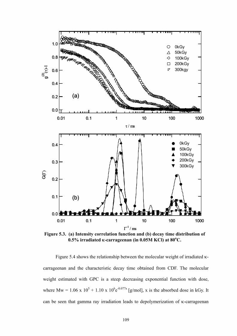
109
Figure 5.3. (a) Intensity correlation function and (b) decay time distribution of 0.5% irradiated -carrageenan (in 0.05M KCl) at 80oC.
Figure 5.4 shows the relationship between the molecular weight of irradiated -
carrageenan and the characteristic decay time obtained from CDF. The molecular
weight estimated with GPC is a steep decreasing exponential function with dose,
where Mw = 1.06 x 10⁵ + 1.10 x 10⁶e-0.077x [g/mol], x is the absorbed dose in kGy. It
can be seen that gamma ray irradiation leads to depolymerization of -carrageenan
0.4
0.3
0.2
0.1
0.0
G(
)
0.01 0.1 1 10 100 1000
-1 / ms
0kGy 50kGy 100kGy 200kGy 300kGy
(a)
(b)
1.0
0.8
0.6
0.4
0.2
0.0
g(2) -
1
0.01 0.1 1 10 100 1000
/ ms
0kGy 50kGy 100kGy 200kGy 300kgy
0.4
0.3
0.2
0.1
0.0
G(
)
0.01 0.1 1 10 100 1000
-1 / ms
0kGy 50kGy 100kGy 200kGy 300kGy
(a)
(b)
1.0
0.8
0.6
0.4
0.2
0.0
g(2) -
1
0.01 0.1 1 10 100 1000
/ ms
0kGy 50kGy 100kGy 200kGy 300kgy

110
quite effectively. A similar dose dependence was found in that of -1 where -1 = 0.83
+ 5.7e-0.061x [ms] taken from an empirical fitting. This clearly indicates that -1 can
also be used as a measure of molecular weight index.
Figure 5.4. Relationship of the molecular weight of -carrageenan (0.5% in 0.05M KCl) and 1/ (T=80oC and a scattering angle of 90o) with absorbed dose.
5.3.1.2. Coil-helical transition of irradiated -carrageenan
The characteristic decay time distribution function was also determined for
irradiated and non-irradiated -carrageenan at decreasing temperatures. The CDFs are
shown in Figures 5.5 a-c. The figures illustrate a typical shifting of peaks towards
slower relaxation modes with decreasing temperatures. This is expected as the
solution increases in viscosity with decreasing temperature. At high temperatures (i.e.,
76 °C and above), sharp peaks were observed in the CDFs. These peaks started to
broaden as temperatures were lowered. Peak broadening started at around 74oC
(Figure 5.5a) for non-irradiated -carrageenan. This transition may be due to
conformational changes from coil to helix. Coil-helix transition has already been
1.4
1.2
1.0
0.8
0.6
0.4
0.2
0.0
200150100500
7
6
5
4
3
2
1
0
Mo
lec
ula
rwe
igh
t(g
/mo
l) x
10
6
-1
/ m
s
Absorbed Dose (kGy)

111
0.4
0.3
0.2
0.1
0.0
G
0.01 0.1 1 10 100 1000
-1/ ms
75.3oC
69.9oC
62.6oC
56.6oC
51.5oC
47.7oC
Sharp peak
Sharp peaks
(a)
(b)
(c)
0.14
0.12
0.10
0.08
0.06
0.04
0.02
0.00
G
0.01 0.1 1 10 100 1000
-1ms
76.2oC
74.0oC
71.0oC
65.1oC
58.8oC
55.4oC
70
60
50
40
30
20
10
0
0.01 0.1 1 10 100 1000
-1
/ ms
76.6oC
70.4oC
64.7oC
56.8oC
52.2oC
47.7oCG
x
10-3
Figure 5.5. Decay time distribution function of 0.5% irradiated -carrageenan (in 0.05M KCl) at varying temperatures at doses of (a) 0 kGy (b) 150 kGy (c) 300 kGy.

112
discussed in Chapters 2.3.5 and 3.3.3. Helical structure can be promoted by adding an
appropriate electrolyte to a sufficient ionic strength and/or by lowering the
temperature. he ordered conformation with added K+ could develop either into a
double helix or a dimer, stabilized by electrostatic attractive forces involving the -
carrageenan ion and by hydrogen bonding [7]. The conformational transition
temperature (CTT, observed as the transition from sharp peak to broad peak or when
the width of the base of peak becomes twice as that at š 75o C in Figure 5.5)
decreased with increasing doses as shown in Figure 5.6. A sharp decrease in CTT was
observed from 25 kGy to 50 kGy which leveled off beyond this dose at a temperature
of around 55oC to 51oC. These temperatures are quite close to CTT values reported by
Chronakis, et al., which is between 55oC to 58oC for concentrations of 0.35% - 1.6%
KC in 0.2M NaI [8]. The leveling off of CTT is consistent with the leveling off of
changes in molecular weight with dose as shown in Figure 5.4 whereby decrease in
molecular weight started to be suppressed also at 50 kGy. As the size of carrageenan
Figure 5.6. Conformational transition temperature as a function of absorbed dose.
80
70
60
50
40200150100500
Co
nfo
rma
tion
al T
ran
sitio
n
Tem
pe
ratu
re (
oC
)
Absorbed Dose (kGy)

113
molecules are shortened with irradiation, the molecules are easier to disentangle from
its helical state forming the coil conformation such that lower CTT are observed with
increasing doses. Studies done by Meunier, et.al. [9] also show a peak broadening and
a shift to lower temperatures of the coil-helix transition with decreasing molar mass
obtained from sonication of -carrageenan
No changes in conformation were observed at a dose of 175 kGy and above
(Figure 5.5c). High absorbed doses may not only result in the cleavage of the polymer
but may also destroy the galacto-pyranose rings of -carrageenan leading to ring
opening. As a consequence, a longer polymer chain with some pendant molecules is
formed which increases the possibility of entanglement of -carrageenan. In addition,
the presence of these straight chain polymers with pendant molecules and some intact
galacto-pyranose rings produces a heterogeneous system. Thus, a broad peak was seen
in the CDF of irradiated -carrageenan at doses of 200 kGy and above. Mechanisms
for the radiation induced ring opening of the pyranose ring in some polysaccharides
have been proposed as shown in equations 5.3 to 5.5. Though -carrageenan was
irradiated in solid state, the presence of polymer bound water may have induced also
some hydrolytic reaction similar to these schemes and consequently resulted in
randomized ring openings. Figure 5.7 would illustrate the radiolytic reactions of -
carrageenan.

114
In Chitosan [10]
5.3
In pectin [11]
5.4

115
In cycloamylose D-glucose [12]
5.5
Figure 5.7. Effect of gamma radiation on the structure of -carrageenan.
(KC)
-O3SO
CH2OH
O
O
OH
H2C
O
O
O
OH
-O3SO
CH2OH
O
O
OH
H2C
O
O
O
OH
-O3SO
CH2OH
O
O
OH
H2C
O
O
O
OH
-O3SO
CH2OH
O
O
OH
H2C
O
O
O
OH
(KC)
n n-xi→iietc.
OH
OH
-O3SO
O
CH2
OH
O
H
O
OH
3SO
O
CH2OH
O
H
-O
O
OH
3SO
OO
CH 2OH
OH
O
KCn
-O
O
OH
3SO
O
CH 2OH
H
OH
-O

116
5.3.1.3. Gelation temperatures of -carrageenan as a function of dose
There is controversy regarding the gelling mechanism of -carrageenan.
According to the first model of Rees and coworkers [13], the junctions are double
helices which when they are cation-mediated forms aggregates of double helices
(Figure 5.8a, domain model) [14]. Another model suggests a cation-specific, nested,
single-helix (Figure 5.8b) [15, 16]. The various levels of aggregation of -carrageenan
helices leading to gelation using the different models have been studied by several
authors [8, 17-25]. The double helices (double helix model) described are aggregated
to higher order assemblies to create a three dimensional network known as the double
helix domain. The single helical model suggests that helical regions along the polymer
chain associate into larger stretches upon increasing salt concentration and / or
polymer and / or decreasing temperature. Figures 5.9a-c show the ICFs of irradiated
-carrageenan at lower temperatures. A power law behavior was observed below 39oC
with non-irradiated -carrageenan. This indicates that the system is undergoing
gelation. It is known that the sol-gel transition point can be observed at the
temperature where power law behavior in ICF appears [3-5, 26-27]. Taking the double
helix model during gelation, -carrageenan-water system starts to decompose into two
phases with different network concentrations. This creates concentration fluctuations.
The double helix aggregate forms a separate phase by excluding water from their
domains. As a result the contrast between -carrageenan and water phases can scatter
light and dramatically reduce its intensity [18]. The gelation temperatures decreased
with increasing dose from 39oC to 36oC down to 28oC for 0 kGy, 25 kGy and 50 kGy
respectively (not illustrated). This phenomenon seems to be reasonable because the
probability of aggregation becomes less by lowering the molecular weight. At 100
kGy and above, no gelation occurred. Figures 5.9b-c show ICFs which do not have
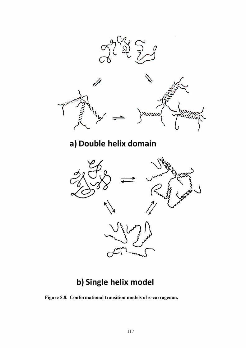
117
Figure 5.8. Conformational transition models of -carragenan.
a) Double helix domain
b) Single helix model

118
Figure 5.9. ICF 0.5% irradiated -carrageenan (in 0.05M KCl) at varying
temperatures at doses of (a) 0 kGy (b) 100 kGy and (c) 200 kGy.
(b)
Gelation
(a)
(c)
5
67
10-1
2
3
4
5
67
100
10-2
10-1
100
101
102
103
104
/ ms
43.9oC
39.7oC
36.5oC
32.1oC
24.5oC
21.1oC
6
810-1
2
4
6
8100
g(2) (
) -1
10-2
10-1
100
101
102
103
104
/ ms
40.2oC
37.5oC
34.1oC
29.5oC
26.0oC
25.7oC
g(2) (
) -1
6
810-1
2
4
6
8100
g(2) (
) -1
10-2
10-1
100
101
102
103
104
/ ms
46.6oC
43.1oC
40.3oC
39.0oC
38.8oC
38.7oC

119
any phase transition at 100 kGy and above. The molecular sizes at these doses are not
big enough to form gels. Table 5.1 summarizes the conformational transition
temperatures and gelation temperatures with dose.
Table 5.1. Molecular weight (Mw), conformational transition temperature
(CTT) and gelation temperature (GT) of -carrageenan at varying doses.
5.3.1.4. Fast relaxation mode peaks corresponding to optimum biological activity
of -carrageenan
Figures 5.10a-f show the CDFs at higher doses of -carrageenan. Figures
5.10a-b indicate broadening of peaks which are also clear manifestations of a sol-gel
transition. At doses of 75 kGy and above (Figures 5.10c-e), instead of a gelation, a
new fast mode peak appeared at around 10-1 ms below the CTT temperatures. It was
largest at 100 kGy which decreased beyond this dose. These peaks started to disappear
at 175 kGy and above (Figure 5.10f). This result can well be related to the optimum
growth promoting effect in plants for -carrageenan (100 kGy). The appearance of the
fast mode peaks corresponds to the lower molecular size oligomers formed from
irradiated carrageenan which may be responsible for the enhancement of the
biological effect. The molecular weight of these oligomers is estimated to be 5.0 x 10³
to 10.0 x 10³ from the peak position of the “new peak” in Figure 5.10d by assuming a
phenomenological linear-relationship between the molecular weight and -1 (Figure
Dose (kGy)
-carrageenanMw CTT
(oC) GT (oC)
0 1.2106 74 39 50 1.3105 56 28
100 1.1105 55 none 200 6.6104 none none

120
Figure 5.10. Decay time distribution function of 0.5% irradiated -carrageenan (in
0.05M KCl) at varying temperatures at doses of (a) 0 kGy (b) 50 kGy (c) 75 kGy (d) 100 kGy (e) 150 kGy and (f) 200 kGy.
5.6). Note that the molecular weight of the original -carrageenan is 1.9 x 10⁶
according to the GPC data. Though the large molecular size -carrageenan (broad
slow relaxation mode peaks) has also a biological effect but due to its size, this cannot
easily diffuse into the plant system as much as these low molecular size oligomer
fragments. The quantity of these oligomers becomes maximum at 100 kGy. At higher
doses, the chemical structure of the galacto-pyranose ring is destroyed, as mentioned
in earlier discussion, such that biological effect decreases. The relationship between
0.15
0.10
0.05
0.00
G
10-2
10-1
100
101
102
103
104
-1/ ms
40.2oC
37.5oC
34.1oC
29.5oC
26.0oC
25.7oC
New Peak
(d)
0.10
0.08
0.06
0.04
0.02
0.00
G
10-2
10-1
100
101
102
103
104
-1/ ms
43.9oC
39.7oC
36.5oC
32.1oC
24.5oC
21.1oC
(f)
80x10-3
60
40
20
0G
10-2
10-1
100
101
102
103
104
-1/ ms
43.5oC
39.6oC
36.2oC
32.0oC
25.6oC
22.6oC
(e)
New Peak
80x10-3
60
40
20
0
G
10-2
10-1
100
101
102
103
104
-1/ ms
47.7oC
43.8oC
39.4oC
35.3oC
28.1oC
Gelation
(b)
0.10
0.08
0.06
0.04
0.02
0.00
G
10-2
10-1
100
101
102
103
104
-1ms
43.5oC
39.6oC
36.2oC
32.0oC
25.6oC
22.6oC
(c)
New Peak
0.10
0.08
0.06
0.04
0.02
0.00
G
10-2
10-1
100
101
102
103
104
-1/ ms
46.6oC
43.1oC
40.3oC
39.0oC
38.8oC
38.7oC
(a)
Gelation
0.15
0.10
0.05
0.00
G
10-2
10-1
100
101
102
103
104
-1/ ms
40.2oC
37.5oC
34.1oC
29.5oC
26.0oC
25.7oC
New Peak
(d)
0.15
0.10
0.05
0.00
G
10-2
10-1
100
101
102
103
104
-1/ ms
40.2oC
37.5oC
34.1oC
29.5oC
26.0oC
25.7oC
New Peak
(d)
0.10
0.08
0.06
0.04
0.02
0.00
G
10-2
10-1
100
101
102
103
104
-1/ ms
43.9oC
39.7oC
36.5oC
32.1oC
24.5oC
21.1oC
(f)
0.10
0.08
0.06
0.04
0.02
0.00
G
10-2
10-1
100
101
102
103
104
-1/ ms
43.9oC
39.7oC
36.5oC
32.1oC
24.5oC
21.1oC
(f)
80x10-3
60
40
20
0G
10-2
10-1
100
101
102
103
104
-1/ ms
43.5oC
39.6oC
36.2oC
32.0oC
25.6oC
22.6oC
(e)
New Peak
80x10-3
60
40
20
0G
10-2
10-1
100
101
102
103
104
-1/ ms
43.5oC
39.6oC
36.2oC
32.0oC
25.6oC
22.6oC
(e)
New Peak
80x10-3
60
40
20
0
G
10-2
10-1
100
101
102
103
104
-1/ ms
47.7oC
43.8oC
39.4oC
35.3oC
28.1oC
Gelation
(b)
80x10-3
60
40
20
0
G
10-2
10-1
100
101
102
103
104
-1/ ms
47.7oC
43.8oC
39.4oC
35.3oC
28.1oC
Gelation
(b)
0.10
0.08
0.06
0.04
0.02
0.00
G
10-2
10-1
100
101
102
103
104
-1ms
43.5oC
39.6oC
36.2oC
32.0oC
25.6oC
22.6oC
(c)
New Peak
0.10
0.08
0.06
0.04
0.02
0.00
G
10-2
10-1
100
101
102
103
104
-1ms
43.5oC
39.6oC
36.2oC
32.0oC
25.6oC
22.6oC
(c)
New Peak
0.10
0.08
0.06
0.04
0.02
0.00
G
10-2
10-1
100
101
102
103
104
-1/ ms
46.6oC
43.1oC
40.3oC
39.0oC
38.8oC
38.7oC
(a)
Gelation
0.10
0.08
0.06
0.04
0.02
0.00
G
10-2
10-1
100
101
102
103
104
-1/ ms
46.6oC
43.1oC
40.3oC
39.0oC
38.8oC
38.7oC
(a)
Gelation
0.10
0.08
0.06
0.04
0.02
0.00
G
10-2
10-1
100
101
102
103
104
-1/ ms
46.6oC
43.1oC
40.3oC
39.0oC
38.8oC
38.7oC
(a)
0.10
0.08
0.06
0.04
0.02
0.00
G
10-2
10-1
100
101
102
103
104
-1/ ms
46.6oC
43.1oC
40.3oC
39.0oC
38.8oC
38.7oC
(a)
GelationGelation
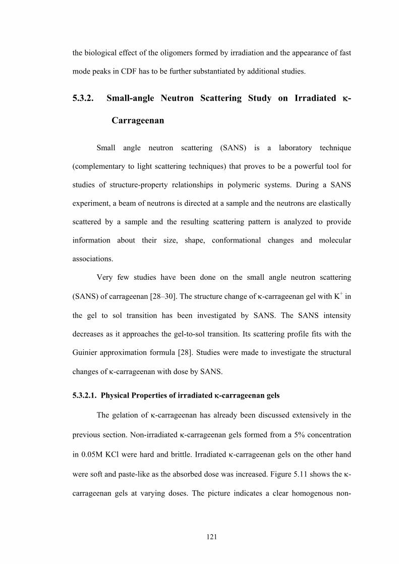
121
the biological effect of the oligomers formed by irradiation and the appearance of fast
mode peaks in CDF has to be further substantiated by additional studies.
5.3.2. Small-angle Neutron Scattering Study on Irradiated -
Carrageenan
Small angle neutron scattering (SANS) is a laboratory technique
(complementary to light scattering techniques) that proves to be a powerful tool for
studies of structure-property relationships in polymeric systems. During a SANS
experiment, a beam of neutrons is directed at a sample and the neutrons are elastically
scattered by a sample and the resulting scattering pattern is analyzed to provide
information about their size, shape, conformational changes and molecular
associations.
Very few studies have been done on the small angle neutron scattering
(SANS) of carrageenan [28–30]. The structure change of -carrageenan gel with K+ in
the gel to sol transition has been investigated by SANS. The SANS intensity
decreases as it approaches the gel-to-sol transition. Its scattering profile fits with the
Guinier approximation formula [28]. Studies were made to investigate the structural
changes of -carrageenan with dose by SANS.
5.3.2.1. Physical Properties of irradiated -carrageenan gels
The gelation of -carrageenan has already been discussed extensively in the
previous section. Non-irradiated -carrageenan gels formed from a 5% concentration
in 0.05M KCl were hard and brittle. Irradiated -carrageenan gels on the other hand
were soft and paste-like as the absorbed dose was increased. Figure 5.11 shows the -
carrageenan gels at varying doses. The picture indicates a clear homogenous non-

122
Figure 5.11. -carrageenan gels in 0.05M KCl at 20oC.
irradiated -carrageenan gel. Turbidity increased with increasing dose up to a dose of
100 kGy. Beyond this dose, very clear gels were obtained. Radiation randomly
cleaves off the glycosidic linkages of -carrageenan resulting in a very heterogeneous
system. Further cutting of the glycosidic linkages would result in a more homogenous
molecular size distribution as it reaches the minimum molecular size possibly
attainable. DLS measurements discussed in earlier section indicated a flattening of
molecular weight beyond a dose of 100 kGy. The sol-gel transitions of 5% -
carrageenan are shown in Figure 5.12. Addition of K+ ions increased the gelation
temperatures. Due to its high concentration, gelation temperature (47oC – 50oC) did
not vary so much up to a dose of 150 kGy. A drastic decrease in gelation temperature
of 29oC was observed only at 200 kGy. A similar trend was observed in -
carrageenan without KCl for the same concentration. Gelation temperature started to
decrease at 150 kGy (33oC). At 200 kGy, no gelation was observed. DLS studies
0 kGy 50 kGy 100 kGy 150 kGy 200 kGy0 kGy 50 kGy 100 kGy 150 kGy 200 kGy
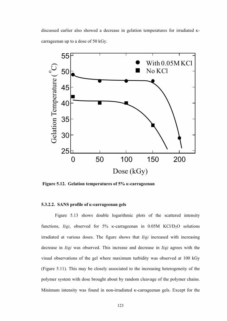
123
discussed earlier also showed a decrease in gelation temperatures for irradiated -
carrageenan up to a dose of 50 kGy.
Figure 5.12. Gelation temperatures of 5% -carrageenan
5.3.2.2. SANS profile of -carrageenan gels
Figure 5.13 shows double logarithmic plots of the scattered intensity
functions, I(q), observed for 5% -carrageenan in 0.05M KCl/D2O solutions
irradiated at various doses. The figure shows that I(q) increased with increasing
decrease in I(q) was observed. This increase and decrease in I(q) agrees with the
visual observations of the gel where maximum turbidity was observed at 100 kGy
(Figure 5.11). This may be closely associated to the increasing heterogeneity of the
polymer system with dose brought about by random cleavage of the polymer chains.
Minimum intensity was found in non-irradiated -carrageenan gels. Except for the
55
50
45
40
35
30
25
Gel
atio
nTe
mpe
ratu
re (
o C)
200150100500Dose (kGy)
With 0.05M KClNo KCl
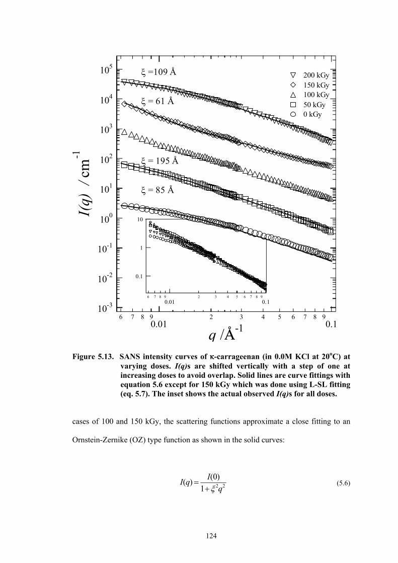
124
Figure 5.13. SANS intensity curves of 6-carrageenan (in 0.0M KCl at 20oC) at
varying doses. I(q)s are shifted vertically with a step of one at increasing doses to avoid overlap. Solid lines are curve fittings with equation 5.6 except for 150 kGy which was done using L-SL fitting (eq. 5.7). The inset shows the actual observed I(q)s for all doses.
cases of 100 and 150 kGy, the scattering functions approximate a close fitting to an
Ornstein-Zernike (OZ) type function as shown in the solid curves:
I(q) I(0)
1 2q2 (5.6)
0.1
1
10
6 7 8 9
0.012 3 4 5 6 7 8 9
0.1
= 85 Å
= 195 Å
= 61 Å
=109 Å
10-3
10-2
10-1
100
101
102
103
104
105
I(q)
/ c
m-1
6 7 8 90.01
2 3 4 5 6 7 8 90.1
q /C-1
200 kGy 150 kGy 100 kGy 50 kGy 0 kGy
125
where I(q) is the scattering intensity, I(0) is the 0-q intensity and is the correlation
lengths indicating the extent of correlated concentration fluctuations. The solid lines
in Figure 5.13 are the fit of the experimental data with this equation. Initially, for 0
kGy was 85Å which increased to 195Å at 50 kGy. Further irradiation at 200 kGy
decreased to 109 Å. No correlation length was evaluated from the carrageenan at
100 kGy since it had a power law behavior.
While the fitting curves were essentially Lorentzian, i.e., an OZ function,
certain deviations to this function were observed especially with -carrageenan
irradiated at 100 and 150 kGy. The double logarithmic plot indicated a power law
behavior exclusively at a dose of 100 kGy. Figure 5.14 shows a very close power law
fitting of -carrageenan at 100 kGy. The scattering exponent was -1.84. Non–
irradiated -carrageenan and -carrageenan irradiated at other doses did not exhibit
any power law behavior. Introduction of the squared-Lorentzian function was also
necessary for 150 kGy to give a successful fitting curve that closely reproduced the
observed I(q)s (equation 5.7).
(5.7)
where is the other correlation length describing the extent of spatial
inhomogeneities [31].
This phenomenological behavior can well be explained by looking at their
collective fluctuations as illustrated in Figure 5.15. The concentration at 0 kGy is high
enough to form a polymer solution that fills the entire space with polymer chains
overlapping each other (C >> C*), where C* is the chain overlap concentration. Since
-carrageenan is a natural polymer, concentration fluctuations are strongly suppressed
222221
)0(
1
)0()(
q
I
q
IqI SLL
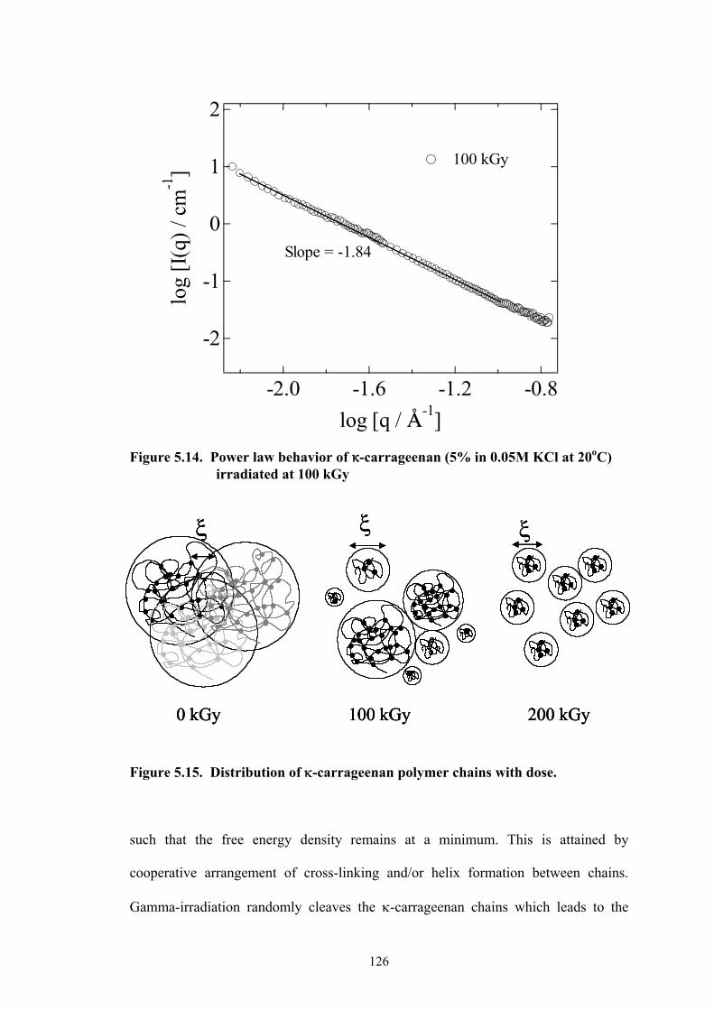
126
Figure 5.14. Power law behavior of 6-carrageenan (5% in 0.05M KCl at 20oC)
irradiated at 100 kGy
Figure 5.15. Distribution of -carrageenan polymer chains with dose.
such that the free energy density remains at a minimum. This is attained by
cooperative arrangement of cross-linking and/or helix formation between chains.
Gamma-irradiation randomly cleaves the -carrageenan chains which leads to the
-2
-1
0
1
2
log
[I(q
) / c
m-1
]
-2.0 -1.6 -1.2 -0.8
log [q / C-1]
Slope = -1.84
100 kGy
0 kGy
100 kGy
200 kGy
0 kGy
0 kGy
100 kGy
100 kGy
200 kGy
200 kGy
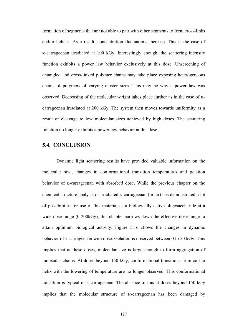
127
formation of segments that are not able to pair with other segments to form cross-links
and/or helices. As a result, concentration fluctuations increase. This is the case of
-carrageenan irradiated at 100 kGy. Interestingly enough, the scattering intensity
function exhibits a power law behavior exclusively at this dose. Unscreening of
entangled and cross-linked polymer chains may take place exposing heterogeneous
chains of polymers of varying cluster sizes. This may be why a power law was
observed. Decreasing of the molecular weight takes place further as in the case of -
carrageenan irradiated at 200 kGy. The system then moves towards uniformity as a
result of cleavage to low molecular sizes achieved by high doses. The scattering
function no longer exhibits a power law behavior at this dose.
5.4. CONCLUSION
Dynamic light scattering results have provided valuable information on the
molecular size, changes in conformational transition temperatures and gelation
behavior of -carrageenan with absorbed dose. While the previous chapter on the
chemical structure analysis of irradiated -carrageenan (in air) has demonstrated a lot
of possibilities for use of this material as a biologically active oligosaccharide at a
wide dose range (0-200kGy), this chapter narrows down the effective dose range to
attain optimum biological activity. Figure 5.16 shows the changes in dynamic
behavior of -carrageenan with dose. Gelation is observed between 0 to 50 kGy. This
implies that at these doses, molecular size is large enough to form aggregation of
molecular chains. At doses beyond 150 kGy, conformational transitions from coil to
helix with the lowering of temperature are no longer observed. This conformational
transition is typical of -carrageenan. The absence of this at doses beyond 150 kGy
implies that the molecular structure of -carrageenan has been damaged by

128
Figure 5.16. Changes in gelation and conformational transition behavior of irradiated -carrageenan at varying doses.
irradiation. Thus, the range between 75 to 150 kGy can be considered as the effective
range for optimum biological activity. It is as this range wherein the molecular size is
small enough not to form any aggregation but still retaining its molecular structure.
DLS experiment also indicates that at this range, a new fast mode in the characteristic
decay time distribution function appears. This fast mode is found to be maximum at
100 kGy with an estimated Mw of 5-10 kDa. This dose very well coincides with the
optimum plant growth promoter effect in -carrageenan and from literatures that
specify that oligomers with an Mw of < 10 kDa are needed as plant growth promoters.
The optimum plant growth promoter effect for other polysaccharides such as chitosan
and alginates is also found at 100 kGy. Thus, for plant growth promoter, what is
needed is a basic chemical structure of an oligosaccharide. DLS confirms that a size
of an Mw <10 kDa with intact molecular structure is appropriate for plant growth
promoter. Other biological functions such as plant elicitor, anti-viral and anti-tumor
0 50 100 150 200
Absorbed Dose (kGy)
Carbonyl Group
Reducing Sugar
Free Sulfate
Carboxylic Acid
Destruction of gelation and
conformational transition behavior
Gelation
Still a PolymerMw > 100 kDa
An Oligomer
No gelation but presence of
new fast mode peak with an Mw .5–10 kDa
0.0
15.0
% R
ed
uc
ing
Su
gar
0.0
0.5
A-A
o(2
60
nm
)
0.0
10.0
% F
reeSulfate (as HSO
3‐ )
0.0
10.0
% C
OO
H
129
activities may need specific chemical structures e.g. amide group in chitosan, sulfates
in carrageenan with the appropriate molecular size of the oligomer.
SANS experiments demonstrates the following: -carrageenan is subjected to
degradation by gamma irradiation. The structure factor of non-irradiated -
carrageenan aqueous solutions is similar to those for polymer solutions in semi-dilute
regime, i.e., an Ornstein-Zernike form. A power-law behavior appears in I(q) which
interestingly was observed at 100 kGy irradiated -carrageenan. Again, this coincides
with the optimum biological activity in -carrageenan. While the SANS data obtained
is quite limited, its interpretation in relation to the optimum biological function of
irradiated -carrageenan also remains to be very limited. This initial result opens new
possibilities for further studies in order to find the real significance of the power-law
behavior exclusively at 100 kGy.
REFERENCES:
1. S. Okabe, M. Nagao, T. Karino, S. Watanabe, M. Shibayama, J. Appl. Cryst. 38 (2005) 1035.
2. M. Shibayama, T. Norisuye, Bull Chem Soc Jpn. 75 (2002) 64. 3. J. Martin, J. Wilcoxon, J. Phys. Rev. Lett. 61 (1988) 373. 4. S. Ren, W. Shi, W. Zhang, C. Sorensen, Phys. Rev. A. 45 (1992) 2416. 5. B. Nystrom, J. Roots, A. Carlsson, B. Lindman, Polymer. 33 (1992) 2875. 6. D. Pinder, W. Nash, Y. Hemar, H. Singh, Food Hydocolloids. 17 (2003) 463. 7. M. Nickerson, A. Paulson, F. Hallet, Carbohydr. Polym. 58 (2004) 25. 8. I. Chronakis, J. Doublier, L. Picullel, Int. Biol. Macromol. 28 (2000) 1.
9. V. Meunier, T. Nicolai, D. Durand, Int. J. Biol. Macromol. 28 (2001) 157.
10. P. Ulanski C. Von Sonntag, J. Chem. Soc., Perkin Trans. 2 (2000) 2022.
130
11. H. Zegota, Food Hydrocolloids 13 (1999) 51. 12. P. Baugh, J. Goodall, G. Phillips, C. von Sonntag, M. Dizdaroglu, Carbohydr.
Res. 49 (1976) 315. 13. A. McKinnon, D. Rees, F. Williamson, J. Chem. Soc. D (1969) 701. 14. D. Rees, Pure & Appl. Chem. 53 (1981) 1. 15. O. Smidsrød, H. Grasdalen, Carbohydr. Polym. 2 (1982) 270. 16. O. Smidsrød, I. Andresen, H. Grasdalen, B. Larsen, T. Painter, Carbohydr. Res.,
80 (1980) c11. 17. Y. Yuguchi, T. Thuy, H. Urakawa, K. Kajiwara, Food Hydrocolloids. 16 (2002)
515. 18. S. Kara, C. Tamerler, H. Bermek, O. Pekcan, Int J Biol Macromol 31 (2003)
177. 19. M. Mangione, D. Giacomazzaa, D. Bulonea, V. Martoranaa, P. San Biagioa,
Biophys. Chem. 104 (2003) 95. 20. A. Michel, M. Mestdagh, M. Axelos, Int. J. Biol. Macromol. 21 (1997) 195. 21. Y. Chen, M. Liao, D. Dunstan, Carbohydr. Polym. 50 (2002) 109. 22. M. Nickerson, A. Paulson, Carbohydr. Polym. 61 (2005) 231. 23. P. MacArtain, J. Jacquier, K. Dawson, Carbohydr. Polym. 53 (2003) 395. 24. L. Piculell, J. Borgström, I. Chronakis, P. Quist, C. Viebke, Int. J. Biol.
Macromol. 21 (1997) 141. 25. M. Núñez-Santiago, A. Tecante, Carbohydr. Polym. 69 (2007) 763. 26. T. Norisuye, M. Shibayama, S. Nomura, Polymer. 39 (1998) 2769. 27. J. Martin, J. Wilcoxon, J. Odinek, Phys Rev A. 43 (1991) 858. 28. M. Sugiyama, C. Yuasa, K. Hara, N. Hiramatus, A. Nakamura, Y. Hayakawa,
Y. Maeda, Physica B 999 (1998) 241. 29. G. Evmenenko, E. Theunissen, K. Mortensen, H. Reynaers, Polymer 42 (2001)
2907.

131
30. N. Mischenko, B. Denef, K. Mortensen, H. Reynaers, Physica B 283 (1997) 234.
31. M. Shibayama, Macromol. Chem. Phys. 199 (1998) 1.

132
CHAPTER 6
RADIOLYSIS REACTIONS OF -CARRAGEENAN
6.1. INTRODUCTION
Electron spin resonance is quite a useful tool in determining the transientt free
radicals generated in irradiated chemical systems. Irradiation induced chemical
reactions in simple, neutral sugars such as glucose, and other carbohydrates have been
carefully studied using this method [1-6]. Low-molecular weight products formed
from the disproportionation reactions of the six primary glucose radicals, and the
radicals that are generated from these primary radicals by a number of elimination and
rearrangement reactions have been identified [7-9]. This method has also been used to
identify mechanism for chain scission and crosslinking of polysaccharides 10-13].
Among the polysaccharides, the radicals of cellulose have been well studied. Random
cleavage of glycoside bonds in the main chain, initialized by radicals placed on
macromolecules is considered as the leading reaction. After initial ionization, most of
kicked out electrons thermalize and eventually recombine with their parent ions.
Consequently, excited fragments of the polymer are formed. They decompose with
cleavage of chemical links, i.e., carbon-hydrogen. This leads to the formation of
radicals on polymer chains and hydrogen atoms [10, 14]. Several mechanisms of the
processes have been proposed. The process is similar for most polysaccharides. The
localization of the energy initiates degradation and dehydrogenation reactions. ESR
spectra of irradiated cellulose differ depending on the source of material origin, its
chemical treatment history, kind and conditions of irradiation [15-17].

133
6.2. METHODOLOGY
Electron Spin Resonance (ESR) experiments were performed on -
Carrageenan using a JEOL (JM-FE3) X-band spectrometer with 9.07-9.22 GHz
frequency. Powdered samples of carrageenan were irradiated in air at room
temperature at different doses (5, 50, 160 and 280 kGy) using the gamma irradiation
facility of the Takasaki Advanced Radiation Research Institute of JAEA. These were
placed in a 5mm diameter quartz tubes immediately prior to ESR analysis. Another set
of -carrageenan samples were placed in quartz tubes and sealed under 10-3 Pa
vacuum and irradiated at liquid nitrogen temperature. The temperature was then
slowly increased by submerging the tubes in dry ice, in ice and at room temperature.
The tips of the quartz tubes were then broken to allow air to come in. ESR analyses
were done at every step of the procedure.
6.3. RESULTS AND DISCUSSION
6.3.1. Electron Spin Resonance of -carrageenan
The ESR spectrum recorded at room temperature (around 25oC) for solid -
carrageenan irradiated at 5 kGy to 280 kGy at room temperature in air is presented in
Figure 6.1. The free radicals formed (trapped hydroxyl radicals, hydrogen radicals, -
carrageenan macroradicals, etc.) indicated poorly resolved ESR spectra as shown in
the figure. A single broad peak was observed for all doses. A study on the ESR of
cellulose also shows a single broad peak which contains more than 20 possible
radicals [10]. ESR spectra of polycrystalline ice of citrus pectin show a poorly
resolved composite ESR spectrum [18]. ESR investigation on the irradiated
derivatives of cellulose such as carboxymethyl cellulose [10], methylcellulose and

134
Figure 6.1. ESR of -carrageenan irradiated at varying doses in air.
hydroxyethylcellulose [11] in dry state and in aqueous solution shows some signals
typical of degradation reaction of cellulose and additional signals on side chains of
derivatives. Though the ESR profile of -carrageenan indicated no change in shape
with dose, its intensity varied with dose. Intensity increased at 50 kGy which
decreased beyond this dose. ESR measurements of irradiated dextrin demonstrate that
radical concentration increases with doses [13]. The decrease in intensity of ESR
signals of irradiated -carrageenan beyond 50 kGy may be related to the decrease in
crystallinity of -carrageenan with dose. Fast decay of free radicals is expected as the
degree of crystallinity is lowered with irradiation. Irradiation of polysaccharides such
as cellulose results in the decrease of chain rigidity and the degree of crystallinity of
the material [19]. Carbohydrate radicals are stabilized in a polycrystalline matrix [18].
2 mT
5kGy 50kGy 160kGy 280kGy

135
ESR of -carrageenan irradiated in vacuum at 50 kGy at increasing
temperatures is shown in Figure 6.2. A single broad peak was also observed. Similar
results were also observed in -carrageenan irradiated at a lower dose of 5 kGy (not
shown). There were no essential changes of signals from liquid nitrogen temperature
to ice temperature. Increase in intensity and change in shape were observed at room
temperature (RT) and when irradiated -carrageenan was exposed to air. The ESR
signal became more intense and sharper. Peak signals start to decrease slightly after
30 mins. of annealing at room temperature. Previous study on dextrin reports that the
spin density is not deeply affected by time due to the presence of carbonyl groups
which are long lived free radicals [13]. Comparing the ESR profile of solid -
carrageenan irradiated in air (at RT) and in vacuum (in liquid nitrogen), a broader
peak was observed in the latter (Figure 6.3). This is expected due to the fast decay of
some free radicals at RT.

136
Figure 6.2. ESR of -carrageenan at varying temperatures irradiated in vacuum
Figure 6.3. ESR of -carrageenan irradiated in vacuum and in air at 5 and
50 kGy
2 mT
Liq N2
Dry Ice Ice RT air RT air after 30 mins.
mT
315 320 325 330 335 340 345-600
-400
-200
0
200
400
600
-10
-8
-6
-4
-2
0
2
4
6
8
10
5kGy vacuum, liquid N 2
50kGy vacuum, liquid N2
5kGy in air50kGy in air
Re
lati
ve in
ten
sit
y i
n a
ir
Re
lati
ve
inte
ns
ity
in
va
cu
um
, Liq
uid
N2
137
6.3.2. Proposed Mechanism for the Radiolysis Reaction of -carrageenan
The current ESR results did not allow the identification of transient species or
macroradicals formed from the radiolysis of -carrageenan. This could have provided
the possible mechanisms for the radiolysis reaction. However, several schemes for the
radiation degradation of polysaccharides have already been proposed by several
authors e.g. chitosan [19, 20], cellulose [14], pectin [18], glucose [22, 4],
cycloamylose [23]. Based on the results of both spectral and chemical analyses of the
radiolytic products of irradiated carrageenan, similar schemes could be applied / and
or adopted to -carrageenan.
It is known that radiolytic effect of carbohydrates results in the abstraction of
carbon-bonded hydrogens which are the precursors of product formation. These are
caused either by the direct effect of ionizing radiation on carbohydrates or by the
indirect attack of the free radicals (OH and H radicals) generated from the water
radiolysis reaction in aqueous solutions as shown in equations 6.1 to 6.3. It can be
expected that abstraction of hydrogen from -carrageenan would yield 12 different
radicals as shown in scheme 1 (eqs. 6.4 to 6.9)
The main reaction in the radiolysis of polysaccharides is the O-glycoside bond
rupture [24]. Thus, of the 12 different radicals, only those macroradicals that are
directly located at the glycosidic bond or can be transferred by rearrangement into a
position adjacent to this bond undergo transformation to form scission products. For
-carrageenan with an (1,3) and (1-4) linkages, C(1) and C(3) of the D-galactose-4-
sulfate unit and C(1) and C(4) of the 3,6-anhydro-D-galactose unit participate in this
reaction. The degradation mechanism for irradiated -carragenan in solid state can be
outlined as proposed in scheme 2 (eqs. 6.10 to 6.13).

138
In aqueous solution, the reaction of .H atoms with many carbohydrates is
slower than that of hydroxyl radicals. Attack by .OH free radicals causes scission of
the glycosidic bond induced either by hydrolysis or by fragmentation. Scission of the
l-4 and 1-3 glycosidic bonds is caused by rearrangement of radicals localized on
C(1), C(4), C(3) and C(5) carbon atoms. The selectivity of .OH radical attack towards
carbohydrates is low and they react almost randomly with a slight preference for C(1)
carbon atom in pyranose ring of glucose. Schemes 3a and 3b (eqs. 6.14 to 6.24)
presents the proposed radiolytic cleavage of the glycosidic linkages of -carrageenan
in aqueous solution. The formed free aldehydic groups constitute the reducing sugars.
By analogy with previous work on cycloamyloses, it can be assumed that the
locations of radiation-induced keto groups in the -carrageenan residues coincide with
the sites of initial attack, i.e., the primary-radical sites. These keto groups arise in
irradiated -carrageenan by reaction steps involving (a) disproportionation and,
possibly, hydrogen-abstraction reactions of primary and secondary radicals; (b)
radical rearrangement; (c) eliminations of water, HOR, and carbon monoxide; and (d)
hydrolysis of the acetal bond if the radical site is at a neighbouring carbon atom.
In the presence of oxygen (in air) the hydrated electrons and .H atoms react
rapidly with O2 giving HO2. and O2
. radicals. These peroxy radicals can also abstract
the carbon-bonded hydrogens in the carbohydrates forming an ROO. radical (equation
6.25). In carbohydrates, the α-hydroxyalkyl peroxyl radicals readily undergo HO2. and
O2. elimination. In -carrageenan system, this would be the peroxyl radicals at C(2)
and C(6) in the D-galactose-4-sulfate unit and C(1) in the 3,6-anhydro-D-galactose
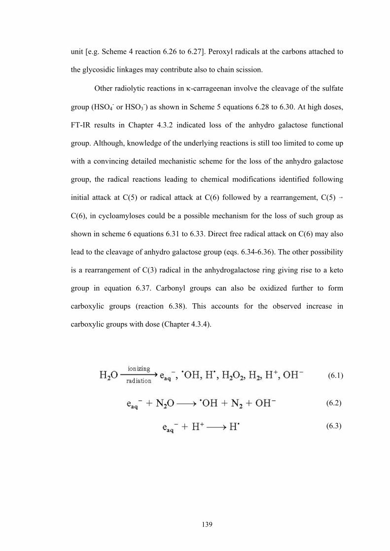
139
unit [e.g. Scheme 4 reaction 6.26 to 6.27]. Peroxyl radicals at the carbons attached to
the glycosidic linkages may contribute also to chain scission.
Other radiolytic reactions in -carrageenan involve the cleavage of the sulfate
group (HSO4- or HSO3
-) as shown in Scheme 5 equations 6.28 to 6.30. At high doses,
FT-IR results in Chapter 4.3.2 indicated loss of the anhydro galactose functional
group. Although, knowledge of the underlying reactions is still too limited to come up
with a convincing detailed mechanistic scheme for the loss of the anhydro galactose
group, the radical reactions leading to chemical modifications identified following
initial attack at C(5) or radical attack at C(6) followed by a rearrangement, C(5) 6
C(6), in cycloamyloses could be a possible mechanism for the loss of such group as
shown in scheme 6 equations 6.31 to 6.33. Direct free radical attack on C(6) may also
lead to the cleavage of anhydro galactose group (eqs. 6.34-6.36). The other possibility
is a rearrangement of C(3) radical in the anhydrogalactose ring giving rise to a keto
group in equation 6.37. Carbonyl groups can also be oxidized further to form
carboxylic groups (reaction 6.38). This accounts for the observed increase in
carboxylic groups with dose (Chapter 4.3.4).
(6.1)
(6.2)
(6.3)

140
OO3SO
O
OH
CH2OH
OO
O
OH
‐
OO3SO
O
OH
CH2OH
OO
O
OH
‐
.
OO3SO
O
OH
CH2OH
OO
O
OH
‐
.
OO3SO
O
OH
CH2OH
OO
O
OH
‐
.
OO3SO
O
OH
CH2OH
OO
O
OH
‐
.
OO3SO
O
OH
CH2OH
OO
O
OH
‐ .
‐.
OO3SO
O
OH
CHOH
OO
O
OH
.OH-H2O
.
.
.
.
.
.
.H
(6.4)
(6.5)
(6.6)
(6.7)
(6.8)
(6.9)
Scheme 1. Abstraction of hydrogen from -carrageenan

141
(6.10)
(6.11)
(6.12)
(6.13)
Scheme 2. Cleavage of the glycosidic linkage in solid -carrageenan
OO3SO
O
OH
CH2OH
OO
O
OH
‐
.
‐ OO3SO
O
OH
CH2OH
O
O
OH
+O .
‐ OO3SO
O
OH
CH2OH
O
O
OH
+OO3SO
O
OH
CH2OH
OO
O
OH
‐
. O.
O
O
OH
O
‐
.
OO3SO
OH
CH2OH
O
O
OH
. +
‐ OO3SO
OH
CH2OH
O
O
OH
O
.
‐ OO3SO
OH
CH2OH
O
O
OH
O
‐ OO3SO
OH
CH2OH
.+
C‐1 (G4S)
C‐4 (A)
C‐3 (G4S)
C‐1 (A)

142
OO3SO
O
OH
CH2OH
OO
O
OH
‐
.
‐
.OO3SO
O
OH
CH2OH
OH
O
O
OH
OH
+
‐ OO3SO
O
OH
CH2OH
O
O
OH
+O .
H2OC‐1 (G4S)
+
‐ OO3SO
O
CH2OH
OH
OH
.O
O
OH
OH
C‐4 (A)
OO3SO
O
OH
CH2OH
OO
O
OH
‐
.
+
O
O
OH
O
‐ OO3SO
O
OH
CH2OH
.
H2O
‐ . OO3SO
O
OH
CH2OH
OO
O
OH
+
OH
CO
OH
CH2OH
O
H
SO‐O3
.O
O
OH
OH
CO
OH
CH2OH
OH
H
OSO‐O3
+.
O
O
OH
OH
+CO
OH
CH2OH
O
H
SO‐O3 O.
O
O
OH
OH
H2O
C‐5 (G4S)
(6.14)
(6.15)
(6.16)
(6.17)
(6.18)
(6.19)
(6.20)
Scheme 3a. Cleavage of the 1-4 glycosidic linkage in aqueous -carrageenan
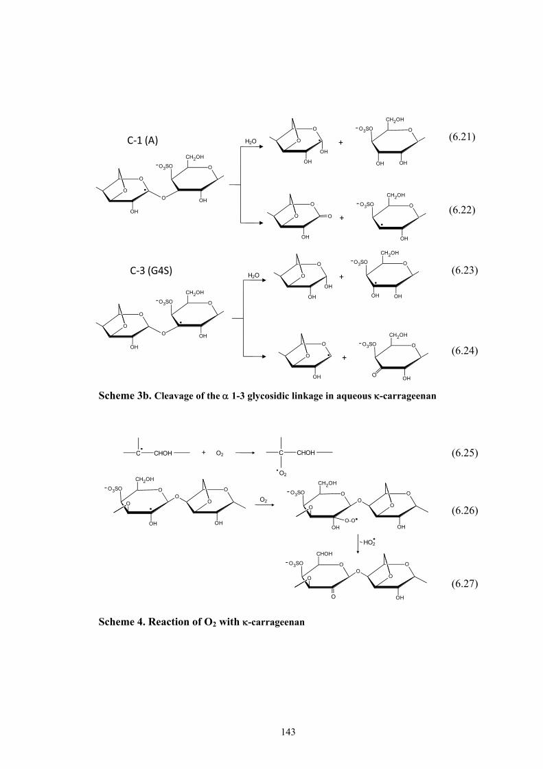
143
O
O
OH
O
‐
.
OO3SO
OH
CH2OH
O
O
OH
. +
O
O
OH
OH.
‐ OO3SO
OH
CH2OH
OH
+H2O
‐ OO3SO
OH
CH2OH
C‐3 (G4S)
O
O
OH
O
.
‐ OO3SO
OH
CH2OH
H2O
O
O
OH
OH
.‐ OO3SO
OH
CH2OH
OH
+
O
O
OH
O
‐ OO3SO
OH
CH2OH
.+
C‐1 (A)
(6.21)
(6.22)
(6.23)
(6.24)
Scheme 3b. Cleavage of the 1-3 glycosidic linkage in aqueous -carrageenan
(6.25)
(6.26)
(6.27)
Scheme 4. Reaction of O2 with -carrageenan
+C CHOH O2 C CHOH
O2
‐
.
OO3SO
O
OH
CH2OH
OO
O
OH
‐ OO3SO
O
H
OO
O
OHOO-O.
CH2OH
‐ OO3SO
O
CHOH
OO
O
OHO
O2
HO2

144
(6.28)
(6.29)
(6.30)
Scheme 5. Cleavgae of the sulfate group in -carrageenan
(6.31)
(6.32)
(6.33)
Scheme 6. Cleavage of the anhydrogalactose unit in -carrageenan
O
O
OH
CH2OH
OO
O
OH
OO3SO
O
OH
CH2OH
OO
O
OH
‐
-HSO4+
O
O
OH
CH2OH
OO
O
OH
.H.
H.
OO3SO
O
OH
CH2OH
OO
O
OH
‐
. H. O
O
OH
CH2OH
OO
O
OH
O -HSO3+
O
O
OH
‐
.
O
OO3SO
OH
CH2OH
OHOH
O
O
OO3SO
OH
CH2OH
OH
.CH2
OHOH
O
O
OO3SO
OH
CH2OH
OH
CH3
OHOH
O
CH3
C
O
H
3SOO O
OH
CH2OH
OH
+
H2O
H.
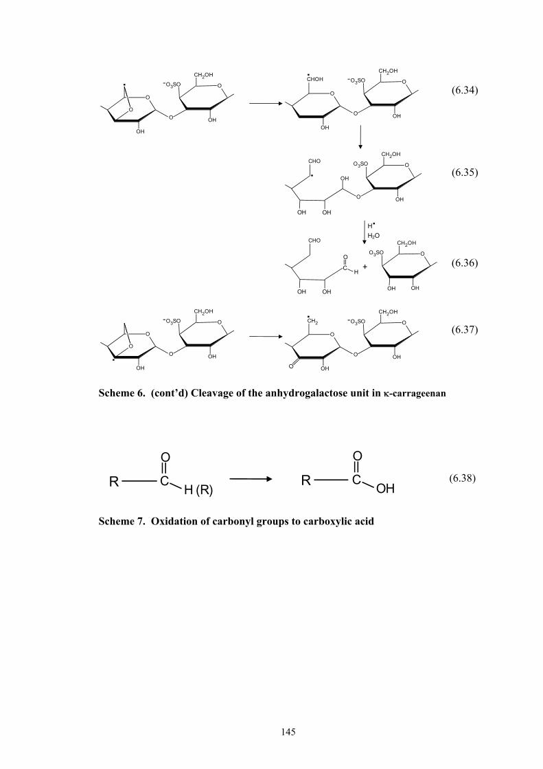
145
C
O
H (R)R C
O
OHR
(6.34)
(6.35)
(6.36)
(6.37)
Scheme 6. (cont’d) Cleavage of the anhydrogalactose unit in -carrageenan
Scheme 7. Oxidation of carbonyl groups to carboxylic acid
O
O
OH
‐.
O
OO3SO
OH
CH2OH
O
OH
‐.
O
OO3SO
OH
CH2OH
CHOH
OHOH
O
OO3SO
OH
CH2OH
OH.
CHO
OHOH
CHO
C
O
H
OO3SO
OH
CH2OH
OH
+
H2O
H.
O
O
OH
‐
. O
OO3SO
OH
CH2OH
O
OH
‐.
O
OO3SO
OH
CH2OH
CH2
(6.38)

146
REFERENCES:
1. I. Shkrob, M. Depew, J. Wan, Chem. Phys. Lett. 202 (1993) 133. 2. M. Kuwabara, Z. Zhang, O. Inanami, G. Yoshii, Radiat. Phys. Chem. 24 (217)
1984. 3. M. Kuwabara, Z. Zhang, G. Yoshii, Radiat. Phys. Chem. 21 (1983) 451. 4. R. Muzzarelli, F. Tanfani, G. Scarpini, M. Muzzarelli, Biochem. Bio. Ph. Res.
Co. 89 (1979) 706. 5. T. Matsuyama, H. Menhofer, H. Heusinger, Int. J. Radiat. Appl. Instrum. Part C.
Radiat. Phys. Chem. 32 (1988) 735. 6. S. Çakir, F. Köksal, R. Tapramaz, O. Çakir, Int. J. Radiat. Appl. Instrum. Part C.
Radiat. Phys. Chem. 38 (1991) 17. 7. S. Kawakishi, Y. Kito, M. Namiki, Carbohydr. Res. 39 (1975) 263. 8. C. von Sonntag, Adv. Carbohydr. Chem. Biochem. 37 (1980) 7. 9. C. von Sonntag, H. Schuchmann, Radiation Chemistry. Present Status and
Future Trends, C. D. Jonah, B. S. M. Rao (Eds), Elsevier Science, Amsterdam, 2001, Chapter Carbohydrates, p. 481.
10. R. Wach, H. Mitomo, N. Nagasawa, F. Yoshii, Radiat. Phys. Chem. 68 (2003)
771. 11. R. Wach, H. Mitomo, F. Yoshii, J. Radioanal. Nucl. Ch. 261 (2004) 113. 12. A. Alberti, S. Bertini, G. Gastaldi, N. Iannaccone, D. Macciantelli, G. Torri, E.
Vismara Eur. Polym. J. 41 (2005) 1787. 13. S. Lotfy, Int. J. Biol. Macromol. 44 (2009) 57. 14. Y. Nakamura, Y. Ogiwara, G. Phillips, Polym. Photochem. 6 (1985) 135. 15. A. Mohanty, B. Singh, Polym.-Plast. Technol. Eng. 27 (1988) 435–466. 16. A. Rukhlya, E. Petrayaev, O. Shadyro, J. Appl. Chem. USSR, 56 Part 2 (1983)
911. 17. Y. Ogiwara, H. H. Kubota, J. Appl. Polym. Sci., 18 (1974) 2057.
18. H. Zegota, Food Hydrocolloids 13 (1999) 51.

147
19. C. Delides, C. Panagiotalidis, O. Legapanagiotalidis, Textile Res. J. 51 (1981)
31.
20. P. Ulanski, J. Rosiak, Radiat. Phys. Chem. 39, (1992) 53.
21. P. Ulanski C. Von Sonntag, J. Chem. Soc., Perkin Trans. 2 (2000) 2022.
22. G. Phillips, G. Moody, Int. J. Appl. Radiat. Is. 6 (1959) 78.
23. P. Baugh, J. Goodall, G. Phillips, C. von Sonntag, M. Dizdaroglu, Carbohydr. Res. 49 (1976) 315.
24. I. Idimecheva, R. Kisel, O. Shadyro, K. Kazem, H. Murase, T. Kagiya. J. Radiat.
Res. 46 (2005) 319.
148
CHAPTER 7
SUMMARY AND CONLUSIONS
Kappa carrageenan is easily degraded by radiation with a higher degradation
yield than most other known natural polysaccharides (Gd = 2.5 for solid in air, 1.7 in
vacuum and 1.2 x 10⁻⁷ mol J-1 in 1% aqueous irradiation). Irradiation of -carrageenan
in solid or aqueous solution, yields higher Gd in the presence of O2. Radiation in
aqueous solution is initialized by reaction with OH• radical at a rate constant k = 1.2 x
109 M-1s-1. Both Gd and kkc are affected by conformational changes from coil to helix
especially in the presence of ions such as Na+. Irradiation of -carrageenan reduces its
viscosity due to degrease in DP but yields radiolytic products that can decrease the
reactive sites for OH• radical interaction and consequently decreases kkc for irradiated
-carrageenan. On the other hand, hydrolysis of -carrageenan by chemical means,
yields lower molecular weight polymer/oligomer fragments that increases its kkc.
The chemical analyses of the radiolytic products indicate increasing reducing
sugars, carbonyl, carboxylic acids, and sulfates with increasing doses which reach a
maximum level at a certain dose depending on the irradiation condition. Values are
very much lower in solid irradiation (in vacuum and in air) as compared to aqueous
irradiation. NMR spectra of -carrageenan irradiated at 100 kGy indicate that the
fraction with an Mw (3-10) kGy has an intact oligomer structure (13)-4 sulfate--D
galactose (14)-3, 6 anhydro-"-D-galactose.
The dynamic behavior of -carrageenan indicates that absorbed dose of up to
50 kGy still undergoes sol-gelation transition. Above 50 kGy, no gelation takes place,

149
instead appearance of fast relaxation mode in CDF is observed at doses of 75–150
kGy. Optimum peak intensity was found at 100 kGy (Mol wt .5 – 10 kDa) which
coincides with the optimum plant growth promoter effect in -carrageenan. At a dose
beyond 150 kGy, the conformational transition temperature from coil to helix is no
longer observed. In addition, SANS experiment indicates a unique structure form at
100 kGy wherein a power law behavior with a fractal dimension -1.84 is observed.
The significance of all data obtained can be summarized in Figure 7.1. The
figure shows the spectrum of possible applications of irradiated -carrageenan based
on the functional groupings and molecular size of the radiolytic products. The color
intensity from light to dark denotes increasing effectivity.
Optimum biological activity can be obtained at a dose between 75 to 150 kGy
which is maximum at 100 kGy in solid. In 1% aqueous solution, a dose of less than 10
kGy and minimum at 2 kGy would be appropriate for the biological activity. For plant
growth promoter, it requires only an intact oligomeric backbone with a glycosidic
linkage (common to all polysaccharides) with an Mw of 5-10 kDa. This can be
obtained by irradiating -carrageenan at 100 kGy either in air or in vacuum or at 2
kGy in 1% aqueous solution. Applications related to the other biological functions of
-carrageenan, e.g. plant elicitor, anti viral (anti-herpes and anti-HIV), and anti-tumor
require more specificity. It has to have an intact -carrageenan oligomer structure
with an intact functional sulfate group ((13)-4 sulfate--D galactose (14)-3, 6
anhydro-"-D-galactose.). Thus, irradiating it in solid and in vacuum may be more
appropriate for this purpose considering that the chemical changes in -carrageenan at
this condition are so minimal.
150
The gelation property of -carrageenan can be utilized for the making of
hydrogels in general. -carrageenan can be irradiated up to a dose of 50 kGy without
destroying its gelation properties. Synthetic water soluble polymers when combined
with natural polymers can substantially improve the physico-mechanical properties of
the hydrogel (swelling and tensile strength).
Stimuli responsive hydrogels undergo abrupt volume change in response to
changes in environmental parameters such as pH, ions, temperature. These hydrogels
normally undergo conformational chain transitions which directly affects the
swelling-deswelling behavior of the hydrogel. In the case of -carrageenan, this
conformational transition from helix to coil is influenced by changes in temperature or
presence of metal ions. Thus, irradiated -carrageenan can also be useful as stimuli
responsive gels when combined or grafted to some synthetic water soluble polymers.
The effective dose range for this purpose is less than 175 kGy. At doses below 50
kGy the conformational transition temperature of -carrageenan is rather high
(>70oC) which drops to 50oC at doses between 50 to 175 kGy. Stimuli responsive gels
are commonly used as matrix for the slow release of drugs or as industrial censors. -
carrageenan may be grafted unto water soluble polymers to make hydrogels of this
type.
The presence of reducing sugars in irradiated -carrageenan gives possibilities
for its utilization as antioxidant. The reducing sugars are easily oxidized to carboxylic
acid. In solid irradiation, the number of reducing sugar increases with dose for even as
high as 200 kGy. On the other hand, for 1% aqueous solution, reducing sugar reaches
a maximum high level at 50 kGy. This can be useful as polysaccharide-based edible
coatings for food protection and preservation in fruits and vegetables. Irradiation of -
151
carrageenan with amino groups at this effective dose range may produce a powerful
antioxidant (Maillard reaction products) which involves the formation of brown
pigments by condensation between carbonyl groups of reducing sugars, aldehydes or
ketones and amine groups of amino acids.
The amount of free sulfates released from -carrageenan with irradiation can
be utilized for purposes of developing radiation dose indicators. This application has
already been described in the introduction. In solid irradiation, free sulfates levels off
at 50 kGy while at 1% aqueous solution, this levels off at 100 kGy with higher acidity
obtained in aqueous solution.
To conclude, Figure 7.2 gives an over-all view of the importance of this study
and some future researches that still need to be done. All the data obtained from this
research are new and valuable information on absorbed dose and conditions for the
development of noble materials from radiation processed natural polymer (-
carrageenan) has been obtained. This fundamental study would serve as a theoretical
basis and would prove to be most useful especially in the formulation and
development of new products from radiation modified -carrageenan for various
applications. This data should further be supplemented with actual results from
applied researches of developed materials. Furthermore, while this research is limited
to -carrageenan, investigation should be extended to other types of carrageenan (
and ). In fact, -carrageenan oligomer is known to be more effective as an anti-viral
and anti-tumor agent. This technique may also be applied to other natural polymers.
The proposed radiolytic reaction mechanism should also be confirmed by doing a
more detailed ESR study. Further elucidation of the radiolytic products by ESR and
other known techniques could be interesting.
152
Lastly, this is the first research which has employed the use of techniques such
as dynamic light scattering and small angle neutron scattering in understanding the
radiation chemistry of polysaccharides. It is hoped that more works along this area
would be done to understand better the effect of radiation on the structure and
dynamic behavior of polysaccharides.

153
Figure 7.1. Effective dose range of irradiated - carrageenan for bio-based materials development
Absorbed Dose (kGy)
PossibleUses Conditions
Biological Activity: Plant Growth Promoter Plant Elicitor Anti‐ viral e.g. anti‐HIV,anti‐herpetic Tumor inhibition
Solid
1% aqueous
Hydrogels Solid
Stimuli Responsive Gels Solid
Anti‐oxidantSolid
1% aqueous
Radiation Dose IndicatorSolid
1% aqueous
0 50 100 150 200
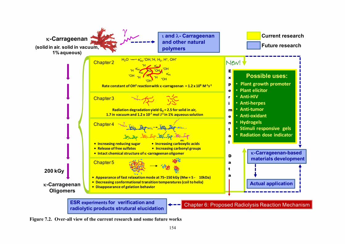
154
200 kGy
-CarrageenanOligomers
-Carrageenan(solid in air. solid in vacuum,
1% aqueous)
H2O eaq, OH, H, H2, H+, OH-- . .
H. OH.eaq-
H. H.
H.
H.
H.
eaq-
eaq-
eaq-
eaq-
OH.
OH.
OH.
OH.
Chapter 2
Radiation degradation yield Gd = 2.5 for solid in air, 1.7 in vacuum and 1.2 x 10‐7 mol J‐1 in 1% aqueous solution
Chapter 3
etc.
O
O
OH
O
O
OH
(KC)nO
O
OH
O
O
OH
-O3 SO
CH2OH
O
OH
O
O
H2C
O
O
O
O
OH
-O3SO-O3SO
CH2OHCH2OH
O
O
OO
O
O
OH
CH2OHCH2OH
O
O
O
O
OH
-O3SO-O3SO-O3SO-O3SO
CH2OHCH2OHCH2OHCH2OH
O
O
OOOO
O
O
OH
CH2OHCH2OHCH2OHCH2OH
O
O
O
O
O
OH
-O3SO-O3SO
CH2OHCH2OH
O
O
H2CH2C
O
O
O
OHOO
O
O
O
O
O
OH
-O3SO-O3SO-O3SO-O3SO
CH2OHCH2OHCH2OHCH2OH
O
O
H2CH2CH2CH2C
O
O
O
OHOO
-O3SO
O
O
O
O
OH
-O3SO
CH2OHCH2OH
O
O
OO
O
O
OH
CH2OHCH2OH
OO
-O3SO
O
O
O
O
OH
-O3SO
CH2OHCH2OHCH2OHCH2OH
O
O
OOOO
O
O
OH
CH2OHCH2OHCH2OHCH2OH
OOOO
O
O O
-O3SO-O3SO
CH2OHCH2OH
O
O
OO
O
OHOH
CHCH2OH
OOO
O O
-O3SO-O3SO-O3SO-O3SO
CH2OHCH2OHCH2OHCH2OH
O
O
OOOO
O
OHOHOHOH
CHCH2OHCHCH2OH
OOOO
CH2OHO
O
OH
O
O
O
OH
CH2OHO
O
OH
O
O
O
OH
(KC)n-xCH2OH
O
O
OH
O
O
O
OH
CH2OHO
O
OH
H2CH2CH2CH2CO
O
O
OH
-O3 SO
i ii→
• Increasing reducing sugar • Increasing carboxylic acids• Release of free sulfates • Increasing carbonyl groups• Intact chemical structure of ‐carrageenanoligomer
Chapter 4
• Appearance of fast relaxation mode at 75–150 kGy (Mw = 5 ‐ 10kDa)• Decreasing conformational transition temperatures (coil to helix)• Disappearance of gelation behavior
Chapter 5
Chapter 6: Proposed Radiolysis Reaction Mechanism
Possible uses:• Plant growth promoter• Plant elicitor• Anti‐HIV• Anti‐herpes• Anti‐tumor• Anti‐oxidant• Hydrogels• Stimuli responsive gels• Radiation dose indicator
Experimental
Data
Rate constant of OH• reactionwitk ‐carrageenan = 1.2 x 10⁹ M‐1s‐1
New!
Current research
Future research
Actual application
and - Carrageenanand other natural polymers
ESR experiments for verification andradiolytic products strutural elucidation
-Carrageenan-basedmaterials development
Figure 7.2. Over-all view of the current research and some future works
155
Publications related to the dissertation 1. L.V. Abad, H. Kudo, S. Saiki, N. Nagasawa, M. Tamada, Y. Katsumura, C.T.
Aranilla, L.S. Relleve and A.M. De La Rosa, 2009. Radiation degradation studies of carrageenans. Carbohyd. Polym. 78, 100-106.
2. L.V. Abad , S. Okabe, M. Shibayama, H. Kudo, S. Saiki, C. Aranilla, L. Relleve, and A.M. De la Rosa, 2008. Comparative studies on the conformational change and aggregation behavior of irradiated carrageenans and agar by dynamic light scattering. Int. J. Biol. Macromol. 42, 55-61.
3. L.V. Abad, S. Saiki, H. Kudo, Y. Muroya, Y. Katsumura and A.M. De la Rosa, 2007. Rate constants of reactions of κ-carrageenan with hydrated electron and hydroxyl radical. Nuclear Inst. and Methods in Physics Research, B. 265, 410-413.
4. L.V. Abad, S. Okabe, S. Koizume, M. Shibayama, 2006. Small-angle neutron
scattering on irradiated kappa carrageenan. Physica B. 381, 103-108.
5. L.V. Abad, I. Nasimova, C. Aranilla, M. Shibayama, 2004. Light scattering studies of irradiated - and -carrageenan. Rad. Phys. Chem. 23, 29-37.
6. L.V. Abad, I. Nasimova, L. Relleve, A.M. Dela Rosa, M. Shibayama, 2004.
Dynamic light scattering studies of irradiated kappa carrageenan. Int. J. Biol. Macromol. 34, 81-88.
Poster and Paper Presentations to Conferences, Symposiums and Meetings Related to the Dissertation
1. L. V. Abad, L.S. Relleve, C. T. Aranilla and A. M. de la Rosa “Radiation
Processed Materials from Carrageenan for Agricultural Applications” 1st
Regional Cooperative Meeting (RCM) on “Development of radiation-processed products of natural polymers for application in agriculture, health care, industry and environment”, 21-25 April 2008, Vienna, Austria,.
2. L. V. Abad, H. Kudo, S. Saiki, N. Nagasawa, M. Tamada, H. Fu, Y. Muroya,
M. Lin, Y. Katsumura, and A. M. deLaRosa “Gamma-rays irradiation and electron-beam pulse radiolysis study on kappa-carrageenans”, The 2nd Asia Pacific Symposium on Radiation Chemistry, 29 August -1 September 2008, Waseda University, Tokyo, Japan
3. L.V. Abad, S. Saiki, H. Kudo, Y. Muroya, Y. Katsumura and A.M. De la Rosa
“Pulse Radiolysis of -Carrageenan, 7th International Symposium on Ionizing Radiation and Polymers, 23-28 September 2006, Antalya, Turkey.
4. L.V. Abad, S. Saiki, H. Kudo, Y. Muroya, Y. Katsumura and A.M. De la Rosa
“Pulse Radiolysis of -carrageenan” 49th Annual Meeting of Japanese Society of Radiation Chemistry, 12-14 October 2006, Takasaki, Gunma, Japan.

156
5. L. Abad, I. Nasimova, S. Koizumi, M. Shibayama “Structure and Dynamics of Irradiated Kappa Carrageenan”, 53rd SPSJ Annual Meeting, 25-27 May 2004, Kobe International Conference Center, Kobe-shi, Japan
6. L. Abad, I. Nasimova and M. Shibayama “Dynamic Light Scattering of
Irradiated Kappa Carrageenan”, 第 15 回高分子ゲル研究討論会, 14-15 January 2004, University of Tokyo, Japan.
7. L. V. Abad, L. S. Relleve, C. T. Aranilla, and A. M. Dela Rosa, “Properties of
Radiation Synthesized Water Soluble Polymer-Carrageenan Hydrogels and their Applications”, 5th Gel Symposium, Polymer Gels: Fundamentals and Nano-Fabrications, 18 November 2003, Kashiwa Campus, University of Tokyo, Japan.
Other Publications
1. L. Relleve, N. Nagasawa, L.Q. Luan, T. Yagi, C. Aranilla, L. Abad, T. Kume, F. Yoshii, A. dela Rosa, 2005. Degradation of carrageenan by radiation. Polym. Degrad. Stab. 87, 403-410.
2. L.V. Abad, L.S. Relleve, C.T. Aranilla, A.M. Dela Rosa, I. Nasimova, S. Okabe, and M. Shibayama, Structure and Dynamics of Irradiated Kappa Carrageenan, Proceedings of the 20th Philippine Chemistry Congress, 12 March 2005, Baguio City, Philippines.
3. L.V. Abad, L.S. Relleve, C.T. Aranilla, and A.M. Dela Rosa, 2003. Properties
of radiation synthesized PVP-kappa carrageenan hydrogel blends. Rad. Phys. Chem. 68, 901-908.
4. L.V. Abad, L.S. Relleve, C.T. Aranilla, A.K. Aliganga, C.M. San Diego, and
A.M. dela Rosa, 2002. Natural antioxidants for radiation vulcanization of natural rubber latex. Polym. Degrad. Stab. 76, 275-279.
5. Relleve, Lorna S., L. V. Abad, C.T. Aranilla, A. Aliganga, A.M. Dela Rosa, F.
Yoshii, T. Kume and N. Nagasawa, “ Biological Activities of Radiation-Degraded Carrageenan” Proceedings of the Symposium on Radiation Technology in Emerging Industrial Applications, 6-10 November 2000, Beijing, People’s Republic of China.
6. Abad, Lucille V., L.S. Relleve, C.T. Aranilla, A. Aliganga, C.M. San Diego
and A.M. Dela Rosa, “Natural Antioxidants for Radiation Vulcanized Natural Rubber Latex”, Proceedings of the Symposium on Radiation Technology in Emerging Industrial Applications, 6-10 November 2000, Beijing, People’s Republic of China,.
7. L. V. Abad, C. San Diego, C.T. Aranilla, L.S. Relleve and A. M. dela Rosa,
“Natural Antioxidants for RVNRL Part II”, IAEA Regional Cooperative Meeting Report on the Improvement of the Physical Properties of RVNRL, 18-

157
22 October 1999, Takasaki Radiation Chemistry Establishment, Takasaki, Japan.
8. A. M. dela Rosa, L.S. Relleve, C.T. Aranilla, L.V. Abad and C. San Diego,
“Preparation of New Hydrogels from Carrageenan via Radiation Crosslinking” Proceedings of the 15th Philippine Chemistry Congress, 26-29 May 1999, Waterfront Cebu City Hotel, Cebu City, Philippines.
9. L.V. Abad, A. M. dela Rosa, I. Janik, and J.M. Rosiak, “Sol/Gel Analysis of
Crosslinked Polymers”, Proceedings of the 15th Philippine Chemistry, 26-29 May 1999, Waterfront Cebu City Hotel, Cebu City, Philippines.
10. L.V. Abad, C.M. San Diego, L.P. Relleve, C.O. Aranilla, and A.M. Dela Rosa,
“Natural Antioxidants for RVNRL”, IAEA Regional Cooperative Meeting Report on the Improvement of the Physical Properties of RVNRL, 7-11 September 1998, Bangkok, Thailand.
11. A.M. dela Rosa, L.V. Abad, L.P. Relleve, C.O. Aranilla and C.L. Pascual,
“Radiation Response of Philippine Natural Rubber Latex”, Proceedings of the International Symposium on Radiation Technology in Conservation of the Environment, 8-12 September 1997, Zakopane, Poland.
12. L.V. Abad, A.M. dela Rosa, K. Makuuchi, and F. Yoshii, "The Role of Proteins
on the Thermal Oxidative Aging of Radiation Vulcanized Natural Rubber Latex", Proceedings of the Second International Symposium on RVNRL, 15-17 July 1996, Kuala Lumpur, Malaysia.
13. A.M. dela Rosa, L.V. Abad, L.P. Relleve, C.O. Aranilla and C.L. Pascual,
"Radiation Vulcanization of Philippine Natural Rubber Latex", Proceedings of the 12th Philippine Chemistry Congress, 23-25 May 1996, Sarabia Manor Hotel, Iloilo City, Philippines.
14. A.M. dela Rosa, L.P. Relleve, Aranilla C.O., Pascual C.L. and L.V. Abad,
"Polymerization of Coconut Oil and Lauryl Alcohol with Methylmethacrylate by Gamma Radiation", Proceedings of the12th Philippine Chemistry Congress, 23-25 May 1996, Sarabia Manor Hotel, Iloilo City, Philippines.
15. L.V. Abad, "The Effect of Proteins on the Aging Properties of Radiation
Vulcanized Natural Rubber Latex", Thesis presented to the University of Santo Tomas (UST), 18 March 1993, University of Santo Tomas, Manila, Philippines.
16. A.M dela Rosa, L.V. Abad, and R.B. Banzon Michel, "Radiation Enhanced
Biomass Conversion of Agricultural Cellulosic Wastes", Proceedings of the 6th Philippine Chemistry Congress, 25 May 1990, University of San Carlos, Cebu City, Philippines.
17. R.B. Banzon, L.V. Abad and A. M. dela Rosa, 1988. Genotoxic Studies in
Irradiated Mangoes Using Short Term Mutagenicity Assays. Phil. Journal Food Science and Technology. 12, 1.

158
18. L.V. Abad, R.B. Banzon, A.M. Dela Rosa,"Composition of the Enzymatic and Acid Hydrolyzates of Gamma Irradiated Rice Straw", Proceedings of the 2nd ASEAN Science and Technology Week, 1989, Vol 2, 124-134.
19. A.M. Dela Rosa, and L.V. Abad, 1986. "Structural Analysis of Gamma
Irradiated Cellulose and Agricultural Cellulosic Wastes", PAEC (C) IB 86005. 20. A.M. Dela Rosa, R.B. Banzon, L.V. Abad, Z.F. Simbul Nuguid, and A.DM.
Bulos, 1985. Effect of Gamma Irradiation on the Saccharification of Cellulose. The Nucleus. 23, 1-10.
21. A.M. Dela Rosa, A.DM. Bulos, R.B. Banzon, Z.F. Simbul Nuguid, and L.V.
Abad, 1983. Radiation Pretreatment of Agricultural Cellulosic Wastes for Energy Production. PAEC (C) IB 83006.
Other Lectures and Presentations
1. L.V. Abad, “Achievements on the IAEA Regional Cooperative Agreement (RCA) RAS 8/106 Project in the Philippines”, IAEA/RCA Project Final Technical Review Meeting for RAS/8/106 on “Radiation Processing Applications for Health and the Environment” and First Technical Planning and Coordination for RAS 8/109 “Supporting Radiation Processing of Polymeric Materials for Agricultural Applications and Environmental Remediation”, 16 - 20 March 2009, Daejeon, Korea.
2. L.V. Abad, “IAEA/RCA Activities on Radiation Processing of Natural Polymers” and “IAEA/RCA RAS 8/106 Activities”, FNCA 2008 Workshop on Application of Electron Accelerator-Radiation Processing of Natural Polymer, 26 – 31 October 2008, Shanghai, China.
3. L. Abad, L. Relleve, C. Aranilla, A. dela Rosa, “RAS 8/106 Country Report”,
IAEA/RCA Mid-Term Progress Meeting of RAS/8/106 on “Radiation Processing Applications for Health and the Environment”, 7 - 12 April 2008, Manila, Philippines.
4. L.V. Abad, “IAEA/RCA Activities on Radiation Processing”, Forum for
Nuclear Cooperation in Asia (FNCA) 2007 Workshop on Application of Electron Accelerator-Radiation Processing of Natural Polymer, 22 – 26 October 2007, Kimdo Hotel, 133 Nguyen Hue Ave., Dist. 1, Ho Chi Minh City, Vietnam.
5. L. V. Abad, “Introduction to Radiation Chemistry of Water and Polymers”;
“Radiation processing applications of polysaccharides”; and “Natural Polymer Modification and Characterization”, IAEA Regional Training Course (RTC) on Radiation Processing for Basic and Medium Level Personnel, 6 -10 August 2007, Malaysian Nuclear Agency (Nuclear Malaysia), Bangi. Malaysia.

159
6. L.V. Abad, “Radiation Processing of Natural Polymers”, IAEA/RCA Regional Training Course RAS/8/106 on Promotion of Radiation Technology Utilization 9 - 13 July 2007, The Linden Suites, Manila, Philippines.
7. L. Abad, L. Relleve, C. Aranilla, C. Bisnar, and A. dela Rosa, Radiation
Processing of Natural Polymers RAS 8/098, IAEA/RCA Project Planning Meeting of RAS/8/106, 23-27 April 2007, Thailand, Bangkok.
8. L.V. Abad, Radiation Processing of Carrageenan, IAEA/RCA Regional
Meeting On “Health Care Stimuli Responsive Radiation Processed Materials”, 26-30 June 2006, Hotel Sheraton, Dhaka, Bangladesh,
9. L.V. Abad, “Radiation Processing of Carrageenan using Electron Beam” and
“Prospects of Electron Beam Treatment of Flue Gases in the Philippines”, FNCA 2004 Workshop on Application of Electron Accelerator, 7 September 2004, Beijing, China.
10. Alumanda M. Dela Rosa, Lucille V. Abad, Lorna S. Relleve, Charito T.
Aranilla, and Cristina L. Pascual, “Radiation-Modified Carrageenan for Agricultural and Health Care Applications”, IAEA Project Coordination Meeting on Radiation Processing of Chitin/Chitosan, 19 March 2002, Bangkok, Thailand.
11. L.V. Abad, “Hydrogels for Biomedical Applications”, February 2002, Poveda
Learning Centre, Quezon City, Philippines.
12. L.V. Abad, “New Applications of Radiation Modified Carrageenan”, 3rd Annual De La Salle University Science and Technology Conference (Physics Session), 9 March 2001, De la Salle University, Manila, Philippines.
13. L.V. Abad, “Radiation Processing of Natural Polymers and Its Applications”, 28th Atomic Energy Week, 13 December 2000, Philippine Nuclear Research Institute, Quezon City, Philippines.
14. L.V. Abad, C.M. San Diego, L.P. Relleve, C.O. Aranilla, and A.M. Dela Rosa,
“Natural Antioxidants for RVNRL”, Regional Cooperative Meeting on the Improvement of the Physical Properties of RVNRL, 7-11 September 1998, Bangkok, Thailand.
15. L.V. Abad, Lecture-Forum on Gas Chromatography and High-Performance
Liquid Chromatograph, 30 August 1996, Far Easten University, Manila, Philippines.
16. L.V. Abad, K. Makuuchi, F. Yoshii and A.M. dela Rosa, "The Role of Proteins
on the Thermal Oxidative Aging of Radiation Vulcanized Natural Rubber Latex", Diliman Community Celebration of the National Science and Technology Week, 22 July 1993, Philippine Nuclear Research Institute, Diliman, Quezon City, Philippines.

160
17. L.V. Abad, K. Makuuchi and F. Yoshii, "The Effect of Proteins on the Ageing Properties of RVNRL, Final report presented to the staff of the Takasaki Radiation Chemistry Research Establishment, 17 March 1992, TRCRE, Gunma ken, Japan.
18. L.V. Abad, R.B. Banzon and A.M. Dela Rosa, "Composition of the Enzymatic
and Acid Hydrolyzates of Gamma Irradiated Rice Straw", First ASEAN Talent Search for Young Scientists", 18 January 1989, Philippine International Convention Center, Manila.
Other Professional Activities
1. Philippine Patent “PVP-carrageenan hydrogel for burn/wound dressing” (№ 1-2000-02471), Philippine Patent Office
2. Philippines’ National Project Coordinator (NPC), IAEA RAS 8/109 “Radiation
Processing of Polymeric Materials for Agricultural Applications and Environmental Remediation (RCA)”, 2009-2011.
3. IAEA Expert to the FNCA 2008 Workshop on Application of Electron
Accelerator-Radiation Processing of Natural Polymer, 26 – 31 October 2008, Shanghai, China.
4. IAEA Expert to the FNCA 2007 Workshop on Application of Electron
Accelerator-Radiation Processing of Natural Polymer, 22 – 26 October 2007, Kimdo Hotel, 133 Nguyen Hue Ave., Dist. 1, Ho Chi Minh City, Vietnam.
5. Project Leader, Radiation Processed Materials from Carrageenan for
Agricultural Applications, IAEA CRP Project on “Development of Radiation-Processed Products of Natural Polymers for Application in Agriculture, Healthcare, Industry and Environment”, 2007 to present.
6. Philippines’ NPC and Project Lead Country Coordinator, IAEA RAS 8/106
“Radiation Processing Applications for Health and Environment (RCA)”, 2007-2008.
7. Course Director, IAEA/RCA Regional Training Course RAS/8/106 on
Promotion of Radiation Technology Utilization, 9 - 13 July 2007, The Linden Suites, Manila, Philippines
8. IAEA Expert Lecturer to the IAEA Regional Training Course (RTC) on
Radiation Processing for Basic and Medium Level Personnel, 6 -10 August 2007, Malaysian Nuclear Agency (Nuclear Malaysia), Bangi. Malaysia.
9. Project Leader, Philippine DOST, TECHNICOM Project “Semi-
Commercialization of PVP-Carrageenan Hydrogels for Burn/Wound Dressing and Bed Sores (Phase II) : Production and Market Acceptability”, 2007-2008.

161
10. Philippines’ NPC and Project Lead Country Coordinator, IAEA RAS 8/098 “Radiation Technology for Development of Advanced Materials and for Protection of Health and the Environment”, 2005-2006.
11. Philippine Project Coordinator , IAEA CRP on the Improvement of Physical
Properties of Radiation Vulcanized Natural Rubber Latex, 1997-1999.