rapamycin attenuates airway hyperreactivity, goblet … · treatment protocols on the major...
TRANSCRIPT

of July 2, 2018.This information is current as
Experimental Allergic AsthmaHyperreactivity, Goblet Cells, and IgE in Rapamycin Attenuates Airway
Brandt, Gurjit K. Khurana Hershey and Timothy D. Le CrasElizabeth M. Mushaben, Elizabeth L. Kramer, Eric B.
http://www.jimmunol.org/content/187/11/5756doi: 10.4049/jimmunol.1102133October 2011;
2011; 187:5756-5763; Prepublished online 21J Immunol
MaterialSupplementary
3.DC1http://www.jimmunol.org/content/suppl/2011/10/21/jimmunol.110213
Referenceshttp://www.jimmunol.org/content/187/11/5756.full#ref-list-1
, 14 of which you can access for free at: cites 46 articlesThis article
average*
4 weeks from acceptance to publicationFast Publication! •
Every submission reviewed by practicing scientistsNo Triage! •
from submission to initial decisionRapid Reviews! 30 days* •
Submit online. ?The JIWhy
Subscriptionhttp://jimmunol.org/subscription
is online at: The Journal of ImmunologyInformation about subscribing to
Permissionshttp://www.aai.org/About/Publications/JI/copyright.htmlSubmit copyright permission requests at:
Email Alertshttp://jimmunol.org/alertsReceive free email-alerts when new articles cite this article. Sign up at:
Print ISSN: 0022-1767 Online ISSN: 1550-6606. Immunologists, Inc. All rights reserved.Copyright © 2011 by The American Association of1451 Rockville Pike, Suite 650, Rockville, MD 20852The American Association of Immunologists, Inc.,
is published twice each month byThe Journal of Immunology
by guest on July 2, 2018http://w
ww
.jimm
unol.org/D
ownloaded from
by guest on July 2, 2018
http://ww
w.jim
munol.org/
Dow
nloaded from
The Journal of Immunology
Rapamycin Attenuates Airway Hyperreactivity, Goblet Cells,and IgE in Experimental Allergic Asthma
Elizabeth M. Mushaben,*,† Elizabeth L. Kramer,*,† Eric B. Brandt,†,‡
Gurjit K. Khurana Hershey,†,‡ and Timothy D. Le Cras*,†
The mammalian target of rapamycin (mTOR) signaling pathway integrates environmental cues, promotes cell growth/dif-
ferentiation, and regulates immune responses. Although inhibition of mTOR with rapamycin has potent immunosuppressive
activity, mixed effects have been reported in OVA-induced models of allergic asthma. We investigated the impact of two rapamycin
treatment protocols on the major characteristics of allergic asthma induced by the clinically relevant allergen, house dust mite
(HDM). In protocol 1, BALB/c mice were exposed to 10 intranasal HDM doses over a period of 24 d and treated with rapamycin
simultaneously during the sensitization/exposure period. In protocol 2, rapamycin was administered after the mice had been sen-
sitized to HDM (i.p. injection) and prior to initiation of two intranasal HDM challenges over 4 d. Airway hyperreactivity (AHR),
IgE, inflammatory cells, cytokines, leukotrienes, goblet cells, and activated T cells were assessed. In protocol 1, rapamycin blocked
HDM-induced increases in AHR, inflammatory cell counts, and IgE, as well as attenuated goblet cell metaplasia. In protocol 2,
rapamycin blocked increases in AHR, IgE, and T cell activation and reduced goblet cell metaplasia, but it had no effect on inflam-
matory cell counts. Increases in IL-13 and leukotrienes were also blocked by rapamycin, although increases in IL-4 were unaffected.
These data demonstrated that rapamycin can inhibit cardinal features of allergic asthma, including increases in AHR, IgE, and
goblet cells, most likely as a result of its ability to reduce the production of two key mediators of asthma: IL-13 and leukotrienes.
These findings highlight the importance of the mTOR pathway in allergic airway disease. The Journal of Immunology, 2011, 187:
5756–5763.
Asthma prevalence has increased substantially in recentyears, especially in children (1–3). Allergic asthma is themost common form and is characterized by airway in-
flammation, airway hyperreactivity (AHR), goblet cell metaplasia,and increases in IgE and Th2 cytokines (1, 4, 5). Although glu-cocorticoids and bronchodilators are the mainstay of asthma treat-ment, these therapies are not effective in all asthmatics (1).The discovery of the drug rapamycin (6, 7) has led to intense study
of its target: the mammalian target of rapamycin (mTOR). mTORis downstream of the PI3K-signaling cascade and signals via twocomplexes: mTOR complex (mTORC)1 and mTORC2 (8, 9). Ac-tivation of mTORC1, which is sensitive to rapamycin, leads tophosphorylation and activation of the p70 ribosomal S6 kinase 1and, subsequently, S6 ribosomal protein (S6), which promotes ri-
bosomal protein synthesis (8). Although several reports indicatedthat mTORC2 is not inhibited by rapamycin, there is evidenceshowing that rapamycin can inhibit mTORC2 activity, depending onthe specific cell type, duration, and dose of rapamycin treatment (10).mTOR is known to play a major role in regulating cell metabolism,growth/differentiation, and survival in many cell types (8, 11).Dysregulation of this pathway has been implicated in various dis-eases, including cancer and type 2 diabetes (9, 12, 13). Rapamycin isused as an immunosuppressant drug to prevent transplant rejection(14, 15); however, its effects on inflammation in OVA-inducedmodels of asthma are mixed (16–18). In addition, studies in OVAmodels (16–18) did not address whether mTOR inhibition alters IL-13 and leukotrienes, which are important mediators of allergicasthma responses, including AHR and goblet cell metaplasia.The goal of our study was to determine whether rapamycin
would attenuate key characteristics of allergic asthma (AHR, in-flammation, goblet cell metaplasia, IgE) and important mediators(IL-13 and cysteinyl leukotrienes) in a clinically relevant modelinduced by exposure to house dust mite (HDM). We hypothesizedthat inhibition of mTOR with rapamycin would attenuate allergicairway disease via reductions in these key mediators. To test thishypothesis, mice were either exposed to HDM and treated withrapamycin simultaneously or sensitized to HDM by systemic in-jection and then treated with rapamycin during subsequent intra-nasal (i.n.) HDM challenges. Multiple end points were assessed,including sensitization, AHR, inflammation, goblet cells, T cells,cytokines, and leukotrienes.
Materials and MethodsAnimals
Animal protocols and procedures were approved by the Animal Care andUse Committee at the Cincinnati Children’s Hospital Research Founda-tion (Cincinnati, OH). Six- to eight-week-old female BALB/c mice were
*Division of Pulmonary Biology, Cincinnati Children’s Hospital, Cincinnati, OH45229; †Department of Pediatrics, University of Cincinnati School of Medicine,Cincinnati, OH 45229; and ‡Division of Asthma Research, Cincinnati Children’sHospital, Cincinnati, OH 45229
Received for publication July 25, 2011. Accepted for publication September 23,2011.
This work was supported by National Institutes of Health Grants U19A170235 (toG.K.K.H.) and HL097135 (to T.D.L.C. and G.K.K.H.), National Institute of Envi-ronmental Health Sciences Grant T32 ES010957 (to E.B.B.), and American HeartAssociation Established Investigator Grant 740069N (to T.D.L.C.).
Address correspondence and reprint requests to Dr. Timothy Le Cras, Division ofPulmonary Biology, Cincinnati Children’s Hospital Medical Center, 3333 BurnetAvenue, Cincinnati, OH 45229-3039. E-mail address: [email protected]
The online version of this article contains supplemental material.
Abbreviations used in this article: AHR, airway hyperreactivity; BALF, bronchoal-veolar lavage fluid; CLCA3, chloride channel, calcium activated, family member 3;HDM, house dust mite; i.n., intranasal(ly); mTOR, mammalian target of rapamycin;mTORC, mammalian target of rapamycin complex; Muc5AC, mucin 5AC; P-S6,phosphorylated S6 ribosomal protein; S6, S6 ribosomal protein.
Copyright� 2011 by TheAmericanAssociation of Immunologists, Inc. 0022-1767/11/$16.00
www.jimmunol.org/cgi/doi/10.4049/jimmunol.1102133
by guest on July 2, 2018http://w
ww
.jimm
unol.org/D
ownloaded from

purchased from Charles River Laboratories (Wilmington, MA). Thetreatment protocols used in these studies are described below.
Protocol 1
Mice were exposed to 10 i.n. doses of HDM (50 mg in 20 ml saline; GreerLaboratories, Lenoir, NC) or saline (0.9% NaCl, 20 ml; control group) over24 d (Fig. 1A). In a third study group, mice were exposed to HDM andtreated with rapamycin. Rapamycin (4 mg/kg) (LC Laboratories, Woburn,MA) was administered by i.p. injection, six times a week starting with thefirst HDM exposure and continuing until 1 d after the last HDM exposure,for a total of 20 treatments.
Protocol 2
Mice were sensitized to HDM by three i.p. injections of HDM (50 mg in100 ml saline) at 7-d intervals (Fig. 1B). Seven days after the last i.p. in-jection, two i.n. challenges of HDM (50 mg in 20 ml saline) were given2 d apart. Another group was treated with rapamycin (4 mg/kg, i.p.),starting 1 d prior to the first i.n. HDM challenge and continued until1 d after the second HDM challenge, for a total of six rapamycin treat-ments. Study groups were mice given i.p. saline + i.n. saline (saline/saline),i.p. HDM + i.n. saline (HDM/saline), i.p. HDM + i.n. HDM (HDM/HDM),and i.p. HDM + i.n. HDM + i.p. rapamycin (HDM/HDM+Rapa).
Airway hyperreactivity
AHR was studied in anesthetized mice 48 h after the last HDM chal-lenge. Anesthesia was delivered by i.p. injection of ketamine/xylaxine/acepromazine (4:1:1) solution (0.2 ml/animal). Changes in airway resis-tance to methacholinewere assessed, as previously described (19). Briefly, atracheostomy was performed, and the mouse was connected to a flexiVentsystem (SCIREQ, Montreal, QC, Canada). Airway resistance was mea-sured after nebulization of 13 PBS (baseline) and then increasing doses ofmethacholine (6.25, 12.5, 25, and 50 mg/ml; acetyl-b-methylcholinechloride, Sigma, St. Louis, MO). After the flexiVent studies, mice werekilled by cutting the thoracic aorta. Blood was collected, and serum wasextracted for measurements of IgG1 and IgE.
Allergic sensitization
Total and HDM-specific IgG1 and IgE levels were measured in serum byELISA to assess allergic sensitization, as previously described (20). Briefly,for total IgG1 and IgE, plates were coated overnight with 2 mg/ml anti-mouse IgG1 or IgE in PBS, respectively (Pharmingen, San Jose, CA). ForHDM-specific IgG1 and IgE, plates were coated with 0.01% HDM in PBS.The following day, plates were blocked with 1% BSA in PBS for 1 h andthen coated with samples and IgG1 or IgE standards. Biotin–anti-mouseIgG1 or IgE (2.0 mg/ml, Pharmingen) was used to capture IgG1 and IgEAbs, respectively. Streptavidin-HRP (1:100; R&D Systems, Minneapolis,MN) was added to detect biotin-labeled Abs, and the reaction was de-veloped by tetramethylbenzidine substrate reagent set (1:1) (BD Bio-sciences, San Jose, CA).
Inflammatory cells and cytokines
Bronchoalveolar lavage fluid (BALF) was collected from mice to assess theinflammatory response, as previously described (19). Briefly, the lungswere lavaged via a tracheostomy with 1 ml 13 PBS containing BSA (1%)and EDTA (2 mM). The BALF was centrifuged (5000 rpm), and the su-pernatant was frozen at 280˚C for measurements of cytokines and leu-kotrienes. Cell pellets were resuspended, and RBCs were lysed using RBClysing buffer (Sigma). The remaining cells were resuspended in 13 PBSafter centrifugation. Total inflammatory cells were counted using a hema-cytometer, and cytospins were performed on the remaining cells for dif-ferential cell counts. Diff-Quick staining of cytospin slides (ShandonLipshaw, Pittsburgh, PA) was performed. Three hundred cells werecounted per slide, and the percentages of macrophages, lymphocytes,neutrophils, and eosinophils were calculated. Total differential inflam-matory cell counts were also calculated. Cytokines in the BALF weremeasured using a multiplex biomarker panel (Chemokine Panel I) andLuminex xMAP technology (Millipore, Billerica, MA), following themanufacturer’s instructions. Cysteinyl leukotriene (C4, D4, E4) levels weremeasured by a commercial company (ElisaTech, Aurora, CO) usingELISA kits (Cayman, Ann Arbor, MI), according to the manufacturer’sinstructions.
Western blot analysis
Western blot analysis was performed on lung homogenates using the fol-lowing Abs to assess the levels of phosphorylated S6 (P-S6, 1:1,000; Cell
Signaling, Danvers, MA), total S6 (1:1,000; Cell Signaling), chloridechannel, calcium activated, family member 3 (CLCA3, 1:2,000; Abcam,Cambridge, MA) (21, 22), and b-tubulin (1:1,000; Cell Signaling). Sec-ondary Abs included goat anti-rabbit and goat anti-mouse (1:10,000, Cal-biochem). ECL Plus system was used for chemiluminescence detection(GE Healthcare). An LAS4000 imaging system and Multi Gauge 3 soft-ware (Fujifilm, Tokyo, Japan) were used to image and quantitate the chemi-luminescent signal for each Western blot. Values were normalized tob-tubulin to control for protein loading and transfer efficiency.
Mucin 5AC immunohistochemistry
Lungs were inflation fixed at a constant pressure (25 cm H2O) by trachealinstallation of 4% paraformaldehyde, transferred to 70% ethanol after 24 h,and embedded in paraffin, as previously described (19). Immunostainingfor mucin 5AC (Muc5AC) was performed on 5-mm paraffin-embeddedsections. Slides were incubated with a primary Muc5AC mouse mAb(1:200; Thermo Scientific, Waltham, MA) overnight at 4˚C, followed by agoat anti-mouse IgG1 secondary Ab (1:200; Southern Biotech, Birming-ham, AL). Signal was detected using the diaminobenzidene method ofdetection (19). Digital images of Muc5AC immunostaining were obtainedusing a Zeiss Axioplan 2 microscope and camera (Carl Zeiss Micro-imaging, Thornwood, NY).
Quantitative real-time PCR analysis
RNA for quantitative real-time PCR was isolated using the SV Total RNAIsolation System (Promega, Madison, WI) and reverse transcribed intocDNA using an iScript cDNA synthesis kit (Bio-Rad Laboratories, Hercu-les, CA), following the manufacturer’s instructions. cDNA samples wereamplified with TaqMan, and a primer/probe set specific for Muc5AC wasused (assay ID: Mm01276725_g1; Applied Biosystems, Carlsbad, CA).Quantification of gene expression was analyzed by a 7300 Real Time PCRSystem (Applied Biosystems) in triplicate, and expression levels werenormalized to b-actin mRNA levels.
Flow cytometry for activated and regulatory T cells
The upper right lung lobe was minced and incubated at 37˚C for 25–30 minin 2 ml RPMI 1640 containing Liberase DL (0.5 mg/ml; Roche Diagnostics,Indianapolis, IN) and DNAse I (0.5 mg/ml; Sigma). Lung cells were passedthrough a 70-mm cell strainer, and the strainer was washed with 5 ml RPMI1640 + DNAse I media. Cells were centrifuged and resuspended in 2 mlRPMI 1640 before counting with a hemacytometer and confirming viabilityby trypan blue exclusion. Approximately 500,000 lung cells were trans-ferred to a 96-well V-bottom plate on ice, centrifuged, and resuspended in13 PBS containing FcBlock (2.4G2 mAb). To assess the levels of activatedT cells, lung T cells were stained with CD3e-FITC, CD4-PE/Cy7, CD69-PE, or CD25-allophycocyanin (BioLegend, San Diego, CA). Live and deadcells were labeled with LIVE/DEAD Fixable Aqua Dead Cell Stain Kit,according to the manufacturer’s instructions (Invitrogen by Life Technol-ogies, Carlsbad, CA). To assess the levels of regulatory T cells, intracellularstaining for Foxp3-PerCP5.5 was performed using the classic protocol andreagents from eBioscience (San Diego, CA). Acquisition was done on aFACS Canto III (Becton Dickinson, Mountain View, CA) and analyzed usedFlowJo software (Tree Star, Ashland, OR).
Statistical analysis
Statistical analysis was performed with Prism 5 software (GraphPad Soft-ware, San Diego, CA). Unpaired t tests, one-way ANOVA with the Tukeypost hoc test, and two-way ANOVA tests with the Bonferroni post hoc testwere used to make comparisons. The p values , 0.05 were consideredstatistically significant.
ResultsProtocol 1
Phosphorylated S6. After 10 treatments with i.n. HDM (Fig. 1A),Western blot analysis for P-S6 was performed on lung homoge-nates from saline controls, HDM, and HDM + rapamycin-treatedmice to verify that the dose of rapamycin was sufficient to inhibitsignaling downstream of mTOR. Mice exposed to HDM had a 2-fold increase in P-S6 levels compared with saline controls (Fig. 2).Rapamycin treatment blocked this increase.
AHR and goblet cell metaplasia. To determine whether rapamycintreatment reduced HDM-induced AHR, methacholine challengeswere performed using a flexiVent system. Increases in AHR were
The Journal of Immunology 5757
by guest on July 2, 2018http://w
ww
.jimm
unol.org/D
ownloaded from

observed in HDM-exposed mice at 25 mg/ml (4.4-fold) and 50mg/ml (3.6-fold) methacholine compared with saline controls(Fig. 3A). AHR in HDM-exposed mice treated with rapamycinwas similar to saline controls. Muc5AC immunostaining wasperformed to assess changes in goblet cells. Increased Muc5ACstaining was observed in HDM-exposed mice (Fig. 3B) comparedwith saline controls. Increases in another goblet cell protein,CLCA3, were observed by Western blot in HDM-exposed mice(Fig. 3C). Rapamycin treatment reduced Muc5AC staining in thelungs and decreased CLCA3 protein levels by 80%.
Inflammatory cells and allergic sensitization. To determine theeffects of rapamycin on allergic airway inflammation, total anddifferential inflammatory BALF cell counts were performed. TotalBALF cell count increases with HDM exposure were preventedby rapamycin (data not shown). Specifically, increases in mac-rophages, neutrophils, and eosinophils were all prevented byrapamycin treatment (Fig. 4A). The percentage of macrophages,neutrophils, and eosinophils also changed with HDM exposure;however, there was not a significant change in the percentage ofneutrophils and eosinophils after rapamycin treatment (Fig. 4A).To assess allergic sensitization, HDM-specific IgG1 and IgE levels
were measured in serum, and both were elevated in HDM-exposedmice (Fig. 4B). Rapamycin prevented these increases, so thatHDM-specific IgG1 and IgE levels were similar to saline controls.Rapamycin also blocked increases in total IgG1 and IgE levelsafter HDM exposure (Supplemental Fig. 1).
Protocol 2
IgG1 and IgE. Data from protocol 1 demonstrated that adminis-tration of rapamycin simultaneously with HDM exposure blockedallergic sensitization and disease; however, the protective effect ofrapamycin on these processes may be due to rapamycin preventingallergic sensitization. In protocol 2, allergic sensitization was firstestablished by i.p. injection of HDM. Rapamycin treatment wasinitiated 6 d after the last i.p. injection of HDM and 1 d prior to i.n.HDM challenge (Fig. 1B). Control animals for these experimentswere injected i.p. with either saline or HDM and then treated withi.n. saline (saline/saline or HDM/saline, respectively). HDM/salinemice showed increases in HDM-specific IgE and IgG1 comparedwith saline/saline controls (Fig. 5A, Supplemental Fig. 2). HDM/HDM mice had higher levels of HDM-specific IgE (7-fold) andIgG1 (1.25-fold) compared with HDM/saline control mice. InHDM/HDM+Rapa mice, HDM-specific IgE and IgG1 levels weresimilar to HDM/saline controls. Total serum IgG1 and IgE werealso increased after HDM sensitization (Supplemental Fig. 2).Rapamycin attenuated the increase in total IgE but not total IgG1.
AHR and goblet cells. AHR was increased in HDM/HDM mice at25 mg/ml (4.5-fold) and 50 mg/ml (5.3-fold) methacholine com-pared with HDM/saline controls (Fig. 5B). In HDM/HDM+Rapamice, AHR was similar to both control groups (saline/saline, HDM/saline). To assess goblet cells, CLCA3 protein levels were mea-sured by Western blot analysis. CLCA3 protein was not detectablein HDM/saline control mice, but it was readily detectable in lunghomogenates from HDM/HDM mice. CLCA3 protein was reducedby 41% in HDM/HDM+Rapa mice compared with HDM/HDMmice (Fig. 5C). To further assess the goblet cell response, quanti-tative real-time RT-PCR was performed on lung RNA for the ex-pression of Muc5AC. Muc5AC mRNA levels were normalized tob-actin mRNA. Muc5AC mRNA expression was increased 32-foldin HDM/HDM mice compared with saline/saline and HDM/salinecontrols. Rapamycin reduced Muc5AC mRNA expression by 84%(Fig. 5D). Abundant goblet cells were also detected in the airwaysof HDM/HDM mice by Muc5AC immunostaining, whereas nostaining was detectable in HDM/saline controls (Fig. 5E). Muc5ACstaining for goblet cells was reduced in HDM/HDM+Rapa mice.
Inflammatory cell counts. Total BALF macrophages, neutrophils,and eosinophils (Fig. 6A), as well as the percentage of eosino-phils (Fig. 6B), were increased in HDM/HDM mice compared withHDM/saline controls and remained elevated in HDM/HDM+Rapamice (Fig. 6).
T cells, cytokines, and leukotrienes. To examine the cellular andmolecular mediators of the allergic response, T cells, cytokines, andleukotriene levels were assessed. The total number of CD69+
Foxp32 CD4+ T cells was increased (1.7-fold) in the lungs ofHDM/HDM mice compared with HDM/saline controls (Fig. 7A,Supplemental Fig. 3). CD69+Foxp32 T cell numbers were reducedin HDM/HDM+Rapa mice compared with HDM/HDM mice andwere similar to HDM/saline controls. The total number of Foxp3+
CD25+ T regulatory cells in the lungs of HDM/HDM mice wasincreased (1.6-fold) compared with HDM/saline controls. Foxp3+
CD25+ T regulatory cell numbers in HDM/HDM+Rapa mice werereduced compared with HDM/HDM mice and were similar toHDM/saline controls (Fig. 7B, Supplemental Fig. 3). The ratio ofFoxp3+CD25+ T regulatory cells/CD25+Foxp32 T cells was lower
FIGURE 2. P-S6 levels corrected to total S6 levels in lung homogenates
of mice exposed to i.n. HDM and treated with rapamycin. Western blot
analysis of P-S6 protein, a downstream mediator of mTOR signaling, was
increased after HDM exposure compared with saline controls. Rapamycin
(Rapa) prevented this increase in P-S6 (n = 3 or 4 mice/group). *p , 0.05,
versus saline; ^p , 0.05, versus HDM.
FIGURE 1. Study protocols. A, Protocol 1: mice were exposed to 10 i.n.
HDM doses over 24 d. Rapamycin treatment by i.p. injection was started
simultaneously with HDM exposure and continued 6 d/wk until 1 d after
the last HDM exposure, for a total of 20 injections. B, Protocol 2: mice
were sensitized to HDM by 3 i.p. injections before i.n. challenge with
saline or HDM. Rapamycin treatment was started 6 d after the last i.p.
injection and 1 d prior to i.n. HDM exposure.
5758 RAPAMYCIN ATTENUATES CARDINAL FEATURES OF ALLERGIC ASTHMA
by guest on July 2, 2018http://w
ww
.jimm
unol.org/D
ownloaded from

in HDM/HDM and HDM/HDM+Rapa mice compared with HDM/saline controls (Fig. 7C). IL-4 levels were undetectable in theBALF of HDM/saline controls, but they were readily detectable inHDM/HDM mice (Fig. 8A). IL-4 levels were similar in HDM/HDM+Rapa mice compared with HDM/HDM mice. IL-13 levelswere also undetectable in HDM/saline control mice, but werereadily detectable in HDM/HDM mice (Fig. 8A). IL-13 was notdetectable in rapamycin-treated mice (HDM/HDM+Rapa). IL-5and eotaxin 1 levels were increased in HDM/HDM mice com-pared with HDM/saline controls (Fig. 8B). Rapamycin treatment
decreased IL-5; however, eotaxin 1 levels remained elevated inHDM/HDM+Rapa mice. Cysteinyl leukotriene levels were in-creased in BALF from HDM/HDM mice compared with HDM/saline control mice (Fig. 8C). Leukotriene levels in HDM/HDM+Rapa mice were similar to HDM/saline controls.
DiscussionThe goal of our study was to determinewhether the mTOR inhibitor,rapamycin, would suppress key characteristics and mediators in aclinically relevant asthma model induced by the aeroallergen HDM.
FIGURE 4. Inflammatory cell counts in
BALF and HDM-specific IgG1 and IgE lev-
els in serum. A, HDM exposure induced
increases in total inflammatory cells and the
percentage of neutrophils and eosinophils com-
pared with saline controls. Rapamycin pre-
vented HDM-induced increases in total in-
flammatory cell counts in the BALF (n = 6
mice/group). B, HDM-specific IgG1 and IgE
levels were increased after HDM exposure
compared with saline controls and blocked
by rapamycin (n = 9 mice/group). *p , 0.05,
versus saline; ^p , 0.05, versus HDM.
FIGURE 3. AHR and goblet cells in mice exposed to i.n. HDM and rapamycin (Rapa). A, Rapamycin prevented increases in airway resistance to
methacholine in HDM-exposed mice (n = 7 or 8 mice/group in two independent experiments). B, Rapamycin attenuated increases in Muc5AC immu-
nostaining in airways after HDM exposure. Scale bars, 100 mm. C, Rapamycin attenuated increases in the goblet cell protein, CLCA3, by Western blot in
HDM-exposed mice. n = 3 to 4 mice per group. *p , 0.05, versus saline; ^p , 0.05, versus HDM.
The Journal of Immunology 5759
by guest on July 2, 2018http://w
ww
.jimm
unol.org/D
ownloaded from
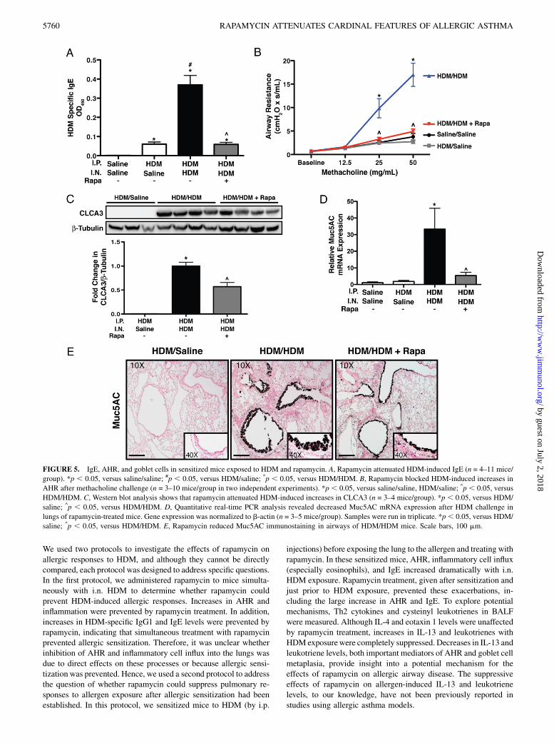
We used two protocols to investigate the effects of rapamycin onallergic responses to HDM, and although they cannot be directlycompared, each protocol was designed to address specific questions.In the first protocol, we administered rapamycin to mice simulta-neously with i.n. HDM to determine whether rapamycin couldprevent HDM-induced allergic responses. Increases in AHR andinflammation were prevented by rapamycin treatment. In addition,increases in HDM-specific IgG1 and IgE levels were prevented byrapamycin, indicating that simultaneous treatment with rapamycinprevented allergic sensitization. Therefore, it was unclear whetherinhibition of AHR and inflammatory cell influx into the lungs wasdue to direct effects on these processes or because allergic sensi-tization was prevented. Hence, we used a second protocol to addressthe question of whether rapamycin could suppress pulmonary re-sponses to allergen exposure after allergic sensitization had beenestablished. In this protocol, we sensitized mice to HDM (by i.p.
injections) before exposing the lung to the allergen and treating withrapamycin. In these sensitized mice, AHR, inflammatory cell influx(especially eosinophils), and IgE increased dramatically with i.n.HDM exposure. Rapamycin treatment, given after sensitization andjust prior to HDM exposure, prevented these exacerbations, in-cluding the large increase in AHR and IgE. To explore potentialmechanisms, Th2 cytokines and cysteinyl leukotrienes in BALFwere measured. Although IL-4 and eotaxin 1 levels were unaffectedby rapamycin treatment, increases in IL-13 and leukotrienes withHDM exposurewere completely suppressed. Decreases in IL-13 andleukotriene levels, both important mediators of AHR and goblet cellmetaplasia, provide insight into a potential mechanism for theeffects of rapamycin on allergic airway disease. The suppressiveeffects of rapamycin on allergen-induced IL-13 and leukotrienelevels, to our knowledge, have not been previously reported instudies using allergic asthma models.
FIGURE 5. IgE, AHR, and goblet cells in sensitized mice exposed to HDM and rapamycin. A, Rapamycin attenuated HDM-induced IgE (n = 4–11 mice/
group). *p , 0.05, versus saline/saline; #p , 0.05, versus HDM/saline; ^p , 0.05, versus HDM/HDM. B, Rapamycin blocked HDM-induced increases in
AHR after methacholine challenge (n = 3–10 mice/group in two independent experiments). *p , 0.05, versus saline/saline, HDM/saline; ^p , 0.05, versus
HDM/HDM. C, Western blot analysis shows that rapamycin attenuated HDM-induced increases in CLCA3 (n = 3–4 mice/group). *p , 0.05, versus HDM/
saline; ^p , 0.05, versus HDM/HDM. D, Quantitative real-time PCR analysis revealed decreased Muc5AC mRNA expression after HDM challenge in
lungs of rapamycin-treated mice. Gene expression was normalized to b-actin (n = 3–5 mice/group). Samples were run in triplicate. *p, 0.05, versus HDM/
saline; ^p , 0.05, versus HDM/HDM. E, Rapamycin reduced Muc5AC immunostaining in airways of HDM/HDM mice. Scale bars, 100 mm.
5760 RAPAMYCIN ATTENUATES CARDINAL FEATURES OF ALLERGIC ASTHMA
by guest on July 2, 2018http://w
ww
.jimm
unol.org/D
ownloaded from

Previous studies used SAR 943, a derivative of rapamycin, andrapamycin in the OVA-induced allergic asthma model. In Brown-Norway rats sensitized to OVA, SAR 943 given prior to OVAchallenge to the lung reduced the number of CD4+ T cells recruitedinto the lung; however, AHR and eosinophil infiltration wereunaffected (16). A similar study in mice showed attenuation ofeosinophils, IL-4, goblet cells, and AHR responses after SAR 943treatment (17) but no effect on serum IgE levels. In a third study,mice sensitized with OVA and treated with rapamycin showedreductions in IgE levels; however, no change in eosinophils orAHR was observed (18). Hence, these OVA studies reportedmixed effects of mTOR inhibitors on allergic responses and, asmentioned earlier, did not assess IL-13 or leukotrienes.Prior studies demonstrated an important role for inflammation
in the development of AHR, with increased inflammatory cellstypically correlating with increases in AHR (23). However, inour second protocol, we observed a marked disconnect betweeninflammatory cell influx and AHR. Despite no effect on inflam-matory cell influx, rapamycin blocked the increase in AHR fol-lowing HDM challenge to the lung. This dissociation betweeninflammatory cells and AHR has been observed previously. Forexample, in patients with allergic asthma, the number of inflam-matory cells in the lung did not correlate with the degree of AHR(23, 24). In our second protocol, eotaxin 1 levels were increased inHDM/HDM mice but unaffected by rapamycin treatment, whichis consistent with the lack of a change in eosinophils levels afterrapamycin treatment, despite decreases in IL-5. This maintenanceof elevated eosinophils is in contrast to the suppressive effects ofrapamycin treatment on AHR, although there are conflicting re-ports on the role of eosinophils in AHR (25–28).Another interesting observation in our study was that rapamy-
cin treatment did not affect IL-4 levels in BALF, despite a majorreduction in IL-13 levels. It is unclear which cell types are re-sponsible for the differences in IL-4 and IL-13 levels that we
observed, because a number of cell types are known to secrete thesecytokines (29). Although mast cells can produce both IL-4 and IL-13, it was reported that rapamycin did not affect degranulation andcytokine release from mouse mast cells (30). It is unlikely thateosinophils are the primary source of IL-4 and IL-13 in our model,because only IL-13 levels are affected by rapamycin, whereas nochange in IL-4 and eosinophil numbers was observed. In addition,although both mast cells and eosinophils can release Th2 cyto-kines, the primary source of cytokines like IL-4 and IL-13 is mostlikely Th2 lymphocytes (29). Interestingly, it was reported thatwhen T cells were costimulated under Th2-skewing conditionsand treated with rapamycin, IL-13 levels were more severely de-creased, and only a modest decrease in IL-4 levels was observed(31). It is possible that other cellular sources are contributing tosustained levels of IL-4 in our model, and future studies will ad-dress this. Interestingly, despite no changes in IL-4, which isknown to be an important mediator of the IgE response, the large
FIGURE 6. Inflammatory cells in BALF from sensitized mice exposed
to HDM and rapamycin. A, Total numbers of macrophages, neutrophils,
and eosinophils were increased with HDM and unaltered by rapamycin
treatment (n = 3–11 mice/group in two independent experiments). B, The
percentage of eosinophils was increased after HDM challenge and unaf-
fected by rapamycin treatment (n = 3–11 mice/group in two independent
experiments). *p , 0.05, versus saline/saline, HDM/saline.
FIGURE 7. Activated T cells and regulatory T cells in lungs of sensitized
mice exposed to HDM and rapamycin. A, Increases in the total number of
CD69+Foxp32 T cells with HDM was attenuated by rapamycin (n = 3–11
mice/group in two independent experiments). *p , 0.05, versus HDM/sa-
line; ^p , 0.05, versus HDM/HDM. B, The total number of Foxp3+CD25+
regulatory T cells was increased after HDM exposure and reduced to the
level of HDM/saline control mice with rapamycin treatment (n = 3–11
mice/group in two independent experiments). *p , 0.05, versus HDM/
saline; ^p , 0.05, versus HDM/HDM. C, The ratio of Foxp3+CD25+ reg-
ulatory T cells/CD25+Foxp32 T cells was lower in HDM/HDM and HDM/
HDM+Rapa mice compared with HDM/saline controls (n = 3–11 mice/
group in two independent experiments). *p , 0.05, versus HDM/saline.
The Journal of Immunology 5761
by guest on July 2, 2018http://w
ww
.jimm
unol.org/D
ownloaded from

increase in IgE detected after allergen challenge was blocked byrapamycin. In addition, AHR was still completely suppressed byrapamycin, suggesting that reductions in IL-13 and/or leukotrieneswere responsible, because these are known to play important rolesin allergic asthma (32–36). However, it was also reported thatrapamycin can inhibit contractile proteins in airway smooth mus-cle cells (37). This raises the possibility that reductions in AHRin our second protocol could be due to direct effects of rapamycinon airway smooth muscle contractility.The importance of mTOR in regulating immune responses is
becoming increasingly apparent. Currently, rapamycin is used inpatients after organ transplant as an immunosuppressant drug (12,14). Early studies demonstrated a role for mTOR in regulating T cellproliferation, especially after cytokine stimulation (38). More recentstudies showed that T cells deficient in mTOR fail to become Th1,Th2, or Th17 effector cells under skewing conditions and insteaddefault to Foxp3+ regulatory T cells (39). These data and othershighlight the importance of mTOR in T cell differentiation (39, 40).Similar to other reports (41, 42), we demonstrated reductions inactivated T cells in the lung with rapamycin treatment. However, incontrast to in vitro studies (39, 43, 44), a decrease in the total
number of T regulatory cells with rapamycin treatment was seen inour in vivo study. This may be due to the fact that total T cellnumbers were reduced by rapamycin treatment. Although T regu-latory cell numbers were decreased with rapamycin, they did not fallbelow those seen in HDM/saline controls. In addition, although thetotal number of T regulatory cells was decreased with rapamycin,the ratio of T regulatory cells/conventional T cells was not differentcompared with HDM/HDM mice.Asthma is a complex disease that manifests differently among
individual patients. Currently, inhaled glucocorticosteroids are themost effective anti-inflammatory therapies for the treatment ofasthma (1), although some patients are refractory to them. Omali-zumab, a relatively new therapy, is being used in the treatment ofasthma and targets IgE. It was shown to be an effective treatment forallergic asthma, as assessed by decreased asthma exacerbations andhospitalizations in patients receiving this therapy (45, 46). In ourstudies, rapamycin treatment suppressed or attenuated key charac-teristics of the asthmatic response, including AHR, IgE, and gobletcell metaplasia, as well as important mediators, including IL-13 andleukotrienes. Additional studies are necessary to elucidate the po-tential contribution of each of these mediators to the effects of
FIGURE 8. Cytokines and cysteinyl leukotrienes levels in BALF of sensitized mice exposed to HDM and rapamycin. A, Rapamycin blocked HDM-
induced increases in IL-13, but it had no effect on IL-4 levels (n = 4–12 mice/group). *p, 0.05, versus HDM/saline; ^p, 0.05, versus HDM/HDM. B, IL-5
and eotaxin 1 levels were increased in HDM/HDM mice. Rapamycin reduced IL-5 levels in the BALF, but it had no effect on eotaxin 1 levels (n = 4–12
mice/group). *p , 0.05, versus HDM/saline; ^p , 0.05, versus HDM/HDM. C, Rapamycin blocked HDM-induced increases in cysteinyl leukotrienes (C4,
D4, E4) (n = 5–10 mice/group). *p , 0.05, versus HDM/saline; ^p , 0.05, versus HDM/HDM.
5762 RAPAMYCIN ATTENUATES CARDINAL FEATURES OF ALLERGIC ASTHMA
by guest on July 2, 2018http://w
ww
.jimm
unol.org/D
ownloaded from
rapamycin on the allergic response and AHR. Understanding thesemechanisms may provide further insight into the role of mTOR inallergic disease.
AcknowledgmentsWe thank Patricia Pastura for technical assistance in these studies and
Dr. Satish Madala for critical review of the manuscript.
DisclosuresThe authors have no financial conflicts of interest.
References1. Bateman, E. D., S. S. Hurd, P. J. Barnes, J. Bousquet, J. M. Drazen,
M. FitzGerald, P. Gibson, K. Ohta, P. O’Byrne, S. E. Pedersen, et al. 2008.Global strategy for asthma management and prevention: GINA executive sum-mary. Eur. Respir. J. 31: 143–178.
2. Hesselmar, B., B. Aberg, B. Eriksson, and N. Aberg. 2000. Asthma in children:prevalence, treatment, and sensitization. Pediatr. Allergy Immunol. 11: 74–79.
3. Masoli, M., D. Fabian, S. Holt, R. Beasley; Global Initiative for Asthma (GINA)Program. 2004. The global burden of asthma: executive summary of the GINADissemination Committee report. Allergy 59: 469–478.
4. Hamid, Q., and M. Tulic. 2009. Immunobiology of asthma. Annu. Rev. Physiol.71: 489–507.
5. Holgate, S. T. 2008. Pathogenesis of asthma. Clin. Exp. Allergy 38: 872–897.6. Sehgal, S. N. 2003. Sirolimus: its discovery, biological properties, and mecha-
nism of action. Transplant. Proc. 35(3, Suppl.)7S–14S.7. Vezina, C., A. Kudelski, and S. N. Sehgal. 1975. Rapamycin (AY-22,989), a new
antifungal antibiotic. I. Taxonomy of the producing streptomycete and isolationof the active principle. J. Antibiot. (Tokyo) 28: 721–726.
8. Reiling, J. H., and D. M. Sabatini. 2006. Stress and mTORture signaling. On-cogene 25: 6373–6383.
9. Laplante, M., and D. M. Sabatini. 2009. mTOR signaling at a glance. J. Cell Sci.122: 3589–3594.
10. Sarbassov, D. D., S. M. Ali, S. Sengupta, J. H. Sheen, P. P. Hsu, A. F. Bagley,A. L. Markhard, and D. M. Sabatini. 2006. Prolonged rapamycin treatmentinhibits mTORC2 assembly and Akt/PKB. Mol. Cell 22: 159–168.
11. Powell, J. D., and G. M. Delgoffe. 2010. The mammalian target of rapamycin:linking T cell differentiation, function, and metabolism. Immunity 33: 301–311.
12. Tsang, C. K., H. Qi, L. F. Liu, and X. F. Zheng. 2007. Targeting mammalian targetof rapamycin (mTOR) for health and diseases. Drug Discov. Today 12: 112–124.
13. Guertin, D. A., and D. M. Sabatini. 2007. Defining the role of mTOR in cancer.Cancer Cell 12: 9–22.
14. Saunders, R. N., M. S. Metcalfe, and M. L. Nicholson. 2001. Rapamycin intransplantation: a review of the evidence. Kidney Int. 59: 3–16.
15. Saemann, M. D., M. Haidinger, M. Hecking, W. H. Horl, and T. Weichhart.2009. The multifunctional role of mTOR in innate immunity: implications fortransplant immunity. Am. J. Transplant. 9: 2655–2661.
16. Eynott, P. R., M. Salmon, T. J. Huang, T. Oates, P. L. Nicklin, and K. F. Chung.2003. Effects of cyclosporin A and a rapamycin derivative (SAR943) on chronicallergic inflammation in sensitized rats. Immunology 109: 461–467.
17. Fujitani, Y., and A. Trifilieff. 2003. In vivo and in vitro effects of SAR 943,a rapamycin analogue, on airway inflammation and remodeling. Am. J. Respir.Crit. Care Med. 167: 193–198.
18. Nagai, H., Y. Maeda, and H. Tanaka. 1997. The effect of anti-IL-4 monoclonalantibody, rapamycin and interferon-gamma on airway hyperreactivity to ace-tylcholine in mice. Clin. Exp. Allergy 27: 218–224.
19. Kramer, E. L., E. M. Mushaben, P. A. Pastura, T. H. Acciani, G. H. Deutsch,G. K. Khurana Hershey, T. R. Korfhagen, W. D. Hardie, J. A. Whitsett, andT. D. Le Cras. 2009. Early growth response-1 suppresses epidermal growthfactor receptor-mediated airway hyperresponsiveness and lung remodeling inmice. Am. J. Respir. Cell Mol. Biol. 41: 415–425.
20. Zhang, X., I. P. Lewkowich, G. Kohl, J. R. Clark, M. Wills-Karp, and J. Kohl.2009. A protective role for C5a in the development of allergic asthma associatedwith altered levels of B7-H1 and B7-DC on plasmacytoid dendritic cells. J.Immunol. 182: 5123–5130.
21. Le Cras, T. D., T. H. Acciani, E. M. Mushaben, E. L. Kramer, P. A. Pastura,W. D. Hardie, T. R. Korfhagen, U. Sivaprasad, M. Ericksen, A. M. Gibson, et al.2011. Epithelial EGF receptor signaling mediates airway hyperreactivity andremodeling in a mouse model of chronic asthma. Am. J. Physiol. Lung Cell. Mol.Physiol. 300: L414–L421.
22. Leverkoehne, I., and A. D. Gruber. 2002. The murine mCLCA3 (alias gob-5)protein is located in the mucin granule membranes of intestinal, respiratory, anduterine goblet cells. J. Histochem. Cytochem. 50: 829–838.
23. Haley, K. J., and J. M. Drazen. 1998. Inflammation and airway function inasthma: what you see is not necessarily what you get. Am. J. Respir. Crit. CareMed. 157: 1–3.
24. Crimi, E., A. Spanevello, M. Neri, P. W. Ind, G. A. Rossi, and V. Brusasco. 1998.Dissociation between airway inflammation and airway hyperresponsiveness inallergic asthma. Am. J. Respir. Crit. Care Med. 157: 4–9.
25. Denzler, K. L., M. T. Borchers, J. R. Crosby, G. Cieslewicz, E. M. Hines,J. P. Justice, S. A. Cormier, K. A. Lindenberger, W. Song, W. Wu, et al. 2001.Extensive eosinophil degranulation and peroxidase-mediated oxidation of airwayproteins do not occur in a mouse ovalbumin-challenge model of pulmonaryinflammation. J. Immunol. 167: 1672–1682.
26. Humbles, A. A., C. M. Lloyd, S. J. McMillan, D. S. Friend, G. Xanthou,E. E. McKenna, S. Ghiran, N. P. Gerard, C. Yu, S. H. Orkin, and C. Gerard. 2004. Acritical role for eosinophils in allergic airways remodeling.Science305: 1776–1779.
27. Lee, J. J., D. Dimina, M. P. Macias, S. I. Ochkur, M. P. McGarry, K. R. O’Neill,C. Protheroe, R. Pero, T. Nguyen, S. A. Cormier, et al. 2004. Defining a link withasthma in mice congenitally deficient in eosinophils. Science 305: 1773–1776.
28. Siegle, J. S., N. Hansbro, C. Herbert, M. Yang, P. S. Foster, and R. K. Kumar.2006. Airway hyperreactivity in exacerbation of chronic asthma is independentof eosinophilic inflammation. Am. J. Respir. Cell Mol. Biol. 35: 565–570.
29. Gessner, A., K. Mohrs, and M. Mohrs. 2005. Mast cells, basophils, and eosino-phils acquire constitutive IL-4 and IL-13 transcripts during lineage differentiationthat are sufficient for rapid cytokine production. J. Immunol. 174: 1063–1072.
30. Kim, M. S., H. S. Kuehn, D. D. Metcalfe, and A. M. Gilfillan. 2008. Activationand function of the mTORC1 pathway in mast cells. J. Immunol. 180: 4586–4595.
31. Jung, U., J. E. Foley, A. A. Erdmann, Y. Toda, T. Borenstein, J. Mariotti, andD. H. Fowler. 2006. Ex vivo rapamycin generates Th1/Tc1 or Th2/Tc2 EffectorT cells with enhanced in vivo function and differential sensitivity to post-transplant rapamycin therapy. Biol. Blood Marrow Transplant. 12: 905–918.
32. Barrett, N. A., and K. F. Austen. 2009. Innate cells and T helper 2 cell immunityin airway inflammation. Immunity 31: 425–437.
33. Finkelman, F. D., S. P. Hogan, G. K. Hershey, M. E. Rothenberg, and M. Wills-Karp. 2010. Importance of cytokines in murine allergic airway disease and hu-man asthma. J. Immunol. 184: 1663–1674.
34. Kuperman, D. A., X. Huang, L. L. Koth, G. H. Chang, G. M. Dolganov, Z. Zhu,J. A. Elias, D. Sheppard, and D. J. Erle. 2002. Direct effects of interleukin-13on epithelial cells cause airway hyperreactivity and mucus overproduction inasthma. Nat. Med. 8: 885–889.
35. Montuschi, P., A. Sala, S. E. Dahlen, and G. Folco. 2007. Pharmacologicalmodulation of the leukotriene pathway in allergic airway disease. Drug Discov.Today 12: 404–412.
36. Vargaftig, B. B., and M. Singer. 2003. Leukotrienes, IL-13, and chemokinescooperate to induce BHR and mucus in allergic mouse lungs. Am. J. Physiol.Lung Cell. Mol. Physiol. 284: L260–L269.
37. Halayko, A. J., S. Kartha, G. L. Stelmack, J. McConville, J. Tam, B. Camoretti-Mercado, S. M. Forsythe, M. B. Hershenson, and J. Solway. 2004. Phophatidyl-inositol-3 kinase/mammalian target of rapamycin/p70S6K regulates contractileprotein accumulation in airway myocyte differentiation. Am. J. Respir. Cell Mol.Biol. 31: 266–275.
38. Dumont, F. J., M. J. Staruch, S. L. Koprak, M. R. Melino, and N. H. Sigal. 1990.Distinct mechanisms of suppression of murine T cell activation by the relatedmacrolides FK-506 and rapamycin. J. Immunol. 144: 251–258.
39. Delgoffe, G. M., T. P. Kole, Y. Zheng, P. E. Zarek, K. L. Matthews, B. Xiao,P. F. Worley, S. C. Kozma, and J. D. Powell. 2009. The mTOR kinase differ-entially regulates effector and regulatory T cell lineage commitment. Immunity30: 832–844.
40. Liu, G., K. Yang, S. Burns, S. Shrestha, and H. Chi. 2010. The S1P(1)-mTORaxis directs the reciprocal differentiation of T(H)1 and T(reg) cells. Nat.Immunol. 11: 1047–1056.
41. Battaglia, M., A. Stabilini, and M. G. Roncarolo. 2005. Rapamycin selectivelyexpands CD4+CD25+FoxP3+ regulatory T cells. Blood 105: 4743–4748.
42. Kang, J., S. J. Huddleston, J. M. Fraser, and A. Khoruts. 2008. De novo inductionof antigen-specific CD4+CD25+Foxp3+ regulatory T cells in vivo followingsystemic antigen administration accompanied by blockade of mTOR. J. Leukoc.Biol. 83: 1230–1239.
43. Strauss, L., T. L. Whiteside, A. Knights, C. Bergmann, A. Knuth, andA. Zippelius. 2007. Selective survival of naturally occurring human CD4+CD25+Foxp3+ regulatory T cells cultured with rapamycin. J. Immunol. 178: 320–329.
44. Valmori, D., V. Tosello, N. E. Souleimanian, E. Godefroy, L. Scotto, Y. Wang,and M. Ayyoub. 2006. Rapamycin-mediated enrichment of T cells with regu-latory activity in stimulated CD4+ T cell cultures is not due to the selectiveexpansion of naturally occurring regulatory T cells but to the induction of reg-ulatory functions in conventional CD4+ T cells. J. Immunol. 177: 944–949.
45. Bousquet, J., P. Cabrera, N. Berkman, R. Buhl, S. Holgate, S. Wenzel, H. Fox,S. Hedgecock, M. Blogg, and G. D. Cioppa. 2005. The effect of treatment withomalizumab, an anti-IgE antibody, on asthma exacerbations and emergencymedical visits in patients with severe persistent asthma. Allergy 60: 302–308.
46. D’Amato, G., A. Salzillo, A. Piccolo, M. D’Amato, and G. Liccardi. 2007. Areview of anti-IgE monoclonal antibody (omalizumab) as add on therapy forsevere allergic (IgE-mediated) asthma. Ther. Clin. Risk Manag. 3: 613–619.
The Journal of Immunology 5763
by guest on July 2, 2018http://w
ww
.jimm
unol.org/D
ownloaded from