rapamycininhibitscytoskeletonreorganizationandcell ... · rapamycin (100 ng/ml) in the presence or...
TRANSCRIPT

Rapamycin Inhibits Cytoskeleton Reorganization and CellMotility by Suppressing RhoA Expression and Activity*
Received for publication, May 4, 2010, and in revised form, September 9, 2010 Published, JBC Papers in Press, October 11, 2010, DOI 10.1074/jbc.M110.141168
Lei Liu‡1, Yan Luo‡1, Long Chen‡, Tao Shen‡, Baoshan Xu‡, Wenxing Chen‡, Hongyu Zhou‡, Xiuzhen Han‡,and Shile Huang‡§2
From the ‡Department of Biochemistry and Molecular Biology, §Feist-Weiller Cancer Center, Louisiana State University HealthSciences Center, Shreveport, Louisiana 71130-3932
The mammalian target of rapamycin (mTOR) functions incells at least as two complexes, mTORC1 and mTORC2. Inten-sive studies have focused on the roles of mTOR in the regula-tion of cell proliferation, growth, and survival. Recently wefound that rapamycin inhibits type I insulin-like growth factor(IGF-1)-stimulated lamellipodia formation and cell motility,indicating involvement of mTOR in regulating cell motility.This study was set to further elucidate the underlying mecha-nism. Here we show that rapamycin inhibited protein synthe-sis and activities of small GTPases (RhoA, Cdc42, and Rac1),crucial regulatory proteins for cell migration. Disruption ofmTORC1 or mTORC2 by down-regulation of raptor or rictor,respectively, inhibited the activities of these proteins. How-ever, only disruption of mTORC1 mimicked the effect of rapa-mycin, inhibiting their protein expression. Ectopic expressionof rapamycin-resistant and constitutively active S6K1 partiallyprevented rapamycin inhibition of RhoA, Rac1, and Cdc42expression, whereas expression of constitutively hypophos-phorylated 4E-BP1 (4EBP1-5A) or down-regulation of S6K1 byRNA interference suppressed expression of the GTPases, sug-gesting that both mTORC1-mediated S6K1 and 4E-BP1 path-ways are involved in protein synthesis of the GTPases. Expres-sion of constitutively active RhoA, but not Cdc42 and Rac1,conferred resistance to rapamycin inhibition of IGF-1-stimu-lated lamellipodia formation and cell migration. The resultssuggest that rapamycin inhibits cell motility at least in part bydown-regulation of RhoA protein expression and activitythrough mTORC1-mediated S6K1 and 4E-BP1-signalingpathways.
The mammalian target of rapamycin (mTOR),3 a memberof the phosphoinositide 3�-kinase-related kinase family, is acentral controller of cell proliferation, growth, and survival
(1). Rapamycin can form a complex with FK506 binding pro-tein 12 and then bind mTOR, selectively inhibiting its kinaseactivity and function (1). Recently, two mTOR complexes(mTORC1 and mTORC2) have been identified in mammaliancells (1). mTORC1 is composed of mTOR, mLST8 (alsotermed G-protein �-subunit-like protein, G�L, a yeast homo-log of LST8), PRAS40 (proline-rich Akt substrate 40 kDa),and raptor (regulatory-associated protein of mTOR) and israpamycin-sensitive (2–8). In response to growth factors andnutrients, mTORC1 regulates cell proliferation and growth bymodulating many processes, including protein synthesis andribosome biogenesis through downstream effectors like 4E-BP1 (eukaryotic initiation factor 4E-binding protein 1) andS6K1 (ribosomal p70 S6 kinase 1) (1, 9). mTORC2 consists ofmTOR, mLST8, mSin1 (mammalian stress-activated proteinkinase-interacting protein 1), rictor (rapamycin-insensitivecompanion of mTOR), and protor (protein observed with ric-tor; also named PRR5 (proline-rich protein 5)), and is rapamy-cin-insensitive (10–16). mTORC2 phosphorylates Akt andPKC, signals to small GTPases (RhoA and Rac1), and controlscytoskeleton organization (11, 17–20). Most recently,mTORC2 has been reported to phosphorylate SGK1 (serumand glucocorticoid-inducible kinase 1) (21), although this re-mains controversial (22). Both mTORC1 and mTORC2 inter-act with a negative regulator DEPTOR (23).Clinical trials have demonstrated that rapamycin and its
analogs (CCI-779, RAD001, and AP23573) (termed rapalogs)are promising anticancer drugs. They share the same mecha-nism and specifically block the function of mTOR, inhibitinggrowth of numerous solid tumors (renal, breast, prostate, co-lon, and brain cancers) with only mild side effects (9). Inten-sive studies have focused on the crucial roles of mTOR incontrolling cell proliferation, growth, and survival. Recentlythis laboratory and others have further revealed its pivotalrole in regulation of tumor cell migration and cancer metasta-sis (18, 24–26). We found that rapamycin suppresses IGF-1-stimulated F-actin reorganization and migration in varioustumor cell lines by inhibiting mTORC1-mediated 4E-BP1 andS6K1 pathways (24). This is in part associated with rapamycininhibition of S6K1-mediated phosphorylation of focal adhe-sion proteins (FAK, paxillin, and p130Cas) (25). However, howmTOR regulates F-actin reorganization and cell motility, par-ticularly how 4E-BP1 pathway regulates cell motility, remainsto be elucidated.RhoA, Rac1, and Cdc42 are Rho-family small GTPases that
cycle between an active GTP-bound form and an inactive
* This work was supported, in whole or in part, by National Institutes ofHealth Grant CA115414 (to S. H.). This work was also supported by Ameri-can Cancer Society Grant RSG-08-135-01-CNE (to S. H.).
1 Both authors contributed equally to this work.2 To whom correspondence should be addressed: Dept. of Biochemistry
and Molecular Biology, Louisiana State University Health Sciences Center,1501 Kings Highway, Shreveport, LA 71130-3932. Tel.: 318-675-7759; Fax:318-675-5180; E-mail: [email protected].
3 The abbreviations used are: mTOR, mammalian target of rapamycin; 4E-BP1, eukaryotic initiation factor 4E binding protein 1; IGF-1, type I insulin-like growth factor; mTORC1/2, mTOR complex 1/2; raptor, regulatory-associated protein of mTOR; rictor, rapamycin (Rapa)-insensitivecompanion of mTOR; S6K1, ribosomal p70 S6 kinase 1.
THE JOURNAL OF BIOLOGICAL CHEMISTRY VOL. 285, NO. 49, pp. 38362–38373, December 3, 2010© 2010 by The American Society for Biochemistry and Molecular Biology, Inc. Printed in the U.S.A.
38362 JOURNAL OF BIOLOGICAL CHEMISTRY VOLUME 285 • NUMBER 49 • DECEMBER 3, 2010
by guest on March 25, 2020
http://ww
w.jbc.org/
Dow
nloaded from

GDP-bound form and were identified as key regulators of ac-tin cytoskeletal dynamics and cell motility (27). Specifically,RhoA induces formation of actin stress fibers and focal adhe-sions, Rac1 stimulates formation of lamellipodia and mem-brane ruffles, and Cdc42 promotes formation of filopodia andactin microspikes (27, 28). Recent studies further show thatRhoA also spatiotemporally regulates tail detachment andlamellipodia formation (29, 30). In NIH 3T3 and HeLa cells,reduced Rac1 and RhoA activity was observed by disruptionof mTORC2 using siRNAs to rictor, mLST8, or mTOR (11).Short time (30 min) rapamycin treatment does not affect Rac1activity in HeLa cells (26), but prolonged rapamycin treat-ment (28 days) does inhibit RhoA protein expression and ac-tivity in an ex vivo organ culture model of human internalmammary arteries (31). However, the role of the smallGTPases in mTOR-mediated cell motility and F-actin reorga-nization has not been studied.Here we show that rapamycin inhibited protein expression
and activities of RhoA, Cdc42, and Rac1 by suppressing theirprotein synthesis in a panel of tumor cells. Both mTORC1and mTORC2 regulated the activities of these proteins. How-ever, only disruption of mTORC1 mimicked the effect of ra-pamycin, inhibiting their protein expression. mTORC1-medi-ated 4E-BP1 and S6K1 pathways were essential for theexpression of these small GTPases. Constitutively activeRhoA, but not Cdc42 and Rac1, prevented rapamycin inhibi-tion of IGF-1-stimulated F-actin reorganization and cellmotility. The results suggest that rapamycin inhibits F-actinreorganization and cell motility at least in part by down-regu-lation of RhoA protein expression and activity throughmTORC1-mediated S6K1- and 4E-BP1-signaling pathways.
EXPERIMENTAL PROCEDURES
Cell Lines and Cultures—Human rhabdomyosarcoma Rh30and Ewing sarcoma Rh1 (gifts from Dr. Peter J. Houghton,Nationwide Children’s Hospital, Columbus, OH) were grownin antibiotic-free RPMI 1640 medium (Mediatech, Herndon,VA) supplemented with 10% fetal bovine serum (FBS) (Hy-clone, Logan, UT) at 37 °C and 5% CO2. Human cervical ade-nocarcinoma (HeLa), prostate carcinoma (PC-3), and glio-blastoma (U-373) (American Type Culture Collection,Manassas, VA) were grown in antibiotic-free Dulbecco’smodified Eagle’s medium (DMEM) (Mediatech) supple-mented with 10% FBS at 37 °C and 5% CO2. Human embry-onic kidney 293 (American Type Culture Collection), 293TD,and 293A cells (Invitrogen) were grown in antibiotic-freeDMEM (Mediatech) supplemented with 10% heat-inactivatedFBS and nonessential amino acid (Mediatech) at 37 °C and 5%CO2. For experiments where cells were deprived of serum,cell monolayers were washed with phosphate-buffered saline(PBS) and incubated in the serum-free DMEM.Recombinant Adenoviral Constructs and Infection of Cells—
The recombinant adenoviruses expressing HA-tagged 4E-BP1–5A (Ad-4EBP1-5A), HA-tagged constitutively active andrapamycin-resistant S6K1 (Ad-S6K1-ca), and the control vi-rus expressing green fluorescence protein (GFP) alone (Ad-GFP) were described (24). To generate recombinant adenovi-ruses expressing mTOR mutants, FLAG-tagged mTOR
(S2035T) and mTOR (S2035T/D2357E) were excised fromexpression vectors pIRESNeo-FLAG-mTOR-T andpIRESNeo-FLAG-mTOR-TE (32) (gifts from Dr. Jie Chen,University of Illinois, Urbana, IL) and subcloned to pShuttle-CMV vector. The recombinant adenoviruses were generatedand amplified using “Ad-Easy system” (33). To construct re-combinant adenoviruses expressing FLAG-tagged RhoA,Cdc42, and Rac1 mutants, DNA fragments encoding corre-sponding mutants were excised from retroviral vectormIEG3-RhoA-L63, mIEG3-RhoA-N19, mIEG3-Cdc42-L28,mIEG3-Cdc42-N17, mIEG3-Rac1-L61, and mIEG3-Rac1-N17(34, 35) (gifts from Dr. Yi Zheng, University of Cincinnati,Cincinnati, OH) and then subcloned to FLAG-taggedpENTR11 shuttle vector. The recombinant adenoviruses includ-ing the control virus expressing LacZ were generated and ampli-fied using ViraPowerTM Adenoviral GatewayTM Expression kit(Invitrogen) following themanufacture’s instructions. All adeno-viruses were amplified, titrated, and used as described (24).Lentiviral shRNA Generation and Infection—Lentiviral
shRNA constructs targeting raptor and rictor were gifts fromDr. David M. Sabatini (Massachusetts Institute of Technol-ogy, Cambridge, MA). Lentiviral shRNA to GFP and S6K1were generated as described (24). Lentiviruses expressing theindicated shRNAs were produced, titrated, and used as de-scribed (25).Semiquantitative RT-PCR—Total RNA from cells was iso-
lated using TRIzol� reagent (Invitrogen) following the manu-facturer’s instructions. cDNA was constructed by usingMML-VII reverse transcriptase (Invitrogen) andoligo(dT)12–18 primer (Invitrogen). PCR was performed using10 ng of cDNA, Tag DNA polymerase (New England Biolabs,Ipswich, MA), and specific primer pairs for RhoA, Cdc42, andRac1, respectively, including RhoA forward (5�-CTGGTGAT-TGTTGGTGATGG) and reverse, (5�-GCGATCATAATCT-TCCTGCC), Cdc42 forward (5�-GTGCCTGAGATAACT-CACCA) and reverse (5�-GTAGGTGCAGGGCATTTGTC),and Rac1 forward (5�-GCTGTAGGTAAAACTTGCCT) andreverse (5�-CTGTTTGCGGATAGGATAGG). �-Actin for-ward (5�-TGACGGGGTCACCCACACTGTGCCCAT) andreverse (5�-CTAGAAGCATTTGCGGTGGACGATG-GAGGG) was used as an internal control. The amplificationwas done for 30 cycles (94 °C 30 s, 60 °C 30 s, and 72 °C 40 s).PCR products were separated on 1% agarose gel, and thebands were recorded with a digital camera.Western Blot Analysis—Western blotting was performed as
described previously (24). The primary antibodies used in-cluded antibodies to FLAG, �-tubulin (Sigma), raptor, rictor(Bethyl Laboratories, Montgomery, TX), HA, p-S6K1 (Thr-389), S6K1, eIF4E, p-Akt (Thr-308), Akt, mTOR, RhoA,Cdc42, Rac1 (Santa Cruz Biotechnology, Santa Cruz, CA),4E-BP1 (Zymed Laboratories Inc., South San Francisco, CA),p-Akt (Ser-473), p-S6 ribosomal protein (Ser-235/236), andS6 ribosomal protein (Cell Signaling, Beverly, MA).Protein Synthesis and Degradation Assay—To determine
the effect of rapamycin on protein synthesis of RhoA,Cdc42, and Rac1, in vivo [35S]Met/Cys labeling was used.Briefly, Rh30 or Rh1 cells grown in 100-mm dishes in se-rum-free DMEM medium were pretreated with or without
Rapamycin Inhibits RhoA Expression and Activity
DECEMBER 3, 2010 • VOLUME 285 • NUMBER 49 JOURNAL OF BIOLOGICAL CHEMISTRY 38363
by guest on March 25, 2020
http://ww
w.jbc.org/
Dow
nloaded from

rapamycin (100 ng/ml) in the presence or absence of IGF-1(10 ng/ml) for 24 h. Subsequently, the cells were washedwith PBS twice, switched to 3 ml of Met/Cys-free DMEM(Mediatech) containing 10 �M MG-132 (Calbiochem), aproteasome inhibitor (to prevent protein from degrada-tion), and then pulsed with 0.3 mCi/ml [35S]Met/Cys (MPBiomedicals, Solon, OH) for up to 4 h in the presence orabsence of rapamycin (100 ng/ml) and IGF-1 (10 ng/ml), asdescribed previously (36). Subsequently, the labeled cellswere lysed in 50 mM Tris, pH 7.2, containing 150 mM NaCl,1% sodium deoxycholate, 0.1% SDS, 1%Triton X-100, 10mM NaF, 1 mM Na3VO4, and protease inhibitor mixture(1:1000, Sigma). RhoA, Cdc42, and Rac1 and �-GAPDH (ascontrol) were immunoprecipitated from cell lysates andseparated on 12% SDS-PAGE gel followed by autoradiogra-phy. The rate of protein synthesis in cells was normalizedby �-GAPDH values.To determine the effect of rapamycin on protein degrada-
tion, cells were grown in complete medium and pretreatedwith/without rapamycin (100 ng/ml) for 24 h and then incu-bated with or without cycloheximide (50 �g/ml). After incu-bation for �12 h, cell lysates were collected followed by West-ern blot analysis using antibodies to RhoA, Cdc42, and Rac1and �-tubulin (as control). The rate of protein degradation incells was normalized by �-tubulin values.Small GTPase Activity Assay—Activities of RhoA, Cdc42,
and Rac1 were measured using Rho assay kit and Rac/Cdc42assay kit (Millipore, Billerica, MA) following the manufactur-er’s instructions. Briefly, agarose beads conjugated with gluta-thione S-transferase (GST)-PAK1 (for Cdc42 and Rac1) andGST-rhotekin (for RhoA) were used to pull down endogenousRac1-GTP, Cdc42-GTP, and RhoA-GTP from cell lysates.The active form of RhoA, Cdc42, or Rac1 was detected byimmunoblotting with antibodies to individual GTPases,respectively.Cell Motility Assay—The single cell motility assay and the
wound healing assay were performed as described (24).F-actin Staining—F-actin staining and visualization were
performed as described (25).Statistical Analysis—Results were expressed as the mean �
S.E.). Statistical analysis was performed by Student’s t test(STATISTICA, Statsoft Inc, Tulsa, OK). A level of p � 0.05was considered to be significant.
RESULTS
Rapamycin Inhibits Protein Expression and Activities ofRhoA, Cdc42, and Rac1—The small GTPases are criticalfor cell motility and F-actin reorganization (27). Becauseprolonged treatment with rapamycin inhibits RhoA proteinexpression and activity in an ex vivo organ culture model ofhuman internal mammary arteries (31), we hypothesizedthat mTOR may regulate cell motility and F-actin reorgani-zation in part by mediating the expression and activity ofthe small GTPases. To test this hypothesis, first we exam-ined the expression and activities of RhoA, Cdc42, andRac1 upon rapamycin and IGF-1 treatment. As shown inFig. 1, IGF-1 (10 ng/ml, 22 h) stimulation markedly ele-vated both the active (GTP-bound) and total protein levels
of RhoA, Cdc42, and Rac1 in Rh30 cells. Treatment withrapamycin (100 ng/ml, 24 h) reduced the basal or IGF-1-stimulated expression and activities of these proteins. Thisis not a cell-type context, as similar results were also ob-served in other tumor cell lines, including those derivedfrom cervical cancer (HeLa), prostate cancer (PC-3) (Fig.1), Ewing sarcoma (Rh1), and glioblastoma (U373) (datanot shown).Rapamycin Inhibits Protein Expression of RhoA, Cdc42, and
Rac1 by Inhibiting Their Protein Synthesis Rather Than Alter-ing Their mRNA Levels or Protein Degradation—mTORC1controls protein synthesis and coordinates transcription bynuclear RNA polymerases (37–39). It has been reported thatrapamycin inhibits cyclin D1 protein expression at transcrip-tional, translational and post-translational levels (40). To elu-cidate how rapamycin inhibits expression of RhoA, Cdc42,and Rac1, we investigated whether rapamycin influencesmRNA levels, protein synthesis, and degradation of thesesmall GTPases. By semiquantitative RT-PCR, we found thatrapamycin (100 ng/ml, 24 h) or IGF-1 (10 ng/ml, 22 h) did notalter their mRNA levels in Rh30 cells (Fig. 2, A and C), sug-gesting that rapamycin does not suppress expression of thesesmall GTPases at transcriptional levels. However, when Rh30cells, pretreated with or without rapamycin (100 ng/ml) in thepresence or absence of IGF-1 (10 ng/ml) for 24 h, were la-beled with [35S]Met/Cys for 4 h, IGF-1 enhanced the incorpo-ration of [35S]Met/Cys into RhoA, Cdc42, and Rac1, whichwas remarkably suppressed by rapamycin treatment (Fig. 2, Band D). Furthermore, when Rh30 cells, grown in 10% FBS-RPMI medium and pretreated with or without rapamycin for24 h, were exposed for �12 h to cycloheximide (50 �g/ml), aninhibitor of eukaryotic protein synthesis by preventing initia-tion and elongation on 80S ribosomes, we found that rapamy-cin treatment did not obviously alter their protein turnoverrate (Fig. 2, E and F). The results suggest that rapamycin in-hibits expression of RhoA, Cdc42, and Rac1 by suppression oftheir protein synthesis.Inhibition of mTOR Kinase Activity Is Necessary for Rapa-
mycin Inhibition of Expression of Small GTPases—Duringskeletal myogenesis, mTOR regulates the production ofIGF-II mRNA, which is independent of its kinase activity (41).To determine whether rapamycin inhibition of expression ofRhoA, Cdc42, and Rac1 is through inhibition of mTOR activ-ity, we generated recombinant adenoviruses encoding FLAG-tagged rapamycin-resistant but kinase active mTOR (S2035T,designated mTOR-T) or kinase-dead mTOR-T (S2035T/D2357E, designated mTOR-TE) (32). The function of adeno-viral constructions was confirmed by determining the expres-sion of FLAG-tagged mTOR mutants and phosphorylationstatus of S6K1 (Thr-389) and 4E-BP1. Consistent with previ-ous observation (42), ectopic expression of FLAG-taggedmTOR-T, but not FLAG-tagged mTOR-TE or GFP (control),conferred high resistance to rapamycin inhibition of phos-phorylation of 4E-BP1 and rendered mild resistance to rapa-mycin inhibition of phosphorylation of S6K1 in Rh30 cells(Fig. 3A). Of interest, expression of FLAG-mTOR-T, but notmTOR-TE or GFP, rendered high resistance to rapamycininhibition of expression of RhoA, Cdc42, and Rac1 in the
Rapamycin Inhibits RhoA Expression and Activity
38364 JOURNAL OF BIOLOGICAL CHEMISTRY VOLUME 285 • NUMBER 49 • DECEMBER 3, 2010
by guest on March 25, 2020
http://ww
w.jbc.org/
Dow
nloaded from

cells, indicating that rapamycin inhibits expression of RhoA,Cdc42, and Rac1 in an mTOR kinase activity-dependent man-ner (Fig. 3, A and B).Both mTORC1 and mTORC2 Regulate the Small GTPases
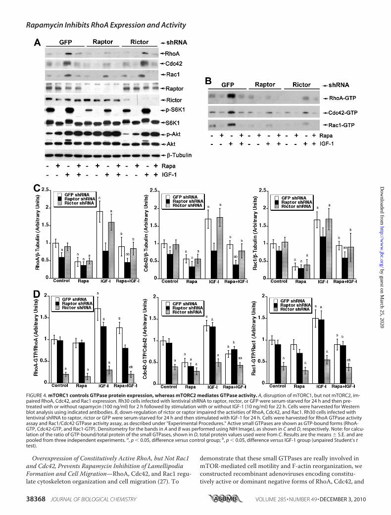
Activities, but Only mTORC1 Controls Their Cellular ProteinExpression—Because mTORC2, which was originally thoughtto be rapamycin-insensitive, has recently been found to besensitive to prolonged (�24 h) rapamycin treatment (17), wenext determined which of the mTOR complexes is responsi-ble for rapamycin inhibition of small GTPase expression. Tothis end, mTORC1 and mTORC2 were disrupted, respec-tively, by knocking down raptor or rictor in cultured cells us-ing corresponding lentiviral shRNAs. As expected, down-reg-ulation of raptor and rictor inhibited phosphorylation ofmTORC1-mediated S6K1 (Thr-389) and mTORC2-mediatedAkt (Ser-473), respectively (Fig. 4A). However, only down-
regulation of raptor, but rictor, inhibited the basal or IGF-1-stimulated protein expression of RhoA, Cdc42, and Rac1 (Fig.4, A and C). Next we assessed the activity of these smallGTPases in Rh30 cells with disrupted mTORC1 or mTORC2.Knocking down raptor reduced the total activity of RhoA,Cdc42, or Rac1 in the cells (Fig. 4B), which was consistentwith inhibition of protein expression of the small GTPases(Fig. 4A). However, down-regulation of raptor did not signifi-cantly alter the GTP-bound form (ratio of GTP-bound/totalprotein) of RhoA, Cdc42, or Rac1 in the cells (Fig. 4D). Incontrast, knocking down rictor did not affect protein expres-sion of RhoA, Cdc42, or Rac1 (Fig. 4, A and C) but decreasedGTP-bound forms of the small GTPases sharply (Fig. 4, B andD). The results indicate that the two mTOR complexes regu-late the small GTPases through different mechanisms, i.e.mTORC1 controls protein expression of RhoA, Cdc42, and
FIGURE 1. Rapamycin inhibits IGF-1-stimulated protein expression and activities of RhoA, Cdc42, and Rac1. Indicated cells were serum-starved for24 h and then pretreated with or without rapamycin (100 ng/ml) for 2 h followed by stimulation with or without IGF-1 (10 ng/ml) for 22 h. Cells were har-vested for the GTPase activity assay (A) and Western blot analysis (B) for RhoA, Cdc42, and Rac1 as described under “Experimental Procedures.” Densitome-try for the bands in A and B was performed using NIH ImageJ, as shown in C and D, respectively. Note: for calculation of the ratio of GTP-bound/total proteinof the small GTPases, shown in C, total protein values used were from D. Results are the means � S.E. and are pooled from three independent experiments.a, p � 0.05, difference versus control group; b, p � 0.05, difference versus IGF-1 group (unpaired Student’s t test).
Rapamycin Inhibits RhoA Expression and Activity
DECEMBER 3, 2010 • VOLUME 285 • NUMBER 49 JOURNAL OF BIOLOGICAL CHEMISTRY 38365
by guest on March 25, 2020
http://ww
w.jbc.org/
Dow
nloaded from

FIGURE 2. Rapamycin inhibits IGF-1-stimulated cellular protein expression of RhoA, Cdc42, and Rac1 by suppressing their protein synthesis.A, Rh30 cells were serum-starved for 24 h and then pretreated with or without rapamycin (100 ng/ml) for 2 h followed by stimulation with or without IGF-1(10 ng/ml) for 22 h. Cells were harvested for semiquantitative RT-PCR. PCR products were separated on 1% agarose gel and stained with ethidium bromide.Bands were visualized under UV light and photographed with a digital camera. The effects of rapamycin on protein synthesis (B) and degradation (E) inRh30 cells were determined as described under “Experimental Procedures.” Semiquantitative data for A, B and E by densitometry using NIH ImageJ areshown in C, D and F, respectively. Results are the means � S.E. and are pooled from three independent experiments. a, p � 0.05, difference versus controlgroup; b, p � 0.05, difference versus IGF-1 group (unpaired Student’s t test). CHX, cycloheximide.
Rapamycin Inhibits RhoA Expression and Activity
38366 JOURNAL OF BIOLOGICAL CHEMISTRY VOLUME 285 • NUMBER 49 • DECEMBER 3, 2010
by guest on March 25, 2020
http://ww
w.jbc.org/
Dow
nloaded from

Rac1, and mTORC2 mediates the activities of these smallGTPases.Both mTORC1-mediated 4E-BP1 and S6K1 Pathways Are
Involved in Rapamycin Inhibition of RhoA, Cdc42, and Rac1Expression—Our recent data indicate that rapamycin sup-presses IGF-1-stimulated F-actin reorganization and migra-tion, in part through inhibition of S6K1-mediated phos-phorylation of focal adhesion proteins (FAK, paxillin, andp130Cas) (25). It is not clear how the 4E-BP1 pathway regu-lates cell motility and F-actin reorganization. Because 4E-BP1/eIF4E pathway is crucial for protein synthesis (1), wehypothesized that this pathway may regulate cell motilityby controlling protein synthesis of the small GTPases. Totest this hypothesis, Rh30 cells were infected with recombi-nant adenoviruses expressing HA-tagged constitutivelyhypophosphorylated 4E-BP1 (4EBP1-5A) and Ad-GFP (ascontrol). 4EBP1-5A expressed in Rh30 cells, as evidenced byWestern blot analysis of tagged HA and elevated protein lev-els of 4E-BP1 (Fig. 5A). As expected, forced expression of
4EBP1-5A, which tightly binds eIF4E and suppresses its activ-ity (24), substantially down-regulated the basal or IGF-1-stim-ulated protein levels of RhoA, Cdc42, and Rac1 in the cells(Fig. 5, A and D).As the S6K1 pathway is also important for protein syn-
thesis (1), next we investigated whether the S6K pathwaycontrols protein synthesis of the small GTPases as well. Asshown in Fig. 5, B and E, lentiviral shRNA to S6K1, but notto GFP (control), down-regulated protein expression ofS6K1 by �90%. Similar to forced expression of 4EBP1-5A,down-regulation of S6K1 also suppressed expression ofRhoA, Cdc42, and Rac1. In contrast, cells expressing con-stitutively active and rapamycin-resistant S6K1 (S6K1-ca),but not GFP (control), showed partial resistance to rapa-mycin inhibition of expression of the small GTPases (Fig.5, C and F). Collectively, our results suggest that both 4E-BP1 and S6K1 pathways are essential for mTORC1-regu-lated RhoA, Cdc42, and Rac1 expression, mediating cellmotility.
FIGURE 3. Inhibition of mTOR kinase activity is necessary for rapamycin inhibition of GTPase expression. Rh30 cells infected with recombinant adeno-virus expressing GFP or FLAG-tagged mTOR-T and mTOR-TE mutants were serum-starved for 24 h and then pretreated with or without rapamycin (100 ng/ml) for 2 h followed by stimulation with IGF-1 (10 ng/ml) for 22 h. Cells were harvested for Western blot analysis using indicated antibodies (A). Semiquanti-tative data for A by densitometry using NIH ImageJ are shown in B. Results are the means � S.E. and are pooled from three independent experiments. a,p �0.05, difference versus control group; b, p � 0.05, difference versus IGF-1 group (unpaired Student’s t test).
Rapamycin Inhibits RhoA Expression and Activity
DECEMBER 3, 2010 • VOLUME 285 • NUMBER 49 JOURNAL OF BIOLOGICAL CHEMISTRY 38367
by guest on March 25, 2020
http://ww
w.jbc.org/
Dow
nloaded from
Overexpression of Constitutively Active RhoA, but Not Rac1and Cdc42, Prevents Rapamycin Inhibition of LamellipodiaFormation and Cell Migration—RhoA, Cdc42, and Rac1 regu-late cytoskeleton organization and cell migration (27). To
demonstrate that these small GTPases are really involved inmTOR-mediated cell motility and F-actin reorganization, weconstructed recombinant adenoviruses encoding constitu-tively active or dominant negative forms of RhoA, Cdc42, and
FIGURE 4. mTORC1 controls GTPase protein expression, whereas mTORC2 mediates GTPase activity. A, disruption of mTORC1, but not mTORC2, im-paired RhoA, Cdc42, and Rac1 expression. Rh30 cells infected with lentiviral shRNA to raptor, rector, or GFP were serum-starved for 24 h and then pre-treated with or without rapamycin (100 ng/ml) for 2 h followed by stimulation with or without IGF-1 (10 ng/ml) for 22 h. Cells were harvested for Westernblot analysis using indicated antibodies. B, down-regulation of rictor or raptor impaired the activities of RhoA, Cdc42, and Rac1. Rh30 cells infected withlentiviral shRNA to raptor, rictor or GFP were serum-starved for 24 h and then stimulated with IGF-1 for 24 h. Cells were harvested for RhoA GTPase activityassay and Rac1/Cdc42 GTPase activity assay, as described under “Experimental Procedures.” Active small GTPases are shown as GTP-bound forms (RhoA-GTP, Cdc42-GTP, and Rac1-GTP). Densitometry for the bands in A and B was performed using NIH ImageJ, as shown in C and D, respectively. Note: for calcu-lation of the ratio of GTP-bound/total protein of the small GTPases, shown in D, total protein values used were from C. Results are the means � S.E. and arepooled from three independent experiments. a, p � 0.05, difference versus control group; b, p � 0.05, difference versus IGF-1 group (unpaired Student’s ttest).
Rapamycin Inhibits RhoA Expression and Activity
38368 JOURNAL OF BIOLOGICAL CHEMISTRY VOLUME 285 • NUMBER 49 • DECEMBER 3, 2010
by guest on March 25, 2020
http://ww
w.jbc.org/
Dow
nloaded from

FIGURE 5. Both 4E-BP1 and S6K1 pathways are involved in rapamycin inhibition of RhoA, Cdc42, and Rac1 expression. Rh30 cells infected with re-combinant adenovirus expressing GFP or constitutively hypophosphorylated 4EBP1-5A (A) with lentiviral shRNA to GFP or S6K1 (B) or with recombinantadenovirus expressing GFP, wild type S6K1 (S6K1-wt), or constitutively active S6K1 (S6K1-ca) (C) were serum-starved for 24 h and then pretreated with orwithout rapamycin (100 ng/ml) for 2 h followed by stimulation with or without IGF-1 (10 ng/ml) for 22 h. Cells were harvested for Western blot analysis us-ing indicated antibodies. Semiquantitative data for A–C by densitometry using NIH ImageJ are shown in D–F, respectively. Results are the means � S.E. andare pooled from three independent experiments. a, p � 0.05, difference versus control group; b, p � 0.05, difference versus IGF-1 group (unpaired Student’s ttest).
Rapamycin Inhibits RhoA Expression and Activity
DECEMBER 3, 2010 • VOLUME 285 • NUMBER 49 JOURNAL OF BIOLOGICAL CHEMISTRY 38369
by guest on March 25, 2020
http://ww
w.jbc.org/
Dow
nloaded from
Rapamycin Inhibits RhoA Expression and Activity
38370 JOURNAL OF BIOLOGICAL CHEMISTRY VOLUME 285 • NUMBER 49 • DECEMBER 3, 2010
by guest on March 25, 2020
http://ww
w.jbc.org/
Dow
nloaded from

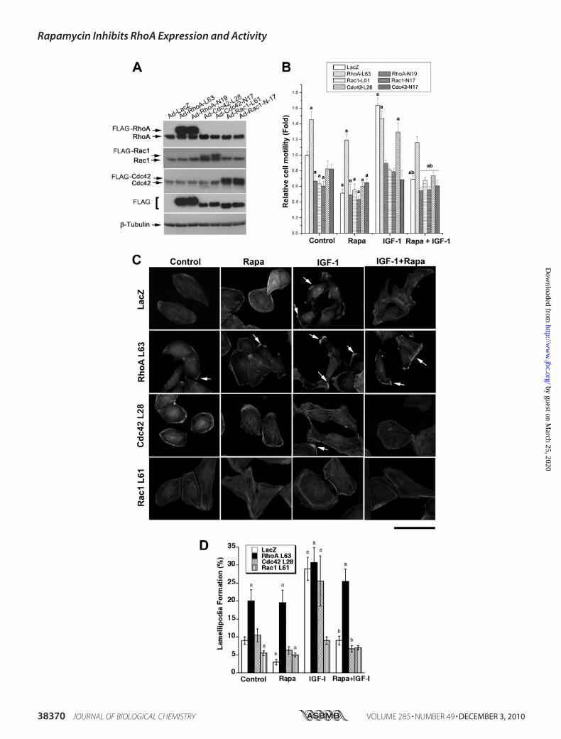
Rac1. Rh30 cells infected with these adenoviruses weretreated with or without rapamycin with and without IGF-1followed by cell motility assays and F-actin staining. As shownin Fig. 6A, considerable levels of constitutively active RhoA(RhoA-L63), Cdc42 (Cdc42-L28), and Rac1 (Rac1-L61) anddominant negative RhoA (RhoA-N19), Cdc42 (Cdc42-N17),and Rac1 (Rac1-N17) were, respectively, expressed in Rh30cells, as detected by Western blotting using antibodies toFLAG and individual GTPases. Expression of RhoA-L63 mim-icked the effect of IGF-1 stimulation and increased the basalcell motility by �150% and conferred resistance to rapamycininhibition of the basal and IGF-1-stimulated cell motility (Fig.6B). To our surprise, expression of Rac1-L61 and Cdc42-L28inhibited the basal cell motility by �35 and 20%, respectively,and did not render significant resistance to rapamycin inhibi-tion of the basal or IGF-1-stimulated cell motility. Further-more, IGF-1 was able to stimulate the motility of cells ex-pressing RhoA-L63 and Cdc42-L28 but not Rac1-L61 (Fig.6B). In addition, expression of RhoA-N19, Cdc42-N17, orRac1-N17 not only inhibited the basal cell motility, but alsosuppressed the IGF-1-stimulated cell motility.Consistent with the above findings, expression of RhoA-
L63 not only increased the lamellipodia formation even in theabsence of IGF-1 but also conferred resistance to rapamycininhibition of IGF-1 stimulated lamellipodia formation (Fig. 6,C and D). Expression of RhoA-N19 mimicked the effect ofrapamycin, blocking IGF-1 stimulated lamellipodia formation(data not shown). Expression of Cdc42-L28 had no apparenteffect on either IGF-1-stimulated or rapamycin-inhibited la-mellipodia formation (Fig. 6, C and D). Expression of Rac1-L61 inhibited IGF-1-stimulated lamellipodia formation andnoticeably increased the cellular stress fibers (Fig. 6, C and D),suggesting a distinct role of Rac1 in regulating cytoskeletonorganization in the cells. The data indicate that rapamycinmay target RhoA, inhibiting IGF-1-stimulated F-actin reorga-nization and cell motility.
DISCUSSION
Recently we demonstrated that rapamycin inhibits F-actionreorganization and cell motility by inhibition of mTOR kinaseactivity (24, 25). mTOR controls synthesis of a variety of pro-teins, such as cyclin D1, c-Myc, ornithine decarboxylase, vas-cular endothelial growth factor, etc. (43). Small GTPases(RhoA, Rac1, and Cdc42) are crucial for cytoskeleton organi-zation and cell migration (27). Therefore, we hypothesizedthat rapamycin may inhibit mTOR-mediated protein synthe-sis or activities of the small GTPases, leading to inhibition ofF-action reorganization and cell motility. To test this hypoth-esis, we first studied the effect of rapamycin on cellular pro-tein levels and activities of RhoA, Rac1, and Cdc42. By West-
ern blotting and small GTPase activity assay, we found thatrapamycin did inhibit the basal and IGF-1-stimulated activi-ties and protein expression of RhoA, Rac1, and Cdc42 in Rh30cells. The inhibitory effect of rapamycin on expression of thesmall GTPases was also observed in other tumor cell lines,including those derived from cervical cancer (HeLa), prostatecancer (PC-3) (Fig. 1), Ewing sarcoma (Rh1), and glioblastoma(U-373) (data not shown), suggesting that this is not celltype-dependent.It is well known that rapamycin selectively inhibits mTOR
kinase activity and function (1). However, studies have alsoshown that rapamycin inhibits differentiation of C2C12 cellsin mTOR kinase activity-independent manner (44), althoughthis remains controversial (45). During skeletal myogenesis,mTOR regulates the production of IGF-II mRNA, which isalso independent of the kinase activity of mTOR (41). Thisprompted us to study whether rapamycin inhibition of smallGTPase expression is through inhibition of mTOR kinaseactivity. Here we found that rapamycin failed to inhibit RhoA,Cdc42, and Rac1 expression in cells expressing rapamycin-resistant mTOR (mTOR-T) but not in control cells expressingGFP or rapamycin-resistant but kinase dead mTOR (mTOR-TE), suggesting that the kinase activity of mTOR is essentialfor expression of RhoA, Cdc42, and Rac1. This is consistentwith our previous finding that the kinase activity of mTOR isnecessary for IGF-1-stimulated F-actin reorganization andcell motility (24, 25).mTOR may control protein expression at transcriptional,
translational, or post-translational levels (40). In this study wefound that rapamycin did not alter mRNA levels or proteinturnover of RhoA, Cdc42, and Rac1 but inhibited protein syn-thesis of these small GTPases, suggesting that mTOR controlsprotein expression of RhoA, Cdc42, and Rac1 at translationallevel.Previous studies only showed that mTORC2 regulates the
activities of RhoA and Rac1 in NIH 3T3, HeLa cells, and hu-man umbilical vein endothelial cells (11, 18, 26). Here for thefirst time we demonstrate that mTOR not only controls thecellular activities of RhoA, Rac1, and Cdc42 but also regulatesthe cellular protein expression of these small GTPases. Ourdata support the notion that mTORC1 mediates protein syn-thesis of RhoA, Cdc42, and Rac1; mTORC2 regulates the ac-tivities of these proteins. This is supported by the findingsthat disruption of mTORC1 by down-regulation of raptorinhibited the expression and activities of the small GTPases,whereas disruption of mTORC2 by down-regulation of rictoronly inhibited the activities of RhoA, Cdc42, and Rac1. This isconsistent with the previous findings that a 20–30% decreasein GTP-bound Rac1 was observed in mTOR-, mLST8-, or
FIGURE 6. Overexpression of constitutively active RhoA, but not Cdc42 or Rac1, prevents rapamycin inhibition of IGF-1-stimulated lamellipodiaformation and cell migration. Rh30 cells were infected with recombinant adenovirus expressing FLAG-tagged constitutively active or dominant negativeRhoA (RhoA-L63 or RhoA-N19), Cdc42 (Cdc42-L28 or Cdc42-N17), and Rac1 (Rac1-L61 or Rac1-N17) as well as control adenovirus expressing LacZ for 24 h.Infected cells were used for Western blot analysis (A), wound healing assay (B) and F-actin staining (C). A, Western blots show expression of correspondingGTPase mutants. B, wound healing assay results are the means � S.E. and are pooled from three independent experiments. a, p � 0.05 (unpaired Student’s ttest). C, F-actin staining was visualized and photographed with a Nikon TE300 digital inverted microscope. Representative images are shown. Note: arrowsindicate lamellipodia formation. Bar � 40 �m. Quantitative results for lamellipodia formation are shown in D as the means � S.E. (n � 3). a, p � 0.05 differ-ence versus control group; b, p � 0.05, difference versus IGF-1 group (unpaired Student’s t test).
Rapamycin Inhibits RhoA Expression and Activity
DECEMBER 3, 2010 • VOLUME 285 • NUMBER 49 JOURNAL OF BIOLOGICAL CHEMISTRY 38371
by guest on March 25, 2020
http://ww
w.jbc.org/
Dow
nloaded from

mAVO3-siRNA-transfected NIH 3T3 cells and little-to-nodecrease in GTP-bound Rac1 in raptor siRNA-transfectedcells (11). Recent studies have revealed that mTORC2 regu-lates Rac1 activation through P-Rex1 (26). How mTORC2regulates RhoA and Cdc42 activity remains to be determined.4E-BP1 and S6K1 are the best characterized mTORC1 ef-
fectors, which were found to be involved in the regulation ofF-actin reorganization and cell motility (24, 25). Recently weobserved that only S6K1 pathway regulated phosphorylationof the focal adhesion proteins (FAK, paxillin, and p130Cas)(25), which is related to F-actin reorganization and cell migra-tion (46). It was not clear how 4E-BP1/eIF4E pathway regu-lates F-actin reorganization and cell motility. To address thisquestion, we used 4EBP1-5A, which is a constitutively hy-pophosphorylated mutant 4E-BP1 (T36A, T45A, S64A, T69A,and S82A) (47) and can tightly binds and sequester eIF4E,inhibiting Cap-dependent translation (47). We found thatexpression of 4EBP1-5A mimicked the effect of rapamycin,remarkably inhibiting expression of RhoA, Cdc42, and Rac1in Rh30 cells stimulated with or without IGF-1. We also in-vestigated the role of S6K1 pathway in the regulation of theGTPase expression. Interestingly, down-regulation of S6K1also impaired the expression of RhoA, Cdc42, and Rac1. Incontrast, expression of a rapamycin-resistant and constitu-tively active of S6K1 mutant (S6K1-F5A-E389-R3A, S6K1-ca)(24, 48) was able to confer partial resistance to rapamycin in-hibition of the GTPase expression. Collectively, we concludethat both 4E-BP1 and S6K1 pathways participate in the regu-lation of expression of RhoA, Cdc42, and Rac1.RhoA, Cdc42, and Rac1 are all critical molecules for cy-
toskeleton organization and cell migration, but they play dif-ferent roles during cytoskeletal dynamics (27, 28). Our dataindicate that although the activities and expression of RhoA,Cdc42, and Rac1 were inhibited by rapamycin, only overex-pression of constitutively active RhoA (RhoA-L63), but notCdc42 (Cdc42-L28) and Rac1 (Rac1-L61), prevented rapamy-cin inhibition of IGF-1 stimulated lamellipodia formation andcell motility, implying a crucial role of RhoA in mTOR-medi-ated cell motility. This is strongly supported by our observa-tions that expression of constitutively active RhoA enhancedthe basal level of lamellipodia formation and cell motility to alevel stimulated by IGF-1, whereas expression of dominantnegative RhoA (RhoA-N19) blocked the effect of IGF-1 stim-ulation. Interestingly, although expression of constitutivelyactive Cdc42 or Rac1 failed to rescue rapamycin inhibition ofIGF-1 stimulated F-actin reorganization and cell migration,expression of dominant negative Cdc42 (Cdc42-N17) or Rac1(Rac1-N17), like dominant negative RhoA (RhoA N19), abol-ished IGF-1-stimulated cell motility. These results suggestthat a certain level of RhoA, Cdc42, or Rac1 activity may berequired for either cell polarization/protrusion or adhesion/de-adhesion, leading to cell migration. In this study we alsonoticed that expression of constitutively active Rac1 actuallyincreased the cellular stress fibers and inhibited IGF-1-stimu-lated lamellipodia formation and cell motility in Rh30 cells(Fig. 6). This is in contrast to the finding in NIH 3T3 cells inwhich expression of constitutively active Rac1 induces forma-tion of membrane ruffles and lamellipodia, whereas expres-
sion of constitutively active RhoA results in formation ofstress fibers (11). The discrepancy may likely be due to differ-ent cell types used. It has been described that Rac1 is essentialfor actin stress fiber formation in primary mouse embryonicfibroblasts (49). In colon carcinoma cells (50), hepatocarci-noma cells (51), and human microvascular endothelial cells(52), activation of RhoA promotes lamellipodia formation andcell migration. Therefore, the roles of the small GTPases inthe regulation of F-actin reorganization and cell motility de-pend on cell types studied.In summary, here we found that rapamycin inhibited RhoA,
Cdc42, and Rac1 expression and activity in an mTOR kinaseactivity-dependent manner. mTORC1 controls the proteinsynthesis, whereas mTORC2 regulates the activities of thesmall GTPases. Both mTORC1-mediated S6K1 and 4E-BP1/eIF4E pathways are essential for RhoA, Cdc42, and Rac1 ex-pression. However, inhibition of RhoA activity is primarilyresponsible for rapamycin inhibition of IGF-1-stimulated la-mellipodia formation and cell motility.
Acknowledgments—We thank Drs. John Blenis, Jie Chen, Peter J.Houghton, John Lawrence, Jr., David M. Sabatini, and Yi Zheng forproviding cell lines or constructs.
REFERENCES1. Guertin, D. A., and Sabatini, D. M. (2007) Cancer Cell 12, 9–222. Fonseca, B. D., Smith, E. M., Lee, V. H., MacKintosh, C., and Proud,
C. G. (2007) J. Biol. Chem. 282, 24514–245243. Hara, K., Maruki, Y., Long, X., Yoshino, K., Oshiro, N., Hidayat, S., To-
kunaga, C., Avruch, J., and Yonezawa, K. (2002) Cell 110, 177–1894. Kim, D. H., Sarbassov, D. D., Ali, S. M., King, J. E., Latek, R. R., Erdju-
ment-Bromage, H., Tempst, P., and Sabatini, D. M. (2002) Cell 110,163–175
5. Kim, D. H., Sarbassov, D. D., Ali, S. M., Latek, R. R., Guntur, K. V., Erd-jument-Bromage, H., Tempst, P., and Sabatini, D. M. (2003)Mol. Cell11, 895–904
6. Loewith, R., Jacinto, E., Wullschleger, S., Lorberg, A., Crespo, J. L.,Bonenfant, D., Oppliger, W., Jenoe, P., and Hall, M. N. (2002)Mol. Cell10, 457–468
7. Sancak, Y., Thoreen, C. C., Peterson, T. R., Lindquist, R. A., Kang, S. A.,Spooner, E., Carr, S. A., and Sabatini, D. M. (2007)Mol. Cell 25,903–915
8. Vander Haar, E., Lee, S. I., Bandhakavi, S., Griffin, T. J., and Kim, D. H.(2007) Nat. Cell Biol. 9, 316–323
9. Fasolo, A., and Sessa, C. (2008) Expert Opin. Investig. Drugs 17,1717–1734
10. Frias, M. A., Thoreen, C. C., Jaffe, J. D., Schroder, W., Sculley, T., Carr,S. A., and Sabatini, D. M. (2006) Curr. Biol. 16, 1865–1870
11. Jacinto, E., Loewith, R., Schmidt, A., Lin, S., Ruegg, M. A., Hall, A., andHall, M. N. (2004) Nat. Cell Biol. 6, 1122–1128
12. Jacinto, E., Facchinetti, V., Liu, D., Soto, N., Wei, S., Jung, S. Y., Huang,Q., Qin, J., and Su, B. (2006) Cell 127, 125–137
13. Pearce, L. R., Huang, X., Boudeau, J., Pawłowski, R., Wullschleger, S.,Deak, M., Ibrahim, A. F., Gourlay, R., Magnuson, M. A., and Alessi, D. R.(2007) Biochem. J. 405, 513–522
14. Sarbassov, D. D., Ali, S. M., Kim, D. H., Guertin, D. A., Latek, R. R., Erd-jument-Bromage, H., Tempst, P., and Sabatini, D. M. (2004) Curr. Biol.14, 1296–1302
15. Yang, Q., Inoki, K., Ikenoue, T., and Guan, K. L. (2006) Genes Dev. 20,2820–2832
16. Woo, S. Y., Kim, D. H., Jun, C. B., Kim, Y. M., Haar, E. V., Lee, S. I.,Hegg, J. W., Bandhakavi, S., Griffin, T. J., and Kim, D. H. (2007) J. Biol.Chem. 282, 25604–25612
Rapamycin Inhibits RhoA Expression and Activity
38372 JOURNAL OF BIOLOGICAL CHEMISTRY VOLUME 285 • NUMBER 49 • DECEMBER 3, 2010
by guest on March 25, 2020
http://ww
w.jbc.org/
Dow
nloaded from

17. Sarbassov, D. D., Guertin, D. A., Ali, S. M., and Sabatini, D. M. (2005)Science 307, 1098–1101
18. Dada, S., Demartines, N., and Dormond, O. (2008) Biochem. Biophys.Res. Commun. 372, 875–879
19. Facchinetti, V., Ouyang, W., Wei, H., Soto, N., Lazorchak, A., Gould, C.,Lowry, C., Newton, A. C., Mao, Y., Miao, R. Q., Sessa, W. C., Qin, J.,Zhang, P., Su, B., and Jacinto, E. (2008) EMBO J. 27, 1932–1943
20. Ikenoue, T., Inoki, K., Yang, Q., Zhou, X., and Guan, K. L. (2008) EMBOJ. 27, 1919–1931
21. García-Martínez, J. M., and Alessi, D. R. (2008) Biochem. J. 416,375–385
22. Hong, F., Larrea, M. D., Doughty, C., Kwiatkowski, D. J., Squillace, R.,and Slingerland, J. M. (2008)Mol. Cell 30, 701–711
23. Peterson, T. R., Laplante, M., Thoreen, C. C., Sancak, Y., Kang, S. A.,Kuehl, W. M., Gray, N. S., and Sabatini, D. M. (2009) Cell 137, 873–886
24. Liu, L., Chen, L., Chung, J., and Huang, S. (2008) Oncogene 27,4998–5010
25. Liu, L., Li, F., Cardelli, J. A., Martin, K. A., Blenis, J., and Huang, S. (2006)Oncogene 25, 7029–7040
26. Hernandez-Negrete, I., Carretero-Ortega, J., Rosenfeldt, H., Hernandez-García, R., Calderon-Salinas, J. V., Reyes-Cruz, G., Gutkind, J. S., andVazquez-Prado, J. (2007) J. Biol. Chem. 282, 23708–23715
27. Heasman, S. J., and Ridley, A. J. (2008) Nat. Rev. Mol. Cell Biol. 9,690–701
28. Tzima, E. (2006) Circ. Res. 98, 176–18529. Kurokawa, K., and Matsuda, M. (2005)Mol. Biol. Cell 16, 4294–430330. Pertz, O., Hodgson, L., Klemke, R. L., and Hahn, K. M. (2006) Nature
440, 1069–107231. Guerin, P., Sauzeau, V., Rolli-Derkinderen, M., Al Habbash, O., Scalbert,
E., Crochet, D., Pacaud, P., and Loirand, G. (2005) J. Vasc. Res. 42, 21–2832. Vilella-Bach, M., Nuzzi, P., Fang, Y., and Chen, J. (1999) J. Biol. Chem.
274, 4266–427233. He, T. C., Zhou, S., da Costa, L. T., Yu, J., Kinzler, K. W., and Vogelstein,
B. (1998) Proc. Natl. Acad. Sci. U.S.A. 95, 2509–251434. Guo, F., Gao, Y., Wang, L., and Zheng, Y. (2003) J. Biol. Chem. 278,
14414–1441935. Guo, F., and Zheng, Y. (2004)Mol. Cell. Biol. 24, 1426–143836. Sunavala-Dossabhoy, G., Fowler, M., and De Benedetti, A. (2004) BMC
Mol. Biol. 5, 137. Mayer, C., and Grummt, I. (2006) Oncogene 25, 6384–639138. Peng, T., Golub, T. R., and Sabatini, D. M. (2002)Mol. Cell. Biol. 22,
5575–558439. Tsang, C. K., and Zheng, X. F. (2007) Cell Cycle 6, 25–2940. Hashemolhosseini, S., Nagamine, Y., Morley, S. J., Desrivieres, S., Mer-
cep, L., and Ferrari, S. (1998) J. Biol. Chem. 273, 14424–1442941. Erbay, E., Park, I. H., Nuzzi, P. D., Schoenherr, C. J., and Chen, J. (2003)
J. Cell Biol. 163, 931–93642. McMahon, L. P., Choi, K. M., Lin, T. A., Abraham, R. T., and Lawrence,
J. C., Jr. (2002)Mol. Cell. Biol. 22, 7428–743843. Huang, S., and Houghton, P. J. (2003) Curr. Opin. Pharmacol. 3,
371–37744. Erbay, E., and Chen, J. (2001) J. Biol. Chem. 276, 36079–3608245. Shu, L., Zhang, X., and Houghton, P. J. (2002) J. Biol. Chem. 277,
16726–1673246. Mitra, S. K., and Schlaepfer, D. D. (2006) Curr. Opin. Cell Biol. 18,
516–52347. Mothe-Satney, I., Brunn, G. J., McMahon, L. P., Capaldo, C. T., Abra-
ham, R. T., and Lawrence, J. C., Jr. (2000) J. Biol. Chem. 275,33836–33843
48. Schalm, S. S., Tee, A. R., and Blenis, J. (2005) J. Biol. Chem. 280,11101–11106
49. Guo, F., Debidda, M., Yang, L., Williams, D. A., and Zheng, Y. (2006)J. Biol. Chem. 281, 18652–18659
50. O’Connor, K. L., Nguyen, B. K., and Mercurio, A. M. (2000) J. Cell Biol.148, 253–258
51. Genda, T., Sakamoto, M., Ichida, T., Asakura, H., Kojiro, M., Narumiya,S., and Hirohashi, S. (1999) Hepatology 30, 1027–1036
52. Abecassis, I., Olofsson, B., Schmid, M., Zalcman, G., and Karniguian, A.(2003) Exp. Cell Res. 291, 363–376
Rapamycin Inhibits RhoA Expression and Activity
DECEMBER 3, 2010 • VOLUME 285 • NUMBER 49 JOURNAL OF BIOLOGICAL CHEMISTRY 38373
by guest on March 25, 2020
http://ww
w.jbc.org/
Dow
nloaded from

Xiuzhen Han and Shile HuangLei Liu, Yan Luo, Long Chen, Tao Shen, Baoshan Xu, Wenxing Chen, Hongyu Zhou,
RhoA Expression and ActivityRapamycin Inhibits Cytoskeleton Reorganization and Cell Motility by Suppressing
doi: 10.1074/jbc.M110.141168 originally published online October 11, 20102010, 285:38362-38373.J. Biol. Chem.
10.1074/jbc.M110.141168Access the most updated version of this article at doi:
Alerts:
When a correction for this article is posted•
When this article is cited•
to choose from all of JBC's e-mail alertsClick here
http://www.jbc.org/content/285/49/38362.full.html#ref-list-1
This article cites 52 references, 23 of which can be accessed free at
by guest on March 25, 2020
http://ww
w.jbc.org/
Dow
nloaded from