rapiscan : epar - public assessment report - european medicines
TRANSCRIPT

7 Westferry Circus ● Canary Wharf ● London E14 4HB ● United Kingdom Telephone +44 (0)20 7418 8400 Facsimile +44 (0)20 7418 8416 E-mail [email protected] Website www.ema.europa.eu An agency of the European Union
© European Medicines Agency, 2010. Reproduction is authorised provided the source is acknowledged.
ASSESSMENT REPORT FOR
Rapiscan
International Nonproprietary Name: regadenoson
Procedure No. EMEA/H/C/ 1176
Assessment Report as adopted by the CHMP with
all information of a commercially confidential nature deleted.

Table of contents Page
Table of contents ................................................................................... 2 Page
1. Background information on the procedure .............................................. 3 1.1. Submission of the dossier.................................................................................... 3 1.1.1. Information on paediatric requirements .............................................................. 3 1.1.2. Licensing status: ............................................................................................. 3 1.2. Steps taken for the assessment of the product ....................................................... 3
2. Scientific discussion ................................................................................ 4 2.1. Introduction ...................................................................................................... 4 2.2. Quality aspects .................................................................................................. 5 2.2.1. Introduction ................................................................................................... 5 2.2.2. Active substance ............................................................................................. 5 2.2.3. Finished Medicinal Product ................................................................................ 7 2.2.4. Discussion on chemical, pharmaceutical and biological aspects............................... 8 2.2.5. Conclusions on the chemical, pharmaceutical and biological aspects ....................... 9 2.3. Non-clinical aspects ............................................................................................ 9 2.3.1. Introduction ................................................................................................... 9 2.3.2. Pharmacology ................................................................................................. 9 2.3.3. Pharmacokinetics .......................................................................................... 10 2.3.4. Toxicology.................................................................................................... 11 2.3.5. Ecotoxicity/environmental risk assessment........................................................ 13 2.3.6. Discussion on non-clinical aspects.................................................................... 13 2.3.7. Conclusion on the non-clinical aspects .............................................................. 14 2.4. Clinical aspects ................................................................................................ 14 2.4.1. Introduction ................................................................................................. 14 2.4.2. GCP............................................................................................................. 14 2.4.3. Pharmacokinetics .......................................................................................... 15 2.4.4. Pharmacodynamics........................................................................................ 17 2.4.5. Discussion on clinical pharmacology ................................................................. 18 2.4.6. Conclusions on clinical pharmacology ............................................................... 18 2.4.7. Clinical efficacy ............................................................................................. 19 2.4.8. Clinical safety ............................................................................................... 28 2.5. Pharmacovigilance............................................................................................ 34 2.5.1. Detailed description of the pharmacovigilance system......................................... 34 2.5.2. Risk management plan................................................................................... 34 2.6. Benefit-risk balance.......................................................................................... 42 2.7. Recommendation ............................................................................................. 43
EMA/455580/2010 Page 2/43

1. Background information on the procedure
1.1. Submission of the dossier
The original applicant CV Therapeutics submitted on 6 May 2009 an application for Marketing
Authorisation to the European Medicines Agency for Rapiscan, through the centralised procedure under
Article 3 (2)(a) of Regulation (EC) No 726/2004. The eligibility to the centralised procedure was agreed
upon by the Agency/CHMP on 25 September 2008. On 12 August 2009, the EMA was notified that
following the acquisition of CV Therapeutics by Gilead Sciences International Ltd. the applicant
changed to Gilead Sciences International Ltd.
The legal basis for this application refers to Article 8.3 of Directive 2001/83/EC, as amended -
complete and independent application.
The application submitted is a complete dossier composed of administrative information, complete
quality data, non-clinical and clinical data based on applicants’ own tests and studies and/or
bibliographic literature substituting/supporting certain tests or studies.
1.1.1. Information on paediatric requirements
Pursuant to Article 7, of Regulation (EC) No 1901/2006 the application included an Agency Decision
P/82/2009 for the following condition:
Myocardial perfusion disturbances
on the agreement of a paediatric investigation plan (PIP) and granting a deferral.
The PIP is not yet completed.
1.1.2. Licensing status:
Rapiscan has been given a Marketing Authorisation in the United States of America on 10 April 2008.
The Rapporteur and Co-Rapporteur appointed by the CHMP and the evaluation teams were:
Rapporteur: Ian Hudson Co-Rapporteur: Concepcion Prieto Yerro
1.2. Steps taken for the assessment of the product
The application was received by the Agency on 6 May 2009.
The procedure started on 27 May 2009.
The Rapporteur's first Assessment Report was circulated to all CHMP members on 13 August 2009.
The Co-Rapporteur's first Assessment Report was circulated to all CHMP members on 20 August
2009.
During the meeting on 20-24 September 2010 the CHMP agreed on the consolidated List of
Questions to be sent to the applicant. The final consolidated List of Questions was sent to the
applicant on 25 September 2009.
The applicant submitted the responses to the CHMP consolidated List of Questions on 19 February
2010.
EMA/455580/2010 Page 3/43

The summary report of the inspection carried out in the USA on 22 October 2009 was issued on 12
April 2010.
The Rapporteurs circulated the Joint Assessment Report on the applicant’s responses to the List of
Questions to all CHMP members on 12 April 2010.
During the CHMP meeting on 19-22 April 2010, the CHMP agreed on a list of outstanding issues to
be addressed in writing by the applicant.
The applicant submitted the responses to the CHMP consolidated List of outstanding issues on 21
May 2010.
The Rapporteurs circulated the Assessment Report on the applicant’s responses to the List of
outstanding issues to all CHMP members on 7 June 2010.
The Rapporteurs circulated the final Assessment Report on the applicant’s responses to the List of
outstanding issues to all CHMP members on 18 June 2010.
During the meeting on 21-24 June 2010, the CHMP, in the light of the overall data submitted and
the scientific discussion within the Committee, issued a positive opinion for granting a Marketing
Authorisation to Rapiscan on 24 June 2010. The applicant provided the letter of undertaking on the
follow-up measures to be fulfilled post-authorisation on 21 June 2010.
2. Scientific discussion
2.1. Introduction
Pharmacologic Stress Agents in Radionuclide Myocardial Perfusion Imaging Single photon emission computed tomography (SPECT) myocardial perfusion imaging (MPI; also known as myocardial perfusion scintigraphy (MPS)) studies with radionuclide agents are used in the detection and functional characterization of ischemic heart disease, providing information that is incremental to and independent from that of other assessments including clinical characteristics, exercise tolerance, electrocardiography, and coronary angiography. The technique is based on the principle that radiopharmaceuticals distribute proportionally to myocardial blood flow. In an MPI study, two images are obtained: one image is obtained at rest and a second under conditions that increase coronary blood flow (CBF), such as exercise or the application of a pharmacologic stress agent (e.g., a coronary vasodilator) which simulates the increase in CBF caused by exercise. During exercise or with administration of a pharmacologic agent, there is an increase in coronary transmural blood flow and collateral redistribution of blood flow. However, in patients with significant coronary artery disease (CAD) a redistribution of CBF occurs with an increase in blood flow to normal coronaries due to vasodilatation, and a relative decrease of blood flow in stenotic arteries. This ‘coronary steal’ translates into less perfused regional myocardium area which can be imaged (detected) with radionuclide tracers. Referral of patients for MPS has increased in all European countries over the last 20 years, but with variations both between and within countries. Three radiopharmaceuticals are approved: thallium (201Tl) chloride, technetium (99mTc) tetrofosmin and technetium (99mTc) sestamibi. A major limitation of thallium (201Tl) is the high false-positive rate attributed to image attenuation artifacts. Technetium-based tracers have improved imaging characteristics of thallium (201Tl) scan. Moreover, ECG gated acquisition (gated SPECT imaging), which is easier with technetium-based agents, allows simultaneous evaluation of left ventricular perfusion and systolic function, identification of attenuation artifacts, and myocardial stunning. Adenosine is a vasodilator of the coronary and peripheral circulation. The effect of adenosine to cause coronary and possibly peripheral vasodilatation is mediated primarily by the A2A adenosine receptor (A2A-AdoR) present in the vascular wall. The sensitivity of the coronary circulation to adenosine and adenosine analogues appears to be greater than that of the peripheral vasculature. This differential sensitivity is likely, in part, due to a larger A2A spare receptor capacity of coronary vessels relative to peripheral resistance vessels. Other adenosine receptor subtypes (e.g., A2B) may play a significant role as mediators of the peripheral vasodilatation caused by adenosine and non-selective agonists. Consequently, heterogeneous receptor expression levels and their relative contributions to the
EMA/455580/2010 Page 4/43

vasodilatation caused by adenosine may account for the differential sensitivity among vascular beds to the action of adenosine and other unselective agonists. The coronary vasodilator effect of adenosine is the basis for its use, in conjunction with radionuclide MPI, to detect underperfused areas of myocardium. Dipyridamole, a nucleoside transport inhibitor, acts by limiting cellular re-uptake of adenosine thereby increasing plasma and tissue levels of adenosine. Adenosine directly, and dipyridamole indirectly, activate all four known AdoR subtypes, designated A1, A2A, A2B, and A3, which mediate a variety of responses in different tissues. Adenosine and dipyridamole are capable of causing significantly greater increases of CBF to myocardial areas perfused by normal, as opposed to stenotic, coronary arteries. Both agents cause a differential distribution of radionuclide between myocardium perfused by normal and stenotic coronary arteries, thereby permitting visualization of myocardial perfusion defects. The safety, reliability, and diagnostic accuracy of pharmacologic stress induced by adenosine and dipyridamole have led to their widespread use for detection of myocardial perfusion defects with radiopharmaceuticals in patients with known or suspected CAD, with paced ventricular rhythm or left bundle branch block and in other clinical scenarios. The current vasodilator stress agents for use in MPI, adenosine and dipyridamole, are administered as IV infusions and are associated with an overall high incidence of side effects. The ability to administer such an agent as a rapid injection would provide the added benefit of increasing the convenience of MPI procedures. Regadenoson was developed as a short-acting pharmacologic stress agent (administered as a single 400 μg iv bolus) in conjunction with radionuclide MPI/MPS because it has low yet selective affinity for the A2A-AdoR, has high potency for increasing CBF, and preferentially causes greater coronary than peripheral vasodilatation in animal models. Thus, regadenoson has the potential to selectively increase CBF, while minimizing some of the side effects caused by the currently approved pharmacologic stress agents, via the selective activation of the receptor responsible for the coronary vasodilatory effect of adenosine (i.e., the A2A-AdoR) and not the other AdoR subtypes (e.g., the A1-AdoR in the heart and the A2B-AdoR on mast cells). In addition, administration of regadenoson via IV bolus in a unit dose is more convenient for the patient and nuclear practitioner than is administration of the existing stress agents using weight-based infusion.
2.2. Quality aspects
2.2.1. Introduction
The medicinal product Rapiscan 0.4 mg/5ml solution for injection is presented as a single use glass vial containing 5 ml of a solution for injection which contains 0.4 mg of the active substance regadenoson. Other ingredients are specified in section 6.1 of the SmPC. Rapiscan is packed in a 5 ml Type I glass vial, closed with a 13 mm rubber stopper and an aluminium flip-off cap with polypropylene disk. Each vial is packaged in an individual carton box.
2.2.2. Active substance
The chemical name of the active substance regadenoson is: adenosine, 2-[4-[(methylamino)carbonyl]-1H-pyrazol-1-yl]-, monohydrate. The International Non Property Name (INN) is regadenoson. The molecular formula is C15H18N8O5.H2O and the molecular weight is 408.37 g/mol. The structural formula is shown below:
.H2O
EMA/455580/2010 Page 5/43

EMA/455580/2010 Page 6/43
Regadenoson is a white to off-white powder. It is practically insoluble in water, soluble in dimethylacetamide, slightly soluble in methanol and ethanol, and very slightly soluble in mixtures of propylene glycol and water. Regadenoson is practically insoluble (<0.1 mg/ml) in buffered aqueous solutions at pH levels between 2 and 7.5. A melting range could not be determined because regadenoson decomposes before melting. Studies on the crystal morphology of regadenoson concluded that regadenoson exists in different polymorphic forms. During the manufacturing process, the crystal monohydrate form of regadenoson is isolated. Thermogravimetric analysis and water content by Karl Fischer titration have indicated that the monohydrate is a stable form of regadenoson and is not hygroscopic. There is no Ph Eur monograph for regadenoson. The chemical structure of regadenoson has been confirmed by elemental analysis, ultraviolet (UV), Infrared (IR), 1H-nuclear magnetic resonance (NMR), 13C NMR, and mass spectra. All data are consistent with the proposed structure.
2.2.2.1.
2.2.2.2.
2.2.2.3.
Manufacture
The manufacture of regadenoson consists of a four-step process, starting from commercially available raw materials. The last step is the recrystallization of regadenoson. Detailed information on the manufacturing process, control of starting materials, reagents and solvents, control of critical steps and intermediates, process development and process validation of the active substance manufacturing have been provided by the applicant. The applicant did not use the active substance master file (ASMF) procedure. All manufacturing steps are adequately described. Adequate in-process controls are in place and appropriate specifications have been adopted for the starting materials, solvents and reagents. All relevant impurities, degradation products and residual solvents have been appropriately characterized. Validation batches results confirm the reproducibility of the manufacturing process to produce the active substance with the quality established. Regadenoson is supplied by one active substance manufacturer.
Specification
As no Ph Eur monograph exists for regadenoson, in-house specifications have been set for the active substance, in accordance with the principles of the relevant ICH guidelines. The active substance specifications include appropriate tests for appearance, identification (IR spectra), assay of regadenoson and related substances (HPLC), residue on ignition (Ph Eur), residual solvents (GC), water content (Karl Fischer), specific rotation, heavy metals, bacterial endotoxins and microbial limit (all as in Ph Eur). Since the finished product, Rapiscan, is a solution for injection, the regadenoson specification does not include the particle size acceptance criterion, which is in line with the ICH Q6A guideline. The analytical test procedures have been satisfactorily described and validated. The impurity limits are acceptable and there is no concern from the point of view of safety. Batch analysis data have been presented and confirm compliance with the predefined active substance specifications. In accordance with EU GMP guidelines1, any confirmed out of specification result, or significant negative trend, should be reported to the Rapporteur and the EMA.
Stability
Stability studies have been performed at long term (25C, 60% RH) and accelerated (40ºC, 75%RH) conditions on three registration and three production scale batches in accordance with the ICH Q1A guideline. Up to 30 months of long term stability data, and up to 6 months of accelerated stability data has been provided, confirming the stability of regadenoson. The batches tested were manufactured at the commercial manufacturing site and packaged in the packaging proposed for marketing. Supporting stability data was provided for phase 3 clinical studies batches. However, the latter batches were not manufactured at the site authorised within the Rapiscan marketing authorisation. The test parameters evaluated in these studies were appearance, water content, assay and organic impurities by HPLC, bacterial endotoxins and microbial limit. The test methods used are the same as the routine controls and are shown to be stability indicating. Forced degradation studies, under basic, acidic, oxidative, heat and humidity conditions, including a photostability study have been performed on regadenoson to identify potential degradation products that could be formed in active substance. Stress studies demonstrate the stability of the drug substance in the solid state. Since the regadenoson is terminally sterilized, a separate study was performed to generate degradation products by autoclaving a methanol/water (50:50) solution of
1 6.32 of Vol. 4 Part I of the Rules Governing Medicinal Products in the European Union

EMA/455580/2010 Page 7/43
regadenoson at 121ºC for 2 hours. Regadenoson has been shown to generate low levels of degradation products in solution at neutral pH under autoclave conditions with temperatures of ~121ºC. The photostability testing has been performed according to ICH Q1B under lighting option 2. Stability of the active substance was evaluated after light exposure to cool white fluorescent light (up to 2.4 million lux-h) and UV-A light (up to 400 watt-h/m2) at 25ºC/60% RH. The attributes monitored were description, assay and organic impurities/degradation products. The results showed that regadenoson in solid state did not degrade under the conditions tested. Based on the stability data provided, the CHMP considers that the proposed retest period is justified when the active substance is stored in the proposed packaging material under the proposed storage conditions.
2.2.3. Finished Medicinal Product
2.2.3.1.
2.2.3.2.
2.2.3.3.
2.2.3.4.
Pharmaceutical Development
The aim of the pharmaceutical development was to develop a sterile solution for injection containing 0.4 mg/5 ml regadenoson which can be administered intravenously as a rapid bolus. Due to the low aqueous solubility of regadenoson, several co-solvents and surfactants have been investigated in the development of the Rapiscan solution for injection. Propylene glycol was identified as an appropriate co-solvent to formulate Rapiscan. An in vitro biomaterial study demonstrated that the concentration of propylene glycol used in the final formulation is non-hemolytic with human whole blood. The use of this excipient in parenteral formulations is well known. Because regadenoson degrades under extreme pH conditions, Rapiscan is developed as a solution for injection with neutral pH. Considering that the active substance is present as a solution, the possible polymorphic changes of the solid regadenoson are not of concern. The selection of the excipients is based on the results of formulation screening studies. The excipients were selected for their abilities to maintain a neutral pH under stress conditions, to act as a co-solvent to prevent precipitation of regadenoson over time and to act as a chelating agent for any trace metal ions to prevent catalytic hydrolysis of regadenoson and to prevent precipitation from a possible interaction of glass vial surface with phosphorus from the phosphate buffer (EDTA). In addition, the compatibility of the selected excipients with regadenoson was investigated. The excipients used in Rapiscan solution for injection are compendial-grade pharmaceutical ingredients commonly used in intravenous medicinal products. The final Rapiscan formulation is shown to be compatible with the terminal sterilization which is proposed for the commercial production process. The results of a freeze-thaw study, performed on the finished product, confirm that no precipitate is formed when Rapiscan is exposed to low temperatures during transportation or external handling.
Adventitious agents
No materials of animal or human origin are used in the manufacture of Rapiscan. Therefore there is no BSE/TSE risk.
Manufacture of the product
The manufacturing process for Rapiscan solution for injection consists of compounding, filtration, filling, stoppering, capping, terminal sterilization, inspection, and packaging. The manufacturing process has been adequately described and validated. Critical steps have been identified and in process controls are in place. The manufacturing process demonstrates to be reproducible and provides a finished product that complies with the finished product specifications.
Product specification
The medicinal product specifications for Rapiscan at batch release include the following tests: appearance, colour, identity (UV, HPLC) and assay (HPLC) of regadenoson and impurities (HPLC), pH, particulate matter, bacterial endotoxins, sterility and volume in container. The proposed test procedures and acceptance criteria follow the principles of the relevant ICH Guidelines (Q6A, Q3C(R3), Q3B(R2) and Q2(R1) and the product specifications are considered suitable to control the quality of the finished product, manufactured with the above manufacturing process.

EMA/455580/2010 Page 8/43
All tests included in the specification have been satisfactorily described and validated. Appropriate data have been presented to justify the release specifications for each quality characteristic that is controlled. All excipients used in the formulation comply with the requirements of the European Pharmacopoeia (Ph Eur) except monobasic sodium phosphate which complies with the USP monograph. Impurities and degradation products have been evaluated and found to be acceptable from the point of view of safety. Batch analysis results comply with the proposed specification and confirm consistency & uniformity of manufacture and indicate that the process is under control. In accordance with EU GMP guidelines2, any confirmed out of specification result, or significant negative trend, should be reported to the Rapporteur and the EMA.
2.2.3.5.
Stability of the product
Stability studies have been carried out under long term (25°C/60% RH) and accelerated (40°C/75% RH) conditions according to the ICH requirements for three registration batches (the scale is 20% of the maximum commercial batch size). Thirty-six months stability data have been provided under long term conditions and six months under accelerated conditions. These batches were manufactured at the proposed site of finished product manufacture, according to the proposed process and with the active substance obtained from the registered active substance manufacturer. The batches were packaged as proposed for marketing (in a 5 mL Type I (clear) glass vial closed with a 13 mm rubber stopper and flip off overseal). The parameters tested and analytical methods used were identical to those at batch release, except from the test for identity and volume in container which are not repeated at the end of shelf-life. In addition to the stability data on registration batches, supplementary long term and accelerated stability data have been provided on one batch manufactured with regadenoson provided by another supplier, not registered in the initial Rapiscan marketing authorisation. Further supplementary stability data was provided for three phase 3 clinical trial batches. The three batches were manufactured at the proposed site of finished product manufacture and placed on stability at accelerated (40°C/75% RH) and long-term storage (25°C/60% RH) conditions. Two batches contained a slightly higher regadenoson concentration than the commercial formula, the third batch was manufactured at the proposed commercial concentration. These batches were all aseptically filled, but not terminally sterilized. All batches were manufactured at 20% of the proposed maximum commercial batch size. The active substance used for manufacturing of these batches was provided by a supplier which is not authorised in the initial Rapiscan marketing authorisation. Furthermore, a photostability study was performed on one registration batch in accordance with ICH Q1B under lighting option 2. Samples were exposed to a total of not less than 1.2 million lux-h under cool white fluorescent light and 200 Watt-h/m2 of near ultraviolet (UVA) lamp light. Light protected control samples were exposed in the same manner and tested in parallel with the light exposed samples. A comparison of the light exposed and control (light protected) samples indicates that Regadenoson Injection in 5 ml Type I clear glass vials is not light sensitive. The secondary package, therefore, does not enhance the stability of the drug product. A freeze/thaw study was conducted on one phase 3 clinical batch. The study was conducted using vials in upright and inverted orientations. Samples were stored at -20°C for 48 hours, then moved to 25°C/60% RH for two hours, then moved to 40°C/75% RH for 48 hours, then moved to 25°C/60% RH for two hours and then moved back to -20°C for another cycle. This cycle was repeated three times. The stability results presented are satisfactory and support the proposed shelf life for the commercially packaged product under the conditions specified in the SmPC.
2.2.4. Discussion on chemical, pharmaceutical and biological aspects
Information on manufacture, control and stability of the active substance and medicinal product have been presented in a satisfactory manner. The excipients are commonly used in this type of formulation and comply with Ph Eur or USP. The packaging material is commonly used and well documented. Physicochemical and biological aspects relevant to the uniform clinical performance of the product have been investigated and are controlled in a satisfactory way. Batch analysis results indicate consistency and uniformity of important product quality characteristics, and these in turn lead to the conclusion that the product should have a satisfactory and uniform performance in the clinic. Stability tests
2 6.32 of Vol. 4 Part I of the Rules Governing Medicinal Products in the European Union
EMA/455580/2010 Page 9/43
performed under ICH conditions indicate that the product is chemically stable for the proposed shelf life.
2.2.5. Conclusions on the chemical, pharmaceutical and biological aspects
The quality of Rapiscan is adequately established. Satisfactory chemical and pharmaceutical documentation has been submitted for marketing authorisation. There are no major deviations from EU and ICH requirements. The quality of this medicinal product is considered to be acceptable when used in accordance with the conditions defined in the SmPC. At the time of the CHMP opinion there are no unresolved quality issues which have a negative impact on the benefit/risk balance of the product.
2.3. Non-clinical aspects
2.3.1. Introduction
Regadenoson is a selective adenosine A2A-receptor agonist with a low affinity for the receptor, which results in a shorter duration of action than higher affinity compounds. The non-clinical development of regadenoson has been designed in accordance with EU/ICH guidelines. The in vitro safety pharmacology studies that evaluated effects on human Ether-à-go-go related gene (hERG) channels and in Purkinje fibres, as well as those studies investigating effects on respiratory and nervous systems were carried out in accordance with relevant guidance and in compliance with Good Laboratory Practice (GLP). Additional studies into the cardiovascular effects were reported under secondary pharmacodynamics and were not conducted in compliance with GLP. These non-GLP studies were not considered to compromise the scientific integrity or affect the experimental results. The pivotal toxicity studies were conducted in accordance with the relevant guidance and in compliance with GLP.
2.3.2. Pharmacology
2.3.2.1. Primary pharmacodynamic studies
In radioligand binding and functional assays in vitro regadenoson has been shown to be a selective adenosine A2A-receptor agonist with low affinity (equilibrium dissociation constant [Ki] ~1.2 μM). In in vitro studies in perfused rat and guinea pig hearts, regadenoson produced dose-dependent increases in coronary vasodilation. A large reserve of A2A-receptors was found in guinea pig heart, with activation of about 4% of the receptors by regadenoson producing half of the maximum possible response. Regadenoson dose-dependently increased coronary blood flow (CBF) in conscious dogs with an ED50 of 0.34 μg/kg and was more potent than adenosine (ED50 of 51 μg/kg), although both were equally effective, producing similar maximal increases in CBF of about 220%. The duration of the 2-fold increase in CBF observed following administration of an iv bolus of regadenoson increased with the dose administered, and with the duration of the bolus injection (10 vs. 30 sec). The increase in CBF was associated with an increase in heart rate and a decrease in mean arterial pressure. In ECG recordings, transient T wave inversion was seen with both regadenoson and adenosine. Tachyphylaxis was not observed following 3 consecutive doses of 1μg/kg. In the study with anesthetized dogs regadenoson caused a 2.6-fold increase in coronary artery average peak velocity (APV), but only a 1.3-fold increase in forelimb artery APV. Adenosine produced a 2.5-fold increase in coronary artery APV and a 2.0-fold increase in forelimb artery APV. In another study with anesthetized dogs regadenoson increased APV by 3.1-, 1.4-, 1.2- and 1.1-fold in coronary, brain, forelimb and pulmonary arteries, respectively.

EMA/455580/2010 Page 10/43
2.3.2.2.
2.3.2.3.
2.3.2.4.
Secondary pharmacodynamic studies
Regadenoson at dose range 6.7–800 mg/kg in awake rats caused a dose- and time-dependent decrease in BP and increase in HR. Plasma norepinephrine (NE) and epinephrine (EPI) levels were increased in a dose-dependent manner. Regadenoson shortened rather than prolonged QT interval in conscious dogs over the dose range 2.5 to 10 μg/kg. Regadenoson had no significant effect on delayed rectifier currents (IKr and IKs) in isolated dog left ventricular myocytes at concentrations up to 10μM and had no effect on action potential duration in isolated rabbit hearts at concentrations up to 30μM, suggesting that there is little risk of ventricular arrhythmias.
Safety pharmacology programme
In addition to the (non-GLP) studies reported above, in vitro studies were conducted in accordance with ICH guidance S7B and in compliance with GLP, to investigate effects on ventricular repolarisation and arrhythmias. Regadenoson at 5μM had no effect on hERG tail current in stably transfected HEK 293 cells, and at concentrations up to 10 μM did not alter action potential parameters in isolated canine Purkinje fibres. Regadenoson had no significant effects on respiratory function in anaesthetised rats at doses up to 200 μg/kg iv. In behavioural studies using Irwin test, regadenoson decreased spontaneous activity at 200 μg/kg in one study and decreased abdominal tone and activity at 400 μg/kg in another. Hypothermia was seen in both studies (from 80 μg/kg). The effects were transient and considered to be related to the reduction in median arterial pressure (MAP). The primary pharmacodynamic studies CVT3146.033-P and CVT3146.055-P suggest that the regadenoson-dependent rank order of effect was coronary >> brain > forelimb > pulmonary artery; study CVT 3146.055-P shows an increased blood flow in the brain at 1 μg/kg.
Pharmacodynamic drug interactions
Non-selective antagonism of adenosine receptors by caffeine at iv doses up to 10 mg/kg did not affect the regadenoson-induced peak increase in CBF in conscious dogs, but dose-dependently decreased the duration of the effect. Caffeine also attenuated the regadenoson-induced decrease in MAP and increase in heart rate. Interactions with dopaminergic drugs are not expected.
2.3.3. Pharmacokinetics
Single iv doses of 2, 20, 200 μg/kg regadenoson administered to male rats showed linear kinetics, rapid clearance, moderate volume of distribution and short terminal half life. In a study using a wider range of doses, the AUC of regadenoson increased greater than proportional to dose at higher doses (200- 800 μg/kg). At dose level 50 μg/kg and above, the peak concentrations and AUC of both norepinephrine and epinephrine increased dose-dependently and it was consistent with the increases in heart rate. In the subsequent repeated-dose toxicity studies in the rat, there was no evidence of drug accumulation or gender differences. In female rabbits, values of AUC for regadenoson generally increased proportionally to the increase in dose over the range of 100–500 μg/kg and clearance, volume of distribution and elimination half-life did not change significantly over this dose range. In dogs following single iv doses of 2, 20 and 200 μg/kg regadenoson exhibited rapid clearance, moderate volume of distribution and short terminal half life. Plasma protein binding of regadenoson was moderate, ranging from 9.6 to 24%. In rat and dog, regadenoson was predominantly distributed into plasma, with blood: plasma ratios <1. In humans, the blood:plasma distribution ratio was slightly greater than 1. Distribution studies in male rats showed rapid and wide distribution of regadenoson, with peaks in most tissues occurring at the first sampling time of 30 minutes post-dose. Lowest levels were seen in the brain. Measurable levels were still found in the lung at 120h post-dose, and in pigmented rats,

EMA/455580/2010 Page 11/43
radioactivity was still associated with the uveal tract and pigmented skin at this time point, suggesting binding to melanin. In vivo, regadenoson appeared to be metabolically stable in dogs, and was metabolised to a minimal extent in rats, with two minor metabolites (a de-ribosylated derivative [CVT-8451] and a glucose conjugate of regadenoson) found in urine, and CVT-8451 also found in bile (<1% of the dose). Regadenoson was not metabolised by human liver microsomes or hepatocytes in vitro, and <2% was converted to metabolites by rat and dog microsomes. The in vitro results with rat and dog microsomes support the in vivo results showing minimal/no metabolism in these species. The incubations were only carried out for 30 minutes, but the in vitro recovery of the regadenoson dose was almost complete within this time in microsomal incubations from rat, dog and humans, and was similar across species. Regadenoson did not inhibit CYP1A2, CYP2C8, CYP2C9, CYP2C19, CYP2D6 or CYP3A4 at concentrations up to 100µM. Excretion of radioactivity was rapid, with biliary elimination being an important route in both rats and dogs.
2.3.4. Toxicology
2.3.4.1.
2.3.4.2.
2.3.4.3.
2.3.4.4.
Single dose toxicity
Regadenoson was well tolerated in rats (i.v. dose up to 1500 μg/kg) and dogs (i.v. doses up to 2400 μg/kg). Pharmacological effects (increases in heart rate and pulse rate, decreases in blood pressure and T wave inversion on ECG) were seen in the dogs. The NOEL for T wave inversion in dogs was established at 1 μg/kg in a subsequent single escalating dose study. In this study, QTc interval increased immediately after dosing, but returned to normal by 5 minutes after the dose. A comparative study was conducted in rats to bridge between the repeated-dose studies where a vehicle containing methyl boronic acid (MBA) was used, and the commercial formulation containing propylene glycol (PG). A reddish discolouration of urine was seen in PG groups and it was consistent with slight hemolysis due to the high volume administered or to possible changes in isotonicity. These effects were not observed at lower administration volumes either of PG or MBA formulations. Subsequent in vitro studies showed that the vehicle was not haemolytic to human blood. The only treatment-related finding with both formulations (PG and MBA) was a dose-related increase in incidence of reversible cardiomyopathy at a high regadenoson dose (200 µg/kg). The toxicity profile of the two formulations was similar. In the pharmacokinetic study in rats supratherapeutic doses of ≥ 200 μg/kg were associated with a marked, sustained, decrease in MAP. The findings are not likely to be relevant at the clinical dose.
Repeat dose toxicity (with toxicokinetics)
Elevations in CK levels were seen in rats at the end of a 7-day study and the end of recovery in a 28-day study, at the high dose of 200 µg/kg/day. In neither case there were any histopathological findings in the skeletal or cardiac muscles and the finding was not considered toxicologically relevant. The NOAEL was therefore the high-dose of 200 µg/kg/day in these studies. In 7-day and 28-day studies in dogs, changes in T wave morphology were observed at 20 and 200 µg/kg in the 7-day study and at 200 µg/kg in the 28-day study. An increased heart rate and shortening of the RR interval was noted during week 3. During week 4, a T wave inversion and prominent elevation and arch of the ST segment were seen at 200 μg/kg/day. No other findings were seen in the dogs. The NOAEL in the dog studies was 200 µg/kg/day.
Genotoxicity
A standard battery of genotoxicity studies was conducted in accordance with relevant guidance and in compliance with GLP. Regadenoson showed no genotoxic potential in this standard battery.
Carcinogenicity
Carcinogenicity studies were not conducted. The absence of carcinogenicity studies is adequately justified. The absence of carcinogenicity studies is justified based on the negative results in the

EMA/455580/2010 Page 12/43
genotoxicity battery and the intended clinical (single) use. The applicant also provided further justification in that there is no concern about carcinogenic potential from the product class, structure-activity relationships, repeated-dose toxicity studies, or long-term tissue retention.
2.3.4.5.
2.3.4.6.
2.3.4.7.
2.3.4.8.
Reproduction Toxicity
Embryo-fetal developmental studies were conducted in rats and rabbits, with doses selected following appropriate dose range-finding studies. In rats, maternal toxicity included decreased motor activity, excessive salivation and decreased body weight and food consumption. In rabbits, maternal toxicity was manifest as tachypnea immediately after dosing, increased incidence of alopecia, altered faeces, and reduced body weight gain and food consumption. Fetal toxicity (reduced fetal weights and delays in ossification of phalanges) was seen in rats at the mid- and high doses (500 and 1000/800 μg/kg, respectively), and ossification was also delayed in the hindlimb metatarsals at the high dose. In rabbits, litter size and the number of live fetuses were reduced, and resorptions increased, at the high dose (500 μg/kg). Fetal weight was also reduced at this dose. Toxicokinetic analysis was not undertaken in the definitive studies because maternal toxicity immediately after dosing precluded collection of blood samples. The maternal AUC at the NOEL for fetotoxicity was 52 ng.h/mL in rats and 592 ng.h/mL in rabbits. In comparison with the range of AUC values (12 to 28 ng.h/mL) obtained in clinical study CVT 5112, these values represent 2- to 4-fold (rats) and 21- to 49-fold (rabbits) exposure margins. Using the mean values of C2min for males and females combined at day 28 in the repeated-dose rat study, the C2min at a dose of 100 μg/kg (the NOEL for fetotoxicity) is estimated to be about 184 ng/mL. In comparison with the range of Cmax values (14 to 24 ng/mL) obtained in clinical studies CVT 5112 and CVT 5121, this provides a margin of about 8- to 13-fold. For rabbits, C2min at 300 μg/kg in the study in non-pregnant females was 2670 ng/mL, which provides a 111- to 190-fold margin compared with the human values of Cmax. On the basis of administered dose/kg body weight, the NOEL in rats and rabbits was 15- and 45-fold higher, respectively, than the clinical dose of 6.7 μg/kg (400 μg administered to a 60 kg person). Therefore the margin between the NOEL for fetotoxicity and the recommended human dose ranged from 2- to 15-fold in rats and 21- to 190-fold in rabbits, depending on the basis of the comparison with clinical values. Regadenoson was not teratogenic in rats or rabbits at the highest doses tested (800 and 500 μg/kg, respectively).
Toxicokinetic data
In the repeated-dose studies in rats and dogs, the highest concentrations of regadenoson were measured about 2 min post-dose. There was no significant gender difference in either species, and values of AUC at the end of the 7- or 28-day studies were similar to those after the initial dose. The exposure multiples in terms of AUC at the NOAEL of 200 μg/kg in rats and dogs were about 3 to 9-fold and 3 to 10-fold, respectively, the AUC values obtained in subjects with normal renal function and with various stages of renal impairment.
Local Tolerance
The proposed vehicle (propylene glycol [PG]) containing 100μg/mL regadenoson was not haemolytic in human whole blood. Regadenoson in the same vehicle was not an irritant to rabbits in local tolerance studies when administered via the intravenous, intra-arterial, perivenous or subcutaneous routes. The intra-arterial, perivenous and subcutaneous studies employed the commercial formulation (80 μg/mL regadenoson in 15% PG).
Other toxicity studies
Process impurity 2-HA is limited in the drug substance to the concentration which is well below the threshold for toxicological concerns. The toxicity of two other drug substance impurities, CVT-3145 and N6-methyl CVT-3146, were investigated in a single-dose toxicity study, in vitro genotoxicity studies, and the 7- and 28-day repeated-dose studies in rats and dogs.

2.3.5. Ecotoxicity/environmental risk assessment
A Phase I estimation of environmental exposure produced a value for PECSURFACE WATER that was lower than the trigger value for a Phase II environmental fate and effects analysis. Consequently regadenoson is not considered to pose a risk to the environment.
2.3.6. Discussion on non-clinical aspects
Two studies in anaesthetised dogs showed regadenoson to be selective for the coronary over the peripheral arteries, and to be more selective than adenosine in this respect. The high potency of regadenoson has been attributed to the ‘spare receptor’ hypothesis. Studies in conscious rats suggest that regadenoson-induced increases in HR are mediated via a direct effect on the sympathetic nervous system, as plasma levels of norepinephrine and epinephrine are increased in a dose-related manner. However, at supratherapeutic doses of regadenoson, the baroreceptor reflex may also be involved, as a result of hypotension resulting from significant peripheral vasodilation. As described in the section on safety pharmacology regadenoson causes decreases in spontaneous activity and in abdominal tone as well as hypothermia. The regadenoson vasodilatory effect on the cerebral arteries at low doses is thought to be responsible for the cephaleas observed in the clinical setting. Spontaneous motor activity, hypothermia and decreased abdominal tone were observed at doses highly exceeding the clinical dose. However, it is considered a limitation that exposure margins have not been provided. Nevertheless, these effects are not expected at regadenoson therapeutic doses. Binding to melanin may suggest that regadenoson has the potential to cause phototoxicity and this has been discussed adequately by the applicant. Regadenoson is selective, but not specific for the A2A-receptor, and the safety pharmacology studies did not indicate a particular concern with regard to respiratory effects, but adverse respiratory effects have been reported in the clinical trials. The mechanism of distribution and relatively slow clearance of radioactivity from the lung of rats is unknown. The widespread distribution of the A2A adenosine receptor is acknowledged, and its involvement in effects such as arterial vasodilatation, sympathomimetic excitation, inhibition of platelet aggregation, inhibition of inflammatory cells (e.g. neutrophils) and processes, including reducing airway hyper-responsiveness, is noted. In the safety pharmacology studies, regadenoson at 200 μg/kg (IV) caused an increase in respiratory rate in mice but no effects on other respiratory parameters measured. The toxicology studies in rats and dogs showed no histopathological lesions following IV dosing for up to 28 days. Therefore the non-clinical studies do not suggest any particular concerns with regard to respiratory effects. The ability of regadenoson to increase plasma epinephrine and norepinephine levels has been demonstrated in the pharmacology studies. Therefore the effects of regadenoson on respiratory function appear to be a result of sympathoexcitation rather than a deleterious effect on pulmonary function. Consequently, although the mechanism of distribution of radioactivity to, and slow clearance from, the lung of rats is not known, there does not appear to be any clinical implication of these findings. As regadenoson is largely cleared from plasma in 30 minutes in all species studied and incubations with human hepatocytes for 4 hours, with appropriate positive controls, showed no metabolism of regadenoson, the conclusion from in vitro data that regadenoson is not metabolised in humans seems reasonable. In addition the absence of in vivo mass balance studies in humans was supported by the argument that it was not considered appropriate to expose individuals to radioactive regadenoson. Given the metabolic stability of regadenoson and the lack of inhibition of CYP isozymes demonstrated in vitro, there seems little likelihood of regadenoson interacting with other drugs that are metabolised by cytochrome P450 isozymes and the absence of pharmacokinetic interaction studies is acceptable. This is adequately covered in section 4.5 of the SmPC. In the single dose toxicity study QTc interval increased immediately after dosing, but returned to normal by 5 minutes after the dose. The transient prolongation of the QTc interval observed after a regadenoson-induced increase in HR was explained mechanistically as the result of a momentary delay (lag) in establishing a stable (shorter) QT interval immediately following such an abrupt increase in HR. That is, shortening of the QT interval lags behind the shortening of the RR interval after a sudden increase in HR (hysteresis), leading to transient prolongation of the QTc interval when not corrected for
EMA/455580/2010 Page 13/43

delay in QT adaptation to the increase in HR. The potential of regadenoson to affect QT prolongation will be further addressed from a clinical point of view. The only toxicity effect reported in the single-dose bridging study with both formulations (PG and MBA) consisted of reversible minimal cardiomyopathy due to the large reduction in blood pressure at high regadenoson dose (200 µg/kg). Repeat-dose toxicity studies with MBA formulation did not reveal any toxic effect. Therefore, considering the general lack of toxic effects and the reproducibility of the only observed effect in the bridging single dose study between the two formulations, it is considered acceptable to extrapolate data from the preliminary to the definitive formulation. The duration of repeated-dose studies in rats and dogs (7 and 28 days respectively) is acceptable to cover the intended single dose administration in man. Fertility studies were not performed because there were no histopathological effects in reproductive organs following repeated intravenous administration of regadenoson for 28 days to rats and dogs. Also effects on postnatal development of the offspring were not evaluated because regadenoson is not intended to be used in women with late-stage pregnancy. In addition, studies involving treatment of juvenile animals are not applicable because regadenoson is not indicated for paediatric use. For regadenoson, with a short duration of pharmacodynamic action when plasma levels are highest, safety margins may be more usefully expressed on the basis of Cmax values rather than AUC. Based on the range of Cmax values in clinical studies the C2min values at the NOAEL in rats and dogs were 8- to 13-fold and 17- to 29-fold higher, respectively, based on average male and female values at 200 μg/kg/day in the 7- and 28-day studies. The proposed clinical dose of 400µg equates to a dose of 6.7 µg/kg for a 60kg person, which has a similar pharmacological effect to the same dose (6.7 µg/kg) in rats. Therefore the margins between the human dose and the NOAEL in the repeated-dose studies may also be expressed on the basis of administered dose per kg body weight, which would give a 30-fold margin. In conclusion, the safety margins between the clinical dose and the NOAEL in the repeated dose toxicity studies ranged from 3 to 30-fold and are considered acceptable.
2.3.7. Conclusion on the non-clinical aspects
The non-clinical studies show that regadenoson has appropriate pharmacology and toxicology for the intended clinical use.
2.4. Clinical aspects
2.4.1. Introduction
The regadenoson program consists of 10 clinical studies that are summarised in the table below. Studies CVT 5131 and CVT 5132 were the pivotal studies for the claimed indication. The dose response studies consisted of three studies - CVT 5111, CVT 5112 and CVT 5121. Clinical trials included healthy volunteers, renal impaired subjects, subjects undergoing cardiac catheterization, patients with mild to moderate asthma, patients with moderate and severe COPD and the target population. No formal Scientific Advice has been given by the CHMP for this medicinal product. The indication as claimed by the applicant was: “Rapiscan is a selective coronary vasodilator for use as a pharmacological stress agent for radionuclide myocardial perfusion imaging in patients unable to undergo adequate exercise stress.” This indication has been endorsed by CHMP.
2.4.2. GCP
The Clinical trials were performed in accordance with GCP as claimed by the applicant. The applicant has provided a statement to the effect that clinical trials conducted outside the community were carried out in accordance with the ethical standards of Directive 2001/20/EC.
EMA/455580/2010 Page 14/43

EMA/455580/2010 Page 15/43
Tabular overview of clinical studies
2.4.3. Pharmacokinetics
2.4.3.1.
2.4.3.2.
Absorption
The intended route of administration of regadenoson is by rapid (≤10 sec) iv injection, and thus no specific absorption studies have been conducted for the program.
Distribution
The initial half-life of regadenoson is 2 to 4 minutes, the intermediate half-life 30 minutes with a large between-subject variability, and the terminal half-life 2 hours. In the initial phase of distribution, the regadenoson plasma concentration declined to less than 50% of the peak concentration within 5 minutes. In CVT 5111, the maximal tolerated dose (MTD) was determined to be 20 g/kg. The observed Cmax concentrations at this dose level ranged from 69–134 ng/mL. At the dose of 30 g/kg, three subjects
EMA/455580/2010 Page 16/43
experienced intolerable adverse events (AEs) consistent with the pharmacologic action of regadenoson. AEs considered intolerable by the subjects formed the basis for the MTD. The regadenoson Cmax concentrations after a dose of 30 g/kg ranged from 98–196 ng/mL (mean 134 ng/mL). Plasma concentrations of regadenoson above 100 ng/mL may, therefore, be associated with poor tolerability. The average Cmax concentration observed in CVT 5112, CVT 5121, CVT 5131, and CVT 5132 after the 400 g dose ranged from 14–24 ng/mL. The PK and safety results demonstrate that, after a 400 g dose of regadenoson, patients are unlikely to achieve plasma regadenoson concentrations shown to be associated with intolerability. In CVT 5112, subjects with varying degrees of renal function received a 400 g iv of regadenoson. Total clearance (CL) of regadenoson was reduced in parallel with the reduction in renal function; however, Cmax as well as the PK parameters associated with distribution, showed only minor differences between the groups.
2.4.3.3.
2.4.3.4.
2.4.3.5.
Elimination
Regadenoson is excreted approximately equally between urine and bile, and almost entirely as unchanged drug. In healthy human subjects administered a single iv dose of regadenoson in CVT 5111 (0.1–30 g/kg) and CVT 5112 (400 g), approximately 57%–65% of the dose was recovered unchanged in urine. Accordingly, the likelihood of drug-drug interactions affecting regadenoson clearance at the metabolic level is expected to be negligible, and therefore PK drug-drug interaction studies and a study in subjects with hepatic impairment have not been conducted with regadenoson. Further, the study conducted in patients with renal impairment (CVT 5112) provides evidence that factors affecting the clearance of regadenoson are unlikely to affect the PK parameters associated with distribution of the drug in the early stages after dosing and associated with peak pharmacologic activity. Regadenoson does not inhibit the major human cytochrome P450 enzymes (CYP1A2, CYP2C8, CYP2C9, CYP2C19, CYP2D6, or CYP3A4) and, therefore, is unlikely to affect the plasma concentrations of other drugs. Regadenoson is moderately bound to human plasma proteins (25%–30%) and does not distribute extensively into red blood cells.
Dose proportionality and time dependencies
In the study CVT 5111 peak plasma concentrations of regadenoson were attained rapidly following dosing (1.02-4.02 min), and, thereafter, there was evidence of a time-dependent multi-exponential decline in plasma concentration. Estimates indicative of systemic exposure increased broadly in proportion to dose, although there was a suggestion of a less than proportional increase as the dose increased from 20 to 30 μg/kg. However, formal statistical analysis of the AUC(0-∞) estimates supported dose proportionality between 0.3 and 30 g/kg. Subjects with lower GFR had lower clearances which resulted in higher AUC values. Asian subjects had marginally lower clearances than the typical subject and would, therefore, have higher exposures.
Special populations
Impaired hepatic function In the study CVT 5111 the predicted regadenoson CL values were 39.4, 28.7, 17.8, and 12.4 L/h for CLCR values of 120, 80, 40, and 20 mL/min, respectively. This is consistent with the results of CVT 5112 showing regadenoson CL values of 35, 26, 20, and 15 L/h for CLCR values for each of the four renal function groups of 97, 60, 35, and 19 mL/min, respectively. Impaired hepatic function Metabolism does not play a significant role in elimination of regadenoson. Accordingly, a study in subjects with hepatic impairment is not considered relevant. Gender The population PK modeling analysis concluded that there is a minor influence of gender on CL with a difference of less than 15% from a reference individual. Race The population PK modeling analysis showed a minor influence of race on CL with a difference of less than 15% from a reference individual of Caucasian race. Weight

EMA/455580/2010 Page 17/43
The population PK modeling analysis concluded that the central volume of distribution increased moderately with weight. There was also minor influence of either body weight or BMI on CL with a difference of less than 15% (point estimate) from a reference individual with weight of 70 kg or BMI of 25 kg/m2, respectively. Elderly The population PK modeling analysis concluded that there is a minor influence of age on CL with a difference of less than 15% (point estimate) from a reference individual. Children No studies have been conducted.
2.4.3.6.
2.4.3.7.
2.4.4.1.
2.4.4.2.
Pharmacokinetic interaction studies
No interaction studies have been carried out. Metabolism does not play a significant role in the elimination of regadenoson, and the drug does not inhibit any of the major human cytochrome P450 enzymes. The likelihood of drug-drug interactions affecting regadenoson clearance at the metabolic level or the plasma concentrations of other drugs is negligible. This is considered an acceptable justification.
Pharmacokinetics using human biomaterials
Plasma protein binding study demonstrated moderate binding to human plasma proteins (25%–30%).
2.4.4. Pharmacodynamics
Mechanism of action
The mechanism of action of Regadenoson is AMP dependent stimulation as with adenosine. Regadenoson is a selective A2A-AdoR agonist, with at least 10-fold lower affinity for the A1-AdoR, and weak, if any, affinity for the A2B- and A3-AdoRs. Activation of the A2A-AdoR by regadenoson produces coronary vasodilatation and increases CBF. Despite low affinity for the A2A-AdoR, regadenoson has high potency for increasing coronary conductance (coronary vasodilatation) in isolated, rat and guinea-pig hearts. Regadenoson increases HR through a direct stimulation of cardiac sympathetic activity and/or an indirect, reflex sympathetic response (or vagal withdrawal) to a decrease in peripheral vascular resistance. A concentration-dependent increase in HR has been observed in all clinical studies with regadenoson. Onset is rapid, and the maximum effect is typically observed within 1 to 2 minutes after drug administration.
Primary and Secondary pharmacology
PK-PD modelling for the HR effect has been performed for CVT 5121, and a population analysis has been conducted with combined data for CVT 5131 and CVT 5132 using the target population. Regadenoson shows the maximum effect on CBF as anticipated. And there appears to be a direct impact on the heart rate based on direct sympathostimulatory effect in the carotid sinus via the baroreceptor mechanisms. A moderate linear relation between heart rate & dose was seen although the interpretation of this is limited. The absence of dose related drop in BP (SBP and DBP) is reassuring and lends support to the specificity of regadenoson action on the coronary vasculature in preference to the peripheral vasculature. This is beneficial in this setting although it could be hypothesized that this may limit ability to induce ischaemia due to lack of effect on cardiac output and stroke volume. This however remains hypothetical given that some subjects had a drop in BP. The applicant was asked and has provided a discussion on the effect of concomitant medications such as betablockers or vasodilators. Patients included in phase III studies were analysed by group of cardioactive treatment (β-blocker, calcium channel blocker, nitrate, ACE Inhibitor, Angiotensin II receptor blockers) for heart rate, blood pressure and first degree A-V block. The results of the analysis by groups shows that there was a lower increase in heart rate and a higher decrease in blood pressure in those who took cardioactive medication compared with those who did not take cardioactive medication. Hypotension was slightly higher in the group with cardioactive medication, especially for β-blockers (2% with cardioactive meds vs <1% without) and first-degree AV block was also slightly higher in the group with cardioactive medication especially for calcium channel blockers (3.2% taken vs 2.8 % not taken). The

use of concomitant vasodilatory medications had no significant additive effect either for efficacy or tolerance of regadenoson (see also safety section). Secondary pharmacology predominantly relates to bronchoconstrictive effect and secondary effect on QT dependent on the change in heart rate. Clinically important bronchial responses to regadenoson were tested in two small clinical studies; CVCT 5124 and CVT 5125. As both studies were small (~50 subjects each), and they could not provide definitive evidence for the effect of regadenoson on bronchoconstriction; however, both studies demonstrated that regadenoson and placebo had similar effects on FEV1. In the asthma study there were subjects with 20 and 30% reduction in FEV1 in the regadenoson group. When viewed in conjunction with symptoms of dyspnoea this raises a question regarding specificity of regadenoson to A2A-AdoR. The dyspnoea noted with regadenoson is most likely due to sympathoexcitation with little impact on A-2A receptors. More importantly, additional studies are planned in patients with obstructive airway disease The issue of lung deposition of regadenoson and the safety aspects have been discussed in the relevant section (non-clinical and clinical safety respectively). Preclinical data provide evidence that regadenoson does not affect ventricular repolarisation or the QT interval when HR is constant. Regadenoson administered by iv bolus to conscious dogs caused a transient increase in HR with an expected concomitant shortening of the QT interval. The results of these two studies (CVT3146.117-hERG channels, CVT3146.118-P dog myocyte) indicate that the drug does not have an effect on ventricular repolarisation. The applicant has discussed the relation between QT/ QTc and the increase in heart rate. The initial apparent marginal change (increase) in QT interval is attributed to a “hysteresis effect” (lag time for increase in HR) and this is plausible. The overall issue of importance is that regadenoson does not have a significant measurable effect on QT interval. The limited interaction studies conducted suggest that aminophylline reverses the effects of regadenoson by binding to the same receptors. Caffeine may reduce effectiveness of regadenoson although the evidence to quantify and support this is not strong. Data from study CVT 5123 in healthy subjects who took oral caffeine (200 mg) or placebo approximately 2 hours prior to regadenoson administration, show that following regadenoson, the median CFR (coronary flow reserve) in caffeinated subjects was at least 80% of the CFR in non-caffeinated subjects. The major implication of interaction with theophyllines remains that aminophylline could be used to reverse effects of regadenoson and also that it may be best to avoid caffeine containing beverages prior to a pharmacological stress test. There are no other specific clinically relevant interactions e.g., betablockers on heart rate and vasodilators on blood pressure that is, when regadenoson is administered.
2.4.5. Discussion on clinical pharmacology
A fixed dosing scheme is proposed at 400µg and this is reasonably well supported. Higher doses (500µg tested) do not improve quality of the imaging. Dose linearity and proportionality have been adequately studied. Dose proportionality and time dependency do not play a significant role except for defining the terminal elimination half life of 2 hours. Regadenoson is metabolically stable with little potential to interact with CYP 450 enzyme system. There does not appear to be an identifiable significant role for transporters. The drug-drug interactions have been studied as required and the reasons for limited studies have been sufficiently explained by the applicant. The applicant has also justified the lack of in vivo data for metabolic and drug-drug interactions which provide sufficient reassurance of safety of regadenoson for single use as needed for MPI. Further reassurance is available from pivotal studies where patients with varied grades of hepatic impairment were included with sparse sampling.
2.4.6. Conclusions on clinical pharmacology
The pharmacology of regadenoson has been detailed adequately in the development programme. The proposed dose of regadenoson (400g, single dose) is adequately justified based on a balance between coronary blood flow achieved and poor tolerability at higher doses.
EMA/455580/2010 Page 18/43

EMA/455580/2010 Page 19/43
2.4.7. Clinical efficacy
2.4.7.1.
2.4.7.2.
2.4.7.2.1.
Dose response study(ies)
The dose response studies consisted mainly of 3 studies - CVT 5111, 5112 and 5121. At doses ≥ 30 μg, a peripheral iv bolus of regadenoson caused increases in APV or CBF similar in magnitude to those observed after administration of 18 μg intracoronary adenosine. At doses ≥ 300 μg, APV was sustained at ≥ 2 times baseline for at least 2 minutes, and at doses of 400 and 500 μg, APV was sustained at ≥ 2.5 times baseline for at least 2 minutes. At the 400 μg dose, mean APV increased to ≥ 2 times baseline by 30 seconds and decreased to less than half of the maximal effect within 10 minutes. The 400 and 500µg doses were chosen for further study and in the pivotal studies, mainly 400µg was used.
Main studies
The Phase 3 studies CVT 5131 and CVT 5132 were randomised, double-blind, double-dummy, active (adenosine)-controlled, multicenter studies of identical design.
Methods
2.4.7.2.1.1. Study Participants
Males and females ≥ 18 years of age who were referred for a clinically indicated pharmacologic stress SPECT MPI were included in the studies. History of coronary revascularization within 6 months prior to enrollment and history of acute myocardial infarction or unstable angina within 3 months prior to enrollment were the main exclusion criteria. The studies were conducted in North America, South America and Europe.
2.4.7.2.1.2. Treatments
Patients randomized to the regadenoson group received a 400 μg dose of Regadenoson Solution for Injection. Regadenoson (5 mL) was administered as an iv bolus over 10 sec. A matching regadenoson placebo solution (5 mL) was administered to patients randomized to receive active adenosine during the randomized scan. Patients randomized to the adenosine group received commercially available Adenoscan (3 mg/mL),manufactured by Astellas Pharma Inc. (formerly Fujisawa Healthcare, Inc., Deerfield, IL). Adenosine was administered as an iv infusion at a rate of 140 μg/kg/min, over a period of 6 minutes. A matching placebo solution, comprised of commercially available saline, was administered to patients randomized to receive active regadenoson during the randomized scan.
2.4.7.2.1.3. Objectives
The primary objective was to demonstrate that the strength of agreement between a regadenoson pharmacologic stress MPI scan and an initial scan acquired with adenosine was not inferior to the strength of agreement between two sequential pharmacologic stress MPI scans obtained with adenosine. The agreement rate was based on the evaluations of three blinded and independent readers in a blinded read.
2.4.7.2.1.4. Outcomes/endpoints
For the primary endpoint, studies were presented to the readers in random order, as rest and stress image pairs, and the readers were not told whether the images were from the initial adenosine study, the randomized adenosine study, or the randomized regadenoson study. The expert readers did not have access to any patient information other than the paired rest and stress images. Each reader scored the 17 anatomical segments on both the rest and stress images using a clinically accepted 5-point semi-quantitative scale for radiotracer uptake as follows: 0 = normal; 1 = mildly reduced or equivocal; 2 = moderately reduced; 3 = severely reduced; or 4 = absent uptake. A particular segment was counted as showing a reversible perfusion defect if the stress score was greater than the rest score and the stress score was ≥2 (moderately reduced). The requirement for a perfusion defect to have a stress score of ≥2 was applied to ensure that mild or equivocal perfusion defects were not

included in the determination of ischemia, because these are often due to artifact, tracer inhomogeneity, and/or clinically insignificant ischemia.
2.4.7.2.1.5. Sample size
Assuming that no fewer than 15% of the patients would ultimately be assigned to the smallest randomized treatment group, and allowing for the exclusion of some patients from the primary analysis data set and some variation in the agreement probabilities, a total of 3 x 247 = 741 patients was chosen as the initial target for each trial.
2.4.7.2.1.6. Randomisation
There was centrally randomised allocation of patients to two parallel groups of patients for the second scan according to a randomisation list. Randomisation to the three categories of ischemia extent was monitored by the Core Imaging Laboratory. Each study discontinued randomization of patients in the ‘no ischemia’ category before study completion in order to adhere to the protocol specified requirements to limit the number of patients in this category to no more than 372 of the first 741 patients randomized. Once randomization had been closed to patients with 0–1 reversible segments, the initial scan was required to have at least 2 segments showing reversible defects by the site’s software assessment of the initial scan. In order to avoid bias in the primary analysis of agreement of image assessment, a similar exclusion was applied to the randomized scan. The order of administration of adenosine and regadenoson was not randomized.
2.4.7.2.1.7. Blinding (masking)
Neither the investigator nor the patient were told which pharmacologic stress agent was to be used during the second stress scan. The pharmacist, however, was not blinded to the randomized stress agent. To maintain the blind for the investigator and patient, each patient was to have 2 iv catheters, one for the 6-minute infusion of either adenosine or placebo and a second for bolus administration of either regadenoson or placebo.
2.4.7.2.1.8. Statistical methods
The median count of reversible defects across the 3 independent readers was used as the primary analysis variable, grouped as follows: 0–1 (no ischemia), 2–4 (small to moderate ischemia), or ≥ 5 (large ischemia). The primary measure of agreement was the unweighted average rate of agreement between the initial and randomized scan images for these three categories. In the primary analysis, the average agreement rate was calculated for the regadenoson and adenosine groups and a 95% confidence interval (CI) for the difference in agreement rates was computed. Noninferiority was to be concluded if the lower limit of the 95% CI was above −13⅓%. On the condition that the primary analysis demonstrated non-inferiority, 7 hypotheses related to safety and tolerability of regadenoson relative to adenosine were formally tested in a pre-specified order to control the overall error rate at 5%. Each test was performed at the 5% significance level and testing was to stop at the first nonsignificant result.
EMA/455580/2010 Page 20/43

EMA/455580/2010 Page 21/43
2.4.7.2.2. Results
Figure 1. Participant flow in the study CVT 5131.
Figure 2. Participant flow in the study 5132.

EMA/455580/2010 Page 22/43

2.4.7.2.2.1. Recruitment
The study CVT 5131 was conducted from October 2003 to August 2006. The study CVT 5132 was conducted from April 2004 to March 2005.
2.4.7.2.2.2. Conduct of the study
Participants underwent an initial SPECT MPI stress study with adenosine at stress and also at rest. Eligible patients were randomized to have a second SPECT stress study with either regadenoson or adenosine (patients undergoing the one-day protocol A were added another rest scan). The second stress scan was acquired no sooner than 1 day and not later than 30 days after the initial stress scan. Protocol deviations were noted in 224 (84%) adenosine patients and 427 (83%) regadenoson patients. Deviations primarily involved the timing or performance of the safety and tolerability assessments. Imaging timing deviations occurred in 19% of adenosine and 23% of regadenoson patients. Deviations regarding caffeine or theophylline levels occurred in 12% of adenosine and 14% of regadenoson patients. Following the initial scan, 9% of adenosine and 10% of regadenoson patients did not meet the additional qualifying criteria for the randomized portion of the study.
2.4.7.2.2.3. Baseline data
The study patients were typical of cardiac patients referred for pharmacologic stress MPI, i.e., predominantly male (69%), Caucasian (75%), older (median age of 66 years) and overweight (median BMI of 29 kg/m2). A high percentage of patients had significant cardiovascular disease and history. Of the three imaging protocols, the two-day 99mTc protocol was the most frequently used (46% adenosine, 44% regadenoson). The median time between the initial adenosine scan and the randomized scan was 7 days for both stress agent groups. Cardioactive medications taken on the day of randomized dosing (prior to dosing) included beta blockers (18% regadenoson, 16% adenosine), calcium channel blockers (10% regadenoson, 8% adenosine) and nitrates (7% regadenoson, 5% adenosine). Table 1. Key demographics and baseline characteristics for primary efficacy analysis in studies CVT 5131 and CVT 5132.
EMA/455580/2010 Page 23/43

2.4.7.2.2.4. Numbers analysed
Numbers analyzed are summarized in the table below.
Table 2. Numbers of patients analysed in CVT 5131 and CVT 5132 studies.
Of those not included in the restricted analysis set (RAS), but dosed with blinded study drug (n = 147), 115 were enrolled after closure to patients with fewer than 2 segments with reversible defects and had randomized studies showing 0–1 reversible segments by the site’s assessment, and 32 did not have image data (either because they were not scanned or their scans were not read by the Core Imaging Laboratory because of technical issues). The Angiography Analysis Set included 280 patients.
EMA/455580/2010 Page 24/43

2.4.7.2.2.5. Outcomes and estimation
Study CVT 5131
The overall agreement rates between the initial adenosine scan and the randomized scans were 0.61 for adenosine and 0.62 for regadenoson, resulting in a difference of 0.01 ± 0.04 (95% CI = −0.075, 0.092). The lower limit of the confidence interval is above the prespecified non-inferiority margin of −0.133.
• The agreement rates for presence (≥ 2 reversible segments) or absence (0–1 reversible segments) of ischemia were 79% for the adenosine group and 80% for the regadenoson group.
• In side-by-side comparisons of the initial and randomized images, 85% of adenosine patients and 83% of regadenoson patients were assessed as having the same extent and severity of ischemia on both the initial and randomized scans.
• The software SSS category (0–3, 4–7, 8–11, ≥ 12) agreement rates were 63% and 60% for adenosine and regadenoson, respectively, and the reader SSS category agreement rates were 69% and 66% for adenosine and regadenoson, respectively.
• By overall findings (normal vs abnormal), agreement rates for adenosine and regadenoson were 83% and 85%, respectively.
• The agreement rates with respect to diagnostic category (normal, scar, ischemia, ischemia plus scar) were the same for both the adenosine and regadenoson groups (71%); when the four diagnostic categories were collapsed into two categories of ischemia and no ischemia, the overall agreement rates were higher than those for the four diagnostic categories (79% for both groups).
• Agreement rates were lower in females than males for both regadenoson and adenosine; agreement rates were similar between regadenoson and adenosine in both subgroups.
• Agreement rates were similar in lower BMI (< 30 kg/m2) and higher BMI (< 30 kg/m2) patients; and were similar between regadenoson and adenosine in both subgroups.
• The image quality was considered good to excellent in 93% of the scans in both groups.
• The sensitivity and specificity were estimated for patients in the angiography subset comparing overall scan finding (normal vs abnormal) to angiography results (normal vs abnormal [either ≥ 70% stenosis or ≥ 50% stenosis]). Since angiography was not required by the protocol, and results were obtained for only 28% of the efficacy-evaluable patients, these estimates are subject to substantial bias. Using the ≥ 70% stenosis standard, regadenoson had an estimated sensitivity of 86% (125/146) and an estimated specificity of 39% (14/36), compared to 82% (69/84) and 50% (7/14), respectively, for adenosine. Using the ≥ 50% stenosis standard, regadenoson had an estimated sensitivity of 83% (132/159) and an estimated specificity of 35% (8/23), compared to 80% (70/88) and 40% (4/10), respectively, for adenosine.
Study CVT 5132
• The overall agreement rates between the initial adenosine scan and the randomized scans were 0.64 for adenosine and 0.63 for regadenoson, resulting in a difference of −0.01 ± 0.05 (95% CI = −0.112, 0.087). The lower limit of the confidence interval is above the prespecified non-inferiority margin of −0.133.
• The agreement rates for presence (≥ 2 reversible segments) or absence (0–1 reversible segments) of ischemia were the same (76%) for the adenosine and regadenoson groups.
• In side-by-side comparisons of the initial and randomized images, 78% of adenosine patients and 79% of regadenoson patients were assessed as having the same extent and severity of ischemia on the initial and randomized scans.
• The software SSS category (0–3, 4–7, 8–11, ≥ 12) agreement rates were 63% and 62% for adenosine and regadenoson, respectively, and the reader SSS category agreement was 57% and 66% for adenosine and regadenoson, respectively.
• By overall findings (normal, abnormal), agreement rates for adenosine and regadenoson were 75% and 82%, respectively.
• The agreement rates with respect to diagnostic category (normal, scar, ischemia, ischemia plus scar) were similar for the adenosine and regadenoson groups (66% ado, 68% reg); when the four diagnostic categories were collapsed into two categories of ischemia and no ischemia, the overall agreement rates were higher than those for the four diagnostic categories (71% ado, 76% reg).
EMA/455580/2010 Page 25/43

EMA/455580/2010 Page 26/43
• Agreement rates were lower in females than males for both regadenoson and adenosine; agreement rates were similar between regadenoson and adenosine in both subgroups.
• The image quality was considered good to excellent in 88% of the regadenoson scans and 90% of the randomized adenosine scans.
• The sensitivity and specificity were estimated for patients in the angiography subset comparing overall scan finding (normal, abnormal) to angiography results (normal, abnormal [either ≥ 70% stenosis or ≥ 50% stenosis]). Since angiography was not required by the protocol, and results were obtained for only 17% of the patients, these estimates are subject to substantial bias. Using the ≥ 70% stenosis standard, regadenoson had an estimated sensitivity of 81% (47/58) and an estimated specificity of 42% (10/24), compared to 85% (23/27) and 60% (6/10), respectively, for adenosine. Using the ≥ 50% stenosis standard, regadenoson had an estimated sensitivity of 78% (50/64) and an estimated specificity of 39% (7/18), compared to 83% (24/29) and 63% (5/8), respectively, for adenosine.
2.4.7.3.
2.4.7.4.
2.4.7.5.
Analysis performed across trials (pooled analyses and meta-analysis)
The two pivotal trials were pooled and the discussion on efficacy has addressed the main issues. .
Clinical studies in special populations
Special populations in the context of use of regadenoson are those with CAD and these form the main population in the pivotal clinical trials. Other special populations such as elderly were already included in the pivotal trials. Paediatric studies were deferred in agreement with PDCO. There are no studies in pregnant women; this is appropriate given the risk of radiation exposure with the radiopharmaceutical. A small study was conducted in those with renal impairment (CVT 5112) and two studies were conducted in those with respiratory disease (Asthma and COPD). These studies are discussed under Pharmacology section. Furthermore studies have started in those with varying grades of renal failure (n=450) and in those with COPD ( n= 900). The applicant has committed that the results will be submitted on completion. In addition, the applicant has provided a commitment to study approximately 100 subjects with hepatic impairment to evaluate potential for unrecognised PD effects in a post-marketing observational study.
Supportive studies
Study CVT 5122 CVT 5122 was a non-randomized, open-label, active (adenosine) controlled multicenter pilot study to compare regadenoson SPECT MPI to adenosine SPECT MPI. The study also assessed the safety and tolerability of regadenoson used conjointly with radionuclides and was used to select a dose of regadenoson (400 or 500 g) that consistently gave SPECT MPI images comparable to images from adenosine MPI. Patients enrolled in CVT 5122 were to have predominantly ischemic defects as determined by an initial, clinically-indicated adenosine SPECT study (140 g/kg/min, for 6 min). Regadenoson was evaluated in two groups of 18 patients each. The first group of 18 patients received a 400 g dose of regadenoson. The second group received a 500 g dose. For each patient, the regadenoson SPECT images were compared to the adenosine SPECT images by a panel of three expert readers. The images were interpreted in a blinded fashion by consensus and by independent interpretations by three readers. Consensus reads were used to classify the images based on presence of ischemia (present, absent or indeterminate). The median of the three independent reads was also used to calculate overall agreement with respect to presence or absence of ischemia; a category assessment of none was considered as no ischemia, and size categories of small, moderate or large were considered as presence of ischemia. Image assessments were obtained for 18 patients dosed at 400 g and 17 dosed at 500 g. Consensus Interpretation: 100% (35/35) of the initial adenosine stress-rest scans showed ischemia; in comparison 89% (16/18) and 82% (14/17) of the scans obtained with either 400 g or 500 g regadenoson showed ischemia, respectively. Independent Interpretation: 16 of the 18 regadenoson patients dosed at 400 g studies and all 18 adenosine studies were classified as showing ischemia, yielding an agreement rate of 16/18 or 89%. Although this result is identical to that for the consensus review, the two regadenoson studies assessed

EMA/455580/2010 Page 27/43
as not showing ischemia were not the same in the two analyses. For the patients dosed at 500 g of regadenoson, three of the initial adenosine studies and three of the regadenoson studies were categorized as not showing ischemia. Thus, the adenosine and the regadenoson marginal rates were identical (14/17). Whether consensus or median independent ratings were used to classify the studies, agreement rates between adenosine and regadenoson at the two doses studied did not differ significantly; however, the study was not powered or designed as a dose comparison study. Patients in the 400 g regadenoson group reported fewer adverse events compared to the 500 g group (61% vs 83%). Thus, the 400 g dose was selected for the Phase 3 program. Study CVT 5126 CVT 5126 was a double-blind, multicenter, randomized, controlled trial to evaluate the safety of regadenoson during pharmacologic low level exercise stress testing. Sixty-two patients were enrolled in this pilot safety study and underwent supine pharmacologic stress testing using adenosine. Sixty patients were then randomized to undergo low level treadmill exercise stress testing with the addition of regadenoson or matching placebo. Previous studies have shown that the addition of low level exercise to pharmacologic stress testing improves image quality and detection of ischemia in addition to improving patient tolerance.
The efficacy of regadenoson was assessed through standardized collection of SPECT images and blinded, central evaluation of those images by three independent readers. Image quality was assessed through comparisons of supine adenosine MPI versus regadenoson with MPI with low level exercise. When combined with low level exercise, regadenoson was similar to adenosine (supine) in detecting perfusion defects. The agreement rate between the two tests for the presence (2 or more reversible segments) or absence of reversible defects was 82% (kappa estimate = 64%). For the 39 patients assessed, the readers’ findings were: present on both, 19; absent on both, 13; present on adenosine supine only, 1; and present on regadenoson plus exercise only, 6. In side-by-side comparisons of images from the adenosine MPI to those from regadenoson plus exercise MPI, the extent of reversible perfusion defects was judged less in 5 patients, the same in 25, and greater in 9. Images obtained with regadenoson in conjunction with low level exercise MPI appeared to be of improved quality compared to those obtained with adenosine-supine MPI as suggested by improved target-to-background radiotracer uptake ratios, better perceived overall image quality by the independent readers, and better perceived image quality with respect to subdiaphragmatic interference as assessed by the independent readers. In a side-by-side comparison of overall image quality, images obtained from regadenoson plus exercise MPI were rated as having similar (74%) or better (26%) quality than images from adenosine-supine MPI. Results for image quality with respect to subdiaphragmatic interference were similar to those noted for overall quality.
2.4.7.6. Discussion on clinical efficacy
Rapiscan is an adjunct to a diagnostic agent, which facilitates a diagnostic test. The clinical program consists of 10 studies and the dose response was evaluated in 3 studies as discussed before. There was no direct comparison of the utility of the Stress images obtained using 400 and 500µg doses. It is understandable that a large study with such a design might be unfeasible for logistical reasons. The consensus and interdependent reads provide opportunity for internal comparisons. However, consensus reads tend to smooth any differences and the small sample size in this case is a significant factor. Notwithstanding all these reasons, the choice of the 400 µg dose is supported. The sensitivity and specificity of the radiopharmaceutical in the scanning procedure, although useful, are not essential for assessing efficacy and are not mandatory as they would be for assessing the efficacy of a diagnostic test. Adenosine is an appropriate reference standard against which to compare a pharmaceutical stress agent. The rationale for the demonstration of efficacy is clear: if regadenoson agrees with adenosine as often as adenosine agrees with itself, then it may be reasonable to conclude that regadenoson is efficacious. As the MPI and angiogram provide different but complementary information, and it is unreasonable to expect all subjects to undergo an angiogram, the method adopted by the applicant of studying a proportion of these as clinically necessary is supported. For both pivotal trials the inclusion and exclusion criteria were appropriate with regards to the scanning requirements. The decision to exclude patients whose first scan with adenosine was not of good quality is understood as a comparison cannot be made between scans. According to the review of the demographics, cardiovascular history, and baseline characteristics of patients included in the primary

EMA/455580/2010 Page 28/43
efficacy analysis shows that the patients randomized were typical cardiac patients who would be referred for pharmacologic stress MPI. The applicant has provided three different weighted kappa estimates for both adenosine and regadenoson as requested and further analysed the weighted kappa statistic in both CVT 5131 and CVT5132 studies. All analyses show a slightly higher kappa value for regadenoson than adenosine, but are broadly similar. All the estimated values of the kappa statistic suggest good agreement (greater than 0.6). The CHMP concluded that adenosine agrees with itself as often as it does with regadenoson. The additional graphical displays provide reassurance that the categorisation of data is not smoothing out important differences between the products. The applicant has provided adequate and appropriate clarifications for other questions raised including the diagnostic impact, patient management and discussion of clinical outcome. Although no formal comparison between adenosine-regadenoson and the corresponding adenosine-adenosine agreement estimates is warranted, weighted kappa estimates seem to support the primary finding of adenosine – regadenoson image comparability even if they have neither been obtained in the efficacy-evaluable analysis set population but in the full analysis set population –which suffers from major protocol deviations- nor adjusted by all relevant identified factors likely influencing the agreement rate. The CHMP concluded that the applicant has demonstrated non-inferiority of regadenoson with adenosine, the kappa statistic has been described adequately and interobserver variability has been clarified.
2.4.7.7.
2.4.8.1.
Conclusions on the clinical efficacy
Efficacy of regadenoson has been established by demonstrating non-inferiority to adenosine in two pivotal trials.
2.4.8. Clinical safety
Patient exposure
Clinical data are presented from all 1,651 subjects dosed with regadenoson and includes a comparative summary of safety data from regadenoson in the two Phase 3 studies (CVT 5131 and CVT 5132). The data collected included changes in physical exams, vital signs, ECGs, clinical laboratory assessments, and the collection of AE and concomitant medication information. ECG collection, interpretation and analysis were performed by Gentiae Clinical Research (San Bruno, CA), using their standards and conventions. The Core Lab determined the ECG intervals (PR, QRS, QT and QTcF) and rhythm and conduction abnormalities. Figure 3. Safety Analysis Groups

EMA/455580/2010 Page 29/43
In the Phase 3 studies, all patients received open-label adenosine for the initial SPECT MPI, and eligible patients were randomized to receive either blinded regadenoson or blinded adenosine for the randomized SPECT MPI. The dose of blinded adenosine was equivalent to 0.84mg/kg or mean dose of 68mg of adenosine (range between 18-126mg). In the Phase 3 studies (Set 2), patients received a 400 μg iv bolus of regadenoson, followed immediately by a saline flush. Adenosine was administered as a 140 μg/kg/min infusion over 6 minutes. All patients who received randomized regadenoson or adenosine also received open-label adenosine (140 μg/kg/min over 6 min) for an initial stress scan. Patients enrolled in the Phase 3 studies (Set 2) were required to be clinically indicated for a pharmacologic stress SPECT MPI study. Patients who could not receive adenosine or were unlikely to have a clinically stable condition were excluded from participation. In the individual studies, safety assessments included collection of AEs, concomitant medications, laboratory assessments, vital signs (HR and BP), and ECGs. For the integrated safety Set 1 population, only AE data were summarized. For Set 2, AEs, laboratory assessments, vital signs, and ECG data were summarized. Because Set 2 represents the target population, subgroup analyses were performed only for Set 2.
2.4.8.2. Adverse events
Eighty percent (80%) of subjects in Set 1 experienced an AE following regadenoson, with the majority (77%) of subjects having AEs considered related to regadenoson. AEs were generally mild (52%) and typically resolved spontaneously, with 9% requiring treatment. The most frequently reported AEs in regadenoson patients were dyspnoea, headache, flushing, chest discomfort, angina pectoris, dizziness, and chest pain. The AEs with incidence > 4% related to study drug included: dyspnoea (28.7%), headache (25.6%), flushing (18.5%), chest discomfort (13.1%), dizziness (10.4%), angina pectoris (7.5%), nausea (7.3%), chest pain (6.1%), feeling hot (4.7%), abdominal discomfort (4.5%), electrocardiogram ST segment depression (4.5%), and palpitations (4.4%). The AEs most frequently considered either possibly or probably related to administration of regadenoson were dyspnoea (27% regadenoson vs 25% adenosine) and headache (25% regadenoson vs 16% adenosine). The majority of AEs in both treatment groups were considered related to study drug (77% in the regadenoson group and 82% in the adenosine group). Table 3. Overview of AE

Most of the AEs reported for regadenoson patients were of mild severity. The maximum severity of AEs was moderate in 21% of regadenoson patients and in 24% of adenosine patients; and was severe in 3% of regadenoson patients and 5% of adenosine patients. AEs which were severe and related to study drug occurred in 2% of regadenoson patients and in 4% of adenosine patients. About 9% of regadenoson patients and 8% of adenosine patients required treatment. The most frequently reported (< 1%) severe drug-related events included headache, dyspnoea, chest discomfort, ECG ST segment depression, palpitations, tachycardia, and dizziness. Patients who prematurely withdrew due to an AE were the same in Set 2 as in Set 1; 2 patients from CVT 5131 prematurely withdrew after they were randomized to receive regadenoson. Table 4 Related AE categorized as very common (frequency > 10%) and common (frequency > 1% to <10%) (Set 1)
Specific AEs of interest were headache, changes in BP, changes in heart rate, ECG changes including heart blocks and bronchoconstrictive effects. The main hemodynamic effects are summarised in the table below. Decrease of blood pressure was seen overall but not significantly different between groups. This decrease was important in some patients (7% of subjects had a decrease > 35mmHg in SBP and 4% > 25mmHg in DBP). Similar numbers were seen in patients on adenosine (8% and 5% of subjects, respectively). Additional analysis of impact of cardioactive medicine on BP was provided in the applicant’s response to CHMP List of Outstanding Issues and it is summarised in the table 5 below. Table 5. HR, BP, hypotension and AV block after regadenoson administration in patients who had taken versus those had not taken a cardioactive medicine.
EMA/455580/2010 Page 30/43

Table 6. Summary of hemodynamic effects
In CVT 5131 and CVT 5132 combined (regadenoson, 1337 patients versus adenosine 678 patients), QTcF (Fridericia correction α = 0.33) changes postdosing were compared between the adenosine and regadenoson dose groups. The median increases from baseline, at 4 minutes after the start of the blinded infusion (4 min after the start of the adenosine infusion and 1.5 min after the regadenoson bolus), were 6 msec and 12 msec for adenosine and regadenoson, respectively. At the time of these QTcF increases, HR was increased by 15 bpm for the adenosine group and by 21 bpm for the regadenoson group, and mean QTc segment (Fridericia’s formula) was 429 msec in the regadenoson group (min: 358; max: 549) and 420 in the adenosine group (min: 360; max: 586). The differences were more pronounced with the QT corrected with the Bazett’s formula (mean increase from baseline: +35 vs +22; mean QT segment: regadenoson 457 msec (min: 374; max: 610) and adenosine 441 msec (min: 367; max: 624). Table 7. Summary of QTc intervals in patients receiving regadenoson and adenosine.
There were no reported cases of torsade or serious ventricular arrhythmia related to QT /QTc interval prolongation in this population, which by itself is at high risk for ventricular arrhythmia with or without effect directly in QT.
EMA/455580/2010 Page 31/43

EMA/455580/2010 Page 32/43
2.4.8.3.
2.4.8.4.
2.4.8.5.
2.4.8.6.
2.4.8.7.
2.4.8.8.
Serious adverse event/deaths/other significant events
SAEs were reported for a total of 20/1651 regadenoson patients (1%) in the integrated Set 1 Population. The only SAE attributed to regadenoson in the clinical program (n = 1,651) was the exacerbation of an ongoing migraine headache. The distribution of SAEs was also similar between regadenoson and comparator. There were 5 deaths reported in the 10 studies of the regadenoson program with 3 in patients who received randomized study drug. The number of deaths are considered to be small and the temporal relation with the dosing does not suggest a direct relation between the medicinal product and the deaths.
Laboratory findings
There were small numbers of laboratory abnormalities of clinical relevance. Glucose levels increased between baseline and study termination for both the regadenoson and adenosine groups. Neutrophil values tended to increase between baseline and termination, with a larger increase after regadenoson than adenosine.
Safety in special populations
The subgroups of gender and body weight (< 60 vs ≥ 60 kg), and to a lesser degree age, did not show clinically significant difference. Overall, females experienced more AEs than males, had a higher frequency of ECG abnormalities (ST segment depression and T wave abnormalities), and had a slightly greater increase in HR than males. Patients with a body weight of < 60 kg tended to have more AEs (including chest pain and chest discomfort) as compared to patients who weighed ≥ 60 kg. The lighter weight patients also had a higher frequency of ST segment depression and had a greater increase in HR. However, observed differences in the safety profile between categories of BMI and body weight was not significant. Therefore dose adjustment by weight is not deemed necessary. The data also suggest that patients with impaired renal function could have more AEs related to the vasodilator effect of regadenoson. However, more data on the effect of renal impairment on regadenoson PK is needed in order to make definitive conclusions. Those with bronchospastic disease did not have significant difference in AEs compared to those without airway obstruction or COPD, notwithstanding the changes in FEV1 results. The surprising aspect of COPD/Asthma is that in the study 5124 & 5125, those receiving placebo had higher reduction of FEV1 ( >20-30% from baseline). This could either imply that regadenoson demonstrates site specific binding tendencies to A2A receptors or this was due to chance. Drug-drug interactions have not been separately studied as detailed in the pharmacodynamic section earlier in this report. The applicant has provided a concomitant medication analysis that does not imply a significantly greater risk although some differences are noted in those with and without vasodilators.
Safety related to drug-drug interactions and other interactions
The drug-drug interactions have not been studied in specific PD studies. Patients who took a beta blocker, calcium channel blocker, ACE inhibitor, ARB, nitrate or digoxin on the same day as regadenoson dosing had a tendency to have less pronounced increases in HR after receiving regadenoson. Patients taking beta blockers on the day of regadenoson dosing had fewer AEs such as angina, ST segment depression, and headache compared to those who did not take a beta blocker. Patients taking a calcium channel blocker on the day of dosing had a lower incidence of AEs such as ST segment depression and ECG ischemic ST segment changes. Table 5 above summarises impact of cardioactive concomitant medication on various parameters.
Discontinuation due to adverse events
Two subjects discontinued the study due to an AE related to infiltration of the dosing solution. The iv solution that infiltrated in both patients was for the delivery of study drug bolus; one after study dose and flush was delivered and one during dose delivery.
Post marketing experience
In post-marketing experience, the most frequent spontaneous adverse drug reports were nausea, headache, vomiting, diarrhoea, dizziness, and dyspnoea. Most commonly reported serious adverse

EMA/455580/2010 Page 33/43
reactions were hypertension, cardiac arrest, loss of consciousness, and syncope. There were also two reports of convulsions, one of TIA and one cerebrovascular accident. Overall, the reports are in line with the identified AEs during the clinical development of the product except for hypertension, convulsion, depressed level of consciousness and TIA that were not observed in the clinical trials. During the procedure a potential safety signal was detected in the Eudravigilance database. Seven cases of severe and serious cardiac disorders revealed by severe respiratory disorders in patients receiving regadenoson were submitted between October 2009 and April 2010. Four of these patients experienced serious sometimes severe cardiac reactions (one case of ventricular fibrillation and one case of dysrhythmia unspecified, two cases of asystole and three reported instances of cardiac arrest) shortly after the administration of regadenoson used for myocardial perfusion imaging. At least two patients also experienced a severe hypotension which required the administration of IV fluids. In addition, these patients experienced some serious respiratory disorders which preceded the cardiac disorders. In particular, 6 patients were reported to have experienced a complete respiratory arrest; only one case may suggest the occurrence of an acute anaphylactic / anaphylactoid reaction (the patient has a past history of asthma and occurrence of a bronchospasm has been reported). Two patients probably experienced decompensation of an underlying cardiac problem when receiving regadenoson; other 5 patients experienced severe sometimes life-threatening cardiac disorders associated with severe respiratory disorders (incl. respiratory arrest). The outcome was favourable in all cases. Three patients received a treatment with aminophylline and two patients received some IV fluids; the events resolved spontaneously in two cases.
2.4.8.9. Discussion on clinical safety
The exposure to regadenoson in the clinical development programme is modest but adequate to address the requirements. The post marketing exposure of nearly 165,000 in just over a year in the US is far greater and has not shown any major safety concerns. Nevertheless, it is difficult to conclude that the clinical development programme would have adequately detected rare adverse events. The main AE of special interest or significance is headache which was more frequent in the regadenoson group in set-2. Other events of special interest did not differ between the two groups. The regadenoson group had lower drop in SBP, but more with higher heart rate suggesting that increase in HR is not only due to changes in haemodynamics but likely to be an intrinsic effect of regadenoson. All parameters relating to vasodilatory effects are lower in the regadenoson group. The main advantage however appears to be its lack of significant bradycardiac effect and reduced AV-blocking ability. The regadenoson or adenosine groups did not differ in the rate of events. The number of deaths is small and the temporal relation with the dosing does not suggest a direct relation between the product and deaths, notwithstanding the fact that some of the subjects might have been high risk while others were at considerably low risk for cardiac events. The distribution of SAEs was also similar and the only SAE attributed to regadenoson was migraine. Based on the data presented by the applicant it appears that age and gender influenced the rate of events, i.e., rates were higher in older individuals and in women. The difference between genders was also influenced by age, i.e., older women had higher rates of AEs. Those with bronchospastic disease did not have significant difference in AEs compared to those without airway obstruction or COPD, notwithstanding the changes observed in FEV1 results in the asthma study. The analysis of haemodynamic effects suggest that regadenoson should not be used in patients with severe hypotension although the use of any vasodilator in those with hypotension is clinically not indicated. The SPC has been amended and the applicant has updated the list of contraindications in the SPC by including severe hypotension and heart failure in section 4.3. The applicant has reanalysed the concomitant use of vasodilatory cardioactive medications and no significant AEs were observed in those taking or not taking cardioactive medications. Data from the two pivotal trials show that regadenoson has a significant effect on QT prolongation compared to adenosine, but the extent of these effects is below what is considered clinically significant in the guideline CHMP/ICH/2/04. The wide range of QTc values in the pooled analysis raises some concerns with respect to the possible existence of a significant number of outliers, since the maximum QTc prolongation at 4 minutes post-dose was 549 msec with the Fridericia’s formula and 624 msec with the Bazett’s formula in patients on regadenoson, both of which are above the 500 msec absolute QTc considered as clinically significant. Overall the CHMP considers that the changes in QT noted in these trials are of limited impact given the pre-clinical data and the results of the QT study.

EMA/455580/2010 Page 34/43
2.4.8.10. Conclusions on the clinical safety
Overall, the issues relating to safety of regadenoson are not considered prohibitive and it can be considered to have a similar safety profile as adenosine. The advantage of regadenoson is that it is administered as a bolus and has a 20 fold lower incidence of AV block in its clinical development programme. While the warning relating to this risk still remains in the SPC and RMP, this lower incidence is still considered an advantage over adenosine and thus the safety profile is acceptable.
2.5. Pharmacovigilance
2.5.1. Detailed description of the pharmacovigilance system
The CHMP considered that the Pharmacovigilance system has deficiencies that should be addressed as
part of the follow up measures.
2.5.2. Risk management plan
The MAA submitted a risk management plan.
Table Summary of the risk management plan Safety issue Proposed pharmacovigilance
activities Proposed risk minimisation activities
Important identified risks SA/AV Nodal Block Routine pharmacovigilance The SPC includes the following information which
is relevant to minimizing the risks of myocardial ischemia and hypotension. This text is presented once in this table to avoid unnecessary duplication under other identified risks. Appropriate cross references tothis section are included. Section 4.2 Posology and Method of Administration Treatment with Rapiscan is restricted to use in a medical facility where cardiac monitoring and resuscitation equipment are available. Aminophylline may be used to attenuate severe and/or persistent adverse reactions to Rapiscan. Rapiscan causes a rapid increase in heart rate. Patients should remain sitting or lying down and be monitored at frequent intervals after the injection until ECG parameters, heart rate and blood pressure have returned to pre-dose levels. Section 4.3 Contraindications • Second or third degree atrioventricular (AV) block or sinus node dysfunction, unless these patients have a functioning artificial pacemaker. • Unstable angina that has not been stabilised with medical therapy. • Severe hypotension. • Decompensated states of heart failure. Section 4.4 Special Warnings and Precautions for Use Rapiscan has the potential to cause serious and life threatening reactions, including those listed below. Continuous ECG monitoring should be performed and vital signs should be monitored at frequent intervals until the ECG parameters, heart rate and blood pressure have returned to pre dose levels. Rapiscan should be used with caution and should only be administered in a medical facility with cardiac monitoring and resuscitation equipment. Aminophylline may be administered in

doses ranging from 50 mg to 250 mg by slow intravenous injection (50 mg to 100 mg over 30-60 seconds) to attenuate severe and/or persistent adverse reactions to Rapiscan. The SPC includes the following additional information which is relevant to minimizing the risk of SA/AV nodal block: Section 4.4 Special Warnings and Precautions for Use Sinoatrial and atrioventricular nodal block: Adenosine receptor agonists including regadenoson can depress the sinoatrial and AV nodes and may cause first, second or third degree AV block, or sinus bradycardia. Section 4.8 Undesirable Effects Rapiscan may cause ….. SA/AV node block leading to first, second or third degree AV block, or sinus bradycardia requiring intervention. Aminophylline may be used to attenuate severe or persistent adverse reactions to Rapiscan Cardiac Disorders: Common: Atrioventricular block Uncommon: Cardiac arrest, Complete AV block, Bradycardia Adenosine receptor agonists, including Rapiscan, can depress the SA and AV nodes and may cause first-, second- or third degree AV block, or sinus bradycardia requiring intervention. In clinical trials first degree AV block (PR prolongation > 220 msec) developed in 3% of patients within 2 hours of Rapiscan administration; transient second degree AV block with one dropped beat was observed in one patient receiving Rapiscan. In postmarketing experience, third degree heart block and asystole have been reported within minutes of Rapiscan administration.
Myocardial Ischemia Routine pharmacovigilance Information in the SPC which is relevant to minimizing the risks of SA/AV nodal block, myocardial ischemia and hypotension are presented under ‘SA/AV block’ above. Additional SPC information which is relevant to minimizing the risk of myocardial ischemia is presented below. Section 4.4 Special Warnings and Precautions for Use Myocardial ischaemia: Fatal cardiac arrest, life-threatening ventricular arrhythmias, and myocardial infarction may result from the ischaemia induced by pharmacologic stress agents like regadenoson. Section 4.8 Undesirable Effects Rapiscan may cause myocardial ischaemia (potentially associated with fatal cardiac arrest, life-threatening ventricular arrhythmias, and myocardial infarction)…… Aminophylline may be used to attenuate severe or persistent adverse reactions to Rapiscan. Cardiac Disorders: Very Common: Electrocardiogram ST segment changes Common: Angina pectoris, ECG abnormalities Uncommon: Cardiac arrest, Myocardial infarction Very Common: Chest pain Fatal cardiac arrest, life-threatening ventricular arrhythmias and myocardial infarction may result from the ischaemia induced by pharmacologic stress agents. Cardiac resuscitation equipment and trained staff should be available before
EMA/455580/2010 Page 35/43
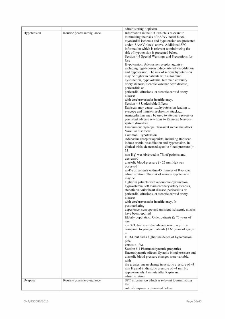
administering Rapiscan. Hypotension Routine pharmacovigilance Information in the SPC which is relevant to
minimizing the risks of SA/AV nodal block, myocardial ischemia and hypotension are presented under ‘SA/AV block’ above. Additional SPC information which is relevant to minimizing the risk of hypotension is presented below. Section 4.4 Special Warnings and Precautions for Use Hypotension: Adenosine receptor agonists including regadenoson induce arterial vasodilation and hypotension. The risk of serious hypotension may be higher in patients with autonomic dysfunction, hypovolemia, left main coronary artery stenosis, stenotic valvular heart disease, pericarditis or pericardial effusions, or stenotic carotid artery disease with cerebrovascular insufficiency. Section 4.8 Undesirable Effects Rapiscan may cause……hypotension leading to syncope and transient ischaemic attacks,… Aminophylline may be used to attenuate severe or persistent adverse reactions to Rapiscan Nervous system disorders: Uncommon: Syncope, Transient ischaemic attack Vascular disorders: Common: Hypotension Adenosine receptor agonists, including Rapiscan induce arterial vasodilation and hypotension. In clinical trials, decreased systolic blood pressure (> 35 mm Hg) was observed in 7% of patients and decreased diastolic blood pressure (> 25 mm Hg) was observed in 4% of patients within 45 minutes of Rapiscan administration. The risk of serious hypotension may be higher in patients with autonomic dysfunction, hypovolemia, left main coronary artery stenosis, stenotic valvular heart disease, pericarditis or pericardial effusions, or stenotic carotid artery disease with cerebrovascular insufficiency. In postmarketing experience, syncope and transient ischaemic attacks have been reported. Elderly population: Older patients (≥ 75 years of age; n = 321) had a similar adverse reaction profile compared to younger patients (< 65 years of age; n = 1016), but had a higher incidence of hypotension (2% versus < 1%). Section 5.1 Pharmacodynamic properties Haemodynamic effects: Systolic blood pressure and diastolic blood pressure changes were variable, with the greatest mean change in systolic pressure of −3 mm Hg and in diastolic pressure of −4 mm Hg approximately 1 minute after Rapiscan administration.
Dyspnea Routine pharmacovigilance SPC information which is relevant to minimizing the risk of dyspnea is presented below:
EMA/455580/2010 Page 36/43

Section 4.7 Effects on ability to drive and use machines No studies on the effects of Rapiscan on the ability to drive and use machines have been performed. Rapiscan administration may result in adverse reactions such as dizziness, headache and dyspnea soon after administration. However, most adverse reactions are mild and transient, resolving within 30 minutes after receiving Rapiscan. Therefore, Rapiscan would be expected to have no or negligible influence on the ability to drive or operate machinery once treatment has been completed and these reactions have resolved. The physician is advised to provide a recommendation for the individual patient Section 4.8 Undesirable Effects Summary of the safety profile Adverse reactions in most patients receiving Rapiscan in clinical trials were mild, transient (usually resolving within 30 minutes after receiving Rapiscan), and required no medical intervention. Adverse reactions occurred in approximately 80% of patients. The most common adverse reactions reported during clinical development in a total of 1,651 patients/subjects were: dyspnoea (29%), headache (27%), flushing (23%), chest pain (19%), electrocardiogram ST segment changes (18%), gastrointestinal discomfort (15%) and dizziness (11%). Respiratory thoracic and mediastinal disorders Very common: Dyspnoea Section 5.1 Pharmacodynamic properties Respiratory effects: The incidence of bronchoconstriction (FEV1 reduction > 15% from baseline) after Rapiscan administration was assessed in two randomised, controlled clinical studies… In both studies, dyspnoea was reported as an adverse reaction following Rapiscan dosing (61% for patients with COPD; 34% for patients with asthma) while no subjects experienced dyspnoea following placebo dosing. Dyspnoea did not correlate with a decrease in FEV1. Safety and tolerability testing: In ADVANCE MPI 1 and ADVANCE MPI 2, the following pre-specified safety and tolerability endpoints comparing Rapiscan to adenosine achieved statistical significance: (1) a summed score of both the presence and severity of the symptom groups of flushing, chest pain, and dyspnoea was lower with Rapiscan (0.9 ± 0.03) than with adenosine (1.3 ± 0.05)..
Headache Routine pharmacovigilance SPC information which is relevant to minimizing the risk of headache is presented below:
EMA/455580/2010 Page 37/43

Section 4.7 Effects on ability to drive and use machines: No studies on the effects of Rapiscan on the ability to drive and use machines have been performed. Rapiscan administration may result in adverse reactions such as dizziness, headache and dyspnea soon after administration. However, most adverse reactions are mild and transient, resolving within 30 minutes after receiving Rapiscan. Therefore, Rapiscan would be expected to have no or negligible influence on the ability to drive or operate machinery once treatment has been completed and these reactions have resolved. The physician is advised to provide a recommendation for the individual patient. Section 4.8 Undesirable effects Summary of the safety profile Adverse reactions in most patients receiving Rapiscan in clinical trials were mild, transient (usually resolving within 30 minutes after receiving Rapiscan), and required no medical intervention. Adverse reactions occurred in approximately 80% of patients. The most common adverse reactions reported during clinical development in a total of 1,651 patients/subjects were: dyspnoea (29%), headache (27%), flushing (23%), chest pain (19%), electrocardiogram ST segment changes (18%), gastrointestinal discomfort (15%) and dizziness (11%). Nervous system disorders Very Common: Headache Description of selected adverse reactions: Headache was reported by 27% of subjects who received Rapiscan in clinical trials. The headache was considered severe in 3% of subjects. Section 5.1 Pharmacodynamic properties: Safety and tolerability testing In ADVANCE MPI 1 and ADVANCE MPI 2, the following pre-specified safety and tolerability endpoints comparing Rapiscan to adenosine achieved statistical significance: the incidence of headache (25% vs 16%) was more frequent with Rapiscan.
Interaction with dipyridamole
Routine pharmacovigilance SPC information which is relevant to minimizing the risk of the interaction with dipyridamole is presented below. Section 4.2 Posology and method of administration When possible, dipyridamole should be withheld for at least two days prior to Rapiscan administration. Section 4.5 Interaction with other medicinal products and other forms of interaction Dipyridamole: Dipyridamole increases blood adenosine levels and the response to regadenoson may be altered when blood adenosine levels are increased. When possible, dipyridamole should be withheld for at least two days prior to Rapiscan administration.
Important potential risks Bronchoconstriction Routine pharmacovigilance SPC information which is relevant to minimizing
the potential risk of bronchoconstriction is presented below. Section 4.2 Posology and method of administration Treatment with Rapiscan is restricted to use in a medical facility where cardiac monitoring and resuscitation equipment are available. Aminophylline may be used to attenuate severe and/or persistent adverse reactions to Rapiscan.
EMA/455580/2010 Page 38/43

Section 4.4 Special warnings and precautions for use Rapiscan has the potential to cause serious and life threatening reactions, including those listed below. Rapiscan should be used with caution and should only be administered in a medical facility with cardiac monitoring and resuscitation equipment Aminophylline may be administered in doses ranging from 50 mg to 250 mg by slow intravenous injection (50 mg to 100 mg over 30-60 seconds) to attenuate severe and/or persistent adverse reactions to Rapiscan. Bronchoconstriction: Adenosine receptor agonists may cause bronchoconstriction and respiratory compromise. For patients with known or suspected bronchoconstrictive disease, chronic obstructive pulmonary disease (COPD) or asthma, appropriate bronchodilator therapy and resuscitative measures should be available prior to Rapiscan administration. 5.1 Pharmacodynamic properties The A2B and A3 adenosine receptors have been implicated in the pathophysiology of bronchoconstriction in susceptible individuals (i.e., asthmatics). In in vitro studies, regadenoson has been shown to have little binding affinity for the A2B and A3 adenosine receptors. The incidence of bronchoconstriction (FEV1 reduction > 15% from baseline) after Rapiscan administration was assessed in two randomised, controlled clinical studies. In the first study in 49 patients with moderate to severe COPD, the rate of bronchoconstriction was 12% and 6% following Rapiscan and placebo dosing, respectively (p = 0.31). In the second study in 48 patients with mild to moderate asthma who had previously been shown to have bronchoconstrictive reactions to adenosine monophosphate, the rate of bronchoconstriction was the same (4%) following both Rapiscan and placebo dosing. In both studies, dyspnoea was reported as an adverse reaction following Rapiscan dosing (61% for patients with COPD; 34% for patients with asthma) while no subjects experienced dyspnoea following placebo dosing. Dyspnoea did not correlate with a decrease in FEV1.
Important Missing Information Safety in Children Routine pharmacovigilance SPC information which is relevant to the lack of
safety information in children is presented below. Section 4.2 Posology and Method of Administration Paediatric population: The safety and efficacy of Rapiscan in children below the age of 18 years have not yet been established. No data are available. Section 5.2Pharmacokinetic properties Paediatric population: The pharmacokinetic parameters of regadenoson have not yet been studied in the paediatric population (< 18 years).
Safety in Pregnancy Routine pharmacovigilance SPC information which is relevant to the lack of safety information in pregnancy is presented below. Section 4.6 Fertility, pregnancy and lactation Pregnancy: There are no adequate data from the use of regadenoson in pregnant women. Animal studies on pre- and post-natal development have not been conducted. Foetotoxicity, but not teratogenicity, was noted in embryo-fetal development studies. The potential risk for humans is unknown. Rapiscan should not be used during pregnancy unless clearly necessary. Section 5.3 Preclinical
EMA/455580/2010 Page 39/43

safety data: Non-clinical data reveal no special hazard for humans based on conventional studies of safety pharmacology, single and repeated dose toxicity, genotoxicity, or embryo-fetal development. Signs of maternal and fetal toxicity were seen in rats and rabbits (reduced fetal weights, delays in ossification [rats], reduced litter size and number of live fetuses [rabbits]), but not teratogenicity. Fetal toxicity was noted following repeated daily administration of regadenoson, but at doses sufficiently in excess of the recommended human dose. Fertility and pre- and post-natal studies have not been conducted.
Safety in lactation Routine pharmacovigilance SPC information which is relevant to the lack of safety information in lactation is presented below. Section 4.6 Fertility, pregnancy and lactation Breast-feeding: It is unknown whether regadenoson is excreted in human breast milk. The excretion of regadenoson in milk has not been studied in animals. A decision should be made whether to discontinue breast-feeding or to abstain from Rapiscan administration taking into account the benefit of breast-feeding for the child and the benefit of therapy for the woman. If Rapiscan is administered, the woman should not breast feed for at least 10 hours (that is, at least 5 times the plasma elimination half life) following Rapiscan administration.
Safety in patients with renal impairment
Routine pharmacovigilance Clinical study 3606 CL-3010. This Phase 4 study will help define the safety profile of regadenoson in patients with renal impairment
SPC information which is relevant to the lack of safety information in patients with renal impairment is presented below. Section 4.2 Posology and Method of Administration Renal impairment: No dose adjustment is necessary. Section 5.2 Pharmacokinetic properties Renal impairment: The disposition of regadenoson was studied in 18 subjects with various degrees of renal function and in 6 healthy subjects. With increasing renal impairment, from mild (CLcr 50 to < 80 ml/min)to moderate (CLcr 30 to < 50 ml/min) to severe renal impairment (CLcr < 30 ml/min), the fraction of regadenoson excreted unchanged in urine and the renal clearance decreased, resulting in increased elimination half-lives and AUC values compared to healthy subjects (CLcr ≥ 80 ml/min). However, the maximum observed plasma concentrations as well as volumes of distribution estimates were similar across the groups. The plasma concentration-time profiles were not significantly altered in the early stages after dosing when most pharmacologic effects are observed. No dose adjustment is needed in patients with renal impairment. The pharmacokinetics of regadenoson in patients on dialysis has not been assessed.
Safety in patients with severe hepatic impairment
Routine pharmacovigilance Retrospective cohort study in patients with severe hepatic impairment (PASS). This PASS, is currently under development to evaluate the hemodynamic and safety profile for regadenoson in patients with severe hepatic impairment
SPC information which is relevant to the lack of safety information in patients with hepatic impairment is presented below. Section 4.2 Posology and Method of Administration Hepatic impairment: No dose adjustment is necessary. Section 5.2 Pharmacokinetic properties Hepatic impairment Greater than 55% of the
EMA/455580/2010 Page 40/43

regadenoson dose is excreted unchanged in the urine and factors that decrease clearance do not affect the plasma concentration in the early stages after dosing when clinically meaningful pharmacologic effects are observed. The pharmacokinetic parameters of regadenoson have not been specifically evaluated in those with varying degrees of hepatic impairment. However, post hoc analysis of data from the two Phase 3 clinical trials showed that the pharmacokinetics of regadenoson were not affected in a small subset of patients with laboratory values suggestive of impaired hepatic function (2.5 fold transaminase elevation or 1.5 fold elevation of serum bilirubin or prothrombin time). No dose adjustment is needed in patients with hepatic impairment.
Safety in patients with bronchoconstrictive disease
Routine pharmacovigilance Clinical study 3606-CL-3001. This Phase 4 study will help define the safety profile of regadenoson in patients with asthma and COPD
See above under the potential risk of bronchoconstriction.
Safety in patients with prolonged QT syndrome
Routine pharmacovigilance SPC information which is relevant to the lack of safety information in patients with prolonged QT syndrome is presented below Section 4.4 Special warnings and precautions for use Long QT syndrome: Regadenoson stimulates sympathetic output and may increase the risk of ventricular tachyarrhythmias in patients with a long QT syndrome. Section 4.8 Undesirable Effects Cardiac Disorders: Common: Other ECG abnormalities including electrocardiogram QT corrected interval prolonged Regadenoson increases sympathetic tone, which causes an increase in heart rate and a shortening of the QT interval. In a patient with a long QT syndrome, sympathetic stimulation can result in less shortening of the QT interval than is normal and may even cause a paradoxical increase in the QT interval. In these patients, the phenomenon of R-on-T syndrome can occur, wherein an extra beat interrupts the T wave of the previous beat, and this increases the risk of a ventricular tachyarrhythmia.
The CHMP, having considered the data submitted in the application, is of the opinion that no additional risk minimisation activities are required beyond those included in the product information.
User consultation
The results of the user consultation with target patient groups on the package leaflet submitted by the
applicant show that the package leaflet meets the criteria for readability as set out in the Guideline on
the readability of the label and package leaflet of medicinal products for human use.
EMA/455580/2010 Page 41/43

EMA/455580/2010 Page 42/43
2.6. Benefit-risk balance
2.6.1.1.
2.6.1.1.1.
2.6.1.1.2.
2.6.1.2.
2.6.1.2.1.
2.6.1.2.2.
2.6.1.3.
2.6.1.4.
Benefits
Beneficial effects
Regadenoson is expected to be used as adjunct to myocardial perfusion imaging as a stress agent. Based on its pharmacological properties this is appropriate. Whilst the pharmacology is similar to adenosine, it has little activity against A1, A2b or A3 receptors with significant specificity at the A2A receptor for agonistic effect. Given this background and the data from the pivotal trials, its non-inferiority to adenosine in performance as a adjunct to radiopharmaceutical has been shown. One of the advantages of regadenoson is fewer risks: less bronchoconstriction and less high grade Av block than with adenosine. From an administration perspective the main benefit of regadenoson over adenosine would be that it can be administered as a bolus instead of an infusion which would increase the convenience for patient and technologist.
Uncertainty in the knowledge about the beneficial effects.
Concerns related to statistical analysis in both pivotal studies have been addressed by the applicant by providing additional analysis. Based on the data provided on regadenoson there is little uncertainty about its performance as a stress agent.
Risks
Unfavourable effects
The risk of AV or sinus node dysfunction in predisposed individuals still exists, although, it is much lower for regadenoson than for adenosine. The major AEs reported are dyspnoea and headache and these have been addressed in the SPC. Bronchospastic tendency appears lower with regadenoson; reduction in FEV1 was still observed. However, this could not be quantified due to the small size of the studies
Uncertainty in the knowledge about the unfavourable effects
Based on the data provided, there are a few uncertainties regarding safety of regadenoson in patients with renal failure and in patients with COPD. The data currently available are from small studies. Post marketing studies are ongoing for further evaluation of risk in renally impaired subjects and in those with COPD (chronic obstructive pulmonary disease). The magnitude of effect will be better judged with increasing exposure as data become available from these studies. The applicant has committed to submit the study reports as follow up measures.
Benefit-risk balance
Based on the data, there are no safety issues; missing information will be addressed by post marketing studies. Non-inferiority to adenosine has been shown in two pivotal trials. Clarifications regarding diagnostic value and the statistic used (kappa) for demonstrating non-inferiority have been adequately addressed by the applicant and overall safety reports including the EU-RMP and Pharmacovigilance systems have been updated. The benefit-risk balance is favourable for regadenoson.
Discussion on the benefit-risk balance
As non-inferiority to adenosine has been shown, it is concluded that regadenoson performs as well as adenosine as an adjunct to the radiopharmaceutical for detection of stress induced ischaemia. Its safety profile is acceptable despite a number of uncertainties, which will be further explored in post marketing studies. The benefit risk balance is considered positive for the indication “Rapiscan is a selective coronary vasodilator for use as a pharmacological stress agent for radionuclide myocardial perfusion imaging (MPI) in patients unable to undergo adequate exercise stress. ” and the CHMP recommends approval as standard marketing authorisation.

EMA/455580/2010 Page 43/43
2.6.1.5.
2.7.1.1.
Risk management plan
A risk management plan was submitted. The CHMP, having considered the data submitted, was of the
opinion that:
Additional pharmacovigilance planning was requested to adequately monitor the safety of the product.
No additional risk minimisation activities were required beyond those included in the product
information.
2.7. Recommendation
Normal opinion
Based on the CHMP review of data on quality, safety and efficacy, the CHMP considered by consensus
that the risk-benefit balance of Rapiscan in the in the following indication:
“Rapiscan is a selective coronary vasodilator for use as a pharmacological stress agent for radionuclide myocardial perfusion imaging (MPI) in patients unable to undergo adequate exercise stress”
was favourable and therefore recommended the granting of the marketing authorisation .