rare brain and cns tumours guidelines - bnos.org.uk · rare brain and cns tumours guidelines . in...
TRANSCRIPT

Rare Brain and CNS Tumours Guidelines
In collaboration with the
National Cancer Action Team
Guidelines on the Diagnosis and Management
of Adult Pineal Area Tumours Officers of the British Neuro-Oncology Society: President: Professor Geoff Pilkington Professor of Cellular & Molecular Neuro-oncology Director of Research, School of Pharmacy & Biomedical Sciences University of Portsmouth Vice President: Professor David Walker Professor of Paediatric Oncology Queen's Medical Centre Nottingham Secretary: Mr David Jellinek Consultant Neurosurgeon Royal Hallamshire Hospital Sheffield Treasurer: Dr Jeremy Rees Consultant Neurologist National Hospital for Neurology and Neurosurgery London
British Neuro-Oncology Society/NCAT Rare Tumour Guidelines (June 2011) www.bnos.org.uk

Guidelines on the diagnosis and management of adult Pineal area tumours
Contents
Section Page Patient pathway..............................................................................................................1 Introduction to Pineal Area Tumours in Adults ...............................................................4 Germ Cell Tumors Of The Central Nervous System (In Children) .................................4 Summary of key recommendations ................................................................................5 Table 1: Histological Varieties of Pineal Tumours ..........................................................6 Background ....................................................................................................................7 Presentation and diagnosis ............................................................................................7 Pineal Area Tumours – Specific Clinical Symptoms ......................................................8 Pineal Area Tumours: Pathology.............................................................................8 Neuroimaging ...............................................................................................................10 CCLG brain tumour imaging protocol .....................................................................11 Follow-up examinations..........................................................................................13 Tumour Markers ...........................................................................................................13 Cerebrospinal fluid........................................................................................................14 Treatment – general principles .....................................................................................14 Organisation of services ...............................................................................................15 Principles of Surgical Management ..............................................................................16 Recommendations..................................................................................................18 Anticipated benefits ................................................................................................18 Pineal Germ-Cell Tumours – treatment........................................................................18 General Aspects .....................................................................................................18 Surgical Treatment .................................................................................................19 Non-surgical treatments .........................................................................................19 Non-germinoma (secreting GCTS) – malignant non-germinomatous germ-cell .........20 tumours (MNGGCT) Chemotherapy Regimens for Intracranial Germ-Cell Tumours ....................................21 Endocrinology...............................................................................................................21 Details of PEI Chemotherapy Administration ...............................................................22 Dose modifications and delays.....................................................................................23 Haematological toxicity...........................................................................................23 Ototoxicity...............................................................................................................23 Nephrotoxicity.........................................................................................................23 Radiotherapy for Non-Germinoma ..............................................................................24 Pineal parenchymal tumours ........................................................................................25 Pineoblastoma........................................................................................................25 Pineocytoma ..........................................................................................................25 Pineal Parenchymal Tumor of Intermediate Differentiation ....................................25 Papillary Tumours of the Pineal Region .................................................................26 Germ-cell Tumours - References for Introduction and Main Text ................................26 Supportive care, Rehabilitation and General palliative care.........................................34 Core Members of Supportive Care services...........................................................34 Extended members of Supportive Care services ...................................................34 Core elements of palliative care .............................................................................37 Specialist palliative care ...............................................................................................38 Survivorship / Living with cancer ..................................................................................40
British Neuro-Oncology Society/NCAT Rare Tumour Guidelines (June 2011) www.bnos.org.uk

British Neuro-Oncology Society/NCAT Rare Tumour Guidelines (June 2011) www.bnos.org.uk
Appendix 1: Chemotherapy Administration Guidelines ...............................................42 Appendix 2: Perioperative management of patients with CNS Germ Cell Tumours ...45 Appendix 3: Endocrinology..........................................................................................46 Clinical assessment and diagnostic tools ...............................................................46 Table: Diagnosis of Hypopituitarism ......................................................................48 Minimal requirements for endocrine follow-up .......................................................49 Appendix 4: Radiotherapy Guidelines (including craniospinal radiotherapy protocol).49 Appendix 5: Symptom related referral pathway to supportive care services for patients with Pineal tumours .............................................................................54 Appendix 6: Additional support for brain tumour patients and carers..........................55

British Neuro-Oncology Society/NCAT Rare Tumour Guidelines (June 2011) www.bnos.org.uk
1
PINEAL AREA TUMOURS – PATIENT PATHWAY
Urgent CT Scan Pineal area tumour/dilated ventricles
Referral to Neurosurgical unit
Neurosurgical unit decision re: Relief of Raised Intracranial Pressure
Appropriate route for biopsy CSF Sampling for cytology
and Tumour Markers: AFP, ß-HCG Endocrine assessment, clinically, basal pituitary
hormone tests (preferably endocrine referral)
Presentation – Headache, Signs and Symptoms of
Raised Intracranial Pressure Parinaud’s Syndrome
MR Scan – Craniospinal Imaging
Emergency relief of raised intracranial pressure Preferably via third ventriculostomy CSF sampling and ? biopsy CSF samples to be sent for analysis in an accredited laboratory
Storage of samples when taken out of hours

British Neuro-Oncology Society/NCAT Rare Tumour Guidelines (June 2011) www.bnos.org.uk
2
BIOPSY DIAGNOSIS: GERM-CELL TUMOUR Referral to Oncology Unit associated with CCLG Centre
Germinoma:
Non-surgical treatment with craniospinal radiotherapy Non-germinomatous GCT (NGGCT)
Chemotherapy, Resection, Radiotherapy
Germinoma “standard” treatment Craniospinal RT 24 Gy in 15 fractions Tumour boost 16 Gy in 10 fractions
(Plus boosts to metastases for metastatic disease)
(European trial of chemotherapy plus ventricular RT in planning stage)
NGGCT or “secreting” GCT “standard” treatment
PEI chemotherapy Tumour resection
Focal RT 54 Gy Craniospinal RT for metastatic disease
BIOPSY DIAGNOSIS: PINEAL PARENCHYMAL TUMOURS Pineocytoma
Pineal Tumours of Intermediate Differentiation Papillary Pineal Tumour
Maximum tumour resection
Consider need for adjuvant radiotherapy
Pineocytoma: adjuvant RT 50-54 Gy or stereotactic radiosurgery Pineal Tumour of Intermediate Differentiation – no clear consensus on
optimum treatment Papillary pineal tumour:

British Neuro-Oncology Society/NCAT Rare Tumour Guidelines (June 2011) www.bnos.org.uk
3
BIOPSY DIAGNOSIS: PINEOBLASTOMA Referral to Oncology Unit associated with CCLG Centre
Treat along same lines as other PNETs and Medulloblastoma
BIOPSY DIAGNOSIS: ASTROCYTIC TUMOUR Management as for astrocytic tumour elsewhere

British Neuro-Oncology Society/NCAT Rare Tumour Guidelines (June 2011) www.bnos.org.uk
4
Introduction to Pineal Area Tumours in Adults
Tumours arising in the pineal area comprise a heterogeneous mix of histologies (Table 1). In the adult age range they are rare, and data from the east of England Cancer Registry would suggest there would be approximately 50 cases annually in England. Pineal area tumours account for between 2% and 8% of paediatric brain tumours. In childhood the majority of pineal area tumours are Germ-Cell Tumours, for which clinical trial protocols and guidelines have been developed. There are no national guidelines or protocols for the management of pineal area tumours in adults.
The brain tumour IOG recommended that national tumour groups for rare CNS tumours should be established to coordinate the approach to care; this should include developing protocols for the investigation, management, registration and clinical research into rare tumours. It was also advised that they should also maintain a national register of all these cases.
The purpose of this report is to provide a schema for the management of adults with pineal area tumours which can be applied nationally, leading to standardised management.
In writing these guidelines, it is acknowledged that since pineal area tumours arise more frequently in children than in adults, many of the principles of management have been adapted from current guidelines for the management of children.
Germ Cell Tumors Of The Central Nervous System (In Children)
Introduction
Pineal area tumours account for between 2% and 8% of paediatric brain tumours and comprise a heterogeneous mix of histologies. In the adult age range they are rare. In childhood the majority of pineal area tumours are Germ-Cell Tumours.
Pineal tumours include the pineal parenchymal tumours, pineoblastoma, astrocytomas, germ cell tumours and the recently recognised papillary tumour of the pineal region.
Because of the rarity of pineal area tumours it is unlikely that these will be suspected as a distinct clinical entity prior to imaging, and more likely the issue will be the suspicion of having a brain tumour.
Because pineal area tumours, particularly intracranial germ-cell tumours, are relatively more frequent in childhood and better studied, many of the principles which apply to the management of these tumours in childhood can be applied to the adult population. Furthermore many case series in the literature report treatment outcomes for a mix of adult and paediatric patients. In the last three decades there have been significant improvements in the prognosis of malignant germ cell tumours (GCT) in all anatomical locations, both in the adult and in the paediatric populations. This can mainly be attributed to platinum-based combination chemotherapy integrated into a multimodality therapeutic approach, including radiotherapy.

British Neuro-Oncology Society/NCAT Rare Tumour Guidelines (June 2011) www.bnos.org.uk
5
Summary of key recommendations
1. Patients should be discussed at full neuro-oncology MDT which collaborates with an MDT for paediatric patients with brain tumours.
2. Patients should have a full endocrine assessment with particular reference to pituitary function.
3. Patients with pineal area tumours should have pre-operative craniospinal MRI.
4. Germ Cell Tumours may secrete specific tumours markers, beta-Human Chorionic Gonadotrophin (ß-HCG) or alpha-fetoprotein (AFP) and assay for these is essential for all patients with pineal area tumours prior to a decision about definitive surgery.
5. Patients with pineal area tumours should have serum (blood and/or CSF) sampled for AFP and ß-HCG pre-operatively as in some cases a diagnosis of secreting germ-cell tumour can be made on tumour markers alone. Blood tests for sarcoidosis should also be performed.
6. The method of choice for ventricular drainage is neuro-endoscopy by third ventriculostomy.
7. Biopsy is required for all patients with negative markers in serum and/or CSF or borderline secretion of markers.
8. Ideally planned surgical resection where appropriate will be carried out in most cases by a surgeon experienced in pineal region surgery.
9. Referral to an ophthalmology service is mandatory early in patient management.
10. Germ-cell tumours are sensitive to radiotherapy and/or chemotherapy and extensive resections as initial management should be avoided.
11. Diabetes is a common complication and should be controlled prior to starting chemotherapy.
12. Selected patients with post-chemotherapy residual disease may require resection of residual disease.
13. The Standard non-surgical treatment for germinoma is craniospinal radiotherapy 24 Gy in 15 fractions with a primary tumour boost of 16 Gy in 10 fractions.
14. For secreting germ cell tumours chemotherapy is based on a combination of Cisplatin, Etoposide and Ifosfamide (PEI) and should commence as soon as possible following diagnosis.
15. Pineoblastomas is within the group of primitive neuro-ectodermal tumours (PNETs). They are managed along the same lines as medulloblastoma and other PNETs.
16. Patients found to have incidental cystic pineal region tumours, in whom there is no CSF pathway obstruction. Where neuroradiology are confident this does not
British Neuro-Oncology Society/NCAT Rare Tumour Guidelines (June 2011) www.bnos.org.uk
6
represent a neoplastic process can be safely managed with interval imaging. We would suggest the first post-diagnostic scan is performed at 6 months and then at annual intervals.
17. Patients require long term multidisciplinary monitoring and coordinated cognitive and physical rehabilitation. Timely referral to rehabilitation and supportive care services is imperative and is dependent on rapid, comprehensive communication between medical and AHP staff.
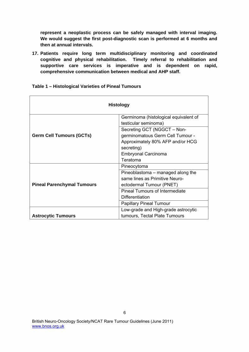
Table 1 – Histological Varieties of Pineal Tumours
Histology
Germinoma (histological equivalent of testicular seminoma)
Germ Cell Tumours (GCTs)
Secreting GCT (NGGCT – Non-germinomatous Germ Cell Tumour - Approximately 80% AFP and/or HCG secreting) Embryonal Carcinoma Teratoma Pineocytoma Pineoblastoma – managed along the same lines as Primitive Neuro-ectodermal Tumour (PNET) Pineal Tumours of Intermediate Differentiation
Pineal Parenchymal Tumours
Papillary Pineal Tumour Astrocytic Tumours
Low-grade and High-grade astrocytic tumours, Tectal Plate Tumours

British Neuro-Oncology Society/NCAT Rare Tumour Guidelines (June 2011) www.bnos.org.uk
7
Background
Intracranial Germ-Cell Tumours (GCTs) - Natural history and histogenesis
GCTs arise from toti-potential primordial germ cells which have the potential for embryonic and extraembryonic differentiation, and as such may exhibit a variety of histological patterns. Yolk sac tumours and choriocarcinoma follow an extra-embryonic differentiation pattern and are characterized by significant secretion of alpha-fetoprotein (AFP) or human choriogonadotropin (ß-HCG) respectively. Embryonal carcinomas represent tumours of immature toti-potential germ-cells. Teratomas display an embryonic differentiation and may mimic organ structures of all germ layers. Intracranial germinomas exhibit histological features analogous to testicular seminoma, displaying morphological features of undifferentiated germinal epithelium.
Presentation and diagnosis
Generic Symptoms and Signs of CNS Tumours (adapted from IOG)
CNS tumours can result in a wide range of physical, cognitive and psychological symptoms. The list of differential diagnoses is considerable, and the incidence of many of these alternatives is usually far greater than that of brain tumours, such that these may be exhaustively explored before the diagnosis of a CNS tumour is considered. Consequently, for some patients and families there is a long delay from first symptoms to reaching a diagnosis, causing considerable stress and anxiety.
There is a significant overlap between the symptoms of pineal area tumours and those of other primary brain tumours. Because of the rarity of pineal area tumours compared with other primary brain tumours it is unlikely that these will be suspected as a distinct clinical entity prior to imaging. Therefore these guidelines have incorporated the IOG recommendations for the investigation of patients who are suspected of having a brain tumour.
Brain tumours
Brain tumours account for the majority of CNS tumours. This group includes tumours of the brain substance itself, many of which arise from the glial or support cells of the brain, for example, glioblastoma multiforme. Also included are tumours that arise from the tissues around the brain, such as tumours of the meninges and metastases from other primary sites that require complex neurological or neurosurgical interventions.
In spite of the variety of brain tumour pathologies, presentation tends to be related to:
headache with cognitive or behavioural symptoms
epilepsy
progressive focal neurological deficits
Or
British Neuro-Oncology Society/NCAT Rare Tumour Guidelines (June 2011) www.bnos.org.uk
8
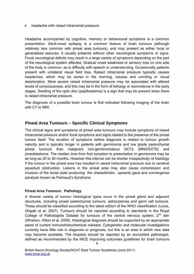
headache with raised intracranial pressure
Headache accompanied by cognitive, memory or behavioural symptoms is a common presentation. Adult-onset epilepsy is a common feature of brain tumours (although relatively less common with pineal area tumours), and may present as either focal or generalised seizures. It usually presents without other neurological symptoms or signs. Focal neurological deficits may result in a large variety of symptoms depending on the part of the neurological system affected. Gradual onset weakness or sensory loss on one side of the body is common, as is difficulty with speech or understanding. Occasionally patients present with unilateral visual field loss. Raised intracranial pressure typically causes headaches, which may be worse in the morning, nausea and vomiting or visual deterioration. More severe raised intracranial pressure may be associated with altered levels of consciousness, and this may be in the form of lethargy or somnolence in the early stages. Swelling of the optic disc (papilloedema) is a sign that may be present when there is raised intracranial pressure.
The diagnosis of a possible brain tumour is first indicated following imaging of the brain with CT or MRI.
Pineal Area Tumours – Specific Clinical Symptoms
The clinical signs and symptoms of pineal area tumours may include symptoms of raised intracranial pressure and/or focal symptoms and signs related to the presence of the pineal tumour itself. The duration of symptoms before diagnosis is related to tumour growth velocity and is typically longer in patients with germinoma and low grade parenchymal pineal tumours than malignant non-germinomatous GCTs (MNGGCTS) and pineoblastoma. The median time from first symptom to presentation in germinoma may be as long as 20 to 30 months. However this interval can be shorter irrespectively of histology if the tumour in the pineal area has resulted in raised intracranial pressure due to cerebral aqueduct obstruction. Lesions in the pineal area may also cause compression and invasion of the tectal plate producing the characteristic upwards gaze and convergence paralysis known as Parinaud’s Syndrome.
Pineal Area Tumours: Pathology
A diverse variety of tumour histological types occur in the pineal gland and adjacent structures, including pineal parenchymal tumours, astrocytomas and germ cell tumours. These should be classified according to the latest edition of the WHO classification (Louis, Ohgaki et al. 2007). Tumours should be reported according to standards in the Royal College of Pathologists Dataset for tumours of the central nervous system, 2nd edn (Wharton, Hilton et al. 2008). Histological diagnosis should be supported by an appropriate panel of current immunohistochemical markers. Cytogenetic and molecular investigations currently have little role in diagnosis or prognosis, but this is an area in which new data may become available. The biopsies should be reported by an accredited pathologist, defined as recommended by the NICE improving outcomes guidelines for brain tumours

British Neuro-Oncology Society/NCAT Rare Tumour Guidelines (June 2011) www.bnos.org.uk
9
(NICE 2006). They should be registered as a neuropathologist or histopathologist, who has specialist expertise in neuro-oncology and takes part in the national External Quality Assurance scheme for neuropathology organised by the British Neuropathological Society (www.bns.org.uk).
The reporting pathologist should be a member of the neuro-oncology MDT and cases should be discussed in that forum, both for biopsy planning and discussion of the diagnosis. Correlation of the pathological findings with neuroimaging and clinical findings is particularly important given the often small biopsies and the difficulties of histological differential diagnosis at this site. The ‘final’ histopathological diagnosis should take account of this information (NICE 2006).
Pineal tumours include the pineal parenchymal tumours, pineoblastoma, astrocytomas, germ cell tumours and the recently recognised papillary tumour of the pineal region. The pineal parenchymal tumours include the pineocytoma WHO grade I, the pineoblastoma WHO grade IV and the pineal parenchymal tumour of intermediate differentiation. The latter has intermediate histological features and is potentially aggressive. It corresponds to WHO grade II to III, but definite grading criteria have not been established, so that there is a need to refine prognostication for this tumour type. The presence of necrosis, the mitotic rate and immunohistochemical expression of neurofilament protein may be predictive factors (Jouvet, Saint-Pierre et al. 2000; Louis, Ohgaki et al. 2007; Arivazhagan, Anandh et al. 2008). Immunohistochemistry may also help with distinction from normal pineal, which has a Ki67 labelling index of 0 and a lobular pattern highlighted by GFAP. Low-grade pineal tumours can occasionally show cytological pleomorphism which can be a worrying feature. However, this feature alone (without elevated mitotic activity) does not appear to be associated with a worse prognosis and should not lead to upgrading (Fevre-Montange, Szathmari et al. 2008). The papillary tumour of the pineal region is a recently defined entity. The behaviour of this rare tumour is yet to be fully defined, but appears to correspond to grades II to III, although grading criteria have not yet been determined.
An array of germ cell tumours can develop in the pineal gland, mostly in younger subjects (Louis, Ohgaki et al. 2007). Some of these tumours, such as the germinoma, are relatively straightforward to diagnose histologically, but the participation of a histopathologist with specialist expertise in germ cell tumour classification in the diagnosis is advisable for more
complex histotypes. Immunohistochemical investigations, including antibodies to -fetoprotein, human chorionic gonadotrophin, placental alkaline phosphatase, cytokeratins, c-kit (CD117), OCT4 and CD30 are of value for their diagnosis and subclassification. Some germinomas develop a florid lymphoid or granulomatous inflammatory reaction that can suggest a differential diagnosis of an inflammatory process. The possibility of an underlying germinoma should therefore be considered in pineal biopsies showing inflammatory changes, and immunohistochemistry is valuable to identify the obscured germinoma tumour cells.
The pineal can be involved by both pilocytic astrocytomas and diffuse astrocytomas. These should be diagnosed and reported according to the same criteria as applied in the more common cerebral locations. In addition to a number of other entities that can arise in this region, tumours can occasionally metastasise to the pineal region. In some cases, this can be the initial presentation of disease (Lassman, Bruce et al. 2006), so that the

British Neuro-Oncology Society/NCAT Rare Tumour Guidelines (June 2011) www.bnos.org.uk
10
possibility of a metastasis should be considered in the initial differential diagnosis of a pineal tumour. In addition to morphology, immunohistochemisty can be of value both in the differential diagnosis from primary CNS tumours, and as a pointer towards a likely primary site in cases where the primary is occult (Becher, Abel et al. 2006).
Neuroimaging
Modern neuro-imaging has greatly contributed to a more precise pre-operative diagnosis of intracranial tumours including pineal area tumours. MRI is the optimum modality although CT scanning is more specific about the presence of calcification. Furthermore CT scanning is usually the first radiological investigation performed in the acute or emergency setting, and therefore is very frequently performed prior to MRI. The correct interpretation of imaging should take into account the MRI appearance of normal anatomy of the pineal area and of that of other benign lesions typical in these sites (e.g. simple pineal cysts – these are simple cysts containing fluid that is hyperintense to CSF on both T1 and T2 weighted sequences).
Germinomas occur in patients typically in the second decade of life and have a strong male predominance (male to female, 10:1). On MRI germ cell tumours usually appear as solid masses that are iso- or hyper intense relative to grey matter and show prominent enhancement following the administration of contrast media. They are iso- to hypointense on T2 weighted images. The presence of fat, calcifications or intra-tumoral cysts suggest the presence of a mature teratomatous component.
On diffusion weighted imaging (DWI) germinomas may show restricted diffusion (hypointense on ADC maps) or appear iso-intense. This is feature of many highly cellular tumours.
In the case of intracranial germinoma, approximately 30% have “bifocal“ disease, namely concurrent primary tumours in pineal and suprasellar/parasellar regions. These are now regarded as having true “bifocal“ primary rather than metastatic disease.
Fewer than 10% of patients have evidence of leptomeningeal metastases at presentation. However for those who have leptomeningeal metastases, management can be very different. The appearances of post-operative spinal MRI can be difficult to interpret owing to the presence of blood tracking along the meninges. Therefore it is strongly recommended that patients with pineal area tumours should have pre-operative MR craniospinal imaging.
Sagittal high resolution 3D T2 weighted imaging is helpful for defining the anatomy of the tumour and planning CSF diversionary treatment such as third ventriculostomy.
Teratomas of the pineal region show a wide variation in maturity of tissues. These occur in a younger age group than germinomas (typically within the first decade of life). Well formed structures including hair, teeth, bone and fat can be seen. They are usually cystic and also have haemorrhagic components. Given the wide variety of histological tissues

British Neuro-Oncology Society/NCAT Rare Tumour Guidelines (June 2011) www.bnos.org.uk
11
present they are very heterogeneous on imaging with evidence of fat blood products and calcification or ossification.
The malignant germ cell tumours are much less common and it can be difficult to separate these radiologically from other pineal region tumours.
Tumours arising from pineal cells are the pineoblastoma (WHO Grade IV) and the pineocytoma (WHO Grade II). The pineoblastoma can be regarded as part of the Primitive Neuro Ectodermal Tumours (PNET) category. This therefore has similar imaging features to medulloblastoma with haemorrhage, necrosis and CSF dissemination. Pineoblastomas are typically seen in childhood.
Pineocytomas typically occur in older patients (usually middle aged patients). These are less aggressive lesions and have an increased T2 signal and may show calcification.
Pineal parenchymal tumour of intermediate differentiation (PPTID) is a new addition to the WHO classification of tumours. This may account for up to 20% of pineal tumours. There is no particular male predominance and they occur in all ages – particularly middle age patients. They have intermediate behaviour being more aggressive than pineocytomas (Anne Osborn, personal communication). It may therefore be appropriate to suggest this diagnosis in pineal masses occurring in middle aged patients.
There is an overlap with the imaging features across many of the pineal region tumours and it is not possible with MRI alone to distinguish some of the sub types of pineal region tumour.
CCLG brain tumour imaging protocol
The Children’s Cancer and Leukaemia Group (CCLG) Brain Tumour Group have developed generic guidelines for imaging brain tumours. Since pineal area tumours in adults have broadly similar clinical features to those arising in children, it is recommended that the paediatric imaging protocol should be followed:
The protocol provided here is based upon the imaging protocol originally published in 2001 but reflects recent advances in imaging techniques (DTI, perfusion MRI, MRS). Not all centres can or will wish to use these newer techniques, and therefore these are given as optional sequences. Many centres will have their own preferred imaging sequences and this protocol is not intended to be prescriptive or to exclude other sequences and techniques, however it is essential that a standardised basic set of sequences is adopted nationally.
Patients with pineal area tumours should have pre-operative craniospinal MRI, as blood tracking along the leptomeninges can make interpretation of post-operative imaging difficult to interpret

British Neuro-Oncology Society/NCAT Rare Tumour Guidelines (June 2011) www.bnos.org.uk
12
New cases
Brain
Standard sequences
Axial T1, T2
Coronal FLAIR
DTI and/or DWI (with ADC maps)
Post Gd Ax, Cor, Sag T1: at 1.5T
Post Gd Ax T1, Ax 3D T1 volume: at 3T
Optional sequences (according to local capacity/availability or CCLG trial involvement)
Cor/SagT2 or FLAIR
Perfusion MRI (requires placement of blue or pink cannula)
ASL
MRS
Spine
Standard sequences
Sag T1 (post Gd)
Ax T1 through any equivocal focal abnormality
Optional:
Sag T2
Immediate post-op (within 48 hours)
Brain
Standard sequences
Ax T1, T2,
Coronal FLAIR
DTI and/or DWI (with ADC maps)
Post Gd Ax, Cor, Sag T1: at 1.5T
Post Gd Ax T1, Ax 3D T1 volume: at 3T

British Neuro-Oncology Society/NCAT Rare Tumour Guidelines (June 2011) www.bnos.org.uk
13
Spine (only if not obtained prior to surgery)
Standard sequences
Sag T1 (pre and post Gd)
Ax T1 through any equivocal focal abnormality
Follow-up examinations
Brain
Standard sequences
Axial T1, T2
Coronal FLAIR
DTI and/or DWI (with ADC maps)
Post Gd Ax, Cor, Sag T1: at 1.5T
Post Gd Ax T1, Ax 3D T1 volume: at 3T
Optional (according to local preference or CCLG trial involvement)
Cor/Sag T2 or FLAIR
Perfusion MRI (requires placement of blue or pink cannula)
ASL
MRS if tumour size >1.0cm (and dependent on tumour type/protocol)
Spine (dependent on tumour type/protocol)
Standard sequences
Sag T1, (post Gd)
Ax T1 through any equivocal focal abnormality
Optional Sag T2
Tumour Markers
Germ Cell Tumours may secrete specific tumours markers, beta-Human Chorionic Gonadotrophin (ß-HCG) or alpha-fetoprotein (AFP).
AFP is a glycoprotein marker, with a serum half-life of five days and levels are measured by immunoassay. AFP levels are normally elevated during gestation, and in the newborn infant. In addition AFFP is a marker for liver tumours.

British Neuro-Oncology Society/NCAT Rare Tumour Guidelines (June 2011) www.bnos.org.uk
14
HCG is produced by the placenta and has a half-life of 16 hours. HCG may be a marker for placental tumours
AFP is a marker for secreting germ-cell tumours. In the presence of histological evidence of germinoma, elevation of AFP level indicates the presence of a mixed tumour, whereas mild elevation of ß-HCG is consistent with a diagnosis of germinoma.
In patients with secreting GCTs high tumour marker levels (>50 for ß-HCG and >25 for alpha-fetoprotein) are associated with a worse prognosis and the need for more aggressive treatment.
In MNGGCTS the presence of markers elevation at diagnosis is very frequent (80% in the serum, >60% in CSF).
The presence of positive markers is assumed to be an unequivocal sign of presence of a secreting GCT and it is justified to start therapy without any histological confirmation in the presence of the clinical and neuro-radiological picture of a GCT.
Markers determination is also very useful during treatment and follow-up in order to monitor response to chemotherapy or remission status.
Cerebrospinal fluid
Assay of CSF for markers has became standard practice in pre-operative evaluation of patients who may have an intracranial GCT and may avoid the need for aggressive surgery. Some patients have elevated marker levels in the CSF and not serum. CSF samples can be obtained during surgical procedures to treat hydrocephalus, e.g. third venticulostomy.
In the presence of secreting GCT, marker levels may be elevated in the CSF and not in the serum. Therefore it is essential to sample CSF for markers as part of the investigation of patients with pineal area tumours.
Treatment – general principles
A diverse variety of tumour histological types occur in the pineal gland and adjacent structures, including pineal parenchymal tumours, astrocytomas and germ cell tumours. Biopsy and definitive histological diagnosis is therefore recommended in most cases to allow optimal therapeutic planning (Blakeley and Grossman 2006). Germ cell tumours generally also require histological confirmation as they cannot reliably be differentiated from other tumour types by neuroimaging. However, cases with characteristic elevations of
Patients with pineal area tumours should have serum sampled for AFP and ß-HCG pre-operatively as in some cases a diagnosis of secreting germ-cell tumour can be made on tumour markers alone

British Neuro-Oncology Society/NCAT Rare Tumour Guidelines (June 2011) www.bnos.org.uk
15
tumour markers, such as -fetoprotein or βHCG in serum or CSF may be diagnosed by the combination of clinical, radiological and tumour marker findings, obviating the need for biopsy in this subgroup (Echevarria, Fangusaro et al. 2008; Kanamori, Kumabe et al. 2008). Tumour marker assessment should therefore be performed prior to biopsy planning for pineal region tumours.
CSF cytology may be of aid in demonstrating leptomeningeal disease at presentation or recurrence and is useful therefore for diagnosis and staging. The tumour cells may be fragile, so preservation and diagnostic yield should be optimised by ensuring that CSF samples are brought to the laboratory promptly so that cytological preparations can be prepared without delay. Immunocytochemistry can be performed on cytological preparations and may aid in the identification of cell types.
Organisation of services
The IOG for Children and Young People defines standards for service delivery for children and young adults (up to age 24) with cancer. Therefore young adults with brain tumours need to be managed in a setting which complies with these generic standards. Key principles include the following:
Planning, commissioning and funding for all aspects of care for children and young people with cancer, across the whole healthcare system, should be coordinated to ensure that there is an appropriate balance of service provision and allocation of resources. The principle that underpins the guidance is that of age-appropriate, safe and effective services as locally as possible, not local services as safely as possible.
Commissioners should ensure, through cancer networks in partnership with services for children and young people, that:
o there is a clear organisational structure for these services, including a cancer network lead for children with cancer and a cancer network lead for young people with cancer – all aspects of care for children and young people with cancer should be undertaken by appropriately trained staff
o principal treatment centres for each cancer type are identified for children and for young people, with associated referral pathways, including to centres outside the network of residence when necessary
o principal treatment centres are able to provide a sustainable range of services, with defined minimum levels of staffing, as outlined in the guidance
o shared care arrangements are established, which identify a lead clinician and lead nurse and have approved clinical protocols for treatment and care, and defined areas of responsibility with the principal treatment centres
o all sites delivering cancer therapy in this age group should be subject to peer review
o all relevant national guidance is followed.

British Neuro-Oncology Society/NCAT Rare Tumour Guidelines (June 2011) www.bnos.org.uk
16
Care should be delivered throughout the patient pathway by multidisciplinary teams (MDTs), including all relevant specialist staff. Membership and governance of these teams should be explicit and include clearly defined responsibility for clinical and managerial leadership.
Appropriately skilled, professional key workers should be identified to support individual children and young people, and their families, by:
o coordinating their care across the whole system and at all
stages of the patient pathway
o providing information
o assessing and meeting their needs for support.
All care for children and young people under 19 years old must be provided in age-appropriate facilities. Young people of 19 years and older should also have unhindered access to age-appropriate facilities and support when needed. All children and young people must have access to tumour-specific or treatment-specific clinical expertise as required.
Theatre and anaesthetic sessional time should be adequately resourced for all surgical procedures, including diagnostic and supportive procedures, in addition to other definitive tumour surgery. Anaesthetic sessional time should also be assured for radiotherapy and painful procedures. The paediatric surgeon with a commitment to oncology should have access to emergency theatre sessions during routine working hours.
All children and young people with cancer should be offered entry to any clinical research trial for which they are eligible and adequate resources should be provided to support such trials. Participation in trials must be an informed choice.
Children and young people with cancer who are not participating in a clinical trial should be treated according to agreed treatment and care protocols based on expert advice, and resources provided to monitor and evaluate outcomes.
The issues related to the registration of cancers in 15–24-yearolds and the potential value of a dedicated register within the structures of the National Cancer Registries should be addressed urgently.
The need for trained specialist staff across all disciplines, able to work with children and young people with cancer, should be included in workforce development plans by cancer networks, to ensure the provision of a sustainable service.
Specific attention is required to address the shortage of allied health professional expertise in this area and the evaluation of the contribution of such services.

British Neuro-Oncology Society/NCAT Rare Tumour Guidelines (June 2011) www.bnos.org.uk
17
Principles of Surgical Management
Ideally planned surgical resection will be carried out in most cases by a surgeon experienced in pineal region surgery. Biopsy of tumours in this region poses technical difficulties and requires a skilled stereotactic surgeon. The samples obtained, by open biopsy, stereotactic biopsy or endoscopic biopsy, are often very small, particularly those obtained by endoscopy. Intraoperative diagnosis can be performed using smear or frozen sections preparations, but has the disadvantage of potentially wasting tissue required for paraffin section definitive diagnosis. Definitive histological diagnosis should be the priority, so careful consideration should be given before planning intraoperative pathological diagnosis, which should only be performed if felt to be essential to the guidance of the surgical procedure. Specimens should be fixed in formalin for subsequent histopathological evaluation based on paraffin sections.
Surgery for pineal region tumours should where possible only be undertaken by a neurosurgeon experienced in use of stereotaxy and where at all possible with previous experience of open surgery for these challenging lesions. The relative rarity of pineal region tumours will inevitably make the latter improbable in the majority of neurosurgical units.
It is imperative that such cases are discussed preoperatively in detail in an accredited neuroscience MDT setting so if at all possible a diagnosis can be confidently made from CSF analysis or serum tumour markers in the setting of expert interpretation of radiological imaging.
When indicated Stereotactic biopsy has the benefit of relative ease and minimal morbidity but is associated with greater likelihood of diagnostic inaccuracy compared to open surgery where more extensive tissue sampling is possible.
The role of surgical debulking in the management of pineal tumours is most clearly defined for pineal tumours that are benign or low grade, when complete surgical resection may be achievable and constitutes optimal management with excellent long-term recurrence-free survival. The benefits of aggressive surgical resection among malignant tumours are less clear but several studies have correlated degree of tumour removal with improved outcome.
The choice of surgical approach should be decided by the individual surgeon’s experience and local anatomy of key vascular structures around the pineal neoplasm. The morbidity of pineal surgery is significant (10-18%) even in experienced hands, Hancq et al 2002, Hernesniemi et al 2008.
When obstructive hydrocephalus is clearly a significant neurological problem, the option of an endoscopic IIIrd ventriculostomy combined (if possible) with transventricular biopsy should be considered as an adjuvant surgical procedure. Cipri et al 2005. Any surgical procedure to alleviate obstructive hydrocephalus should be combined with CSF sampling for cytological analysis and tumour markers.

British Neuro-Oncology Society/NCAT Rare Tumour Guidelines (June 2011) www.bnos.org.uk
18
Where surgery was performed with the intent of radial resection, if at all possible a post operative MRI scan should be performed within 48 hours of surgery to quantify residual disease.
Early published results on the use of Stereotactic radiosurgery for pineal tumours appear to indicate the technique is a safe therapeutic adjunct. Kano et al 2009, Mori et al 2009.
References
Hancq S, De Witte O, Brottchi J. ‘Pineal region surgery. Experience in 22 patients’, Neurochirurgie. 2002. 48:14-24
Hernesniemi J et al. ‘Microsurgical management of pineal region lesions: personal experience with 119 patients.’ Surg Neurol. 2008 70:576-83.
Cipri S et al. ‘Neuroendoscopic management of hydrocephalus secondary to midline and pineal lesions.’ Neurosurg Sci. 2005 49:97-106
Kano H et al. ‘Role of stereotactic radiosurgery in the management of pineal parenchymal tumors.’ Prog Neurol Surg. 2009 23:44-58
Mori Y et al. ‘Stereotactic radiosurgery for pineal and related tumors.’ Prog Neurol Surg. 2009;23:106-18
Recommendations
For all children and young people, there should be robust mechanisms to ensure that a neurosurgeon, neuroradiologist and oncologist are always available to discuss a given case before a major therapeutic decision is instituted, even if an actual MDT meeting is not possible due to the urgency of the case – the decision should be formally reviewed at the next MDT meeting.
Definitive surgery should be carried out by a surgeon experienced in paediatric CNS tumour surgery, or when necessary by a surgeon (for example, neurosurgeon, ENT, maxillofacial, spinal or trans-sphenoidal surgeons) with specialist skills for lesions in rare anatomical sites with the support of the paediatric team.
The definition of specialist expertise in paediatric CNS tumour surgery should be considered urgently. Treatment of raised intracranial pressure is an emergency and access to staff trained in CSF diversion procedures should be available at all times and provided in locations that are easily accessed. Basic neurosurgical training should allow, when necessary, adult surgeons to institute life-saving measures to enable paediatric patients to be stabilised before transfer to specialised paediatric units.
Children under 15 years old with CNS tumours should be managed in a centre with full paediatric support facilities, including 24-hour paediatric nursing and medical staff, paediatric anaesthetic staff, paediatric intensive care and readily available paediatric neurology, endocrinology, oncology, imaging and neuroradiology. Each centre should have a paediatric neuro-oncology nurse specialist.

British Neuro-Oncology Society/NCAT Rare Tumour Guidelines (June 2011) www.bnos.org.uk
19
There should be at least two such neurosurgeons in the unit supported by colleagues from the adult services for on call purposes.
Anticipated benefits
More accurate staging and careful selection of therapy by the MDT achieved by earlier referral and access to neuroimaging. Careful audit of therapy with appropriate recognition of short- and long-term morbidity, so that therapeutic regimens can be adapted appropriately both to the individual and the disease process.
Long-term functional outcome assessment with neurology, endocrine, educational, neuropsychological and psychological appraisals, occupational therapy, and speech and language therapy assessments to ensure that the quality as well as the length of life is measured.
Pineal Germ-Cell Tumours - treatment
General Aspects
The accuracy of initial diagnosis and staging can influence significantly the management decisions and consequently the probability of cure. Diagnosis of GCTs is based on: clinical symptoms and signs, markers, neuroimaging, cytological (CSF) and histological confirmation. All these features are important and strongly recommended to be examined by a multidisciplinary team (neurosurgeon, oncologist, neuro-radiologist, pathologist) before any treatment.
For patients with advancing symptoms there may be pressure to start therapy with no undue delay. However it is very important to try to undertake correct staging and diagnosis before treatment decisions because this can have an impact on the choice of therapy and outcome.
The treatment of pineal GCTs follows a multimodality approach.
Germinomas are exceptionally sensitive to both irradiation and platinum-based chemotherapy. Platinum-based chemotherapy is also highly effective in malignant nonseminomatous GCTs.
Therapy for malignant intracranial GCT is stratified according to the histologic differentiation (i.e. germinoma vs. secreting GCT) and initial tumour stage (non-metastatic/metastatic).
Surgical Treatment
Although there is a consensus that biopsy is required for all patients with negative markers in serum and CSF or borderline secretion of markers, in view of the chemosensitivity and radiosensitivity of germ-cell tumours the value of up-front extensive surgical resections, especially total or near-total resections is unproven. However selected patients with post-chemotherapy residual disease may require resection of residual disease.

British Neuro-Oncology Society/NCAT Rare Tumour Guidelines (June 2011) www.bnos.org.uk
20
When ventricular drainage is required then the method of choice is neuro-endoscopy by third ventriculostomy. In those patients with involvement of the anterior third ventricle making third ventriculostomy impossible, then ventriculoscopy at a procedure to establish external or internal ventricular drainage will still afford the opportunity to obtain biopsies and CSF sampling.
The use of stereotactic biopsy to obtain histological confirmation has also been one of the standard diagnostic procedure in recent years.
Non-surgical treatments
Germinoma
For many years in the UK standard treatment for intracranial germinoma has been craniospinal irradiation. In the last 10 years there have been efforts to reduce the craniospinal radiotherapy dose. In the early 1990s for patients treated according to the German MAKEI 89 protocol a dose reduction from 36 to 30 Gy to the craniospinal axis was performed. The 5 years relapse-free survival rate was 88%. The recently closed SIOP CNS GCT 96 protocol evaluated two different therapeutic options in intracranial germinoma with regard to both their outcome and predicted long-term toxicity. In the recently closed SIOP GCT 96 study the craniospinal radiotherapy dose was 24 Gy with a 16 Gy boost to the primary tumour. The results of this study have not been published yet. However preliminary results have been presented in 2008. Results are available for 223 germinomas, enrolled up to the end of 2005. Event free survival for localised disease was 0.96±0.02 for craniospinal radiotherapy (n=115) and 0.85±0.05 for chemotherapy with involved field radiotherapy (n=64) – see below, overall survival being 0.97±0.03 and 0.94±0.03 for options craniospinal radiotherapy and chemotherapy with involved field radiotherapy respectively. Event free survival for metastatic disease (n=44) is 0.98±0.02.
The GCT 96 study also included an option for combined chemotherapy with two courses of chemotherapy with carboplatin, etoposide, ifosfamide (CarboPEI) followed by focal RT to a dose of 40 Gy. Although this was not favoured in the UK, many centres in France followed this approach..With this combined chemotherapy and focal radiotherapy approach a 3-year relapse free survival of 96% and an overall 3 year survival of 98% has recently been reported, but in this study two of the four observed events occurred after the evaluated 3 year observation period. A recent analysis of recurrence in the French SFOP studies and of the institutional experience of the Milan Cancer Institute revealed that most relapses after combined treatment and focal irradiation appeared in the ventricular area. In summary it appears that chemotherapy followed by involved field RT is associated with an unacceptable risk of leptomeningeal relapse in the ventricular system. Strategies that employed chemotherapy only have resulted in poor outcomes.
The future European SIOP study for intracranial germinoma will invlve a combined chemotherapy and radiotherapy approach with the aim of reducing the dose an volume of radiotherapy. Two cycles of CarboPEI chemotherapy will be followed by whole venticular RT 24 Gy. For patients who do not achieve a CR there wil an additional boost of 16 Gy to the primary tumour bed. Patients presenting with leptomeningeal metastases will continue to receive craniospinal RT.

British Neuro-Oncology Society/NCAT Rare Tumour Guidelines (June 2011) www.bnos.org.uk
21
For radiotherapy regimen for intracranial germinoma see appendix 4
Non-germinoma (secreting GCTS) – malignant non-germinomatous germ-cell tumours (MNGGCT)
These show an inferior prognosis compared with germinoma. The most effective chemotherapy and the optimal irradiation dose should provide the opportunity for cure in a great majority of patients. A multimodal therapy combining chemotherapy and irradiation appears to be most promising.
In the SIOP CNS GCT 96 protocol for MNGGCTs , the effect of a combined treatment with PEI and risk adapted radiotherapy was examined.
In these patients 4 cycles of cisplatin-based chemotherapy with cisplatin, etoposide and ifosfamide (PEI) were followed by delayed tumour resection and radiotherapy. The radiotherapy was stratified according to the initial staging. Non-metastatic tumours received focal irradiation (54 Gy), whereas patients with intracranial or spinal metastases or tumor cells in the CSF receive a craniospinal irradiation (30 Gy plus 24 Gy tumour boost).
Chemotherapy Regimens for Intracranial Germ-Cell Tumours
PEI (Non-Germinoma)
Chemotherapy is based on a combination of Cisplatin, Etoposide and Ifosfamide (PEI) and should commence as soon as possible following diagnosis. This is the same chemotherapy as in SIOP CNS GCT 96, but additional guidelines are provided in Appendix 1 to facilitate delivery of this complex chemotherapy regimen.
Each course of PEI consists of:
Cisplatin 20 mg/m²/day days 1, 2, 3, 4, 5 Etoposide 100 mg/m²/day days 1, 2, 3 Ifosfamide 1500 mg/m²/day days 1, 2, 3, 4, 5
A total of 4 courses should be given at 21 day intervals, subject to count recovery. Surgery should be considered for residual tumour after chemotherapy.
“STANDARD” NON-SURGICAL TREATMENT OF GERMINOMA: Craniospinal radiotherapy 24 Gy in 15 fractions Primary boost: 16 Gy in 10 fractions

British Neuro-Oncology Society/NCAT Rare Tumour Guidelines (June 2011) www.bnos.org.uk
22
Endocrinology
Endocrine function in patients with rare CNS tumours may be affected either by direct impact of the tumour on the hypothalamic-pituitary axis (HPA) or secondarily as a consequence of treatment with surgery and/or radiotherapy and/or chemotherapy.
The major secondary damage to the HPA is by cranial irradiation whereas a potential damage by chemotherapy is currently undefined. Radiation effects are delayed and may surface up to 10 - 15 years after initial therapy. The likelihood of a radiotherapy induced HPA dysfunction critically depends on the total hypothalamic / pituitary irradiation dose and its fractionation. Young children are more sensitive to irradiation than adolescents or adults. A threshold dose of > 60 Gy or a fraction dose of > 1.8 Gy to the HPA leads to a 80-100 % chance of pituitary dysfunction. The various axes differ in sensitivity with growth hormone deficiency being most sensitive and the posterior pituitary function most resistant to irradiation.
The gonadotropic axis is peculiar as low doses in prepubertal children may induce precocious puberty predominantly in girls whereas higher dosage > 60 Gy induce gonadal failure.
Late metabolic changes are increasingly recognized following treatment.
The effects of chemotherapy are not yet conclusively defined. Large series of childhood cancer survivors suggest an increased frequency of endocrine late effects in patients treated with a combination of radiotherapy and chemotherapy. However, a consistent and independent direct effect of chemotherapy on the hypothalamic-pituitary regulation has not been determined. Only the gonadal axis appears to be sensitive to damage via certain chemotherapeutic agents inducing primary gonadal failure (Schmiegelow 2001, Gurney 2003). The toxicity is dose-dependent and associated with alkylating agents (including procarbazine, cisplatin, and vinblastine) or with drugs acting directly on the gonads (including doxorubicin, cyclophosphamide, melphalan, and chlorambucil) (Stava 2007). In addition, the posterior pituitary function may be altered by cancer therapy (Yeung 1998). Cytotoxic treatments with vinca alkoids, cisplatin, cyclophosphamide, and melphalan may stimulate secretion of antidiuretic hormone (ADH) (Stava 2007).
The diagnosis of a defect in the HPA may be suggested by the clinical scenario, although symptoms of pituitary insufficiency may be non-specific particularly in adults (e.g. fatigue). Dynamic tests are necessary to assess the endocrine axes and to decide on replacement therapy. In children endocrine assessment are necessary every 6 months whereas in adults yearly to biannual intervals are sufficient. Especially signs of precoccious puberty as a common problem in children with GCTs should be sought. Further, diabetes Insipidus (DI) as a common complication encountered in the treatment of malignant CNS GCTs should be closely controlled prior and during chemotherapy. A joint follow-up with a specialist endocrinologist should be attempted (for details see Appendix 3).

British Neuro-Oncology Society/NCAT Rare Tumour Guidelines (June 2011) www.bnos.org.uk
23
Details of PEI Chemotherapy Administration
A double (or triple) lumen central venous line is essential for the delivery of this chemotherapy.
Etoposide (100 mg/m2, days 1, 2, 3) should be diluted to <0.3mg/ml in 0.9% saline (NaCl) and given over one to four hours (according to institutional practice), prior to cisplatin and ifosfamide.
Cisplatin (20 mg/m2, days 1, 2, 3, 4, 5) should be given over one hour, and must be accompanied by an adequate diuresis. In the absence of diabetes insipidus, this should be achieved with a forced mannitol diuresis, which should be administered as a one hour infusion of 40ml/m2 20% mannitol during each cisplatin infusion and approximately 3-4 and 6-7 hours afterwards. For patients with significant diuresis secondary to diabetes insipidus, mannitol is unlikely to be needed, and it is suggested that it should be omitted if a urinary output of at least 400 ml/m2 over 6 hours is maintained.
Ifosfamide (1500 mg/m2, days 1, 2, 3, 4, 5) is given after cisplatin, over 3 hours by continuous infusion with hydration and mesna (uromexitan), to prevent bladder toxicity. Mesna should be given at a dose of 1800mg/m2/day (120% of the daily Ifosfamide dose) and continued for at least 12 hours following completion of the last dose of ifosfamide. It is recommended that this is given as a continuous infusion (alongside or added to hydration fluid).
Other nephrotoxic drugs, including aminoglycoside antibiotics, should be used with caution with ifosfamide and cisplatin.
Hydration fluid should commence at least three hours before ifosfamide and continue throughout the infusions of cisplatin and ifosfamide, at a total rate (including chemotherapy) of at least 125ml/m2/hour (3l/m2/day) and continue until 24 hours from the end of the cisplatin infusion. 2.5% dextrose 0.45% saline should be used with potassium, magnesium and calcium additives. The following concentrations are recommended:
20mmol KCl per litre 10mmol MgSO4 per litre 0.6mmol Ca Gluconate per litre In the rare event of hemorrhagic cystitis, mesna or hydration should be increased and diuretics added, according to institutional practice. Particular attention must be paid to urine output and plasma electrolytes in patients with diabetes insipidus. NB If etoposide is used in place of hydration fluid, care must be taken to ensure that the volume is sufficient to provide fluid at the required rate. Depending on the volumes used for drugs, the total fluid volume in addition to the hydration fluid is likely to be significant. Consideration should be given to capping this at 3.5l/m2/day or 4l/m2/day.
A total of four courses are given. The fourth course of PEI is followed by radiotherapy, but, if there is residual tumour, surgery should be considered after chemotherapy.

British Neuro-Oncology Society/NCAT Rare Tumour Guidelines (June 2011) www.bnos.org.uk
24
Dose modifications and delays
Haematological toxicity
Courses of chemotherapy should delayed until haematological recovery from the previous course has taken place, (neutrophils ≥ 1.0 x 10⁹/l and platelets 100 x 109/l).
Ototoxicity
Previous experience suggests that ototoxicity is unlikely with this treatment regimen. In the event off significant toxicity (≥ Grade 2), consideration should be given to substituting carboplatin for cisplatin, in discussion with members of the UKCCSG GCT working group.
Modifications in treatment are based on the Brock / CTC (SIOP) Grading. If any dose alterations are required on the basis of ototoxicity, audiological assessment should be performed before each subsequent course of chemotherapy.
Grade Subjective hearing loss Audiometry (PTA) (Bilateral)
0 None (no change) Loss <40 db on all frequencies
1 None (no change) Loss at least 40 db at 8000 Hz
2 Tinnitus Loss at least 40 db at 4000 Hz
3 Interfering with function, correctable with hearing aid
Loss at lease 40 db at 2000 Hz
4 Deafness not correctable Loss at least 40 db at 1000 Hz
Note: Grading for Audiometry is based on loss in both ears – Thus the grading (including that for modification of chemotherapy) is based on the Lowest Grading i.e. the ‘better ear’.
Nephrotoxicity
Previous experience suggests that nephrotoxicity severe enough to warrant treatment modification is unlikely.
If GFR is < 80 ml/min per 1.73 m2 before a course of chemotherapy delay chemotherapy for one week, and repeat GFR
If repeat GFR still < 80 ml/min per 1.73 m2
Cisplatin may need to be substituted by carboplatin for the next course, as for ototoxicity. Discuss with member of working group

British Neuro-Oncology Society/NCAT Rare Tumour Guidelines (June 2011) www.bnos.org.uk
25
Radiotherapy for Non-Germinoma
This should start as soon as possible after the last course of chemotherapy usually within 3-4 weeks following adequate haematological recovery (and after surgery if undertaken). Detailed planning recommendations are given in Appendix 4.
For localised disease at diagnosis, craniospinal radiotherapy is not indicated. It is delivered only to the tumour bed at a total dose of 54 Gy in 30 fractions of 1.8 Gy over 6 weeks. Three dimensional computerised planning is mandatory.
For metastatic disease, the dose to the craniospinal axis is 30 Gy, in 20 fractions, followed by a boost of 24 Gy, in 15 further fractions of 1.6 Gy, to the primary tumour and all sites of macroscopic intracranial metastatic disease. Boost doses to spinal macroscopic metastatic deposits should be limited to 16 Gy in 10 fractions.
Non-metastatic disease (negative CSF-cytology, negative spinal MRI) Number of
fractions Dose per Fraction
Total dose Duration (weeks)
Tumour bed 30 1.8Gy 54.0Gy 6
Metastatic disease (positive CSF-cytology and / or positive spinal MRI) Number of
fractions Dose per Fraction
Total dose Duration (weeks)
Brain 20 1.5 Gy 30.0 Gy 4 Spinal axis 20 1.5 Gy 30.0 Gy 4 Tumour boost (CNS) +15 1.6 Gy 24.0 Gy +3 Tumour boost (spine) +10 1.6 Gy 16.0 Gy +2
Total 35 30.0 Gy to CS axis 46.0 Gy to spinal mets 54.0 Gy to primary tumour and intra-cranial metastases
7

British Neuro-Oncology Society/NCAT Rare Tumour Guidelines (June 2011) www.bnos.org.uk
26
Pineal parenchymal tumours
In childhood although approximately half of all pineal area tumours are germ-cell tumors, a third are pineal parenchymal tumors, and most of the others are astrocytomas. Pineal parenchymal tumors arise from pineocytes. These are cells with neuroendocrine and photo-sensory functions. The WHO classification includes pineocytoma (WHO grade II), pineoblastoma (WHO grade IV), pineal parenchymal tumors of intermediate differentiation and the recently recognised entity, papillary pineal tumours.
Pineoblastoma
Pineoblastomas are malignant primitive embryonal tumours which are now considered to be within the group of primitive neuro-ectodermal tumours (PNETs). They are managed along the same lines as other PNETs and are recommendations for their management are included in the guidelines for medulloblastoma and other PNETs.
Pineocytoma
Pineocytomas are typically slowly growing and are composed of small uniform mature cells resembling pineocytes, with occasional large pineocytomatous rosettes. In paediatric cases they generally occur in ‘older children’, particularly in the teenage years. Patients often present with symptoms and signs of raised intracranial pressure. As with other pineal area tumours, patients often present with Parinaud’s Syndrome, which consists of paralysis of upward gaze, nystagmus, eyelid retraction and pupils which react more to light than to accommodation. On MRI, pineocytomas are generally well-circumscribed contrast enhancing, hypointense on T1- and hyperintense on T2 images. Leptomeningeal spread is rare (Schild et al 1993), but has been described (D’Andrea et al).
Treatment should be with surgical resection where feasible. If complete or subtotal resection is achieved the outcome is generally good and it is uncertain whether adjuvant treatment is appropriate in these circumstances. Following partial resection or biopsy post-operative radiotherapy is generally employed using focal fields to a dose of 50 - 55 Gy. Following this approach, 5-year survival of 86% has been achieved. There are recent reports of good outcomes following stereotactic radiosurgery (SRS). Following single fraction SRS 5-year local control rates of 85% (Mori et al, 2009) and 89% (Kano et al, 2009) have been reported.
Pineal Parenchymal Tumor of Intermediate Differentiation
Pineal parenchymal tumors of intermediate differentiation are rare. In a series of 135 pineal tumours (including germ-cell tumours) only 4 had parenchymal tumours of intermediate differentiation (Schild et al, 1996). Reported cases have included long-term survivors and also cases with leptomeningeal dissemination. Management has varied from surgery alone to craniospinal radiotherapy. It is still unclear as to how these rare tumours are best managed.

British Neuro-Oncology Society/NCAT Rare Tumour Guidelines (June 2011) www.bnos.org.uk
27
Papillary Tumours of the Pineal Region
Papillary pineal tumours are rare and occur in both adults and children. In a French multicentre series (Fevre-Montange et al, 2006) of 31 patients ages ranged from 5 through to 66, with a median of 29. The majority of reports in the literature consist of single case reports. Based on these reports the prognosis is variable. Long-term survival has been reported following complete resection alone and following surgery and postoperative radiotherapy. In the French multicentre retrospective series (Fevre-Montange et al, 2006) gross total resection could be achieved in 21 of 31 cases with 15 patients receiving postoperative radiotherapy. Despite this the majority of patients experienced local recurrences with 5-year OS of 73% and PFS of only 27%. In view of small numbers in the literature it is difficult to draw firm conclusions as to optimum management. However in view of the high risk of local recurrence maximum surgical resection and postoperative focal radiotherapy would appear appropriate based on current literature.
Germ-cell Tumours - References for Introduction and Main Text
Introduction to Germ-Cell Tumours and Pathogenesis
Kaatsch P, Kaletsch U, Michaelis J. Annual Report 1997; German Childhood Cancer Registry. Mainz, Germany: Deutsches Kinderkrebsregister; 1998.
Teilum G. Classification of endodermal sinus tumour (mesoblastoma vitellinum) and so-called "embryonal carcinoma" of the ovary. Acta Pathol Microbiol Scand.64:407-429.1965
Gonzalez-Crussi F. Extragonadal teratomas. Atlas of tumor pathology, second series, fascicle 18. Washington, D.C.: AFIP; 1970.
Gonzalez-Crussi F, Winkler RF, Mirkin DL. Sacrococcygeal teratomas in infants and children: relationship of histology and prognosis in 40 cases. Arch Pathol Lab Med. 102:420-425, 1978
Hawkins E, Heifetz SA, Giller R, Cushing B. The prepubertal testis (prenatal and postnatal): its relationship to intratubular germ cell neoplasia: a combined Pediatric Oncology Group and Children's Cancer Study Group. Hum Pathol.28:404-410,1997
van Gurp RJ, Oosterhuis JW, Kalscheuer V, Mariman EC, Looijenga LH. Biallelic expression of the H19 and IGF2 genes in human testicular germ cell tumors. J Natl Cancer Inst.;86:1070-1075, 1994
Miura K, Obama M, Yun K, et al. Methylation imprinting of H19 and SNRPN genes in human benign ovarian teratomas. Am J Hum Genet.65:1359-1367, 1999.
Papillary Pineal Tumour: Associated with a high risk of local recurrence Maximum surgical resection and post-operative RT probably the most appropriate management

British Neuro-Oncology Society/NCAT Rare Tumour Guidelines (June 2011) www.bnos.org.uk
28
Bussey KJ, Lawce HJ, Olson SB, et al. Chromosome abnormalities of eighty-one pediatric germ cell tumors: sex- , age-, site-, and histopathology-related differences--a Children's Cancer Group study. Genes Chromosomes Cancer. 25:134-146, 1999.
Chaganti RS, Houldsworth J. Genetics and biology of adult human male germ cell tumors. Cancer Res.60:1475-1482, 2000
Perlman EJ, Hu J, Ho D, Cushing B, Lauer S, Castleberry RP. Genetic analysis of childhood endodermal sinus tumors by comparative genomic hybridization. J Pediatr Hematol Oncol. 2000;22:100-105, 2000.
Schneider DT, Schuster AE, Fritsch MK, et al. Genetic analysis of mediastinal nonseminomatous germ cell tumors in children and adolescents. Genes Chromosomes Cancer, 2001.
Germ-Cell Tumours – Principles of Management
Baranzelli MC, Bouffet E, Quintana E, Portas M, Thyss A, Patte C. Non-seminomatous ovarian germ cell tumours in children. Eur J Cancer. 36:376-383, 2000
Bosl GJ. Germ Cell Tumor Clinical Trials in North America. Sem Surg Oncol. 17:257-262, 1997
Kiltie AE, Gattamaneni HR: Survival and Quality of Life of Pediatric Intracranial Germ Cell Tumor Patients treated at the Christie Hospital, 1972-1993, MPO 25: 450-456, 1995
Sutton LN, Radcliffe J, Goldwein JW et al: Quality of life of germinomas treated with craniospinal irradiation. Neurosurgery 45 (6): 1292-7; discussion 1297-1298, 1996
Kanamori M. Kumabe T. Tominaga T.
Is histological diagnosis necessary to start treatment for germ cell tumours in the pineal region?. Journal of Clinical Neuroscience. 15:978-87, 2008
Diez B, Balmaceda C, Matsutani M, Weiner HL. Germ Cell tumours of the CNS in children: recent advances in therapy. Child’s Nerv Syst 15:578-585, 1999
Rosemblum MK, Matsutani M, Vanmeir EG. CNS Germ cell tumours in Pathology and Genetics. Tumours Of the nervous System. Kleihues P, WK Cavenee editors IARC Lyon 2000
Packer RJ, Cohen BH, Coney K. Intracranial germ cell tumors. Oncologist 5:312-320, 2000
Hoffman HJ, Otsubo H, Hendrick EB, Humprheys RP, Drake JM, Becker LE, Greenberg M, Jenkin D. Intracranial germ-cell tumours in children. J Neurosurg 74:545-551,1991
Legido A, Packer RJ, Sutton LN, D’angio G, Rorke LB, Bruce DA, Schut L. Suprasellar germinomas in childhood. A reappraisal. Cancer 63:340-344,1989
Packer RJ, Sutton LN, Rosenstock JG. Pineal region tumours of childhood. Pediatrics 74:97-102,1984

British Neuro-Oncology Society/NCAT Rare Tumour Guidelines (June 2011) www.bnos.org.uk
29
Moon WK, Chang KH, Han MH, Kim IO. Intracranial germinomas: correlation of imaging findings with tumor response to radiation therapy. AJR Am J Roentgenol 172:713-716, 1999
Nam DH, Cho BK, Ahn HS. Treatment of intracranial non-germinomatous malignant germ cell tumors in children: the role of each treatment modality. Child’s Nerv Syst 15: 185-191, 1999
Herrmann HD,Westphal M, Winkler K, Laas RW, Schulte FJ. Treatment of non-germinomatous germ-cell tumors of the pineal region. Neurosurgery, 34 (3):524-529, 1994
Bamberg M, Kortmann RD, Calaminus G, et al. Radiation therapy for intracranial germinoma: results of the German cooperative prospective trials MAKEI 83/86/89. J Clin Oncol. 17:2585-2592,1999
Bouffet E, Baranzelli MC, Patte C, et al. Combined treatment modality for intracranial germinomas: results of a multicentre SFOP experience. Societe Francaise d'Oncologie Pediatrique. Br J Cancer.79:1199-1204,1999
Cefalo G, Gianni MC, Lombardi F, Fossati-Bellani F: Intracranial germinoma: does a cisplatinum-based chemotherapeutic regimen permit to avoid whole CNS irradiation. Med Ped Oncol, 25: 303, 1995
Alapetite C, Carrie C, Brisse E, Thiesse P, Habrand JL, Cuilliere JC, Moncho V, Gaborland G, Frappaz D, Baranzelli MC, Patte C. Patterns of relapse following focal irradiation for intracranial germinoma. Critical review of TGM-TC90-SFOP protocol. Med Pediatr Oncol 37(3), 249 (abstr.),2001
Balmaceda C, Heller G, Rosenblum M, et al. Chemotherapy without irradiation--a novel approach for newly diagnosed CNS germ cell tumors: results of an international cooperative trial. The First International Central Nervous System Germ Cell Tumor Study. J Clin Oncol.;14:2908-2915, 1996
Calaminus G, Bamberg M, Baranzelli MC, et al. Intracranial germ cell tumors: a comprehensive update of the European data. Neuropediatrics.25:26-32, 1994.
Yoshida J, Sugita K, Kobayashi T: Treatment of Intracranial Germ Cell Tumors: Effectiveness of Chemotherapy with Cisplatin and Etoposide (CDDP and VP 16) Acta Neurochir (Wien) 120: 111-117, 1993
Germ-Cell Tumours – Endocrine Considerations
Pomarede R et al . Endocrine aspects and tumoral markers in intracrania germinoma: an attempt to delineate the diagnostic procedure in 14 patients. J Pediatr 101:374-8, 1982
Aida T et al. Endocrine functions in children with suprasellar germinoma. Neurol Med Chir (Tokio) 33:152-7,1993
Buchfelder M et al. Endocrine disturbances in suprasellar germinomas. Acta Endocrinol 120:337-42,1989

British Neuro-Oncology Society/NCAT Rare Tumour Guidelines (June 2011) www.bnos.org.uk
30
Maghine M, Cosi GL, Genovese E, Manica-Bitti ML, Cohen A, Zecca S, Tinelli C, Gallucci M, Bernasconi S, Boscherini B, Severi F,Aricò M. Central Diabetes insipidus in children and young adults. N Engl J Med 343:998-1007, 2000
Radiology of Germ-Cell Tumours
Wilson JT Primary diffuse chiasmatic germinomas: differentiation from optic chiasm gliomas. Pediatr Neurosurg 23: 1-5,1995
Cohen DN, Steinberg M, Buchwald R. Suprasellar germinomas: diagnostic confusion with optic gliomas. Case Report. J Neurosurg 41:490-493,1974
Zimmerman RA, Bilaniuk LT. Age-related incidence of pineal calcification detected by computed tomography. Radiology 142:659-662,1982
Satoh H, Uozumi T, Kiya K, Kurisu K, Arita K, Sumida M, Ikawa F. MRI of pineal region tumours: relationship between tumours and adjacent structures. Neuroradiology 37:624-630, 1995
Fujimaki T, Matsutani M, Funada N, Kirino T, Takakura K, Nakamura O, Tamura A, Sano K. CT and MRI features of intracranial germ cell tumors. J Neurooncol 19:217-226, 1994
Tien RD, Barkovich AJ, Edwards MS. MR imaging of pineal tumors. AJR Am J Roentgenol. 155:143-151,1990
Sumida M, Uozumi T, Kiya K, Mukada K, Arita K, Kurisu K, Sugiyama K, Onda J, Satoh H, Ikawa F, Migita K. MRI of intracranial germ cell tumours. Neuroradiology 37:32-37,1995
Lee L. Saran F. Hargrave D. Bodi I. Bassi S. Hortobagyi T.
Germinoma with synchronous lesions in the pineal and suprasellar regions.
Childs Nervous System. 22:1513-8, 2006
Stephens AW, Gonin R, Hutchins GD, Einhorn LH. Positron emission tomography evaluation of residual radiographic abnormalities in postchemotherapy germ cell tumor patients. J Clin Oncol. 14:1637-1641, 1996
Germ-Cell Tumours – Tumour Markers
Saller B, Clara R, Spottl G, Siddle K, Mann K. Testicular cancer secrets intact human choriogonadotropin (hCG) and its free β-subunit: evidence that hCG (+hCG-β) assays are the most reliable in diagnosis and follow-up. Clin Chem 36(2):234-239,1990
Mann K, Karl HJ. Molecular heterogeneity of human chorionic gonadotropin and its subunits in testicular cancer. Cancer 52:654-660,1983
Oosterom R, Stoter G. Standardizing human chorionic gonadotropin measurements in serum of testicular cancer patients. Clin Chem 38 (4):601-602,1992
Allen JC, Nisselbaum J, Epstein F, Rosen G, Schwartz MK. Alphafetoprotein and human chorionic determination in cerebrospinal fluid: an aid to the diagnosis and management of intracranial germ-cell tumours. Child’s Nerv Syst 51: 368-374,1979

British Neuro-Oncology Society/NCAT Rare Tumour Guidelines (June 2011) www.bnos.org.uk
31
Shinoda J, Yamada H, Sakai N, Ando T, Miwa Y. Placental alkaline phosphatase as a tumour marker for primary intracranial germinoma. J. Neurosurgery 68:710-720,1988
Murphy BA, Motzer RJ, Mazundar M, Vlamis MA, Nisselbaum J, Bajiorin, Bosl GJ. Serum tumour marker decline is an early predictor of treatment outcome in germ cell tumour patients treated with cisplatin and ifosfamide salvage chemotherapy. Cancer 73: 2520-2526,1994
Trigo JM, Tabernero JM, Paz-Ares L, Garcia-Liano JL, Mora J, Lianes P, Esteban E, Salazar R, Lopez-Lopez JJ, Cortes-Funes H. Tumour markers at the time of recurrence in patients with germ cell tumours. Cancer 88: 162-168, 2000
Gregory JJ, Finlay JL. α-Fetoprotein and β-Human Chorionic Gonadotropin. Their clinical significance as tumour markers. Drugs 57: 463-467,1999
Schneider DT, Calaminus G, Göbel U. Diagnostic value of alpha1-fetoprotein and human chorionic gonadotropic hormone in infancy and childhood. Pediatr Hematol Oncol. 2001;18:11-26, 2001
Blohm ME, Vesterling-Hörner D, Calaminus G, Göbel U. Alpha 1-fetoprotein (AFP) reference values in infants up to 2 years of age. Pediatr Hematol Oncol.15:135-142, 1998
Surgery and Diagnosis
Pople IK, Athanasiou TC, Sandeman DR, Coakham HB. The role of endoscopic biopsy and third venrticulostomy in the management of pineal region tumors. Br J Neurosurg; 15:305-311, 2001
Macarthur DC, Buxton N, Vioeberghs M, Punt J. The effectiveness of neuroendoscopic interventions in children with brain tumors. Child’s Nerv System, 17: 589-594, 2001
Germ-Cell Tumours - Chemotherapy
Einhorn LH, Donohue JP. Chemotherapy for disseminated testicular cancer. Urol Clin North Am.;4:407-426, 1977.
Garre ML, El-Hosainy MO, Fondelli P, Göbel U, Brisigotti M, Donati PT, Nantron M, Ravegnani M, Garaventa A, De Bernardi B. Is chemotherapy effective therapy for intracranial immature teratoma? A case report. Cancer 1,77(5):977-982 (1996)
Göbel U, Calaminus G, Engert J, Kaatsch P, Gadner H, Bökkerink JP, Haas RJ, Waag K, Blohm ME, Dippert S, Teske C, Harms D. Teratoma in infancy and chiildhood. Med Pediatr Oncol 31 (1) 8-15, 1998
Dalgleish AG, Woods RL, Levi JA. Bleomycin pulmonary toxicity: its relationship to renal dysfunction. Med Pediatr Oncol. 12:313-317, 1984
Goldinger PL, Schweizer O. The hazards of anesthesia and surgery in bleomycin-treated patients. Semin Oncol. 6:121-124, 1979
Pratt CB, Meyer WH, Jenkins JJ, et al. Ifosfamide, Fanconi's syndrome, and rickets. J Clin Oncol. 9:1495-1499, 1991

British Neuro-Oncology Society/NCAT Rare Tumour Guidelines (June 2011) www.bnos.org.uk
32
Mann JR, Raafat F, Robinson K, et al. The United Kingdom Children's Cancer Study Group's Second Germ Cell Tumor Study: Carboplatin, Etoposide, and Bleomycin Are Effective Treatment for Children With Malignant Extracranial Germ Cell Tumors, With Acceptable Toxicity. J Clin Oncol. 18:3809-3818, 2000
Schneider DT, Hilgenfeld E, Schwabe D, et al. Acute myelogenous leukemia after treatment for malignant germ cell tumors in children. J Clin Oncol. 17:3226-3233, 1999
Stenning SP, Parkinson MC, Fisher C, et al. Postchemotherapy residual masses in germ cell tumor patients: content, clinical features, and prognosis. Medical Research Council Testicular Tumour Working Party. Cancer.83:1409-1419, 1998
Kretschmar C. Kleinberg L. Greenberg M. Burger P. Holmes E. Wharam M.
Pre-radiation chemotherapy with response-based radiation therapy in children with central nervous system germ cell tumors: a report from the Children's Oncology Group. Pediatric Blood & Cancer. 48:285-91, 2007
Villano JL. Propp JM. Porter KR. Stewart AK. Valyi-Nagy T. Li X. Engelhard HH. McCarthy BJ.
Malignant pineal germ-cell tumors: an analysis of cases from three tumor registries.
Neuro-Oncology. 10:121-30, 2008
Calaminus G. Bamberg M. Jurgens H. Kortmann RD. Sorensen N. Wiestler OD. Gobel U. Impact of surgery, chemotherapy and irradiation on long term outcome of intracranial malignant non-germinomatous germ cell tumors: results of the German Cooperative Klinische Padiatrie. 216:141-9, 2004
Pineocytoma References
Mori Y. Kobayashi T. Hasegawa T. Yoshida K. Kida Y. Stereotactic radiosurgery for pineal and related tumors. Progress in Neurological Surgery. 23:106-18, 2009.
Role of stereotactic radiosurgery in the management of pineal parenchymal tumors. Progress in Neurological Surgery. 23:44-58, 2009.
Sato K. Kubota T. Pathology of pineal parenchymal tumors. Progress in Neurological Surgery. 23:12-25, 2009.
Fevre-Montange M. Szathmari A. Champier J. Mokhtari K. Chretien F. Coulon A. Figarella-Branger D. Polivka M. Varlet P. Uro-Coste E. Fauchon F. Jouvet A.
Pineocytoma and pineal parenchymal tumors of intermediate differentiation presenting cytologic pleomorphism: a multicenter study. Brain Pathology. 18:354-9, 2008
Amendola BE. Wolf A. Coy SR. Amendola MA. Eber D. Pineal tumors: analysis of treatment results in 20 patients. Journal of Neurosurgery. 102 Suppl:175-9, 2005
Hasegawa T. Kondziolka D. Hadjipanayis CG. Flickinger JC. Lunsford LD. The role of radiosurgery for the treatment of pineal parenchymal tumors. Neurosurgery. 51:880-9, 2002

British Neuro-Oncology Society/NCAT Rare Tumour Guidelines (June 2011) www.bnos.org.uk
33
Kobayashi T. Kida Y. Mori Y. Stereotactic gamma radiosurgery for pineal and related tumors. Journal of Neuro-Oncology. 54:301-9, 2001
Hirato J. Nakazato Y. Pathology of pineal region tumors. Journal of Neuro-Oncology. 54:239-49, 2001
Nakamura M. Saeki N. Iwadate Y. Sunami K. Osato K. Yamaura A. Neuroradiological characteristics of pineocytoma and pineoblastoma. Neuroradiology. 42:509-14, 2000
Jouvet A. Saint-Pierre G. Fauchon F. Privat K. Bouffet E. Ruchoux MM. Chauveinc L. Fevre-Montange M. Pineal parenchymal tumors: a correlation of histological features with prognosis in 66 cases. Brain Pathology. 10:49-60, 2000
Tsao MN. Wara WM. Larson DA. Radiation therapy for benign central nervous system disease. [Review] [99 refs] Seminars in Radiation Oncology. 9:120-33, 1999
Tsumanuma I. Tanaka R. Washiyama K. Clinicopathological study of pineal parenchymal tumors: correlation between histopathological features, proliferative potential, and prognosis. Brain Tumor Pathology. 16:61-8, 1999
Kreth FW. Schatz CR. Pagenstecher A. Faist M. Volk B. Ostertag CB. Stereotactic management of lesions of the pineal region. Neurosurgery. 39:280-9; 1996
Chiechi MV. Smirniotopoulos JG. Mena H. Pineal parenchymal tumors: CT and MR features. Journal of Computer Assisted Tomography. 19:509-17, 1995
Mena H. Rushing EJ. Ribas JL. Delahunt B. McCarthy WF. Tumors of pineal parenchymal cells: a correlation of histological features, including nucleolar organizer regions, with survival in 35 cases. Human Pathology. 26:20-30, 1995
Jouvet A. Fevre-Montange M. Besancon R. Derrington E. Saint-Pierre G. Belin MF. Pialat J. Lapras C.
Structural and ultrastructural characteristics of human pineal gland, and pineal parenchymal tumors. Acta Neuropathologica. 88:334-48, 1994
Schild SE. Scheithauer BW. Schomberg PJ. Hook CC. Kelly PJ. Frick L. Robinow JS. Buskirk SJ. Pineal parenchymal tumors. Clinical, pathologic, and therapeutic aspects. Cancer. 72:870-80, 1993
Linggood RM. Chapman PH. Pineal tumors Journal of Neuro-Oncology. 12:85-91, 1992
Sakoda K. Uozumi T. Kawamoto K. Fujioka Y. Hasada J. Hatayama T. Nakahara T. Responses of pineocytoma to radiation therapy and chemotherapy--report of two cases. Neurologia Medico-Chirurgica. 29:825-9, 1989
Disclafani A. Hudgins RJ. Edwards MS. Wara W. Wilson CB. Levin VA. Pineocytomas. Cancer. 63:302-4, 1989
Trojanowski JQ. Tascos NA. Rorke LB. Malignant pineocytoma with prominent papillary features. Cancer. 50:1789-93, 1982

British Neuro-Oncology Society/NCAT Rare Tumour Guidelines (June 2011) www.bnos.org.uk
34
Pineal Tumours of Intermediate Differentiation - References
Sato K. Kubota T. Pathology of pineal parenchymal tumors. Progress in Neurological Surgery. 23:12-25, 2009
Fevre-Montange M. Szathmari A. Champier J. Mokhtari K. Chretien F. Coulon A. Figarella-Branger D. Polivka M. Varlet P. Uro-Coste E. Fauchon F. Jouvet A. Pineocytoma and pineal parenchymal tumors of intermediate differentiation presenting cytologic pleomorphism: a multicenter study. Brain Pathology. 18:354-9, 2008
Senft C. Raabe A. Hattingen E. Sommerlad D. Seifert V. Franz K. Pineal parenchymal tumor of intermediate differentiation: diagnostic pitfalls and discussion of treatment options of a rare tumor entity. Neurosurgical Review. 31:231-6, 2008
Anan M. Ishii K. Nakamura T. Yamashita M. Katayama S. Sainoo M. Nagatomi H. Kobayashi H. Postoperative adjuvant treatment for pineal parenchymal tumour of intermediate differentiation. Journal of Clinical Neuroscience. 13:965-8, 2006
Sasaki A. Horiguchi K. Nakazato Y. Pineal parenchymal tumor of intermediate differentiation with cytologic pleomorphism. Neuropathology. 26:212-7, 2006
Pusztaszeri M. Pica A. Janzer R. Pineal parenchymal tumors of intermediate differentiation in adults: case report and literature review. Neuropathology. 26:153-7, 2006
Lutterbach J. Fauchon F. Schild SE. Chang SM. Pagenstecher A. Volk B. Ostertag C. Momm F. Jouvet A. Malignant pineal parenchymal tumors in adult patients: patterns of care and prognostic factors. Neurosurgery. 51:44-55, 2002
Jouvet A. Saint-Pierre G. Fauchon F. Privat K. Bouffet E. Ruchoux MM. Chauveinc L. Fevre-Montange M. Pineal parenchymal tumors: a correlation of histological features with prognosis in 66 cases. Brain Pathology. 10:49-60, 2000
Schild SE. Scheithauer BW. Haddock MG. Wong WW. Lyons MK. Marks LB. Norman MG. Burger PC. Histologically confirmed pineal tumors and other germ cell tumors of the brain. Cancer. 78:2564-71, 1996
Jouvet A. Fevre-Montange M. Besancon R. Derrington E. Saint-Pierre G. Belin MF. Pialat J. Lapras C. Structural and ultrastructural characteristics of human pineal gland, and pineal parenchymal tumors. Acta Neuropathologica. 88:334-48, 1994
Schild SE. Scheithauer BW. Schomberg PJ. Hook CC. Kelly PJ. Frick L. Robinow JS. Buskirk SJ. Pineal parenchymal tumors. Clinical, pathologic, and therapeutic aspects.
Cancer. 72:870-80, 1993
Papillary Pineal Tumours - References
Sato TS. Kirby PA. Buatti JM. Moritani T. Papillary tumor of the pineal region: report of a rapidly progressive tumor with possible multicentric origin. Pediatric Radiology. 39:188-90, 2009
Inoue T. Kumabe T. Kanamori M. Sonoda Y. Watanabe M. Tominaga T. Papillary tumor of the pineal region: a case report. Brain Tumor Pathology. 25:85-90, 2008.

British Neuro-Oncology Society/NCAT Rare Tumour Guidelines (June 2011) www.bnos.org.uk
35
Buffenoir K. Rigoard P. Wager M. Ferrand S. Coulon A. Blanc JL. Bataille B. Listrat A. Papillary tumor of the pineal region in a child: case report and review of the literature. Childs Nervous System. 24:379-84, 2008
Boco T. Aalaei S. Musacchio M. Byrne R. Cochran E. Papillary tumor of the pineal region. Neuropathology. 28:87-92, 2008
Kawahara I. Tokunaga Y. Yagi N. Iseki M. Abe K. Hayashi T. Papillary tumor of the pineal region. Neurologia Medico-Chirurgica. 47:568-71, 2007
Roncaroli F. Scheithauer BW. Papillary tumor of the pineal region and spindle cell oncocytoma of the pituitary: new tumor entities in the 2007 WHO Classification. Brain Pathology. 17(3):314-8, 2007 Jul.
Fevre-Montange M. Hasselblatt M. Figarella-Branger D. Chauveinc L. Champier J. Saint-Pierre G. Taillandier L. Coulon A. Paulus W. Fauchon F. Jouvet A. Prognosis and histopathologic features in papillary tumors of the pineal region: a retrospective multicenter study of 31 cases. Journal of Neuropathology & Experimental Neurology. 65(10):1004-11, 2006 Oct.
Kern M. Robbins P. Lee G. Watson P. Papillary tumor of the pineal region--a new pathological entity. Clinical Neuropathology. 25(4):185-92, 2006 Jul-Aug.
Supportive care, Rehabilitation and General palliative care
Provision of appropriate supportive care is mandatory in all patients. Patients treated for pineal tumours must be discussed in the appropriate site specific MDT(s). Any tertiary treatment centre has to demonstrate a robust pathway for accessing and providing the necessary support services.
When considering the supportive care for patients with primary CNS lymphoma, PNET, optic glioma, and pineal tumour, few centres and clinicians will gain wide experience in their management because of their rarity (NICE 2006). This means these patients present particular problems in management and service co-ordination. They may need long-term monitoring due to problems associated with their disease and/or its treatment e.g. physical, visual and cognitive impairment.
Patients diagnosed with these tumours require input from a well co-ordinated multi-professional team (NICE 2006:Table 8: 37), to support their complex changing care needs throughout the patient pathway. This approach does not differ from other patient groups with disease affecting their CNS (NICE 2004; NSF 2005; NICE 2006; RCP, NCPC & BSRM 2008)..
“Supportive Care is an umbrella term encompassing the work of a broad range of healthcare professionals to address the changing needs of patients, their relatives and carers throughout the patient journey” (NICE 2006: 100). This has been extensively described in previous (NICE 2004: Ch10; 2006: Ch8) and current national guidance (NCAT 2010) in order to optimise patients’ quality of life. Support services will include Allied Health Professionals (AHP) and other professionals within the multi professional team:

British Neuro-Oncology Society/NCAT Rare Tumour Guidelines (June 2011) www.bnos.org.uk
36
Core Members of Supportive Care services
Physiotherapists
Occupational Therapists
Speech & Language Therapists
Dietitians
Extended members of Supportive Care services
Nurses
Primary healthcare team
Neuropsychology, neuropsychiatry and psychological therapy
Social services and care managers/ continuing care manager
Orthotic/appliance officer
Wheelchair and other equipment services
Chaplaincy and bereavement services
Ophthalmologist services
Complementary therapy services (NICE 2006:111)
Referral to supportive care services should not be dependent on diagnosis, but on patient need (NCAT 2010, NICE 2006). Intervention may be required at any stage of the patient’s disease trajectory dependent on presentation of their symptoms: pre – diagnosis, diagnosis, initial treatment, post treatment, disease progression and end of life care (NCAT 2010, NICE 2006, NICE 2004) – see appendix 5 (Symptom related referral pathway to supportive care services for patients with CNS tumours). Observational studies show patients with 1° CNS tumours can benefit from rehabilitation intervention (NCAT 2010). This is predominantly identified in inpatient mutli-professional units (Geler-Kulcu et al 2009, Tang et al 2008, Greenberg et al. 2006, Giordana & Clara 2006, Garrard et al 2004, Huang et al 1998, 2000, 2001 a & b, Mukand et al 2001, Marciniak et al 2000, Sliwa & Marciniak 1999, O’Dell et al 1998, Marciniak et al 1996) and some out patient settings (Giovagnoli et al 1999, Sherer et al 1997). All demonstrate the importance of rehabilitation to improve function and facilitate discharge to the community for these patients. Current evidence regarding timing of rehabilitation in relation to medical treatment e.g. radiotherapy treatment, is inconclusive (Marciniak 1996, Huang et al. 2001). However, based on theoretical evidence concerning neuroplasticity and brain tumours (Duffau 2006, 2008) and parallel literature concerning therapy intervention (DH 2007), rehabilitation intervention should begin as early as possible to optimise rehabilitation opportunities and functional participation in society (NCAT 2010).
The importance of timely access to appropriate rehabilitation services is dependent on rapid, comprehensive communication between AHP’s. This is discussed in NICE 2006 and

British Neuro-Oncology Society/NCAT Rare Tumour Guidelines (June 2011) www.bnos.org.uk
37
echoed in other evidence based guidance for patients with long-term neuro-degenerative conditions (MNDA 2004; MSS 2008; NCP & BSRM 2008; RCP; NCAT 2010). These recommend early referral to specialist rehabilitation services when patients present with symptoms affecting their independence and/or participation in their current environment. They advocate ongoing, comprehensive assessment and provision of support according to patient’s changing needs. This may include integrated care planning by health, social services and the voluntary sector.
According to their individual diagnosis and treatment, the particular clinical features of these patients will fluctuate, change and ultimately deteriorate. These may be as a consequence of the patient’s disease, prognosis and/or treatment related side effects. To ensure a holistic approach, it is essential that local service provision provides specialist rehabilitation including: vocational/leisure interests, equipment, environmental adaptation, and psycho-social support (RCP, NCPC & BSRM 2008, DH & Macmillan Cancer Support 2009). Ongoing re-assessment at key stages of the patient pathway is recommended (NICE 2004; NICE 2006; NCAT 2010). NICE 2004 also acknowledges the need for patients to obtain reliable information about complementary therapy services and empower them to make informed decisions regarding their use.
The need for psychological support services including neuropsychology and neuropsychiatry for patients with CNS disease is advocated in previous guidance (NICE 2004; NSF 2005; NICE 2006; MSS 2008; NCPC & BSRM 2008; RCP).
The emotional and spiritual needs of the patient, family and carers must be recognised by the multi professional team throughout the patient pathway from pre-diagnosis to end of life care. Additionally, patients may substantially benefit from early contact (as soon after diagnosis as possible)with dedicated brain tumour-specific charities and not-for-profit organisations which offer face-to-face, telephone and online support opportunities as well as a wide range of comprehensive, practical information regarding brain tumours. Talking through the challenges of brain tumours with other patients and caregivers who are on the same journey can provide a unique level of emotional support and hope. Appropriate local spiritual support and bereavement care services support should be accessed (NICE 2004, NICE 2006).
References
NCAT (2010) Cancer and Palliative Care Rehabilitation Care Pathways (draft). Cancer Action Team. London
Cole, R.P. Scialla, S.J. Bednarz, L. (2000) Functional recovery in cancer rehabilitation. Archives of Physical Medicine and Rehabilitation 81(5) pp 623-7
DH (2007) National Stroke Strategy ,Strategy, Department of Health. London. available at http://www.dh.gov.uk/en/Publicationsandstatistics/Publications/PublicationsPolicyAndGuidance/DH_081062 [accessed online 28.7.09]
DH and Macmillan Cancer Support (2009) Vocational Rehabilitation Strategy Paper: A proposed model of vocational rehabilitation in cancer. National Cancer Survivorship Initiative Work and Finance Workstream, London November 2009
British Neuro-Oncology Society/NCAT Rare Tumour Guidelines (June 2011) www.bnos.org.uk
38
DH 2009 National Cancer survivorship initiative
Duffau H (2006) Brain plasticity: from pathophysiological mechanisms to therapeutic applications. Journal of Clinical Neuroscience 13:885-897
Duffau H (2008) Brain Plasticity and tumours. Advances and Technical Standards in Neurosurgery 330: 3-33 - DOI-10.1007/978-3-211-72283-1_1
Garrard, P et al (2004) Rehabilitation of the Cancer Patient: Experience on a Neurological Unit Neurorehabilitation & Neural Repair 18 (2) pp 76-79
Geler-Kulcu,D Gulsen, G Buyukbaba Ozkan, D (2009) Functional recovery of patients with brain tumour or active stroke after rehabilitation: a comparative study Journal of Clinical Neuroscience 16:74-78
Giordana,MT and Clara,E (2006) functional rehabilitation and brain tumours patients. A review of outcome. Journal of Neurological Sciences 27:240-244
Giovagnoli, AR (1999) Quality of life in patients with stable disease after surgery, radiotherapy, and chemotherapy for malignant brain tumour Journal of Neurology, Neurosurgery and Psychiatry 67 pp 358-363
Greenberg, E. Treger, I.Ring, H. (2006) Rehabilitation outcomes in patients with brain tumours and acute stroke. American Journal of Physical Medicine and Rehabilitation 85(7) pp 568-73
Huang, ME, Cifu DX, Marcus, LK (1998) Functional outcome following brain tumour and acute stroke: a comparative analysis Physical Medicine & Rehabilitation 79 pp1386-90
Huang, ME, Cifu DX, Ketser-Marcus, L (2000) Functional outcomes in patients with brain tumour after in-patient rehabilitation: Comparison with traumatic brain injury Physical Medicine & Rehabilitation 79 (4) pp 327-335
Huang, ME Wartella, J Kreutzer, J Broaddus, W Lyckholm, L (2001a) Functional outcomes and quality of life in patients with brain tumours: a review of the literature. Brain Injury 10 pp843-56
Huang, ME Wartella, JE Kreutze, JS (2001b) Functional outcomes and quality of life in patients with brain tumours: a preliminary report Archives of Physical Medicine and Rehabilitation 82 pp1560-6
Marciniak, C.M. Sliwa, J.A. Spill, G.E. Heinemann, A.W. Semik, P.E. (1996) Functional outcome following rehabilitation of the cancer patient. Archives of Physical Medicine and Rehabilitation 77 pp 54-7
MSS (2008) Translating the NICE and NSF guidance into practice: A guide for physiotherapists Second Edition. London Available online at http://www.mssociety.org.uk [accessed 3.8.09]
Mukand JA, Blackinton DD, Crincoli MG, Lee JJ, Santos BB (2001) Incidence of neurological deficits and rehabilitation of patients with brain tumours American Journal of Rehabilitation Medicine 80 pp346-50
NICE (2004) Guidance on Cancer Services: Improving Supportive and Palliative Care for Adults with Cancer chapter 10.National Institute for Clinical Excellence, London.

British Neuro-Oncology Society/NCAT Rare Tumour Guidelines (June 2011) www.bnos.org.uk
39
NICE (2006) Guidance on Cancer Services: Improving outcomes for people with tumours of the brain and central nervous system, National institute for Clinical Excellence, London
NSF (2005) The National Service Framework for Long- term conditions. Department of Health. London
O’Dell, MW Barr, K Spainer, D Warnick, RE (1998) Functional Outcome of inpatient rehabilitation in persons with brain tumours Archives of Physical Medicine 79 pp 1530-4
RCP, BSRM, NCPC (2008) Long-term neurological conditions: management at the interface between neurology, rehabilitation and palliative care: concise guidance to good practice No 10. Royal College of |Physicians, British Society of rehabilitation Medicine, National Council for palliative Care Services. London
Sherer, M Meyers, CA Bergloff, P (1997) Efficacy of post acute brain injury rehabilitation for patients with primary malignant brain tumour Cancer 80(2) pp250-7
Sliwa, JA Marciniak, C (1999) Physical rehabilitation of the cancer patient Cancer Treatment and Research 100 pp 75-89
Tang V, Rathbone M, Dorsay JP, Jiang S, Harvey D (2008) Rehabilitation in priomarty and metastatic brain tumours. Impact of functional outcomes on survival. J Neurol 255: 820-827
Core elements of palliative care (adapted from C&YP IOG)
Timely and open communication and information
Choices/options in all aspects of care, including complementary therapies
Death in the place of choice
Coordination of services at home, where this is the chosen place of care, including provision of specialist equipment
Expert symptom management, including radiotherapy and chemotherapy
Access to 24-hour specialist advice and expertise
Emotional, spiritual and practical support for all family members
Respite care, with medical and nursing input, when required
Specialist palliative care
The World Health Organization (WHO) has defined palliative care as “an approach that improves the quality of life of patients and their families facing the problem associated with life-threatening illness, through the prevention and relief of suffering by means of early identification and impeccable assessment and treatment of pain and other problems, physical, psychosocial and spiritual” [1]. Palliative care “is applicable early in the course of illness, in conjunction with other therapies that are intended to prolong life, such as chemotherapy or radiation therapy, and includes those investigations needed to better

British Neuro-Oncology Society/NCAT Rare Tumour Guidelines (June 2011) www.bnos.org.uk
40
understand and manage distressing clinical complications” [1]. This philosophy is endorsed in the NHS Cancer Plan [2], the Improving Supportive and Palliative Care for Adults with Cancer document [3], and the Improving Outcomes for People with Brain and Other Central Nervous System Tumours document [4].
Palliative care specialists have a particular role in the management of “difficult-to-control” symptoms, and in the planning and provision of end of life care. The remit of specialist palliative care services is discussed in detail in the Improving Supportive and Palliative Care for Adults with Cancer document [3], whilst the Department of Health’s guidance on end-of-life care is set out in detail in the End of Life Care Strategy document [5].
The management of difficult-to-control symptoms involves adequate assessment, appropriate treatment, and adequate re-assessment (i.e. review of the efficacy and tolerability of the treatment). The objective of assessment is to determine the aetiology of the symptom. Thus, many of the symptoms associated with pineal tumours are non-specific (e.g. headache, nausea and vomiting), and patients may also experience these symptoms as a consequence of the anticancer treatment, the supportive care treatment, or a co-existent medical condition [6,7]. Inadequate assessment may result in the initiation of inadequate or inappropriate treatment, which will inevitably result in continuation of the problem (and possibly loss of confidence in the ability of members of the MDT by the patient and their carers).
Headache due to tumour or raised intracranial pressure may be effectively managed in the short term by corticosteroids, and / or conventional analgesic drugs. However, corticosteroids are rarely effective in other causes of headache (e.g. migraine, “tension type headache” [6]), and although conventional analgesic drugs may be effective for many causes of headache, they may not be the most appropriate treatment for specific causes of headache [6]. It should be noted that there is almost no data on the management of specific symptoms in patients with adult pineal tumours, and so treatment strategies need to be extrapolated from patients with other CNS tumours (and indeed patients with other types of cancer).
In addition to providing advice and assessment of difficult-to-control physical symptoms throughout the disease trajectory, referral may be particularly beneficial in patients with advanced disease. In this situation, management of challenging physical symptoms (e.g. pain, sleep disturbance, seizures at the end of life) and any associated psychosocial or spiritual symptoms can be addressed. Planning for the future is imperative as patients with pineal tumours may undergo progressive cognitive impairment, personality changes and communication difficulties. Advance care planning – the voluntary process of discussing wishes and preferences for future care, should be offered early whilst the patient has the capacity to make those decisions. National guidance is available on how to manage advance care planning in clinical practice from the Royal College of Physicians [8] and Advanced Care Planning section of the National End of Life Care Programme [9].
When a patient has entered the terminal phase of their illness and it is recognised that a patient is actively dying, integrated pathways for the care of the dying, such as the Liverpool Care Pathway of the Dying Patient [10], should be considered. These can be used in any setting, and the use of such pathways has been recommended by the End of Life care programme and more recently in the End of Life Care Strategy [5]. There is a
British Neuro-Oncology Society/NCAT Rare Tumour Guidelines (June 2011) www.bnos.org.uk
41
paucity of data specific to the management of patients with brain tumours and the end of life. Local palliative care teams can provide guidance on specific symptoms e.g continuing regular opioid analgesia or anticonvulsant medication via the subcutaneous route when the oral route is not possible.
Patients with advanced pineal tumours who are approaching end of life may still be on long term maintenance doses of steroids. If they become unable to take oral medication, the decision needs to be taken whether or not the steroids should be discontinued abruptly, weaned or given parenterally. There is no evidence on the best practice and the decision needs to be made on an individual basis, although symptoms that might arise as a result of withdrawal can usually be dealt with by adjusting the patient’s other medication (e.g. in a subcutaneous syringe driver), thus ensuring optimal symptom control continues.
References
[1]. World Health Organization. National Cancer Control Programmes. Policies and Managerial Guidelines, 2nd edn. Geneva: WHO; 2002.
[2]. Department of Health. The NHS Cancer Plan: A Plan for Investment, a Plan for Reform. London: DOH; 2000.
[3]. National Institute for Clinical Excellence. Improving Supportive and Palliative Care for Adults with Cancer. London: NICE; 2004.
[4]. National Institute for Health and Clinical Excellence. Improving Outcomes for People with Brain and Other Central Nervous System Tumours. London: NICE; 2006.
[5]. Department of Health. End of Life Care Strategy. Promoting high quality care for all adults at the end of life. London: DOH, 2008.
[6]. Headache Classification Subcommittee of the International Headache Society. The International Classification of Headache Disorders, 2nd edn. Oxford: Blackwell Publishing; 2004.
[7]. Mannix KA. Palliation of nausea and vomiting. In: Doyle D, Hanks G, Cherny N, Calman K (eds). Oxford Textbook of Palliative Medicine, 3rd edn. Oxford: Oxford University Press; 2004. pp.459-68.
[8]. Royal College of Physicians, National Council for Palliative Care, British Society of Rehabilitation Medicine, British Geriatrics Society, Alzheimer’s Society, Royal College of Nursing, Royal College of Psychiatrists, Help the Aged, Royal College of General Practitioners. Advance Care Planning. Concise Guide to Good Practice, No12. London: RCP, 2009.
[9]Advance Care Planning: A guide for Health and Social Care Staff. London: NHS End of Life Care Programme publication supported by NCPC. Updated August 2008. Gateway
Reference: 7793. Accessed via: http://www.endoflifecare.nhs.uk/eolc/acp.htm
[10] Liverpool Care Pathway for the Dying Patient (LCP). Available from:
http://www.mcpcil.org.uk/liverpool-care-pathway/index.htm
British Neuro-Oncology Society/NCAT Rare Tumour Guidelines (June 2011) www.bnos.org.uk
42
Survivorship / Living with cancer
The supportive care issues and recommendations outlined in the NICE guidance ‘Supportive Care and Continuing Care of People with Brain and Other CNS Tumours’ (NICE 2006) and ‘Improving Supportive and Palliative Care for Adults with Cancer’ (NICE 2004) should be referred to and followed for adults with pineal area tumours. Similarly, the Cancer and Palliative Care Rehabilitation Care Pathways (NCAT 2010), due for publication, should be followed.
The Department of Health National Cancer Survivorship Initiative Vision document (2010) sets out that all cancer survivors should have:
A personalised assessment and care plan;
Support to self-manage their condition;
Information on the long-term effects of living with and beyond cancer; and
Access to specialist medical care for complications that occur after cancer.
The specialised requirements for treatment of this rare tumour type require a key worker to co-ordinate treatment across both local and potentially distant specialised treatment centres, in order to develop and deliver such a personalised approach to the care of the brain tumour patient.. This role should be available throughout the patient pathway, and the patient and their family should be informed if their key worker changes.
Ongoing emotional support is required for these patient groups and their families/carers. In addition, well co-ordinated treatment and appointments are essential, especially if patients require treatment at different centres and departments. Patient hand-held records may clarify who is responsible for various aspects of their care, and identify who to contact if they have changes in symptoms or concerns of any kind.
To ease the general financial burden, proactive advice should include comprehensive and supportive information. If treatment is required at a non-local specialised centre, travel and accommodation costs warrant discussion with patients and their families.
The key worker role should provide support and signposting to appropriate services:
local health authority,
charitable institutions which may provide grants for such purposes.
state benefits
Disability employment advisors at local Job Centres, for those patients fit enough to return to work
The welfare and support of the patient’s primary carer and immediate family need to be considered at key points throughout the patient pathway. This must include appropriate management of the point of diagnosis, the end of each round of treatment, disease recurrence, the terminal phase and bereavement care (ref NICE 2004).
British Neuro-Oncology Society/NCAT Rare Tumour Guidelines (June 2011) www.bnos.org.uk
43
References:
NCAT (2010) Cancer and Palliative Care Rehabilitation Care Pathways (draft). Cancer Action Team. London
National Cancer Survivorship Initiative Vision (2010) Department of Health, Macmillan Cancer Support and NHS. Improvement available for download from http://www.dh.gov.uk/en/Publicationsandstatistics/Publications/PublicationsPolicyAndGuidance/DH_111230 or www.ncsi.org.uk
National Institute for Clinical Excellence (2004) Improving supportive and palliative care for adults with cancer. NICE cancer service guidance. Available from: www.nice.org.uk/csgsp
National Institute for Clinical Excellence (2006) Improving Outcomes for People with Brain and Other CNS Tumours. NICE cancer service guidance. Available from:
http://guidance.nice.org.uk/CSGBraincns

British Neuro-Oncology Society/NCAT Rare Tumour Guidelines (June 2011) www.bnos.org.uk
44
Appendices
Appendix 1: Chemotherapy Administration Guidelines
Recommendations for delivery of PEI chemotherapy using double lumen line
Day 1 Line A Line B
T = 0 hr Etoposide 100mg/m2 over 4hr
Dilute in at least 333ml/m2 (based T = +2hr Mesna 5g/500ml 5%w/v dextrose
on max concentration of 0.3 mg/ml)
Run at 7.5ml/m2/hr (1800mg/m2/day) Use as pre-hydration with sodium
until 12hrs post day 5 Ifosfamide chloride 0.9%w/v as diluent T = +4hr
Commence hydration at 125 ml/m2/hr**
T = +4hr Cisplatin 20mg/m2 x 1hr
Dex 2.5%w/v Saline 0.45%w/v T = +4hr Mannitol 20%w/v 40ml/m2 x 1hr
KCl 20mmol/L if diuresis <400ml/m2 in 6hrs MgSO4 10mmol/L Y-connect to Cisplatin Ca gluconate 0.6mmol/L
Y-connect to mesna T = +5hr Ifosfamide 1500mg/m2 x 3hrs
**Hydration continues until 24 hrs after T = +8hr Mannitol 20%w/v 40ml/m2 x 1hr
end of cisplatin infusion (T +5 hr D6). if diuresis <400ml/m2 in 6hrs Rate should provide total daily IV fluid
volume (including drugs) of at least T = +11hr Mannitol 20%w/v 40ml/m2 x 1hr
3l/m2 but not more than 4l/m2 if diuresis <400ml/m2 in 6hrs
Day 2 Line A Line B
T = 0 hr Etoposide 100mg/m2 over 4hr
Continue mesna at 7.5ml/m2/hr Dilute in at least 333ml/m2 (based
until 12hrs post day 5 Ifosfamide on max concentration of 0.3 mg/ml)
Continue hydration at 125 ml/m2/hr** T = +4hr Cisplatin 20mg/m2 x 1hr
Dex 2.5%w/v Saline 0.45%w/v T = +4hr Mannitol 20%w/v 40ml/m2 x 1hr
KCl 20mmol/L if diuresis <400ml/m2 in 6hrs MgSO4 10mmol/L Y-connect to Cisplatin Ca gluconate 0.6mmol/L

British Neuro-Oncology Society/NCAT Rare Tumour Guidelines (June 2011) www.bnos.org.uk
45
Y-connect to mesna T = +5hr Ifosfamide 1500mg/m2 x 3hrs
**Hydration continues until 24 hrs after T = +8hr Mannitol 20%w/v 40ml/m2 x 1hr
end of cisplatin infusion (T +5 hr D6). if diuresis <400ml/m2 in 6hrs Rate should provide total daily IV fluid
volume (including drugs) of at least T = +11hr Mannitol 20%w/v 40ml/m2 x 1hr
3l/m2 but not more than 4l/m2 if diuresis <400ml/m2 in 6hrs
Day 3 Line A Line B
Mesna 5g/500ml 5%w/v dextrose T = 0 hr Etoposide 100mg/m2 over 4hr
Continue at 7.5ml/m2/hr Dilute in at least 333ml/m2 (based
until 12hrs post day 5 Ifosfamide on max concentration of 0.3 mg/ml)
Continue hydration at 125 ml/m2/hr** T = +4hr Cisplatin 20mg/m2 x 1hr
Dex 2.5%w/v Saline 0.45%w/v T = +4hr Mannitol 20%w/v 40ml/m2 x 1hr
KCl 20mmol/L if diuresis <400ml/m2 in 6hrs MgSO4 10mmol/L Y-connect to Cisplatin Ca gluconate 0.6mmol/L
Y-connect to mesna T = +5hr Ifosfamide 1500mg/m2 x 3hrs
**Hydration continues until 24 hrs after T = +8hr Mannitol 20%w/v 40ml/m2 x 1hr
end of cisplatin infusion (T +5 hr D6). if diuresis <400ml/m2 in 6hrs Rate should provide total daily IV fluid
volume (including drugs) of at least T = +11hr Mannitol 20%w/v 40ml/m2 x 1hr
3l/m2 but not more than 4l/m2 if diuresis <400ml/m2 in 6hrs
Day 4 Line A Line B Continue mesna at 7.5ml/m2/hr until 12hrs post day 5 Ifosfamide
Continue hydration at 125 ml/m2/hr** T = +4hr Cisplatin 20mg/m2 x 1hr
Dex 2.5%w/v Saline 0.45%w/v T = +4hr Mannitol 20%w/v 40ml/m2 x 1hr
KCl 20mmol/L if diuresis <400ml/m2 in 6hrs MgSO4 10mmol/L Y-connect to Cisplatin Ca gluconate 0.6mmol/L
Y-connect to mesna T = +5hr Ifosfamide 1500mg/m2 x 3hrs

British Neuro-Oncology Society/NCAT Rare Tumour Guidelines (June 2011) www.bnos.org.uk
46
**Hydration continues until 24 hrs after T = +8hr Mannitol 20%w/v 40ml/m2 x 1hr
end of cisplatin infusion (T +5 hr D6). if diuresis <400ml/m2 in 6hrs Rate should provide total daily IV fluid
volume (including drugs) of at least T = +11hr Mannitol 20%w/v 40ml/m2 x 1hr
3l/m2 but not more than 4l/m2 if diuresis <400ml/m2 in 6hrs
Day 5 Line A Line B Mesna 5g/500ml 5%w/v dextrose Continue at 7.5ml/m2/hr until 12hrs post day 5 Ifosfamide ( ie until T +20hr)
T = +4hr Cisplatin 20mg/m2 x 1hr
Continue hydration at 125 ml/m2/hr** T = +4hr Mannitol 20%w/v 40ml/m2 x 1hr
Dex 2.5%w/v Saline 0.45%w/v if diuresis <400ml/m2 in 6hrs KCl 20mmol/L Y-connect to Cisplatin MgSO4 10mmol/L
Ca gluconate 0.6mmol/L T = +5hr Ifosfamide 1500mg/m2 x 3hrs
Y-connect to mesna
T = +8hr Mannitol 20%w/v 40ml/m2 x 1hr
**Hydration continues until 24 hrs after if diuresis <400ml/m2 in 6hrs end of cisplatin infusion (T +5 hr D6).
Rate should provide total daily IV fluid T = +11hr Mannitol 20%w/v 40ml/m2 x 1hr
volume (including drugs) of at least if diuresis <400ml/m2 in 6hrs
3l/m2 but not more than 4l/m2
Day 6 Line A Line B
Continue hydration at 125 ml/m2/hr** Dex 2.5%w/v Saline 0.45%w/v KCl 20mmol/L MgSO4 10mmol/L Ca gluconate 0.6mmol/L Y-connect to mesna until 24 hrs after end of cisplatin infusion (T+5hr)

British Neuro-Oncology Society/NCAT Rare Tumour Guidelines (June 2011) www.bnos.org.uk
47
Appendix 2: Perioperative management of patients with CNS Germ Cell Tumours
Close liaison with a Paediatric Endocrinologist is essential.
Given the various approaches of anaesthetists, surgeons and endocrinologists, it is very difficult to develop a unique protocol for perioperative management. Consequently, the aims of those guidelines are mainly to underline some specific aspects in the management of those patients:
To provide cortisol emergency cover during the perioperative period, if patient is not on high doses of corticosteroids.
To administer DDAVP, if the patient has diabetes insipidus (DI), prior to surgery
To keep a close monitoring of fluid intake and urinary output during surgery and replace losses ml for ml.
Postsurgery
Keep accurate 6-8 hourly fluid balance, inserting a urinary catheter if necessary. Where catheterized, hourly urinary output and specific gravity; otherwise specific gravity on all urine samples
Check paired plasma and urine osmolality, plasma electrolytes and glucose immediately postoperative and afterwards 8 hourly. Changes in plasma sodium >5 mmol/L require more frequent measurements (4-6 hourly).
Daily weight, at 8.00 am before breakfast
Establish whether there is thirst impairment once the patient regains consciousness.
Commence oral fluid intake and remove intravenous infusion as soon as feasible
Continue to monitor fluid balance for at least 10/14 days as in inpatient postsurgical settings, a classical tri-phasic response in anti diuretic hormone (ADH) secretion can occur. This can also be associated with Cerebral Salt Wasting:
a. An initial phase of DI, possibly due to oedema, manifesting within 24 post-operative hours and lasting up to 2 days
b. A second subsequent phase of either normal fluid regulation or of inappropriate ADH secretion (SIADH) lasting 1-14 days. The latter is presumed to be due to surgically-induced vasopressin neuronal necrosis.
c. A third phase of permanent DI can follow, especially after severe and prolonged SIADH.
The above three phases may each also occur independently.
Patients with DI at presentation may require higher Desmopressin doses post-operatively.

British Neuro-Oncology Society/NCAT Rare Tumour Guidelines (June 2011) www.bnos.org.uk
48
d. Cerebral Salt Wasting, due to oversecretion of atrial natriuretic peptide causing natriuresis and diuresis, can also develop as a primary (neuronal insult) or as a secondary response to SIADH (directly via ADH or through plasma volume expansion).
Appendix 3: Endocrinology
Clinical assessment and diagnostic tools
The diagnosis of a defect in the HPA may be suggested by the clinical scenario, although symptoms of pituitary insufficiency may be non-specific particularly in adults (e.g. fatigue). Thus assessment based on questionnaires focussing on symptoms such as those used in the CCSS will underestimate the true rate of HPA deficiencies. However in children, growth failure, weight gain or loss, precocious or delayed puberty may provide clinical clues.
1. Symptoms of pituitary dysfunction a. GH deficiency
i. In all patients muscle mass and strength may be decreased, visceral fat may be increased, patients are fatigued with a decreased quality of life, impairment of attention and memory. Children have a reduced growth velocity.
b. Gonadotrophin deficiency i. Female patients show abnormalities of their cycle with oligo- or
amenorrhea, infertility, loss of libido, and dyspareunia. ii. Males lose their libido and show impaired sexual function. There
may be mood changes and signs like loss of facial, scrotal, and truncal hair and decreased muscle mass.
iii. Children have a delayed or absent puberty. c. ACTH deficiency
i. Patients may complain of weakness, nausea, vomiting, anorexia and/or weight loss. There may be circulatory problems such as hypotension, dizziness or collapse.
ii. Children may fail to thrive. d. TSH deficiency
i. The main symptoms and signs are tiredness, cold intolerance, constipation, hair loss, dry skin, hoarseness and cognitive slowing.
ii. A significant sign in children is a reduced growth velocity and weight gain.
2. Biochemical tests of HPA
a. GH axis i. As measurement of IGF-I alone is not sufficiently sensitive to define
the status of the GH axis, dynamic tests are also necessary to delineate GH function. The insulin tolerance test (ITT) is still regarded as the gold standard for the evaluation of the GH axis. In

British Neuro-Oncology Society/NCAT Rare Tumour Guidelines (June 2011) www.bnos.org.uk
49
brain tumour patients with epilepsy the ITT may be contraindicated. There are a number of other tests such as the arginine and glucagon stimulation tests that can be used, with the latter also being used (like the ITT) to evaluate the adrenal axis.
b. Gonadotrophin secretion i. Delayed or absent puberty with prepubertal levels of gonadotrophins
and sex steroids indicate gonadal dysfunction ii. Precocious puberty may be a direct consequence of low irradiation
doses in prepubertal children. iii. In adults oligoamenorrhoea in females with oestradiol levels of <100
pmol/L and inappropriately low LH and FSH levels or lower than expected gonadotrophin levels in postmenopausal females confirm the diagnosis. In men testosterone levels are decreased (<10–12 nmol/L) with inappropriately low LH and FSH levels.
c. ACTH secretion i. Low morning levels of cortisol (< 100 nmol/l) would suggest the
diagnosis. ii. A stimulation test with a low peak cortisol (< 500 nmol/L in the ITT or
in a short synacthen test with 250 µg ACTH) confirms the diagnosis. d. TSH secretion
i. TSH levels cannot reliably be used as a diagnostic marker. A free thyroxine levels < 11 pmol/L on more than one occasion suggests central hypothyroidism.
e. Prolactin secretion i. An increased prolactin level obtained under stress free conditions
suggest hyperprolactinemia. f. ADH secretion
i. A urine volume of ≥40 ml/kg bodyweight per day with a urine osmolality of <300 mOsm/kg water would suggest diabetes insipidus.
ii. Water deprivation test until 12 noon following complete fluid restriction after midnight can confirm the diagnosis (urine osmolality <700 mOsm/kg; ratio of urine to plasma osmolality <2)
The demanding nature of these tests warrants referral to an endocrinologist whenever symptoms indicate a potential problem.

British Neuro-Oncology Society/NCAT Rare Tumour Guidelines (June 2011) www.bnos.org.uk
50
Diagnosis of Hypopituitarism (Fernandez 2009)

British Neuro-Oncology Society/NCAT Rare Tumour Guidelines (June 2011) www.bnos.org.uk
51
Minimal requirements for endocrine follow-up
It is desirable that pituitary hormones are measured before the initial tumour therapy in all cases where the tumour affects hypothalamic or pituitary structures and may thus have induced pituitary dysfunction.
Patients, who received chemotherapy only, should be scrutinized for
o Disorders of the gonadal axis such as delay in menarche, pubertal development, oligo-, amenorrhea, infertility or loss of libido.
o Uncharacteristic symptoms like fatigue indicative of other pituitary dysfunction such as central hypothyroidism or GH deficiency
In patients treated with brain irradiation
o Weight, blood pressure, serum glucose and lipid levels should be monitored regularly.
o Basal pituitary function should be checked at 2-yearly intervals even in the absence of any symptoms during the first 10 years following radiotherapy. Minimal evaluation in adults should include morning cortisol, TSH, fT4, and IGF-I. Females should be screened for changes in regular menstrual cycles. In males morning testosterone levels needs to be assessed.
o As early HPA dysfunction may be difficult to diagnose further dynamic testing of the pituitary axis may be warranted in all subjects with any of the non-specific clinical symptoms or signs of an endocrine disorder. Referral to a specialist in endocrinology should be mandatory.
10 years after radiotherapy, treatment should be stratified according to symptoms indicative of pituitary dysfunction.
Appendix 4: Radiotherapy Guidelines (including craniospinal radiotherapy protocol)
Timing of Radiotherapy
Patients on treatment for germinoma should begin RT as soon as possible, preferably within four but not later than six weeks of diagnosis. In non-germinomatous tumours, radiotherapy should start as soon as possible after the last course of chemotherapy, following adequate haematological recovery, usually within 3-4 weeks (and after surgery if undertaken).
Equipment
Modality: Photon irradiation from a linear accelerator shall be generally used, but electron beams with suitable energy to treat the spinal cord may be allowed.

British Neuro-Oncology Society/NCAT Rare Tumour Guidelines (June 2011) www.bnos.org.uk
52
Energy
The cranial field shall be treated with megavoltage photons with energies in the range of 4-6 MV. The primary tumour boost will usually be given with a similar energy. Photons of energy 4-6 MV will generally be used for spinal irradiation but electrons of suitable energy can be used as an alternative in very young patients. For patients receiving focal irradiation in nongerminomatous germ cell tumours photon energies of 4-8 MV can be considered.
Simulator
All patients must be simulated on a RT simulator, either a conventional simulator or CT simulator.
Three-dimensional planning
3-D planning is strongly advised for both craniospinal treatments and determination of the primary tumour and tumour boost target volume (and/ or sites of macrosopic metastatic disease).
Treatment Volume Anatomical Description
Target Volume
The target volume for the initial portion of germinoma treatment and for metastatic nongerminomatous tumours is the entire craniospinal axis, which refers to the entire subarachnoid space. The target volume for the tumour boosts and for the focal treatment for nongerminomatous tumours is the primary tumour volume/ postoperative tumour bed and/ or any areas of primary macroscopic metastatic disease.
Craniospinal Axis
The craniospinal axis comprises the whole brain as well as the spinal cord and thecal sac. The whole brain treatment volume should extend anteriorly to include the entire frontal lobe and cribriform plate. The treatment field should extend at least 0.5 cm inferiorly below the cribriform plate and at least 1.0 cm elsewhere below the base of the skull. The volume should extend inferiorly to the superior border of the spinal field. In patients with continuing bone growth the field should extend 0.5 cm anterior to the cervical vertebral body.
The aim is to include the entire subarachnoid space including the extensions along the nerve roots as far as the intervertebral foramena. The spinal treatment field should extend laterally to cover the intervertebral foramina with at least 1 cm margins on either side. It should extend superiorly to form an accurate match with the border with the lower borders of the cranial fields and inferiorly 1 cm below the termination of the thecal sac. This position should be determined from a saggital MR scan and will usually extend inferiorly to
British Neuro-Oncology Society/NCAT Rare Tumour Guidelines (June 2011) www.bnos.org.uk
53
the lower border of the second sacral vertebra but may be modified depending on the extent of the thecal sac as demonstrated on the diagnostic MRI.
Primary Tumour and tumour boost Volume
The Clinical Target Volume (CTV) should encompass all visible residual tumour and/ or tumour bed (Gross Tumour Volume, GTV) plus a 1.0 cm margin. A margin for set-up error is added to the CTV to form the Planning Treatment Volume (PTV). This will vary depending on the immobilization device used (typically 5 mm for a standard immobilization shell).
Conformal planning
The optimum method for defining the PTV for the primary tumour and/ or metastases is either a planning CT scan with i.v. contrast or CT-MR image fusion if available. The CTV is the residual tumour and/ or tumour bed, which is outlined on each slice (recommended slice spacing 0.5 cm or less). The margin for PTV is ‘grown’ isotropically around the CTV. The final PTV may be manually corrected for PTV outside the skull. Organs at risk (OARs) should be outlined based on clinicians discretion. These may include for example the thyroid gland, eyes and lenses.
Treatment Dose - Dose Definition
All doses are specified according to ICRU 50.
No inhomogeneity correction should be made for bone transmission with photon RT. If electrons are used for spinal RT a correction must be made for reduced transmission through bone.
Prescription Point
a) Brain
The brain is usually treated by a pair of parallel opposed fields and dose prescription is to the 100% isodose or the dose should be defined at the midpoint of the central axis if conventionally planned. If asymmetric collimators are used, the dose should be specified at an off-axis point, which corresponds to the midplane point used for a conventional opposed lateral treatment set-up.
b) Spine
The dose to the spine should be prescribed to the 100% isodose along the central axis following the posterior margin of the vertebral bodies as for conventionally planned therapy.
British Neuro-Oncology Society/NCAT Rare Tumour Guidelines (June 2011) www.bnos.org.uk
54
c) Primary Tumour and Tumour Boost
The prescription point should be in the centre of the target volume, i.e. at the 100% isodose at the isocentre.
Total Treatment Dose and fractionation
See sections 4.1.1 for germinoma and 4.2.5 for nongerminomatous GCTs
All fields should be treated once daily - 5 days per week
Rests
There will be no planned rests. Delays due to machine services and bank holidays should be avoided if at all possible.
Dose Uniformity and Reference Points
a) Brain and Posterior Fossa
Homogeneity of +7%, -5% relative to the prescription point is required (ICRU 50).
b) Spine
The maximum dose variation along the longitudinal axis of the spinal cord should be +7% to -5%. Tissue compensations may be required to achieve this degree of dose uniformity or alternative techniques such as top-up fields.
Treatment Technique
Patient Position
For CSRT and the boost the patient can be either prone or supine. The neck should be extended sufficiently to keep the mandible out of the exit beam of the spinal field. A treatment shell or similar immobilisation device must be used.
Customised shielding blocks should be used to shape the brain field at the base of the skull and around the eyes.
Spinal Irradiation
If possible the spinal volume should be treated with a single posterior field. If necessary the spinal field should be treated at an extended FSD.
Junctions
Junctions of abutting fields should be moved either on a daily rotating basis or weekly (moving junction technique).
Primary Tumour and Primary Tumour Boost
The primary tumour/ tumour bed should be treated according to clinician’s choice by a suitable technique enabling the least amount of normal brain tissue and organs at risk exposed to high dose irradiation. If appropriate and required the patient should be treated
British Neuro-Oncology Society/NCAT Rare Tumour Guidelines (June 2011) www.bnos.org.uk
55
in a supine position when the craniospinal axis has been treated prone. Customised divergent beam blocks or multileaf collimators with or without using beam’s eye view facilities should be employed.
Modification of radiotherapy based on haematological toxicity
Treatment interruptions are unlikely in chemotherapy naïve patients treated to moderate doses of craniospinal RT. Treatment should be continued without interruption unless the patient develops a platelet count of <25 x 109/L, CTC grade 4 neutropaenia (neutrophils <0.5 x 10⁹/L) that fails to respond to 3 days of G-CSF or another severe adverse event, which requires a treatment delay.
No RT modification or interruption is required for anaemia or lymphopaenia.
It is however recommended that the haemoglobin level should be maintained above at least 10 gm/dL.

British Neuro-Oncology Society/NCAT Rare Tumour Guidelines (June 2011) www.bnos.org.uk
56
Appendix 5
Symptom related referral pathway to supportive and palliative care services for patients with Pineal tumours
Referral to appropriate supportive care services at any stage of the patient pathway dependent on symptoms/function (refer to NCAT rehabilitation pathways 2010 for more detail):
Stage of pathway (NICE 2004)
Pineal Tumour
Symptom Support services Pre-diagnosis
Visual/gaze problems OS
Diagnosis
Visual/gaze problems emotional issues psychosocial issues
OS PS SS
Initial/during treatment
Visual/gaze problems hair loss emotional issues psychosocial issues
OS A PS/ CT/C/PC SS/PC
Post treatment
Visual/gaze problems RT related Weakness/ /mobility/fatigue RT related ↓ cognition ↓ Activity of Daily Living ? diabetes hair loss emotional/mobility/pain issues psychosocial issues
OS PT/OT OT/SS D A PS/ CT/C SS/PC PC
Disease progression
Visual/gaze problems ↓ Activity of Daily Living emotional /mobility/pain issues psychosocial issues
OS OT/ PS/ CT/C/PC SS/PC
End of life care
↓ function ↓ swallow/nutrition emotional /mobility/pain issues psychosocial issues
PT/OT/PC SLT/D/PC CT/PS/C/PC SS/PC
Key: A = Appliances - requiring appliances – prosthetics/wigs C = Chaplaincy - spiritual/emotional issues CT = Complementary Therapy - emotional/mobility/pain issues D = Dietitian - ↓ appetite/weight changes OS = Orthoptic Services - visual/gaze problems OT = Occupational Therapy - ↓ ADL/ cognitive deficits/ anxiety issues PC = Palliative Care - Symptom Control, Advance Care Planning, end of life
care PT = Physiotherapy - weakness and/ or sensory deficits/↓ balance/ ↓
exercise tolerance/ fatigue affecting functional mobility & arm function
PS = Psychological Support Services - psychological issues/anxiety/ depression SS = Social Services - psychosocial issues & welfare benefits

British Neuro-Oncology Society/NCAT Rare Tumour Guidelines (June 2011) www.bnos.org.uk
57
SLT = Speech & Language Therapy - ↓ speech/language/swallow function

British Neuro-Oncology Society/NCAT Rare Tumour Guidelines (June 2011) www.bnos.org.uk
58
Appendix 6: Additional support for brain tumour patients and carers
Patients and carers may substantially benefit from early contact (as soon after diagnosis as possible) with brain tumour-specific charities and not-for-profit organisations which offer face-to-face, telephone and/or online support opportunities as well as a wide range of comprehensive, practical information regarding brain tumours. Talking through the challenges of brain tumours with other patients and carers who are on the same journey can provide a unique level of emotional support and hope.
Brain tumour charities and not-for-profit organisations:
Brain Tumour Research (an umbrella group of UK brain tumour charities, many of which provide information and support)
http://www.braintumourresearch.org/ 01296 733011
Brain Tumour UK (provides information and support)
http://www.braintumouruk.org.uk/ 0845 4500 386
International Brain Tumour Alliance (IBTA) (maintains links on its website to numerous brain tumour support groups)
http://www.theibta.org 01737 813872
Samantha Dickson Brain Tumour Trust (provides information and support)
http://braintumourtrust.co.uk/ 0845 130 9733
Virtualtrials.com (an international website providing information and online support/discussion groups)
http://www.virtualtrials.com