rashmi thimmapuram presentation
TRANSCRIPT
Rashmi Thimmapuram
Illinois Mathematics and Science Academy
Repeated and unpredictable seizures
Varying intensities of seizures
Treatment Options
Medical treatment
Resection Surgery
Evaluation
Magnetic Resonance Imaging (MRI) or Computed
Tomography (CT)
Intracranial Monitoring using Electrocorticography
(ECoG) and Electrocoritcal Stimulation Mapping (ESM)
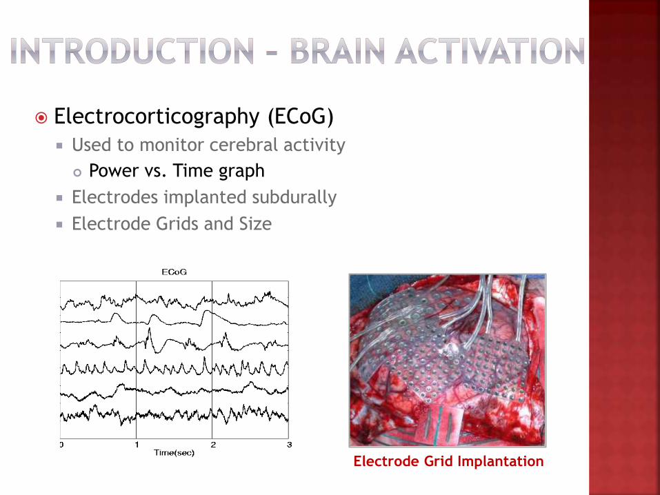
Electrocorticography (ECoG)
Used to monitor cerebral activity
Power vs. Time graph
Electrodes implanted subdurally
Electrode Grids and Size
Electrode Grid Implantation
Functions correspond to regions
Brain reorganization in epileptic patients
Brain Mapping – Relationship between function and
anatomy of brain
Electrocortical Stimulation Mapping (ESM)
Direct electric stimulation of the cortex
“Gold Standard”
Drawbacks
Invasive
Tedious and time consuming
Risk of unnatural seizures
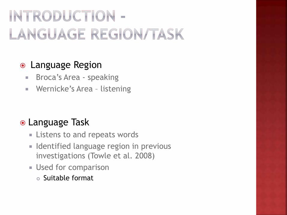
Language Region
Broca’s Area - speaking
Wernicke’s Area – listening
Language Task
Listens to and repeats words
Identified language region in previous
investigations (Towle et al. 2008)
Used for comparison
Suitable format
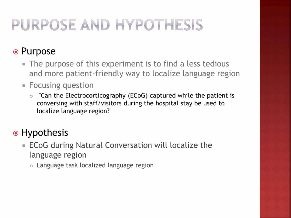
Purpose
The purpose of this experiment is to find a less tedious
and more patient-friendly way to localize language region
Focusing question
"Can the Electrocorticography (ECoG) captured while the patient is
conversing with staff/visitors during the hospital stay be used to
localize language region?"
Hypothesis
ECoG during Natural Conversation will localize the
language region
Language task localized language region

Patient’s video recordings
Video Player
Patient’s ECoG data
PC with MS-Windows &
Ubuntu
Computer Applications
EEGvue
Neuroscan 4.3
Loc3D Jr.
EEG View
MS-Excel
Neuroscan
Loc3D Jr.

Independent Variable Type of method used to localize the language region
Dependent Variable Brain activation (µV)
Constants Patient
Single hospital stay
Electrode grid
Frequency band range
Lobes observed
Comparison Group
Language task
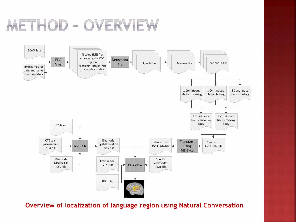
Overview of localization of language region using Natural Conversation
Timestamps for different states from the videos
EEG Vue
Neuroscan 4.3
1 Continuous file for Listening
Neuroscan ASCII Data fileLoc3D Jr
Transpose using
MS-Excel
NeuroscanASCII Data file
CT Scan parameters
INFO file
Electrode Marker File
CSV FileEEG View
1 Continuous file for Talking
1 Continuous file for Resting
1 Continuous file for Listening
Only
1 Continuous file for Talking
Only
Electrode Spatial location
CSV file
Specific electrodesAMP file
REG file
Brain modelVTK file
ECoG data
Epoch FileEpoch FileEpoch FileEpoch FileAverage File
Epoch FileEpoch FileEpoch FileEpoch FileCContinuous File
Nicolet BMSI file containing the EEG
segment<patient>.<state>.<da
te>.<cd#>.<trial#>
Epoch FileEpoch FileEpoch FileEpoch FileEpoch File
CT Scans

Operational definitions for each state
Talking
Listening
Resting
ECoG segments from patient’s videos
Load and position data
Compare and contrast brain activation
CD # Date CD Start CD End Event Start Event End State Duration29 7/27/2004 22:44:48 23:24:21 23:10:26 23:10:43 Listening to Conversation 0:00:17
29 7/27/2004 22:44:48 23:24:21 23:11:52 23:11:55 Listening to Conversation 0:00:03
29 7/27/2004 22:44:48 23:24:21 23:12:45 23:12:53 Talking 0:00:08
29 7/27/2004 22:44:48 23:24:21 23:12:58 23:13:10 Talking 0:00:12
28 7/27/2004 22:05:15 22:44:48 22:20:15 22:20:20 Talking 0:00:05
28 7/27/2004 22:05:15 22:44:48 22:22:21 22:22:41 Listening to Conversation 0:00:20
28 7/27/2004 22:05:15 22:44:48 22:24:44 22:25:16 Listening to Conversation 0:00:32
28 7/27/2004 22:05:15 22:44:48 22:25:18 22:25:25 Talking (w/ hand) 0:00:07
28 7/27/2004 22:05:15 22:44:48 22:25:28 22:25:33 Talking (w/ hand) 0:00:05
28 7/27/2004 22:05:15 22:44:48 22:26:49 22:26:51 Talking 0:00:02
27 7/27/2004 21:25:42 22:05:15 22:01:21 22:01:26 Resting (eyes closed) 0:00:05
27 7/27/2004 21:25:42 22:05:15 22:01:44 22:02:36 Resting (eyes closed) 0:00:52
27 7/27/2004 21:25:42 22:05:15 22:02:40 22:03:02 Resting (eyes closed) 0:00:22
27 7/27/2004 21:25:42 22:05:15 22:03:04 22:03:13 Talking 0:00:09
27 7/27/2004 21:25:42 22:05:15 22:03:38 22:03:53 Talking (w/ hand) 0:00:15
27 7/27/2004 21:25:42 22:05:15 22:04:03 22:04:08 Talking 0:00:05
27 7/27/2004 21:25:42 22:05:15 22:04:18 22:04:23 Talking 0:00:05
26 7/27/2004 20:46:09 21:25:42 20:56:16 20:58:27 Listening to Conversation 0:02:11
25 7/27/2004 20:06:36 20:46:09 20:14:06 20:14:22 Resting (eyes open) 0:00:16
25 7/27/2004 20:06:36 20:46:09 20:14:35 20:16:10 Resting (eyes open) 0:01:35
23 7/27/2004 18:47:30 19:27:03 18:51:25 18:54:45 Resting (eyes open) 0:03:20
23 7/27/2004 18:47:30 19:27:03 18:55:21 18:55:25 Listening to Conversation 0:00:04
23 7/27/2004 18:47:30 19:27:03 18:55:43 18:55:49 Listening to Conversation 0:00:06
21 7/27/2004 17:28:24 18:07:57 17:59:53 18:00:00 Listening to Conversation 0:00:07
21 7/27/2004 17:28:24 18:07:57 18:00:03 18:00:16 Listening to Conversation 0:00:13
20 7/27/2004 16:48:51 17:28:24 16:48:53 16:49:14 Resting (eyes open) 0:00:21
20 7/27/2004 16:48:51 17:28:24 16:49:52 16:50:14 Resting (eyes open) 0:00:22
20 7/27/2004 16:48:51 17:28:24 16:50:17 16:52:51 Resting (eyes open) 0:02:34
18 7/27/2004 15:29:45 16:09:18 15:30:30 15:30:32 Talking 0:00:02
18 7/27/2004 15:29:45 16:09:18 15:30:35 15:30:38 Talking 0:00:03
18 7/27/2004 15:29:45 16:09:18 15:31:05 15:31:20 Talking 0:00:15
18 7/27/2004 15:29:45 16:09:18 15:33:23 15:33:27 Listening to Conversation 0:00:04
18 7/27/2004 15:29:45 16:09:18 15:34:11 15:34:16 Listening to Conversation 0:00:05
17 7/27/2004 14:50:12 15:29:45 14:59:49 14:59:57 Listening to Conversation 0:00:08
17 7/27/2004 14:50:12 15:29:45 15:09:24 15:09:26 Listening to Conversation 0:00:02
17 7/27/2004 14:50:12 15:29:45 15:09:51 15:09:56 Listening to Conversation 0:00:05
16 7/27/2004 14:10:39 14:50:12 14:10:51 14:10:57 Listening to Conversation 0:00:06
16 7/27/2004 14:10:39 14:50:12 14:11:11 14:11:20 Talking 0:00:09
16 7/27/2004 14:10:39 14:50:12 14:11:35 14:11:38 Listening to Conversation 0:00:03
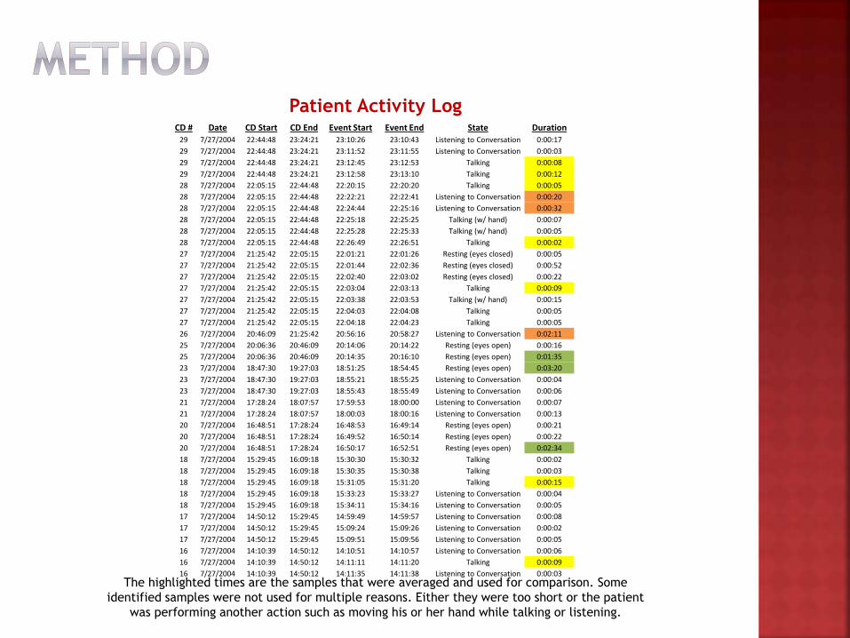
The highlighted times are the samples that were averaged and used for comparison. Some
identified samples were not used for multiple reasons. Either they were too short or the patient
was performing another action such as moving his or her hand while talking or listening.
Patient Activity Log
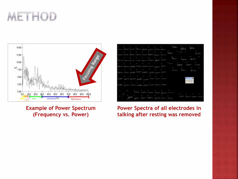
Example of Power Spectrum
(Frequency vs. Power)
Power Spectra of all electrodes in
talking after resting was removed
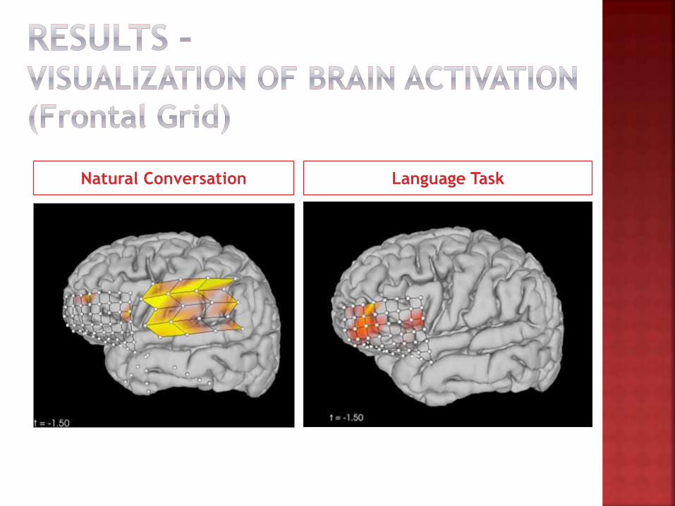
Natural Conversation Language Task
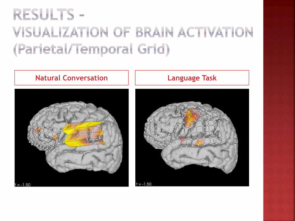
Natural Conversation Language Task

Electrode Activation (Natural Conversation vs. Language Task)
Grid Location Electrode #Natural
ConversationLanguage task
Frontal Grid
33 X -
40 - X
44 - X
45 - X
46 - X
53 X X
Parietal/Upper
Temporal Grid
78 X -
79 - X
80 X X
83 X X
84 X -
88 X -
91 X -
92 X -
95 X X
96 X X
Note: ‘X’ in the column indicates Electrode Activation. ‘–‘ indicates absence of
Electrode Activation. Highlighted in orange are the electrodes that were activated
in both Natural Conversation and Language task.
Hypothesis not supported
Natural conversation could not localize the same
language region that language task did
However, natural conversation activated more regions in
parietal grid
Not ready for clinical applications
Can be used alongside ESM to refine the
technique
Future work
Automated mapping (Ziegler et al. 2011)
Wireless data transmission
Software automation
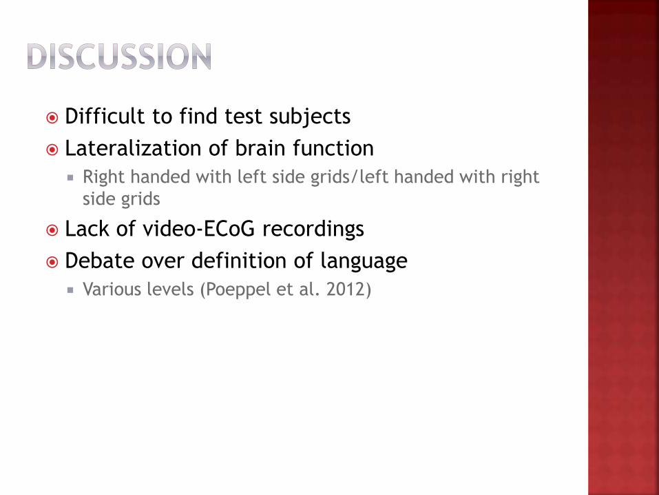
Difficult to find test subjects
Lateralization of brain function
Right handed with left side grids/left handed with right
side grids
Lack of video-ECoG recordings
Debate over definition of language
Various levels (Poeppel et al. 2012)

I would like to thank…
Dr. Vernon Leo Towle, University of Chicago
Falcon Dai & Weili Zheng, University of Chicago
Dr. Judith Scheppler & SIR Department, Illinois
Mathematics and Science Academy
Parents
Bauer P., Vansteensel M., Bleichner M., Hermes D., Ferrier C., Aarnoutse E., & Ramsey N (2013). Mismatch between electrocortical
stimulation and Electrocorticography frequency mapping of language. Brain Stimulation. xxx:1-8.
Borchers S., Himmelbach M., Logothetis N., & Karnath H. (2012). Direct electrical stimulation of human cortex- the gold standard for
mapping brain functions? Nat. Rev. Neurosci. 13:63-70.
Carter R., Aldridge S., Page M., & Parker S. (2009). The Human Brain Book: An illustrated guide to its structure, functions, and
disorders. New York, NY: DK Publishing.
Crone N., Sinai A., Korzeniewska A. (2006). High-frequency gamma oscillations and human brain mapping with electrocorticography.
Prog. Brain Res. 159:275-95.
"Epilepsy". Fact Sheets. World Health Organization. October 2012.
<http://www.who.int/mediacentre/factsheets/fs999/en/index.html> Accessed 24 Jul. 2013.
Gaona C.M., Sharma M., Freudenburg Z.V., Breshears J.D., Bundy D.T., Roland J., Barbour D.L., Schalk G., & Leuthardt E.C. (2011).
Nonuniform high-gamma (60–500 Hz) power changes dissociate cognitive task and anatomy in human cortex. J. Neurosci. 31:2091–2100.
Geschwind N. (1970). The Organization of Language and the Brain. Science. 170:940-944.
Penfield W. (1958). Some mechanisms of consciousness discovered during electrical stimulation of the brain. Proceedings of the National
Academy of Sciences of the United States of America. 44(2):51-56
Poeppel D., Emmorey K., Hickok G., & Pylkkanen L. (2012). Towards a new neurobiology of language. J. Neurosci. 32(41):14125-14131.
Ruescher J., Iljina O., Altenmuller D., Aertsen A., Schulze-Bonhage A., & Ball T. (2013). Somatotopic mapping of natural upper- and
lower-extremity movements and speech production with high gamma Electrocorticography. NeruoImage. 81:164-177.
Sinai A., Bowers C.W., Crainiceanu C.M., Boatman D., Gordon B., Lesser R.P., Lenz F.A., & Crone N.E. (2005). Electrocorticographic high
gamma activity versus electrical cortical stimulation mapping of naming. Brain. 128:1556-1570.
Towle V., Yoon H., Castelle M., Edgar J.C., Biassou N.M., Frim D., Spire J., & Kohrman M. (2008). ECoG gamma activity during a
language task: differentiating expressive and receptive speech areas. Brain. 131:2013-27.
Watkins K. & Paus T. (2004). Modulation of Motor Excitability during Speech perception: The Role of Broca’s Area. Journal of Cognitive
Neuroscience. 16(6):978-987.
Wu M.,Wisneski K., Schalk G., Sharma M., Roland J., Breshears J., Gaona C., Leuthardt E.C. (2010). Electrocorticographic frequency
alteration mapping for extraoperative localization of speech cortex. Neurosurgery 66:407–409.
Ziegler J., Kretzschmar H., Stachniss C., Grisetti G., & Burgard W. (2011). Accurate human motion capture in large areas by combining
IMU- and laser-based people tracking. Intelligent Robots and Systems (IROS). 86–91.