rate normal sinus rhythm st segment … · differential rhythms & arrhythmias etc short pr...
TRANSCRIPT
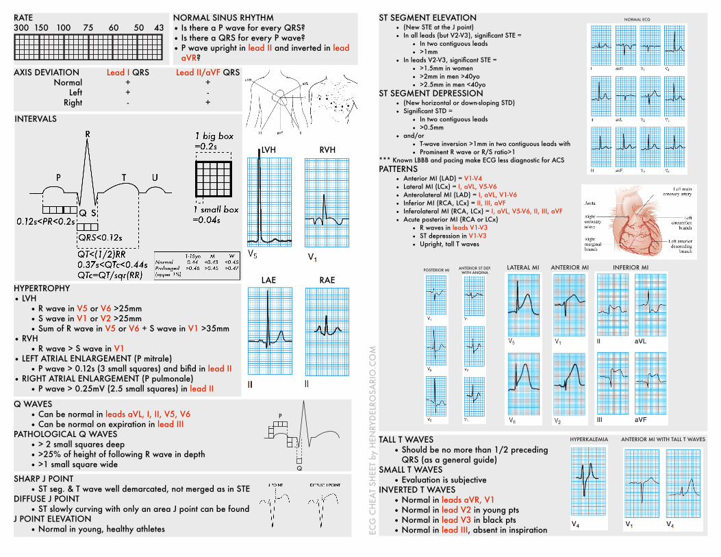
HYPERTROPHY • LVH
• R wave in V5 or V6 >25mm • S wave in V1 or V2 >25mm • Sum of R wave in V5 or V6 + S wave in V1 >35mm
• RVH • R wave > S wave in V1
• LEFT ATRIAL ENLARGEMENT (P mitrale) • P wave > 0.12s (3 small squares) and bifid in lead II
• RIGHT ATRIAL ENLARGEMENT (P pulmonale) • P wave > 0.25mV (2.5 small squares) in lead II
NORMAL SINUS RHYTHM • Is there a P wave for every QRS? • Is there a QRS for every P wave? • P wave upright in lead II and inverted in lead
aVR?
RATE 300 150 100 75 60 50 43
LVH RVH
LAE RAE
AXIS DEVIATION Normal
Left Right
Lead I QRS + + -
Lead II/aVF QRS + - +
Q WAVES • Can be normal in leads aVL, I, II, V5, V6 • Can be normal on expiration in lead III
PATHOLOGICAL Q WAVES • > 2 small squares deep • >25% of height of following R wave in depth • >1 small square wide
ST SEGMENT ELEVATION • (New STE at the J point) • In all leads (but V2-V3), significant STE =
• In two contiguous leads • >1mm
• In leads V2-V3, significant STE = • >1.5mm in women • >2mm in men >40yo • >2.5mm in men <40yo
ST SEGMENT DEPRESSION • (New horizontal or down-sloping STD) • Significant STD =
• In two contiguous leads • >0.5mm
• and/or • T-wave inversion >1mm in two contiguous leads with • Prominent R wave or R/S ratio>1
*** Known LBBB and pacing make ECG less diagnostic for ACS PATTERNS
• Anterior MI (LAD) = V1-V4 • Lateral MI (LCx) = I, aVL, V5-V6 • Anterolateral MI (LAD) = I, aVL, V1-V6 • Inferior MI (RCA, LCx) = II, III, aVF • Inferolateral MI (RCA, LCx) = I, aVL, V5-V6, II, III, aVF • Acute posterior MI (RCA or LCx)
• R waves in leads V1-V3 • ST depression in V1-V3 • Upright, tall T waves
LATERAL MI ANTERIOR MI INFERIOR MIPOSTERIOR MI ANTERIOR ST DEP. WITH ANGINA
NORMAL ECG
TALL T WAVES • Should be no more than 1/2 preceding
QRS (as a general guide) SMALL T WAVES
• Evaluation is subjective INVERTED T WAVES
• Normal in leads aVR, V1 • Normal in lead V2 in young pts • Normal in lead V3 in black pts • Normal in lead III, absent in inspiration
HYPERKALEMIA ANTERIOR MI WITH TALL T WAVES
ECG
CH
EAT
SHEE
T by
HEN
RYD
ELRO
SARI
O.C
OM
Q
P
INTERVALS
SHARP J POINT • ST seg. & T wave well demarcated, not merged as in STE
DIFFUSE J POINT • ST slowly curving with only an area J point can be found
J POINT ELEVATION • Normal in young, healthy athletes
POSTERIOR MI ANTERIOR ST DEP. WITH ANGINA
NORMAL ECG

DIFFERENTIAL
RHYTHMS & ARRHYTHMIAS
ETC
Short PR interval • AV junctional rhythms • WPW syndrome • LGL syndrome
Long PR interval • 1st degree AV block • Ischemic heart disease • Hyperkalemia • Acute rheumatic myocarditis • Lyme disease • Digoxin, quinidine, BB, Ca
blockers
ST segment elevation • ST segment elevation MI • Left ventricular aneurysm • Prinzmetal’s (vasospastic) angina • Pericarditis • High take-off • LBBB • Brugada syndrome
ST segment depression • Acute posterior MI • Myocardial ischemia • Drugs (digoxin, quinidine) • Ventricular hypertrophy + ‘strain’
Pathological Q waves • STEMI • LVH • WPW syndrome • BBB • Pulmonary embolism
Large R or S waves • LVH, RVH • Posterior MI • WPW syndrome • Dextrocardia • BBB
Small QRS complexes • Obesity • Emphysema • Pericardial effusion
Wide QRS complexes • BBB • Ventricular rhythms • Hyperkalemia
Abnormal shaped QRS complexes • Incomplete BBB • Fascicular block • WPW syndrome
J waves present • Hypothermia
Diffuse J point • Early repolarization, LVH with
strain, pericarditis, acute MI
Tall T waves • Hypothermia • Acute MI • Hyperkalemia
Small T waves • Hypokalemia • Pericardial effusion • Hypothyroidism
Inverted T waves • Myocardial ischemia • Myocardial infarction • Ventricular hypertrophy + ‘strain’ • Digoxin toxicity
Short QTc interval • Hereditary short QT syndromes • Hypercalcemia • Digoxin effect • Hyperthermia
Long QTc interval • Hypocalcemia • Drugs (quinidine, procainamide,
amiodarone, sotalol, flecainide, antipsychotics, TCAs, terfenadine, macrolides, quinolones)
• Acute myocarditis • Long QT syndrome
Prominent U waves • Hypokalemia • Hypercalcemia • Hyperthyroidism
Wide P wave • LAE
Tall P wave • RAE
ECG
CH
EAT
SHEE
T by
HEN
RYD
ELRO
SARI
O.C
OM SA nodal rhythms
• Sinus rhythm • Sinus arrhythmia • Sinus tachycardia • Sick sinus syndrome
• Sinus bradycardia • SA block • Sinus arrest
Atrial rhythms • Atrial tachycardia • Atrial flutter • Atrial fibrillation
• P wave for q QRS, QRS for q P wave • HR inc during inspiration • >100bpm • dysfunction of sinus node • <60bpm • P fails, next P where expected • P fails, next P not where expected
• >100bpm, abnormally shaped P waves • sawtooth P, atrial rate 300/min, AV bl. • no P waves, irregularly irregular
Ventricular rhythms • Ventricular tachycardia • Accelerated idioventricular rhythm • Torsades de pointes • Ventricular fibrillation
• broad QRS, 3+ PVCs in a row • broad QRS, HR <120bpm • broad QRS, polymorphic, long QT • no identifiable waves, erratic
SVTs • AV re-entry tachycardia • AV nodal re-entry tachycardia
• narrow QRS, inverted P, P half-buried • narrow QRS, P buried inside QRS
Conduction disturbances • Left bundle branch block • Right bundle branch block • Bifascicular block • Trifascicular block
AV blocks • First-degree • Second-degree
• Mobitz Type I • Mobitz Type II
• Third-degree
• long PR • non-conducted P waves
• progressive lengthening of PR • PR constant
• atria and ventricles are independent
• V1: small Q, R, S; V6: R, S, R’ • V1: tiny R, S, R’; V6: small Q, R, S • left axis dev, left ant. hemiblock, RBBB • bifascicular block, 1st degree AV block
Escape rhythms • AV junctional escape rhythm • Ventricular junct. escape rhythm
• narrow QRS, absent P, 40-60bpm • broad QRS, absent P, 15-40bpm
Ectopic beats • Atrial ectopic beats • AV junctional ectopics • Ventricular ectopics • Bigeminy
• early P wave, abnormal P wave shape • early QRS, narrow QRS • early QRS, broad QRS • ventricular ectopic follows q norm. beat
Pulmonary embolism Pericardial effusion Hypokalemia Hyperkalemia
• S in lead I, Q in III, TWI in III • electrical alternans: variation in R ht. • flat T waves, U waves • peaked T waves, wide QRS, long PR