(rdj downloaded from on february 19, 2019 - published by ... · na2b4o7 11h20perliter. 1 published...
TRANSCRIPT

MANGANESE INTERFERENCE IN THE ABSORPTION ANDTRANSLOCATION OF RADIOACTIVE IRON (FE59)
IN ANANAS COMOSUS (L.) MERR.'
C. P. SIDERIS2
(WITH ONE FIGURE)Received April 24, 1949
IntroductionThe interference of manganese with the availability of iron to pineapple
plants has been explained by JOHNSON (7) and KELLEY (9, 10) as due tothe oxidation of the latter by the former in soil and leaf tissues. Addi-tional studies by SIDERIS, YOUNG and KRAUSS (14) and SIDERIs and YOUNG(17, 20) revealed that iron may be made unavailable in ample amounts tothe leaf tissues after its precipitation in the exodermal root tissues at pHhigher than 5.5, the latter resulting from the absorption by the roots ofanions at higher rates than cations.
All evidence obtained so far indicates that the iron requirements of pine-apple leaves are small; also, differences in the iron content of chlorotic andgreen leaves are, likewise, small.
Therefore, in order to avoid the unicertainty and difficulty of handlingby ordinary chemical analyses the small amounts of iron absorbed by theroots at different periods and to eliminate the interference of preexistingamounts of this element in the tissues of the plants, the latter were grownin solution cultures supplied with iron containing a mixture of the radio-active isotopes Fe55 and Fe59, as well as the other stable isotopes as stated bythe U. S. A. E. C. (28).
Experimental procedureCULTURAL METHODS
The crowns (vegetative organs born on the apex of fruits) of the clonesCayenne and 43-115 and the slips (vegetative organs born on the peduncle)of 10845 and 9429, sent by J. L. Collins to the writer, were suspended innutrient solutions kept in 4-gallon enameled crocks which had been paintedwith asphalt paint to cover possible unenameled spots.
The nutrient solutions, constantly aerated during plant growth, con-tained 0.472 g. Ca (NO3)2 4H20; 0.174 g. K2SO4; 0.080 g. NH4NO3; 0.246g. MgSO4- 7H20; 0.136 g. KH2PO4 0.005 g. ZnSO4 7H20; 0.003 g.Na2B4O7 11H20 per liter.
1 Published with the approval of the Director as Technical Paper No. 179 of thePineapple Research Institute, University of Hawaii.
2 The study was conducted in 1947 during the tenure of a Research Fellowship inthe Division of Plant Nutrition of the University of California, at Berkeley, California.
307
Xf J6 ee. C j4eP(rDJ
www.plantphysiol.orgon May 14, 2019 - Published by Downloaded from Copyright © 1950 American Society of Plant Biologists. All rights reserved.
PLANT PHYSIOLOGY
No iron was added to the nutrient solutions from August 23, the begin-ning day of the experiment, to October 23, the day of the receipt of radio-active iron.3 However, none of the plants showed symptoms of chlorosisexcept clone 43-115, which was of less intense green than the other clones.
On October 23, 1947, two of the plants from a total of 12 per clone pertreatment which were to be treated with radioactive Fe* were placed in2-liter pyrex beakers, one per beaker and others in 4-gallon crocks, five percontainer. The pyrex beakers were shielded from direct sunlight by acover of heavy black paper which was painted on the exposed side withaluminum paint to reflect light. The plants were suspended'for subsequentgrowth in the nutrient solution contained in the pyrex beakers throughholes in a concrete cover which was painted with asphalt paint, the latteremployed to prevent absorption of the solution by the cover.
The sample of the radioactive iron which arrived as FeCl3 in 7.9 ml. ofHCl solution, with 455 mg. of Fe of an activity of 1.5 mc (millicurie)equivalent to 3.31 me per gram and an intensity of radiation (unshielded)of 30 mr/hr at six inches. The total volume of 7.9 ml. of FeCl3 in HClwas poured into a 150 ml. brown bottle containing 20 ml. of 10%7 meta-phosphoric acid (HPO3) to which was added 44.1 ml. of water to raise thetotal volume to 72 ml. Metaphosphoric acid was added to convert ferriciron to the metaphosphate salt which is very soluble, Fe not precip-itable within a wide range of pH. The mixture equivalent to 6.3 mg. ofFe per ml. of solution was employed as a stock solution of Fe* for thenutrient solutions.
The plants grown in the 2-liter pyrex beakers were supplied with 20 yof Fe, and either with lOO.y of manganese as MnSO4 4H20, or without.Those grown in the 4-gallon crocks were supplied with 4.y of Fe, but notwith manganese.
ANALYTICAL METHODS
Sections 3, 4 and the lower portions of 5 of the chlorophyllous regionsof the mature (C) and active (D) leaves and roots were selected for meas-urements of tissue radioactivity on the basis of previous studies (14, 17, 20)which showed that iron accumulations were greater in these than in othertissues of the leaves. The extreme terminal sections of the leaves, althoughricher in iron, were discarded because of the onset of senescence muchearlier in these than in other tis'sues.
Depending on the intensity of the radioactivity of the tissues, the freshweights used for a single determination ranged from one to 25 grams.Such tissues, placed in porcelain evaporating dishes and dried to obtain dryweight values, were then ashed in an oven. The ash, dissolved in 5 ml. of1:1 HICl solution, were transferred to conical' 15 ml. centrifuge tubes
3 This isotope was obtained from the Isotope Division of the U. S. Atomic EnergyCommission at Oak Ridge, Tenn., and contained Fe5' and Fe59, the latter being the moreactive fraction, will be designated in subsequent references as Fe*.
308
www.plantphysiol.orgon May 14, 2019 - Published by Downloaded from Copyright © 1950 American Society of Plant Biologists. All rights reserved.

SIDERIS: 'MANGANESE INTERFERENCE IN IRON ABSORPTION
where sufficient 27Vc NH4OH was added to make the solution alkaline andprecipitate iron. The solution, after standing for 30 minutes, was cen-trifuged at 0° C. The supernatant liquid was discarded and the precipi-tate, after suspension in 2 ml. of acetone, was transferred by a pipette witha wide opening dropwise on to a metal disc of tinned iron 2.5 cm. in diam-eter kept on a hot plate at very low heat. The use of acetone for suspen-sion of the precipitate is for the purpose of rapid evaporation of the mix-ture. In case of an incomplete removal of the precipitate from the cen-trifuge tube and from the pipette, both tube and pipette are treated witha weak solution of HCO placed in the tube, the iron is again reprecipitatedwith NH4OH, centrifuged, and the precipitate transferred to the metaldisc as before.
Great care must be exercised to avoid excessive heating of the metallicdisc because of the danger of losing the precipitate by spattering, which iscaused by a rapid vaporization of the solution. The use of infrared lampsfor drying such precipitates should be preferred to the hot plate method.However, a sheet of asbestos placed over the entire area of the hot plateregulated satisfactorily the heat radiations and the evaporation of the solu-tion. In case of very weak adherence of the dry precipitate to the metallicdisc, a drop or more of 5% sucrose added carefully with a pipette withouttouching the precipitate will prevent possible losses.
The precipitates on metallic discs are ready for measurement of theirradioactivity with a Geiger-Muller counter. The thickness of the micawindow of the tube best suited for such measurements and the distance be-tween the radioactive substance and the tube depend on the intensity andvariety of the radioactive emanations. The thickness of the G-M windowwas approximately 3 mg/cm2, presumably well adapted for the measure-ment of the emanations from Fe59 but not from Fe55.
Due to the much stronger iron content of the root than leaf tissues, theisolated iron from the leaf samples was placed approximately at 0.5 inchfrom the G-M window and that of the root samples 2.0 inches. A radiumstandard placed at the two distances gave 300 and 60 counts per minute forthe 0.5 and 2.0 inch distances, respectively. The background radiationsvaried slightly for one month the entire period of the measurements, rang-ing from seven to nine cts/m. The total ash content of the leaves, consist-ing mostly of K, Ca, Mg and P-salts, was approximately five times as greatas that of the roots. However, the relationship for iron was reversed; thiselement was approximately 20 times as great in the roots as in the leaves,i.e., 3.5 and 70.0 y per cm2 of the metallic disc for the leaves and roots,respectively. Absorption of Fe* by sample thickness, not determined bystandardized quantities of Fe*, was presumably small, due to the smallamounts of total iron contained in the samples.
Cobalt, unavailable at the time of these measurements in the laboratory,could have served as a better standard than radium, which was available,because the latter emits mostly alpha particles and the former beta and
309
www.plantphysiol.orgon May 14, 2019 - Published by Downloaded from Copyright © 1950 American Society of Plant Biologists. All rights reserved.

PLANT PHYSIOLOGY
gamma radiations which, also, are emitted by Fe*. However, for obtain-ing a factor of radiation intensity at different distances between the G-Mcounter and the experimental material, it was thought that the results ob-tained with radium, which is used as aln auxiliary unit for gamma rayemitters (8), could not deviate greatly from those with Co060 or Fe59.
Still a better method would have been the use of Fe* standards of dif-ferent intensities of radioactivity.
The number of counts per minute per 0.01 mg of Fe in solution, 25 daysafter its receipt from Oak Ridge, at 0.5 inch from the G-M tube, was ap-proximately 100. The same distance from the G-M tube was used formeasuring the radioactivity of nearly all the leaf but not the root samples.
The distance for the roots, approximately 2.0 inches, in table II, wasconverted to the 0.5 inch distance by use of the factor obtained with theradium standard. However, the counts for both distances are reported inthis table. It is possible that the converted values in the roots deviate tosome extent due to the difference in the kinds of radiations emitted byFe* and Ra*.
The radiation counts of the leaves all obtained at 0.5 inch distance arereported as such in all tables without conversion by the factor of the Rastandard.
Possible deviations between the leaves of different clones or betweenthe plus Mn and minus Mn cultures of these clones were eliminated by theuse of identical tissue weights and distances from the G-M tube for themeasurement of their radiations.
The radioactivity of the background was determined before and aftereach determination of the radioactivity of the sample. The latter wasmeasured at three successive intervals and the average number of countsper minute minus the average number of the background counts, whichranged from seven to nine per minute, divided by the number of grams offresh weight of the plant tissues, gave the values reported in the varioustables as cts/m/g (counts per minute per gram).
The same conditions of uniformity of tissue weights were adopted forthe roots, except the distance from the G-M counter, which was 2.0 inchesfor all root measurements.
Thus in table II the values under roots, ets/m/g, indicate those actuallyobtained at the 2.0 inch distance and those calculated at the 0.5 inch dis-tance by the factor of the Ra standard.
FRACTIONATION OF CELLULAR COMPONENTS
The possibility of a greater association of iron with certain cellular com-ponents than with others suggested the idea of fractionating leaf tissuesinto classes characterized by certain chemical and physical properties. Forthis purpose, leaf tissues mixed with a solution of potassium hydroxidewere macerated in a Waring Blendor. The resulting pulpy mixture was
310
www.plantphysiol.orgon May 14, 2019 - Published by Downloaded from Copyright © 1950 American Society of Plant Biologists. All rights reserved.

SIDERIS: MANGANESE INTERFERENCE IN IRON ABSORPTION
separated into solid and liquid portions and the proteinaceous matter re-covered from the latter by precipitation with metaphosphoric acid. De-tails of this procedure are, as follows:
Fifty gm. of leaf tissues were suspended in 100 ml. of 0.05N KOH in aWaring Blendor and macerated for one minute. At the end of this period,the acidity of the mixture was examined and more KOR was added to raisethe pH above 7.0 but below 8.0 and the maceration was continued for 10minutes longer. At the end of this period, the mixture was transferred tothe coarse (not nappy) side of a piece of Canton flannel held in a 6-inchfunnel and the liquid was extracted therefrom by manual pressure. Theresidue in the flannel was wetted with 0.05N KOH and squeezed four timesin succession until a final volume of 250 ml. was obtained.
UI
a.
04
,
coA
H
:3
2.5
2.01
1.51
1.01
.5
I I II I I I I I I I I I I I
_~~~~~~~
* /. , a
*/ r .0.80/ ~~~~~~~~~~~~t* 3.098 re
a/ t~- 2.660 fc.,, . ~~~~~~~~atP.01
*/ .
C a
'I rl l l I I l I
0 .1 .2 .3
equiringor significance
I I I.4. .5 .6 .7 .8 .9 1.0 1.1 1.2 1.3 1.4 1.5 1.6
MICROGRAMS O F I R O NFIG. 1. Correlation of iron and protein.
The residue in the flannel, composed mostly of cell wall material ad-mixed with some proteinaceous matter, fibers and other insoluble substances,was carefully removed with a spatula, weighed and divided into two equalportions, one of these used for the determination of proteinaceous nitrogenby the Kjeldahl method, and the other for dry weight with subsequentmeasurement of its radioactivity after ashing by the method as mentionedabove. The latter fraction should have been washed with metaphosphoricacid to remove possible traces of precipitated ferric iron after treatmentwith KOH, but due to oversight in the beginning and lack of ample timein the end of this study, such treatment was omitted.
311
www.plantphysiol.orgon May 14, 2019 - Published by Downloaded from Copyright © 1950 American Society of Plant Biologists. All rights reserved.
PLANT PHYSIOLOGY
The liquid fraction was centrifuged in 250 ml. centrifuge bottles to re-move starch and traces of fibrous and parenchymatous tissues and withsome chloroplastic matter. The supernatant liquid was carefully removed,its volume determined and then divided into two equal parts which wereplaced in separate 250 ml. centrifuge bottles, one intended for the determi-nation of nitrogen and the other, after ashing, for radioactivity in the pro-teinaceous matter. These liquids after treatment with appropriate volumesof 10% metaphosphoric acid to cause complete precipitation of the pro-teinaceous-colloidal matter in suspension were centrifuged for 20 minutesin a centrifuge kept at 0° C. The supernatant colorless or slightly greenishyellow but clear liquid was decanted carefully in a flask and then theproteinaceous precipitate in the centrifuge bottles was treated with 50 ml.of 1% metaphosphoric acid and recentrifuged to remove traces of watersoluble Fe59. At the end of this operation, the supernatant liquid wasplaced in the flask which contained the liquids from the previous operation.
The precipitates in the bottles, loosened by gentle centrifugal move-ments of the hand after the addition of 5 ml. or more of 0.O1N KOH, werepoured in appropriate containers; that for the determination of pro-teinaceous nitrogen in a Kjeldahl flask and the other for radioactivity in a250 ml. porcelain dish. The latter, after ashing and precipitation of Fe59with NH4OH, was transferred to metallic discs and its radioactivity meas-ured as stated above.
The liquid fraction from the separation of the proteinaceous matterwas divided into two equal volumes, each placed in 250 ml. centrifugedbottles and treated with acetone in the ratio of one to two that is, 80 ml. ofthe liquid fra'ction of 160 ml. of acetone. The mixtures in the centrifugebottles, were placed for three hours in a refrigerator at -23° C and thencentrifuged at 00 C for 20 minutes.
The supernatant liquids were carefully decanted into clean 250 ml. cen-trifuge bottles while the precipitates, with strong peroxidase activity, weredissolved in 10 ml. 0.O1N KOH solution and transferred to appropriatecontainers for the determination of proteinaceous nitrogen and radioactiv-ity as mentioned above. The supernatant liquids from this operation,treated in the 250 ml. centrifuge bottles with a few ml. of 27% NH4OHto precipitate Fe59, were centrifuged. The resulting supernatant liquidwas discarded and the precipitate, dissolved in 5-10 ml. of 1: 1 HCI,was transferred to 15 ml. conical centrifuge tubes, where Fe59 was pre-cipitated out with a few ml. of 27% NH4OH, then centrifuged and theprecipitate transferred to metallic discs, as stated above.
The various fractions obtained by the above procedure, although notpure chemical entities but mixtures of related substances, are designated inthe text as cellulose-lignin (cell wall and fibers), protein, peroxidase andinorganic Fe59 fractions.
312
www.plantphysiol.orgon May 14, 2019 - Published by Downloaded from Copyright © 1950 American Society of Plant Biologists. All rights reserved.

SIDERIS: MANGANESE INTERFERENCE IN IRON ABSORPTION
PLANT GROWTH
During the 2-month period of growth (August 23 to October 23) beforethe addition of Fe59 to the cultures, it became obvious that the rate ofgrowth as measured by the length, width and number of leaves per plantwas different for the various clones. Also, the number and length of theroots and other related morphological features such as thickness, lateralroots and root hairs, likewise, differed in the various clones.
Clone 43-115 produced considerably longer leaves, and fewer roots thanCayenne. Also, the roots of 43-115 were thinner and carried fewer lat-erals than Cayenne, which may account in part for certain differences inthe absorption of nutrient elements between these clones, as reported below.Clones 10845 and 9429 produced fewer leaves and roots than Cayenne butthe roots in the former were equipped with more but very slender lateralsthan in the latter.
Plant weights, calculated on the new growth (new tissues) of 10 leavesattained between August 23 and November 3, 1947, are reported in table I.
TABLE IWEIGHTS OF THE NEW GROWTH OF TEN LEAVES PRODUCED BY DIFFERENT CLONES GROWN IN
NUTRIENT SOLUTIONS WITHOUT ADDED IRON AT BERKELEY, CALIFORNIA,FROM AUGUST 23 TO NOVEMBER 3, 1947
LEAF WEIGHTS (FRESH)CLONES
g- s. %*
Cayenne ... .. ... .... 186.0 98.543-115 ....................... 188.2 100.010845..... .. 112.5 59.89429 .... ..... .. 97.5 51.9
* Calculated as per cent. of 188.2 of clone 43-115.
The values were obtained by weighing 10 of the mature (C) and active (D)groups from which small terminal sections representing tissues from growthbefore August 23 had been severed.
The data showing very great differences between the weights of Cayenneand 43-115, on the one side, and of 10845 and 9429 on the other, may havebeen responsible for the different rates of absorption of mineral elementsfrom the nutrient solutions or their dilution in plant tissues.
RADIOACTIVITY OF PLANT TISSUESThe data in table II, indicating the radioactivity of Fe59 in the leaf and
root tissues of different cultures, are reported as counts (ct) per minute(m) per gram (g) of fresh tissue (8).
All data are based on 10 grams of fresh tissue from two leaves perplant and two roots per plant, the latter weighing from one to two gramsafter removal with blotting paper of the superficial water of the nutrientsolution.
313
www.plantphysiol.orgon May 14, 2019 - Published by Downloaded from Copyright © 1950 American Society of Plant Biologists. All rights reserved.
PLANT PHYSIOLOGY
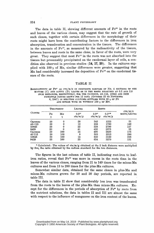
The data in table II, showing different amounts of Fe59 in the rootsand leaves of the various clones, may suggest that the rate of growth ofsuch clones, together with certain differences in the morphology of theirroots might have been the contributing factors to the differences in ironabsorption, translocation and concentration in the tissues. The differencesin the amounts of Fe59, as measured by the radioactivity of the tissues,between leaves and roots in the same clone, in favor of the roots, were verygreat. They suggest that most Fe59 in the roots was not absorbed into thetissues but presumably precipitated on the exodermal layer of cells, a con-dition also observed in previous studies (14, 17, 20). In the cultures sup-plied with 100 y of Mn, similar differences were greater, suggesting thatMn had considerably increased the deposition of Fe59 on the exodermal tis-sues of the roots.
TABLE II
RADIOACTIVITY OF} FE* AS CTS/M/G IN COMPOSITE SAMPLES OF No. 4 SECTIONS OF THEMATURE (C) AND ACTIVE (D) LEAVES OR IN THE ROOTS MEASURED AT 0.5 AND 2.0
INCH DISTANCES, RESPECTIVELY, FOR THE CAYENNE, 43-115, 10845 AND 9429PINEAPPLE-CLONES GROWN FOR 15 DAYS (OCTOBER 23 TO NOVEMBER
6, 1947) IN SOLUTION CULTURES SUPPLIED WITH 20 Y oF' FEAND EITHER WITH OR WITHOUT 100 -y OF MN.
TREATMENT LEAVES ROOTSCLONES CTS/M/G
FE MN 0.5" 2.0" 0.5"f* ROOTS/LEAVEScts/m/g cts/m/g cts/m/g
Cayenne 20 0 58 245 1225 2143-115 20 0 12 358 1790 14910845 20 0 22 558 2790 1279429 20 0 41 455 2275 55Cayenne 20 100 14 490 2450 17543-115 20 100 9 503 2515 28010845 20 100 14 630 3150 2259429 20 100 25 654 3270 131
* Calculated: The values of cts/m/g obtained at the 2 inch distance were multipliedby five, the ratio obtained by the radium standard for the two distances.
The figures in the last column of table II, indicating root-iron to leaf-iron ratios, reveal that Fe59 was more in excess in the roots than in theleaves of the various clones, ranging from 21 to 149 times for the minus-Mncultures and from 13 to 280 times for the plus-Mn cultures.
Somewhat similar data, obtained for the same clones in plus-Mn andminus-Mn cultures grown for 20 and 30 day periods, are reported intable III.
The data in table II show that considerably less iron was translocatedfrom the roots to the leaves of the plus-Mn than minus-Mn cultures. Ex-cept for the differences in the periods of absorption of Fe* by roots fromthe nutrient solutions, the data in tables II and III are almost the samewith respect to the influence of manganese on the iron content of the leaves.
314
www.plantphysiol.orgon May 14, 2019 - Published by Downloaded from Copyright © 1950 American Society of Plant Biologists. All rights reserved.
SIDERIS: MIANGANESE INTERFERENCE IN IRON ABSORPTION
A radioautograph, not presented here, showed that Fe* was distributedthroughout the entire leaf area although in somewhat greater amountstoward the terminal than basal regions. A semicircular zone about 2.5 cm.from the tip of the leaf, indicating the line of demarkation between thetissues of the old and new growth, accumulated more iron than other areas.Although the area of the old growth between the tip and line of demarka-tion contained some iron, the radioautograph did not reveal any greaterconcentrations in the fibrovascular than in the mesophyllic tissues as shownoccasionally in chlorotic leaves by the formation of more chlorophyll alongthe leaf veins, following iron absorption through the roots, than in thenearby mesophyllic tissues. It is possible that the even distribution of theiron radiations in the leaf tissues resulted from the great thickness of theleaf, about 3 mm. which interfered with their outward penetration andrecording on photographic film. Such radiations were highly intensifiedat the edges of the leaf, where the thickness was approximately 1 mm.
TABLE IIIRADIOACTIVITY OF FE* AS CTS/M/G IN THE CHLOROPHYLLOUS SECTIONS OF THE LEAVES OF
CAYENNE, 43-115, 10845 AND 9429 PINEAPPLE CLONES GROWN IN SOLUTION CULTURESFOR 20 OR 30 DAYS (OCTOBER 23 TO NOVEMBER 23, OR OCTOBER 23 TO
NOVEMBER 23, 1947) SUPPLIED WITH 20 y OF FE AND EITHERWITH OR WITHOUT 100 y OF MN.
20-days 30 days
CULTURES FE = 20 CULTURES FE = 20PINEAPPLE --T-CLONES MINUS PLUS RATIOS MINUS PLUS Mn = Oy
Mn Mn MN=O1 y: MN MN MN=Oy:cts/m/g cts/m/g
Cayenne 121 lost ......... 118 44 2.6843-115 46 20 2.30 51 38 1.3410845 32 15 2.13 ........ ...... .....
9429 55 38 1.45 ....... ...... .........
Previous studies (14, 15, 16, 17, 18, 19, 20, 21, 22) have indicated thatiron, chlorophyll and protein gradients increased from the basal to the ter-minal sections of leaves, suggesting presumably some association betweeniron and either chlorophyll or protein, or both. Because of the existenceof various metallo-proteins (metalloporphyrins) in plants and animals, cer-tain analyses were made to obtain pertinent information, which is reportedin table IV.
The data in table IV show that the radioactivity in the protein andperoxidase fractions of the 23-day plants differed considerably from thatof the 30-day plants in the Cayenne clone. Lack of time prevented the writerfrom repeating the analyses to detect the source of disagreement in theperoxidase fraction.
However, the data in the 23-day cultures show considerably greater
315
www.plantphysiol.orgon May 14, 2019 - Published by Downloaded from Copyright © 1950 American Society of Plant Biologists. All rights reserved.

PLANT PHYSIOLOGY
amounts of Fe59 in the various cellular components for the Cayenne thanfor the 43-115 clone, which conform with the results in table III. The pro-tein fraction, which may be considered the least contaminated with inorganicFe59, possessed greater radioactivity than the cellulosic-ligneous fraction,although the latter was four to five times greater in weight than the former.The high values of the peroxidase fraction in the 23-day plants cannot beexplained.
The radioactivity of the 30-day plants, although varying slightly fromthe 23-day plants, shows, except for the peroxidase fraction, the same rela-tive distribution of iron in the various cellular components. The nitrogenvalues, intended to reveal the distribution of proteinaceous matter in thevarious fractions, show that the greatest portion of Fe59 was in the protein;other fractions contained much smaller amounts.
TABLE IVRADIOACTIVITY OF FE* IN VARIOUS CELLULAR COMPONENTS AS CTS/M/G OF FRESH LEAF TIS-
SUE OR OF DRY CELLULAR COMPONENTS AND MILLIGRAMS OF PROTEIN NITROGEN PERGRAM OF FRESH LEAF TISSUE, IN CAYENNE AND 43-115 PINEAPPLE CLONES
GROWN IN SOLUTION CULTURES WITH 4 Y OF FE FOR 23 AND 30 DAYS
23 DAYS 30 DAYS-CAYENNEMATURE (C) LEAVES ACTIVE (D) LEAVES
CELLULAR CAYENNE 43 115 FRESH TISSUES DRY TISSUESCOMPONENTS
N WEIGHT CTS/M/Gcts/m/g cts/m/g cts/m/g rng/g gi. % gis.
Cell. x Lign* 16.50 2.33 17.0 0.266 0.355 81 48.0Proteint 16.80 3.80 11.2 1.410 0.083 19 135.0Peroxidase 16.80** 6.70 4.0 0.112 ......
Inorganic 1.0
* Cell. x Lign. = Cellulosie and Ligneous (cell walls and fibers).** Peroxidase precipitate not ashed but dried and radioactivity measured.
t Impure preparation.
The Fe59 content, as indicated by the radioactivity in the various frac-tions, does not correlate positively with the protein-N of the fractions whencalculated per unit of fresh leaf tissue. However, if such radioactivity iscalculated per unit (gram- weight) of their respective cellular components,as in the last column in table IV, it increases directly with the amounts ofthe proteinaceous fraction, suggesting that iron is associated, although notwith the catholic proteinaceous content of the cell, at least with a certainsmall section of this fraction.
The question has often been raised (2, 7, 9, 10, 26, 27) whether iron isrendered unavailable by oxidation with subsequent precipitation by man-ganese in the nutrient solution or soil before absorption by plant roots orafter absorption. In order to obtain information on this subject, 5 ml. ofthe nutrient solution from the plus-Mn and minus-Mn cultures, exposed to
316
www.plantphysiol.orgon May 14, 2019 - Published by Downloaded from Copyright © 1950 American Society of Plant Biologists. All rights reserved.

SIDERIS: MIANTGANESE INTERFERENCE IN IRON ABSORPTION
the action of plant roots for 23 days at pH 4.5-6.0, were placed in containerswhich were examined under the Geiger-Muller counter for radioactivity.The results of these findings, indicating for the Cayenne 445 cts/m/g inthe minus-Mn and 411 cts/m/g in the plus-Mn cultures, and for clone 43-115, 452 ets/m/g in the minus-Mn and 460 cts/m/g in the plus-Mn cultures,reveal no significant differences between the minus-Mn and plus-Mn cul-tures.
The data might indicate that there were hardly sufficient changes to makecertaini that precipitation of Fe by Mn had taken place in the nutrient solu-tion. Although the conditions were unsuitable for the oxidation and subse-quent precipitation of Fe59 in solution cultures supplied with metaphosphateas the sole source of PO4 and at acidities ranging mostly from pH 4.0 to 5.5,nevertheless, depositions of Fe59 took place on the exodermal root tissuesmore so in cultures supplied with Mn than without. Therefore, the resultssuggest that certain conditions on the root surface, not yet satisfactorily ex-plainable, develop in the absence as well as in the presence of Mn duringthe absorption of iron which are primarily responsible for the oxidation andprecipitation of iron. The presence of Mn increases the amounts of Fe pre-cipitation. However, such precipitated iron may be made slowly availableto plants by the action of acids resulting from a greater rate of absorptionof cations than anions or by reducing agents (14, 17, 20).
Discussion
The results presented above indicate that in nutrient cultures suppliedwith or without manganese, iron was deposited in great amounts in theexodermal tissues of pineapple roots, some of which was translocated to theleaves but the greatest portion remained insoluble and consequently un-available. In cultures with high concentrations of Mn (100 y), the amountsof iron deposited in the exodermal root tissues increased and those trans-located to the leaves decreased.
Because of the deposition of great amounts of iron in the exodermal tis-sues of roots in the absence of Mn, which may be increased considerably inits presence, the causes for the precipitation and deposition of iron cannotbe alone ascribed to the action of Mn. Such causes, inherent in the exo-dermal tissues of the roots, are only augmented in the presence of Mn.
Previous studies (14, 17, 20) showed that high pH values generated bya greater rate of absorption by roots of anions than cations (NO3 vs. K orCa) contributed as much to the precipitation and deposition of Fe in theexodermal tissues of roots and to the low amounts in the leaves as highamounts of Mn in the nutrient solution.
Possible mechanisms for explaining the deposition of iron in the exo-dermal tissues of roots grown in solution cultures supplied with or withoutMn may be attributed to (a) the alkalinity of the products of hydrolysis of
317
www.plantphysiol.orgon May 14, 2019 - Published by Downloaded from Copyright © 1950 American Society of Plant Biologists. All rights reserved.

PLANT PHYSIOLOGY
bicarbonate ions (HOG3) formed from CO2 by respiring roots, and (b) theoxidation of iron by manganese according to the reaction:
Fe++ + Mn+++ Fe+++ + Mn++Both reactions, resulting in the oxidation of Fe++ to Fe+++, are possible
in soils of higher pH than 5.5 and in the exodermal tissues of roots but notas much in the chlorophyllous tissues of leaves with high acidity (pH 3.4-4.6) and reducing agents such as ascorbic acid (15, 18, 21).
The production of chlorosis in soybean plants only by certain iron andmanganese combinations and not by others caused SOMERS and SHIvE (25)to assume that good growth and development of plants free from pathologi-cal symptoms depended on a two to one ratio of iron to manganese withinthe plant tissues and in the nutrient solution, and that ratios with valueshigher than two produced iron toxicity, and lower than two, manganesetoxicity. A similar view has also been shared by TWYMAN (27). However,BENNETT (1) found in tomato plants that ratios of iron to manganese rang-ing from 1.6 to 0.1, with maximum Fe at 50 y and Mn at 80 -y, and withminimum Fe at 5 y and Mn at 5 y produced green leaves, whereas similarratios ranging from 0.08 to 0.01, with maximum Fe at 8 y and Mn at 180 y,and with minimum Fe at 0.5 y and Mn at 50 y produced chlorotic plants.Unpublished data of the writer have shown that iron to manganese ratioshigher than 0.01, with maximum Fe at 5 y and Mn at 50 y, and with mini-mum Fe at 0.5 y and Mn at 5 y produced green plants, but similar ratios of0.001 with Fe at 0.05 y and Mn at 50 y produced chlorotic plants.
The above comparisons show that different plants may require differentiron-manganese ratios because according to previous studies (14, 17, 20),the absorption and translocation of iron may depend on the acidity of theexodermal root tissues, which is affected by the ratio of cations to anionsentering the roots simiultaneously. Moreover, the rate of utilization ofeither Fe or Mn in the tissues for the synthesis of metalloproteins may de-termine the rate of translocation of these elements from roots to leaves. Thetoxicity theory (25, 27) at concentrations 50y of Mn or 5 y Fe, althoughpossible, cannot be easily understood, since the writer witnessed pineappleplants grown by J. P. Bennett in solution cultures supplied with 300 y ofMn and some even with higher amounts, unless such plants were not absorb-ing Mn at the rate of supply.
The results in table IV, showing a certain degree of association of ironto protein, and previous studies (14, 15, 16), summarized in figure 1, indi-cating positive correlation between iron and protein (r = 0.80 and t = 3.098,requiring for significance t = 2.660 at P.01), suggest that iron may be instoichiometric relationship with a certain section but not with the entireproteinaceous fraction of the cell. However, with future improvements inthe subdivision of the proteinaceous fraction, the iron-containing sectionmay be isolated and identified.
Protein, chlorophyll and iron relationships in a variegated variety ofAnanas comosus (L.) Merr. are reported in table V.
318
www.plantphysiol.orgon May 14, 2019 - Published by Downloaded from Copyright © 1950 American Society of Plant Biologists. All rights reserved.

SIDERIS: MANGANESE INTERFERENCE IN IRON ABSORPTION
The data indicate that with greater amounts of chlorophyll in the greenthan chlorotic areas of the same leaves both protein and iron increased, butnot in stoichiometric proportions.
The occurrence of iron in the chloroplasts of various plants has been re-ported by MOOR (13). Similar findings were reported for the chloroplastsof Zea mays and Hordeum distichum by GRIESSMEYER (3), for spinach byLIEBICH (11) and Claytonia by HILL and LEHMAN (5). JACOBSON (6)found in tobacco that from 65 to 85% of the total leaf iron was in the chloro-plasts. Also, MOMMAERTS (12) and JACOBSON (6) found iron in the purifiedchlorophyll protein complex substances of corn and tobacco leaves, whichresults are in agreement with the data in table V for pineapple leaves.
These results and those of others indicating certain relationships betweenchlorophyll, protein and iron suggest that all three substances must bear astoichiometric relationship in a certain section of the proteinaceous complexin the chloroplast, the identity of which has not yet been definitely estab-lished.
TABLE VCHLOROPHYLL, PROTEIN-N AND IRON IN GREEN AND CHLOROTIC AREAS OF THE LEAVES OF A
VARIEGATED STRAIN OF Ananas comosus (L.) MERR.
LEAF AREAS RATIOCELLULAR COMPONENTS
OF DRIED TISSUES GREEN CHLOROTIC GREEN:MG/G MG/G CHLOR.
Chlorophyll ............. ........... 2.76 0.38 7.25Protein ........ .................. 88.00 57.80 1.47Iron ....... .................... 0.0095 0.0041 2.32
SummaryThe following results were obtained from experiments on the absorption
and translocation of radioactive iron (Fe59) in pineapple plants grown innutrient solutions supplied with 20 y or 4.y of Fe59, and either with 100 yor without manganese.
1. Most iron removed from the nutrient solution was deposited in theroots, presumably in the exodermal tissues, more so in the cultures suppliedwith than without manganese.
2. The amounts of translocated iron from the roots to the leaves wasconsiderably lower in the cultures supplied with than without manganese.
3. No precipitation of iron could be detected with certainty in the nutri-ent solutions containing manganese as a result of the reaction Fe++ + M-+++= Fe++ + Mn++.
4. Considerable amounts of the translocated iron remained in the pro-teinaceous matter of the cells and others in the cell walls and substanceswith peroxidase activity.
5. The data suggest that iron may occur in combination with some pro-teinaceous fraction, presumably an enzyme, which activates the formationof certain other proteins intimately related with chlorophyll.
319
www.plantphysiol.orgon May 14, 2019 - Published by Downloaded from Copyright © 1950 American Society of Plant Biologists. All rights reserved.
PLANT PHYSIOLOGY
The writer wishes to express his appreciation and thanks to Drs. J. P.Bennett, L. Jacobson, H. A. Barker, Professor D. R. Hoagland and W. H:Dore of the Plant Nutrition Department and Dr. Joseph Hamilton of theRadiation Laboratories at the University of California, who helped in vari-ous ways for the success of this work.
Also, thanks are due to the Isotopes Division of the U. S. Atomic EnergyCommission at Oak Ridge, Tennessee, for the supply of Fe59.
PINEAPPLE RESEARCH INSTITUTEUNIVERSITY OF HAWAIIHONOLULU, T. H.
LITERATURE CITED
1. BENNETT, J. P. Iron in leaves. Soil Sci. 60: 91-105. 1945.2. GILE, P. L. Chlorosis of pineapples induced by manganese and car-
bonate of lime. Science (N. S.) 44: 855-857. 1916.3. GRIESSMEYER, H. Uber experimentelle Beeinflussung des Eisens im
chloroplasten. Planta 11: 331-358. 1930.4. HANSON, E. A. Some properties of the chlorophyll in relation to its
biological function. Dissert. Univ. Leyden. 1938.5. HILL, R., and LEHMAN, H. Studies on iron in plants with special ob-
servations on the chlorophyll :iron ratio. Biochem. Jour. 35:1190-1199. 1941.
6. JACOBSON, L. Iron in the leaves and chloroplasts of some plants inrelation to their chlorophyll content. Plant Physiol. 20: 233-245.1945.
7. JOHNSON, M. 0. Manganese chlorosis of pineapples: its cause and con-trol. Hawaii Agric. Expt. Sta. Bull. No. 52. 1924.
8. KAMEN, M. D. Radioactive Tracers in Biology. Academic Press Inc.New York, 1947.
9. KELLEY, W. P. The influence of manganese on the growth of pine-apples. Hawaii Agric. Expt. Sta. Press Bull. No. 23. 1909.
10. KELLEY, W. P. The function and distribution of manganese in plantsand soils. Hawaii Agric. Expt. Sta. Bull. No. 28. 1912.
11. LIEBICH, H. A quantitative chemical investigation of the iron, in thechloroplasts and other cell components of Spinacia oleracea.Zeitschr. Bot. 37: 129-157. 1941; Chem. Zentr. 1: 884. 1942.
12. MOMMAERTS, W. F. H. N. Some chemical properties of the plastid-granum. Proc. Kon. Akad. Wetensch. Amsterdam 41: 896-903.1938.
13. MOOR, B. The presence of inorganic compounds in chloroplasts of greencells of plants. Proc. Roy. Soc. (London) B-87: 556-570. 1914.
14. SIDERIS, C. P., YOUNG, H. Y. and KERAUSS, B. H. Effects of iron on thegrowth and ash constituents of Ananas comosus (L.) Merr. PlantPhysiol. 18: 608-632. 1943.
320
www.plantphysiol.orgon May 14, 2019 - Published by Downloaded from Copyright © 1950 American Society of Plant Biologists. All rights reserved.
SIDERIS: MANGANESE INTERFERENCE IN IRON ABSORPTION
15. SIDERIS, C. P. and YOUNG, H. Y. Effects of iron on chlorophyllous pig-ments, ascorbic acid, acidity and carbohydrates of Ananas comnosuts(L.) Merr., supplied with nitrate or ammonium salts. PlantPhysiol. 19: 52-75. 1944.
16. SIDERIS, C. P. and YOUNG, H. Y. Effects of iron on certain nitrogenousfractions of Ananas comosus (L.) Merr. Plant Physiol. 21: 75-94.1946.
17. SIDERIS, C. P. and YOUNG, H. Y. Effects of different amounts of potas-sium on growth and ash constituents of Ananas contosus (L.) Mlerr.Plant Physiol. 20: 609-630. 1945.
18. SIDERIS, C. P. and YOUNG, H. Y. Effects of potassium on chlorophyll,acidity, ascorbic acid and carbohydrates of Ananas comosus (L.)Merr. Plant Physiol. 20: 649-670. 1945.
19. SIDERIS, C. P. and YOUNG, H. Y. Effects of potassium on nitrogenousconstituents of Ananas comosus (L.) Merr. Plant Physiol. 21: 218-232. 1946.
20. SIDERIS, C. P. and YOUNG, H. Y. Effects of nitrogen on growth and ashconstituents of Ananas comosus (L.) Merr. Plant Physiol. 21: 247-270. 1946.
21. SIDERIS, C. P. and YOUNG, H. Y. Effects of nitrogen on chlorophyll,acidity, ascorbic acid and carbohydrates of Ananas comosus (L.)Merr. Plant Physiol. 22: 97-116. 1947.
22. SIDERIS, C. P. and YOUNG, H. Y. Effects of nitrogen on the nitrogenousconstituents of Ananas comosus (L.) Merr. Plant Physiol. 22:127-148. 1947.
23. SIDERIS, C. P. Chlorophyll and protein interrelationships in Ananascomosus (L.) Merr. Plant Physiol. 22: 160-173. 1947.
24. SMITH, E. I. The chlorophyll-protein compounds of the green leaf.Jour. Gen. Physiol. 24: 565-582. 1941.
25. SOMERS, I. I. and SHIVE, J. W. The iron-manganese relation in plantmetabolism. Plant Physiol. 17: 582-602. 1942.
26. TOTTINGHAM, W. E. and BECK, A. J. Antagonism between manganeseand iron in the growth of wheat. Plant World 19: 359-370. 1916.
27. TWYMAN, E. S. The iron-manganese balance and its effect on thegrowth and development of plants. The New Phytol. 45: 18-24.1946.
28. U. S. ATOMIC ENERGY COMMISSION. Availability of radio isotopes withincreased specific activities. Isotopes Division Circular E-11. OakRidge, Tennessee.
321.
www.plantphysiol.orgon May 14, 2019 - Published by Downloaded from Copyright © 1950 American Society of Plant Biologists. All rights reserved.