re-epithelization and density of ... - bio-conferences.org
TRANSCRIPT

Re-epithelization and density of collagen fibers on wound healing of mice’s skin (Mus musculus) that treated with combination of chitosan membrane and eel (Monopterus albus) mucous
Deaoxi Renaschantika Djatumurti1, Afifatur Rafida1, Asha Yonika Putri Manalu2, and Tri
Wahyu Pangestiningsih3
1Undergraduate student, Faculty of Veterinary Medicine, Universitas Gadjah Mada, Jl. Fauna, No. 2,
Karangmalang, Catur Tunggal, Depok, Sleman, Daerah Istimewa Yogyakarta, Indonesia. 55281 2Undergraduate student, Faculty of Medicine, Public Health and Nursing, Universitas Gadjah Mada,
Jl. Farmako Sekip Utara, Sleman, Daerah Istimewa Yogyakarta, Indonesia. 55281 3Anatomy Department, Faculty of Veterinary Medicine, Universitas Gadjah Mada, Jl. Fauna, No. 2,
Karangmalang, Catur Tunggal, Depok, Sleman, Daerah Istimewa Yogyakarta, Indonesia. 55281
Abstract. Untreated skin wound can inhibit the wound healing process.
Chitosan and eel mucous have biodegradable, biocompatible, and
antimicrobial properties. This study aimed to determined the potential of
combination between chitosan membrane and eel mucous extract in skin
wound re-epithelization and collagen fibers deposition. Thirty adult male
mice were divided into 5 groups, 6 animals/group. Skin wound were
perform by punch biopsy in 0.5 cm diameter. Group I-IV respectively
recieved: 10% povidone iodine; eel mucus extract and gauze; combination
of 1 % chitosan membrane and eel mucus; 1% chitosan membrane. Group
V without therapy. Therapy was given once a day for 10 days. At the 5th and
10th half of animals/group were euthanized. Wound skin area were collected
for hematoxylin-eosin and Mallory-azan stainings. Epithelial thickness and
collagen density were observed and analyzed statistically. The results
showed significant difference in epithelial thickness on the 5th and 10th days
(P ≤ 0.05) in combination group. Percentage of collagen density at 10th day
of combination group showed significantly difference (P ≤ 0.05) compared
with control. The conclusion is combination of 1 % chitosan membrane and
eel mucous extract has the most potential to re-epithelization and increase
density of collagen fibers in skin wound healing.
1 Introduction
Wound is an injury to anatomical structure which leads to tissue discontinuity. Wound, if left
untreated, increases risk of infection which hinders it from an effective healing process.
Staphylococcus aureus is a prominent etiology of skin infection that results in pus
Corresponding author: [email protected]
© The Authors, published by EDP Sciences. This is an open access article distributed under the terms of the Creative Commons Attribution License 4.0 (http://creativecommons.org/licenses/by/4.0/).
BIO Web of Conferences 33, 06005 (2021)ICAVESS 2021
https://doi.org/10.1051/bioconf/20213306005

production[1].
The use of 10% iodine solution as antimicrobials has been debatable due to its toxic effect
at cellular level [2]. On the other hand, the diversity of floral and faunal species in tropical
countries provides us with many herbal options as alternatives. Parts of animals and plants
are widely utilized as herbal medicine. Asian swamp eel (Monopterus albus) and snakehead
Murrel (Channa striatus) are widely consumed by Southeast Asians and have been studied
as treatment for kidney disease, erectile dysfunction, and to speed up surgical wound healing
[3].
Asian swamp eel mucus, the primary protective agent to the fish, contains glycoprotein,
lysozyme, immunoglobulin, and lectin which play important role as antimicrobials [4].
Chitosan, found in many crustacean shells, is biocompatible, biodegradable, has antioxidant
and hemostatic properties. However, there are no studies to date that assess the medical
benefit of Monopterus albus mucus and chitosan combined as wound dressing to improved
healing process.
2 Methods and Materials
2.1 Ethical Approval
This study on animal subjects was approved by the Institutional Review Board of the Faculty
of Veterinary Medicine, Universitas Gadjah Mada, Yogyakarta, Indonesia, with registration
number : 0054/EC-FKH/Int./2019
2.2 Tools and Materials
Tools used in this study include animal cages, magnetic stirrer, weighing scale, centrifuge,
biopsy punch (5 mm in diameter), a set of surgical instruments, light microscope. Materials
used in this study were mice (Mus musculus), pellet and drinking water, rice hulls, eels
(Monopterus albus), water, ketamine-xylazine as anesthesia, 10% povidone iodine solution,
sterile gauze, adhesive bandage, cotton, sample pot, and 10% PBS-buffered formaldehyde
solution were used for skin biopsy, care, and sample collection. Hematoxylin-eosin, xylol,
Mallory-Azan, paraffin, ethanol solution (with varying concentrations i.e., 70%, 80%, 90%,
and 100%), mounting medium (Entellan®), Dibuthylphtalate Polystyrene Xylene, object
glass, cover glass, distilled water, physiologic saline solution, Hematoxylin-Eosin and
Mallory-Azan histological dye.
2.3 Animal Model Preparation and Treatment
Thirty mice, 2-months old, male were acclimatized prior to skin wound and treatment. Skin
wound were perform by punch biopsy in 0.5 cm diameter in a sterile manner. Skin wound
was performed to all mice, and then mice were divided into 5 groups. The first group as the
positive control group (mice in this group received 10% iodine solution covered by sterile
gauze). Wounds of mice in group II were treated by eel mucus extract and sterile gauze. Mice
in group III received a combination of chitosan membrane and eel mucus extract. Mice in
group IV were given chitosan membrane only. Group V as the negative control
group. Treatment was given once a day for 10 days. Observation was done twice, on the 5th
and 10th days, where half of the mice within each group were euthanized at each period of
observation before sample was obtained.
2
BIO Web of Conferences 33, 06005 (2021)ICAVESS 2021
https://doi.org/10.1051/bioconf/20213306005

2.4 Mucus Extraction Procedure
Eel mucus was obtained through swab technique. Eel was first rinsed using tap water twice,
using distilled water afterwards. Swab was done on the dorsal surface of the eel to collect the
mucus and then mucus was put in a conical tube. Tube was then centrifuged for 15 minutes
at the speed of 5,000 rpm. Supernatant fraction was collected and kept at -4 centigrade [5].
2.5 Chitosan Membrane Synthesis
A modified chitosan membrane was made by diluting 1 gram of chitosan in 100 mL of 1%
acetic acid solution. Solution was then homogenized using a magnetic stirrer for 15 minutes.
The dope was poured to a flat surface on a clear PVC sheet. The dope was then left to dry on
room temperature for 48 hours [6].
2.6 Skin Wound Procedure
Mice were anesthetized using ketamine at a dose of 40 mg/kg BW and xylazine at a dose of
5 mg/kg BW. The dorsum of the mice at the location of the biopsy was shaved. Skin wound
was then performed using biopsy punch on the dorsum at the diameter of 5 mm with the
depth reaching subcutaneous region (0,2 cm).
2.7 Euthanasia and Organ Isolation
Euthanasia and collection of specimens were performed on the 5th or 10th day after biopsy.
Euthanasia was carried out by administering lethal dose of anesthesia intramuscularly. Skin
excision was performed at the dimension of 1 cm by 1 cm using a sterile blade. Skin was
then fixated in 10% PBS-buffered formaldehyde solution for 24 hours.
2.8 Histopathology Slides Preparation
Preparation began by trimming samples at the central part of the wound. Thus, we were able
to observe the normal skin and the part in the healing phase. Samples were dehydrated with
aethanol at multilevel concentration, i.e., 70%, 80%, 90%, and absolute ethanol, for 60
minutes each. Clearing process using xylene solution and then paraffin infiltration of the
sample was carried out in incubator at 60oC , 3 times, 30 minutes each. Tissue embedding
was done by immersing the tissue in liquid paraffin then leaving it at room temperature to
form a paraffin block. The blocks were sliced with a 5 µm then placed on the surface of
warm water (45oC) then mounted on the object glass.
2.9 Hematoxylin-Eosin (HE) Staining
Staining began with deparaffinization in xylol solution 3 times, then continued by
rehydration in degradation concentration of ethanol from absolute to 70% for 5 minutes
each and then rinsing them with distilled water. Slides were incubated in hematoxylin for 10
minutes and then rinsed in running tap water for 15 minutes. Slides were incubated in eosin
solution for 5 mimutes and continued by dehydration process by soaking the slides for 3
minutes in 70%, 80%, 90% and absolute ethanol, 3 munites each. Clearing proses for the
samples were conducted by incubated the slides in xylene, 3 times, 5 minutes each. Slides
were then covered by cover glass using entelan as the mounting medium. This staining
3
BIO Web of Conferences 33, 06005 (2021)ICAVESS 2021
https://doi.org/10.1051/bioconf/20213306005
procedure conformed to the reference protocol at Laboratory of Microanatomy, Department
of Anatomy, Faculty of Veterinary Medicine, Universitas Gadjah Mada.
2.10 Mallory-Azan Staining
Deparaffinization and rehydration was done in the same procedure as HE staining. Staining
began by immersing the slides in Mallory I solution for 5 minutes then rinsed with distilled
water three times. The staining process continued by immersing the slides in Mallory II
solution for 6 minutes, followed by rinsing using distilled water five times. Lastly, the slides
were once again soaked in Mallory III dye solution for 2 minutes and then rinsed with
distilled water 5 times. Dehydration and cleaning process were then carried out before
mounting the slides.
2.11 Data Collection
Observation was conducted microscopically on HE and Mallory-Azan-stained slides using
Optilab® viewer at 12x40 magnification. Measurement of epithelial thickness was done
using software (Image Raster ® version 3.0) and HE-stained slides, in a position
perpendicular to the basement membrane until the most superficial cell layer that was still
attached to the epidermal layer. Measurements were made on three area, at both edges of
wound and central part of the wound, and the mean value was calculated. Collagen density
measurement was done at dermal area of the skin. Measurement of collagen area was done
through software (Image J ®) and displayed as percentage. Measurement was done in 5 fields
of view, with one field at the center of wound healing, one at each edge of the wound and
one at the middle third of the wound, then the average value was calculated.
2.12 Statistical Analysis
Data were statistically analyzed using SPSS version 16 (IBM, Inc.). Homogeneous, normally
distributed data were analyzed using parametric independent T test or two-way ANOVA test
followed by post hoc analysis. Otherwise, data were analyzed using Kruskal-Wallis test and
Mann-Whitney U test as the post hoc test to observe differences between groups. Statistical
analysis was done at 95% confidence interval. P-value of 0.05 or less was regarded to be
statistically significant.
3 Result
3.1 Epithelial Thickness
Epithelial thicknesses from each group are presented in Table 1.
Table 1. Epithelial thickness at the wound area, five and ten days after biopsy.
Group Epithelial thickness (mean ± SD)
Day 5 Day 10
Positive Control (I) 89,49 ± 2,23a 77,83 ± 3,16a
Mucus only (II) 108,73 ± 2,15a,b 45,47 ± 5,98a,d
Combination (III) 142,89 ± 2,24b 42,43 ± 0,55e
Chitosan Membrane only (IV) 94,38 ± 8,84a 85,84 ± 3,28a
4
BIO Web of Conferences 33, 06005 (2021)ICAVESS 2021
https://doi.org/10.1051/bioconf/20213306005

Negative Control (V) 62,96 ± 5,29c 71, 94 ± 4,82c
Quantitative analysis on epithelial thickness between groups on day 5 using Kruskal-
Wallis test showed statistically significant differences between groups (p = 0,026). Mann-
Whitney test was performed to assess with results displayed in the following table:
Table 2. Mann-Whitney test result on epithelial thickness 5 days after biopsy
Positive
Control Mucus Combination Chitosan
Membrane Negative
Control Positive Control Mucus 0.275 Combination 0.050* 0.127 Chitosan
Membrane 0.827 0.275 0.050*
Negative Control 0.050* 0.050* 0.050* 0.050*
Note: shown above are p-values from Mann-Whitney test. * indicates statistically significant results.
The above results showed that positive control group has significantly different epithelial
thickness compared to combination and negative control group (p ≤ 0.050), wherein
combination group has higher mean of epithelial thickness on day 5 (Table 2). Epithelial
thickness of the negative control group is significantly lower than any other group (Table 2).
Statistically significant differences were also observed between combination and chitosan
only group, in which chitosan only group has thinner epithelium compared to combination
group. Analysis of epithelial thickness on day 10 using ANOVA showed significant differences
with p-value less than 0,05 (< 0,001). Results of post-hoc analysis are shown in table 3.
Table 3. Mann-Whitney test result on epithelial thickness, 10 days after biopsy
Positive
Control Mucus Combination Chitosan
Membrane Negative
Control Positive Control Mucus 0,068 Combination 0,047* 0,861 Chitosan
Membrane 0,644 0,024* 0,016*
Negative
Control 0,735 0,132 0,025*
0,425
Note: * indicates statistically significant results.
Post-hoc analysis showed a statistically significant difference between positive control
group and combination group, in which combination group has thinner epithelium than
positive control group on 10th day (Table 3). Statistically significant difference was also
observed between mucus group and chitosan group where chitosan group has lower epithelial
thickness compared to mucus group. Mice that were treated with the combination of mucus
extract and chitosan membrane had significantly thinner epithelium than mice in the chitosan
membrane only group and negative control group. This study showed that mice in
combination group have the lowest mean value of epithelial thickness on day 10, compared
to other groups.
5
BIO Web of Conferences 33, 06005 (2021)ICAVESS 2021
https://doi.org/10.1051/bioconf/20213306005
Regarding time to wound recovery, Kruskal-Wallis test revealed significant differences
in terms of epithelial thickness on day 5 and day 10 post-biopsy in all groups (p = 0.006),
which showed us association of time and epithelial thickness. Mann-Whitney test performed
to assess differences of epithelial thickness on day 5 and day 10 between mucus group and
combination group also showed significant results (p ≤ 0.05) (Table 1). Mucus group and
combination group had greater decrease in epithelial thickness compared to positive control
group, chitosan membrane group, and negative control group. Mucus group and combination
group had lower mean of epithelial thickness compared to the other group on day 10
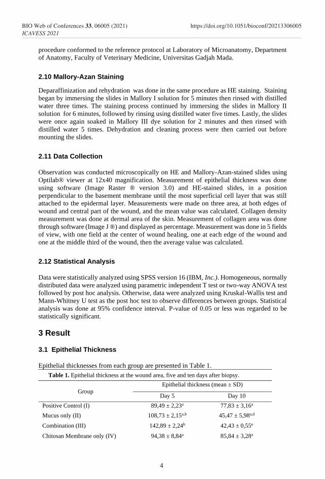
Observation of histopathological skin wound on day 5 using HE staining at low power
magnification (Figure 1) revealed complete coverage of granulation tissue by newly formed
epithelium in mucus group, chitosan group and combination group, while partial coverage
was observed in positive and negative control groups. Accumulation of crusts was observed
microscopically in the mucus group and negative control group.
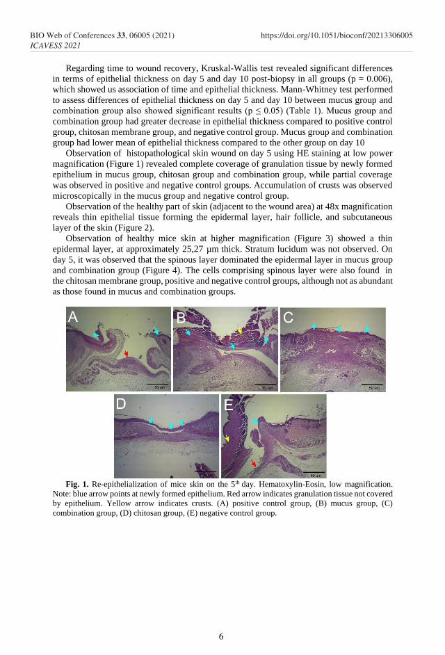
Observation of the healthy part of skin (adjacent to the wound area) at 48x magnification
reveals thin epithelial tissue forming the epidermal layer, hair follicle, and subcutaneous
layer of the skin (Figure 2).
Observation of healthy mice skin at higher magnification (Figure 3) showed a thin
epidermal layer, at approximately 25,27 µm thick. Stratum lucidum was not observed. On
day 5, it was observed that the spinous layer dominated the epidermal layer in mucus group
and combination group (Figure 4). The cells comprising spinous layer were also found in
the chitosan membrane group, positive and negative control groups, although not as abundant
as those found in mucus and combination groups.
Fig. 1. Re-epithelialization of mice skin on the 5th day. Hematoxylin-Eosin, low magnification.
Note: blue arrow points at newly formed epithelium. Red arrow indicates granulation tissue not covered
by epithelium. Yellow arrow indicates crusts. (A) positive control group, (B) mucus group, (C)
combination group, (D) chitosan group, (E) negative control group.
6
BIO Web of Conferences 33, 06005 (2021)ICAVESS 2021
https://doi.org/10.1051/bioconf/20213306005
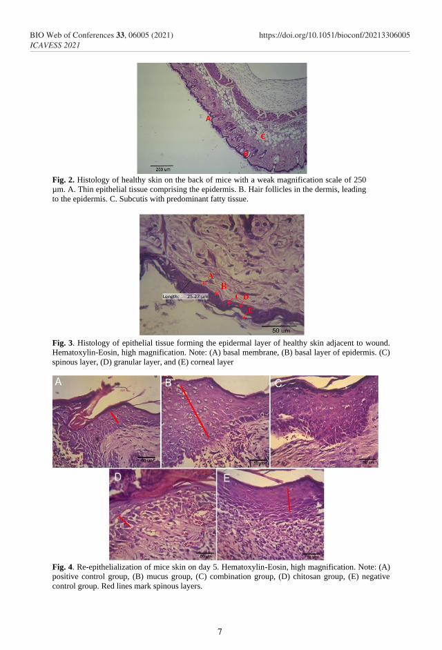
Fig. 2. Histology of healthy skin on the back of mice with a weak magnification scale of 250
µm. A. Thin epithelial tissue comprising the epidermis. B. Hair follicles in the dermis, leading
to the epidermis. C. Subcutis with predominant fatty tissue.
Fig. 3. Histology of epithelial tissue forming the epidermal layer of healthy skin adjacent to wound.
Hematoxylin-Eosin, high magnification. Note: (A) basal membrane, (B) basal layer of epidermis. (C)
spinous layer, (D) granular layer, and (E) corneal layer
Fig. 4. Re-epithelialization of mice skin on day 5. Hematoxylin-Eosin, high magnification. Note: (A)
positive control group, (B) mucus group, (C) combination group, (D) chitosan group, (E) negative
control group. Red lines mark spinous layers.
7
BIO Web of Conferences 33, 06005 (2021)ICAVESS 2021
https://doi.org/10.1051/bioconf/20213306005

Mean of epithelial thickness on the 10th day decreased from what was observed on the 5th
day in all groups (Table 1). However, cells comprising epidermal layers looked more
compacted, resembling more those in normal skin. Hair follicle started forming in the
combination group. This finding was not observed in other groups (Figure 5).
Fig. 5. Re-epithelialization of mice skin on day 10. Hematoxylin-Eosin, high magnification. Note: (A)
positive control group, (B) mucus group, (C) combination group, (D) chitosan group, (E) negative
control group. Red lines mark spinous layers.
3.2 Collagen Fiber Density
Results of collagen density measurement are shown in table 4.
Table 4. Collagen fiber densities in wound area, 5 and 10 days after biopsy
Group Percentage, mean ± SD p-value
Day 5 Day 10
Positive Control 23.02 ±
5.09
21.23 ±
0.72a
0.579
Mucus 16.08 ±
8.84
22.90 ±
4.87a
0.306
Combination 22.93 ±
4.24
37.04 ±
4.60b
0.017*
Chitosan 15.07 ±
5.14
19.31 ±
5.16a
0.371
Negative Control 12.49 ±
1.95
14.55 ±
8.22a
0.695
P value 0.134 0.004*
Note: * indicates significant differences in terms of collagen fiber density between day 5 and day
10. Superscripts flag significant differences between groups. P-values were obtained by post-hoc
analysis.
Results from two-way ANOVA showed significant difference of collagen fiber density
between groups within same day of observation. Percentages of collagen density between
8
BIO Web of Conferences 33, 06005 (2021)ICAVESS 2021
https://doi.org/10.1051/bioconf/20213306005

day 5 and day 10 were also statistically different with P-value of 0.018. Effect of treatment
groups and day of observation on collagen density did not produce significant result (p =
0.164).
Results from between subject-effect test on collagen density on day 10 showed significant
differences among groups (P = 0.004). Post-hoc analysis is presented on table 5 below.
Table 5. Post-Hoc analysis on collagen fiber densities (as percentage of area) on day 10
Positive
Control
Mucu
s
Combinatio
n
Chitosan
Membran
e
Negativ
e
Control
Positive
Control
Mucus 0.705
Combination 0.004
*
0.008
*
Chitosan
Membrane 0.666 0.424 0.002*
Negative
Control 0.153 0.081 0.000*
0.295*
Note: * flags statistically significant difference between groups
Results of post-hoc analysis showed significant difference of collagen fiber density
between combination group and positive control group on day 10, wherein positive control
group had lower percentage of collagen fiber density than the combination group. Collagen
fibers in the mucus group were also significantly less dense than in the combination group.
Collagen fiber in the combination group on day 10 was more packed than in the chitosan
membrane group and negative control group (Table 4). Negative control group had the lowest
percentage of collagen fiber density among other groups. Thereby, the combination group
had the most packed collagen fiber, followed by mucus group, positive control group,
chitosan membrane group, and negative control group.
Difference in percentage of collagen density between day 5 and day 10 of intervention
was further analyzed using an independent T test. This study found statistically significant
differences in collagen density between day 5 and day 10 within the combination group (p-
value of 0.017). Increase in the percentage of collagen fiber density was highest in the
combination group compared to other groups (Table 4).
Qualitative analysis of collagen fiber on day 10 (Figure 6) revealed growth of blue-
colored collagen in all treatment groups. Collagen fiber in combination group (C) were darker
in color than other groups, which resembled the color of collagen fiber found in healthy tissue
sample (F).
9
BIO Web of Conferences 33, 06005 (2021)ICAVESS 2021
https://doi.org/10.1051/bioconf/20213306005
Fig. 6. Collagen fiber in the dermal layer in all groups, 10 days after biopsy. Mallory-Azan staining,
high magnification. Note: (A) positive control group, (B) mucus group, (C) combination group, (D)
chitosan membrane group, (E) negative control group, (F) normal skin. Arrow shows collagen fiber,
which was blue-stained.
4 Discussion
4.1 Epithelial Thickness
We revealed that better epithelial tissue growth in groups of positive control, mucus group,
chitosan, and combination than in negative control group. Progressive healing process
occurred in the combination group, where thickest epithelium was found on day 5 post
biopsy.
The cutaneous epithelial growth pattern in this study was similar to what was found by
[7] where epithelial tissue reached its peak thickness on the 7th day and decreased further
until the 21st day on mice treated with 10% povidone-iodine solution, saliva and physiologic
saline. The decrease in epithelial thickness after 10 days of treatment occurred due to the
epithelial tissue that had covered the wound surface completely and no proliferation.
Epithelial tissue that has covered the whole wound area stops proliferating and starts the
maturation process, characterized by keratin synthesis by corneal layer of epithelium. Study
conducted by Putri and Tasminatun in 2016 [8]also reported peak epithelial thickness on day
7 followed by decrease on the next day. [9] found that epithelial thickening would continue
and then regress until it reaches normal thickness. This study found that 10 days after biopsy,
mice receiving combination of mucus extract and chitosan membrane had the thinnest
epithelium.
Crusts, found at the margin of the wound in this study, are made from dried exudates and
are observable as solid layer on the outside [10]. Debris and exotoxin found in exudates may
slow down the wound healing process and pose a risk of advanced infection [11].
The skin layer observed in this study was in accordance with [12], which stated that
epidermis has varying layers forming it across sites. [13] stated that cells forming spinous and
basal layers have clear mitotic figures to replace the cells lying on top of them. This
observation was also in keeping with data from epithelial thickness measurement wherein
thick epithelia were observed in the combination group and mucus group due to spinous cells
predominance in the epithelial tissues in both groups. Predominance of spinous cells marked
the proliferation process in tandem with the wound healing process. Granular layer was not
10
BIO Web of Conferences 33, 06005 (2021)ICAVESS 2021
https://doi.org/10.1051/bioconf/20213306005

observed on day 5. This may partially be due to the keratinocyte deposition process which had
not occurred yet.
Epidermis is the outermost skin layer and becomes a physical barrier of the tissue beneath
it [14]. According to [1], tissue discontinuity due to trauma triggers physiologic response to
recover the structural and functional loss.
[15] stated that in the wound healing process, keratinocytes actively proliferate due to
stimulation by epidermal growth factor (EGF), keratinocyte growth factor (KGF), and
transforming growth factor-α (TGF-α). [16] reported that in traumatic wounds in the buccal
area, human keratinocytes migrate laterally and synthesize basal membrane then halt the
migration process and proliferate actively until the 4th day. Keratinocyte migration has been
shown by [17] through immunofluorescent staining using CK17, which detects keratinocyte
migration from the border of wound to granulation tissue to synthesize basal membrane.
Progressive increase in epithelial thickness in combination group 5 days post biopsy may
partially be due to hemostatic effect of chitosan, therefore able to stop bleeding more swiftly
and provide extracellular matrix for epithelial cells to attach during wound healing process.
Chitosan structure resembles glycosaminoglycan, a polysaccharide found in extracellular
tissue. Lactoferrin, a peptide found in eel mucus, may increase production of proinflammatory
cytokines by macrophages. Inflammatory cytokines may modulate keratinocyte migrations to
wound area, thereby speed up the healing process. Qualitative observation also showed
complete coverage of wound area by epithelial tissue in all mice receiving combination of
mucus extract and chitosan membrane and few mice treated with mucus only or chitosan
membrane only.
Study by [18] to evaluate the performance of chitosan membrane as wound dressing found
that better wound healing process occurred in tissues that had thinnest epithelial tissue, which
marked completed growth stimulation by growth factor and other mediators. [7] found that
epithelial reconstruction was better in animal treated with saliva than with 10% povidone-
iodine solution. Histopathological observation also indicated better epithelial repair in the
combination group supported by the development of hair follicle which was not found in other
groups (Figure 5). Combination of chitosan membrane and eel mucus extract speeds up re-
epithelialization better than 10% povidone-iodine solution.
4.2 Collagen Fiber Density
According to [19] regeneration of wound skin start from matrix formation which encloses the
wound. During proliferation stage, wound form scar tissue in the deep area as the result of
fibroblast activity . [20] stated that synthesize collagen by fibroblast to form new tissue is
stimulate by macrophages that migrating to wound area and secreting transforming growth
factor-β (TGF-β). For that reason, this study observed the effect of intervention on collagen
fiber density at wound site.
Transforming growth factor-β may played a role in the proliferation phase in this study
but it needs to be confirmed in further studies by using more specific staining method.
According to [21], once inflammation phase is finished, macrophages will turn into the M2-
type (anti-inflammatory macrophages) which express anti-inflammatory mediators, namely,
protease, protease inhibitor, vascular endothelial growth factor (VEGF) and TGF-β which
promote protein synthesis and cellular proliferation. Fibroblast migration and proliferation
trigger collagen fiber and fibronectin syntheses which, in turn, increase extracellular matrix.
Collagen fiber synthesis is paramount to strengthen the tissue after injury [19] and [22].
Collagen is secreted to extracellular space in the form of procollagen which forms
tropocollagen. Tropocollagen then unites to form collagen fibers. Collagen synthesis begins
as early as three days after trauma and occurs rapidly then reaches its peak 14 days after
trauma, which is characterized by collagen fiber thickening [23].
11
BIO Web of Conferences 33, 06005 (2021)ICAVESS 2021
https://doi.org/10.1051/bioconf/20213306005
Administration of 10% povidone-iodine solution to wounded tissue may slow down the
proliferation of fibroblast, which in turn decreases the speed of collagen synthesis. This study
showed a decrease in collagen density (displayed as percentage) from day 5 to day 10,
although this decrease was not statistically significant (Table 4). [18] stated that in terms of
wound care, administration of povidone-iodine solution must be minimized due to its
cytotoxic effect on fibroblasts. This study is also in concordance with study by Danastri et al.
in 2014 which assessed the effect of povidone-iodine solution at concentration higher than
0.1% on fibroblasts and polymorphonuclear cells based on fast protein liquid chromatography
(FPLC) method.
The progressive increase of collagen density which was observed in the combination group
supports the previous finding by [24], which stated that chitosan is able to prolong the half-
life of basic fibroblast growth factor (FGF) by protecting it from enzymatic degradation.
Therefore, fibroblasts may function more optimally. On the other hand, Immunoglobulin M,
which is found in mucus of Asian swamp eel, plays an important role to protect the host from
proteolytic degradation [25]. The benefits from these two on wound healing may reflect
potencies for use as treatment for wound, alternative to gauze and 10% povidone-iodine
solution, to speed up the wound healing process.
5 Conclusion
This study concluded that application of chitosan membrane (1% b/v), combined with 100%
Asian swamp eel (Monopterus albus) mucus sped up the re-epithelialization process and
increased the collagen fiber density in mice (Mus musculus) undergoing skin wound. Findings
from this study may be the basis for further research to compare their efficacy as alternatives
to conventional wound dressings.
References
1. V. Kumar, A. Abbas, J. Aster, S. Robbins, J. Perkins, Robbins, Basic Pathology
(Elsevier Saunders, Philadelphia, 2013)
2. A. Muhammad, Konsep Dasar Sistem Pakar (Penerbit Andi, Yogyakarta, 2005)
3. A.B. Atif, M.K. Zahri, A.R. Esa, B.A. Zilfalil, U.S.M. Rao, S. Nordin, J. Appl.
Pharmaceutical Sc. 5, 1 (2015)
4. N.M. Hussin, S.M. Shaarani, M.R. Sulaiman, A.H., Ahmad, C.S.Vairappan, Chemical
Composition and Antioxidant Activities of Catfish Epidermal Mucus, J. Adv.
AgricTech. 4, 1 (2017)
5. M. N. N. M Ikram and B. H. Ridzwan, Inter Res J Pharma and Pharmacol 3, 1 (2013)
6. D.A. Setiawan, B.D. Argo and Y. Hendrawan, Jurnal Keteknikan Pertanian Tropis dan
Biosistem 3, 1 (2015)
7. I.A. Wahyudi, M. Magista and M.Angel, Efektivitas Penggunaan Saliva dibandingkan
Povidone Iodine 10% terhadap Penyembuhan Luka pada Kutaneus Tikus Sprague
dawley, IDJ 2, 1 (2013)
8. F.R. Putri and S. Tasminatun, J. Kedokteran dan Kesehatan 12, 1 (2016)
9. W.K. Fathi, Al – Rafidain Dent J. 12, 1 (2012)
10. W.W. Dorland, Kamus Kedokteran Dorland (EGC, Jakarta, 2002)
11. M.J. Morison, Manajemen Luka, (EGC, Jakarta, 2004)
12. W.J. Banks, Applied Veterinary Histology (Lippincott Williams and Wilkins, New
York, 1993)
12
BIO Web of Conferences 33, 06005 (2021)ICAVESS 2021
https://doi.org/10.1051/bioconf/20213306005

13. S.J. Kalangi, Histofisiologi Kulit, JBM 5, 3 (2013)
14. S. Baranoski and R.N. Ayello, Wound Care Essentia : Practice Principle 3rd Edition
(Lippincott Williams and Wilkins. New York,2012)
15. A.J. Singer and R.A.F. Clark, Cutaneous Wound Healing, NEJM 341, 10 (1999)
16. O.J. Andreasen, F.M. Andreasen and L. Andreasen, Textbook and color atlas of
traumatic injuries to the teeth (Wiley Blackwell, Oxford, 2013)
17. I. Pastar, O. Stojadinovic, N.C. Yin, H. Ramirez, A.G. Nusbaum, A. Sawaya, S.B. Pate,
L. Khalid, R.R. Isseroff, and M.T. Canic, Adv. Wound Care 3, 7 (2014)
18. A.D. Sezer, F. Hatipolu, E. Cevher, Z. Ourtan, A.L. Ba, J.Akbua, AAPS Pharm. Sci.
Tech. 8, 1 (2007)
19. M.M. Pavletic, Atlas of Small Animal Wound Management and Reconstructive
Surgery 3rd Edition (Willey Blackwell, Iowa, 2010)
20. I.M.S. Wijaya, Perawatan Luka dengan Pendekatan Multidisiplin (ANDI publisher,
Yogyakarta, 2018)
21. A.R. Pratama, N. Wathoni, and T. Rusdiana, Jurnal Farmaka 15, 2 (2017)
22. T. Velnar , T. Bayley, and V.Smrkolj, The Wound Healing Proses: an Overview of The
Cellular and Molecular Mechanisms. J Inter Med Res. 37 (2009)
23. A.I.M. Novitasari, R Indraswary, R. Pratiwi, ODONT Dent. J. 4, 1 (2017)
24. K. Masuoka, M. Ishahara, T. Asazuma, H. Hattori, T. Matsui, B. Takase, J.biomaterial
26, 19 (2005)
25. M.A. Esteban, An overview of the Immunological Defense in Fish Skin, ISRN
Immunol 29 (2012)
13
BIO Web of Conferences 33, 06005 (2021)ICAVESS 2021
https://doi.org/10.1051/bioconf/20213306005