3rd interventional hands-on pain relief & … · 3rd interventional hands-on pain relief &...
TRANSCRIPT
3rd INTERVENTIONAL HANDS-ONPAIN RELIEF & NEUROMODULATION
CADAVER WORKSHOP
Anatomical introduction
Gdansk- Poland
Programme of the lecture
Short introduction to the anatomy of:
1.Vertebral column
2.Intervertebral discs
3.Intervertebral foramina
4.Vascular supply of the vertebral column and spinal cord
5.Spinal nerve
6.Autonomic nervous system
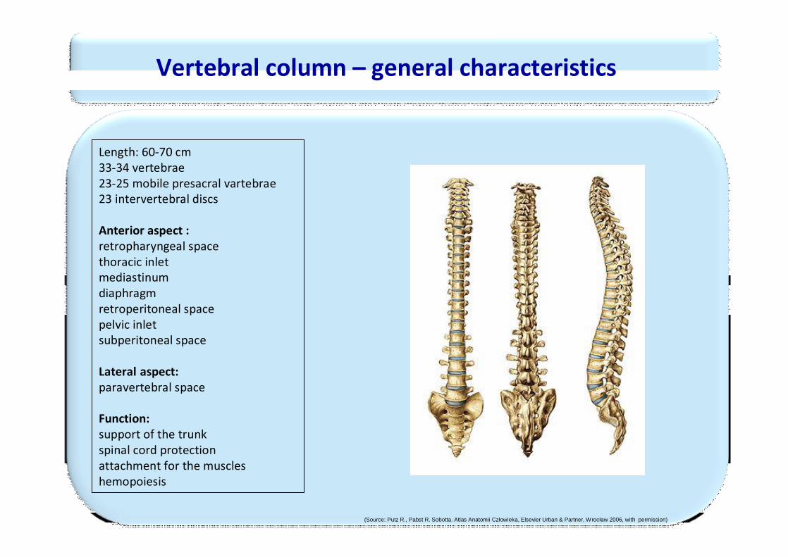
Vertebral column – general characteristics
Length: 60-70 cm33-34 vertebrae23-25 mobile presacral vartebrae23 intervertebral discs
Anterior aspect :retropharyngeal spacethoracic inletmediastinumdiaphragmretroperitoneal spacepelvic inletsubperitoneal space
Lateral aspect:paravertebral space
Function:support of the trunkspinal cord protectionattachment for the muscleshemopoiesis
(Source: Putz R., Pabst R. Sobotta. Atlas Anatomii Człowieka, Elsevier Urban & Partner, Wrocław 2006, with permission)
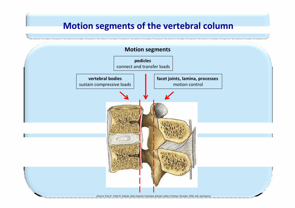
Motion segments of the vertebral column
Motion segments
vertebral bodiessustain compressive loads
pediclesconnect and transfer loads
facet joints, lamina, processes motion control
(Source: Putz R., Pabst R. Sobotta. Atlas Anatomii Człowieka, Elsevier Urban & Partner, Wrocław 2006, with permission)
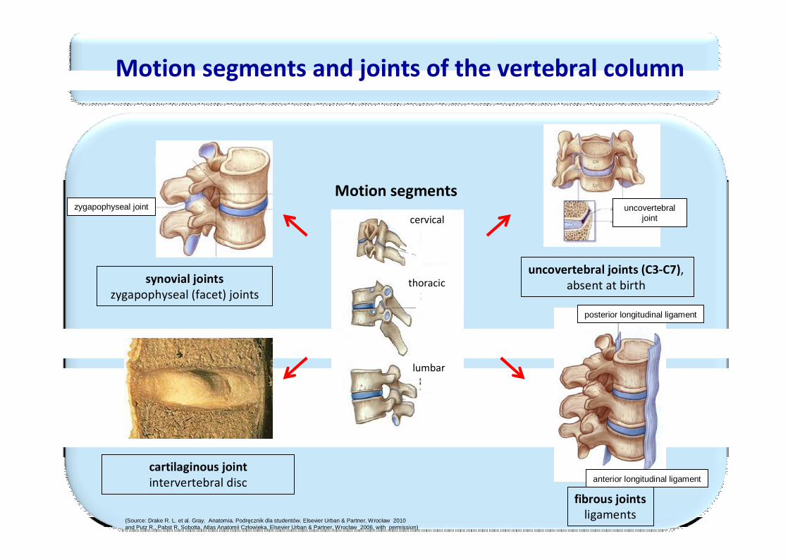
Motion segments and joints of the vertebral column
cervical
thoracic
lumbar
Motion segments
synovial jointszygapophyseal (facet) joints
cartilaginous jointintervertebral disc
uncovertebral joints (C3-C7), absent at birth
fibrous jointsligaments
zygapophyseal joint uncovertebraljoint
(Source: Drake R. L. et al. Gray. Anatomia. Podręcznik dla studentów. Elsevier Urban & Partner, Wrocław 2010 and Putz R., Pabst R. Sobotta. Atlas Anatomii Człowieka, Elsevier Urban & Partner, Wrocław 2006, with permission)
posterior longitudinal ligament
anterior longitudinal ligament
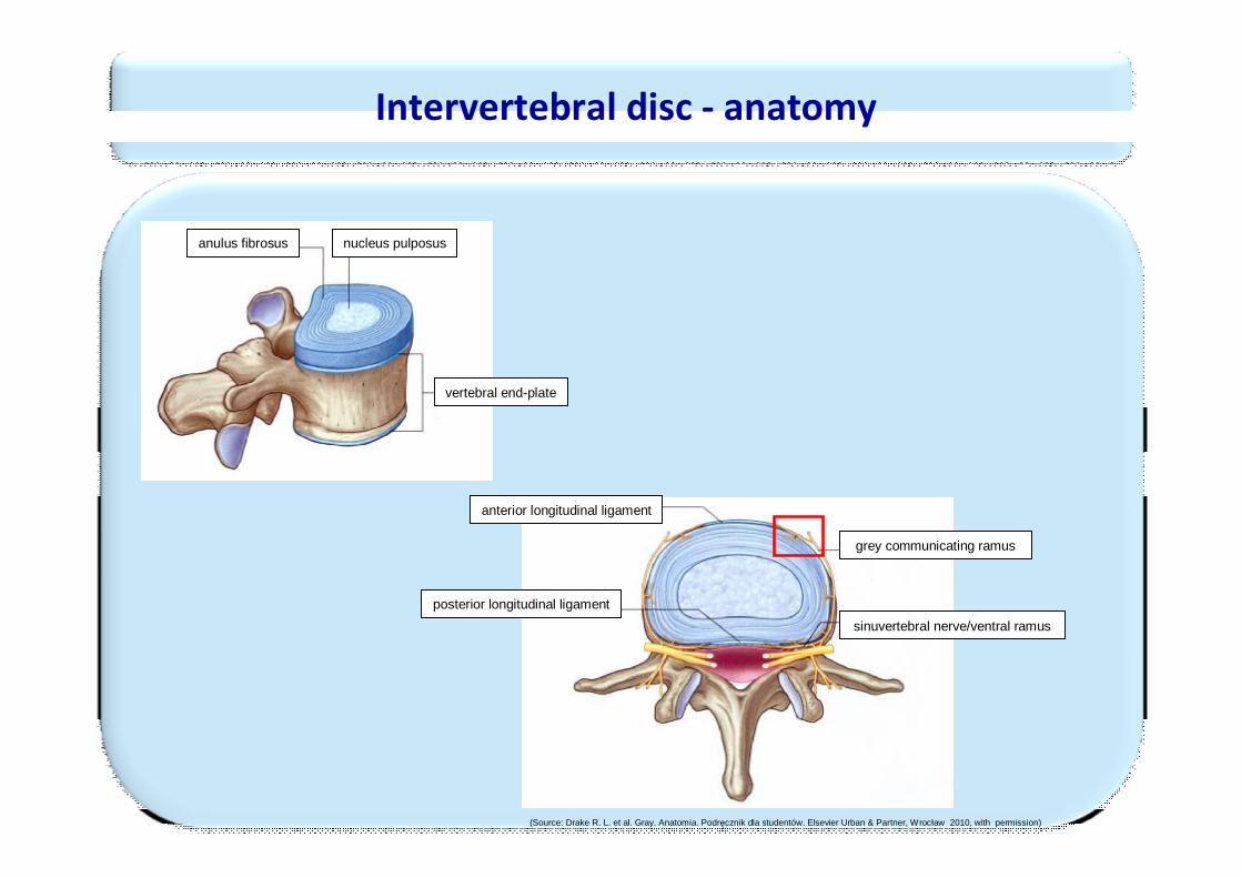
Intervertebral disc - anatomy
anulus fibrosus nucleus pulposus
vertebral end-plate
grey communicating ramus
sinuvertebral nerve/ventral ramusposterior longitudinal ligament
anterior longitudinal ligament
(Source: Drake R. L. et al. Gray. Anatomia. Podręcznik dla studentów. Elsevier Urban & Partner, Wrocław 2010, with permission)
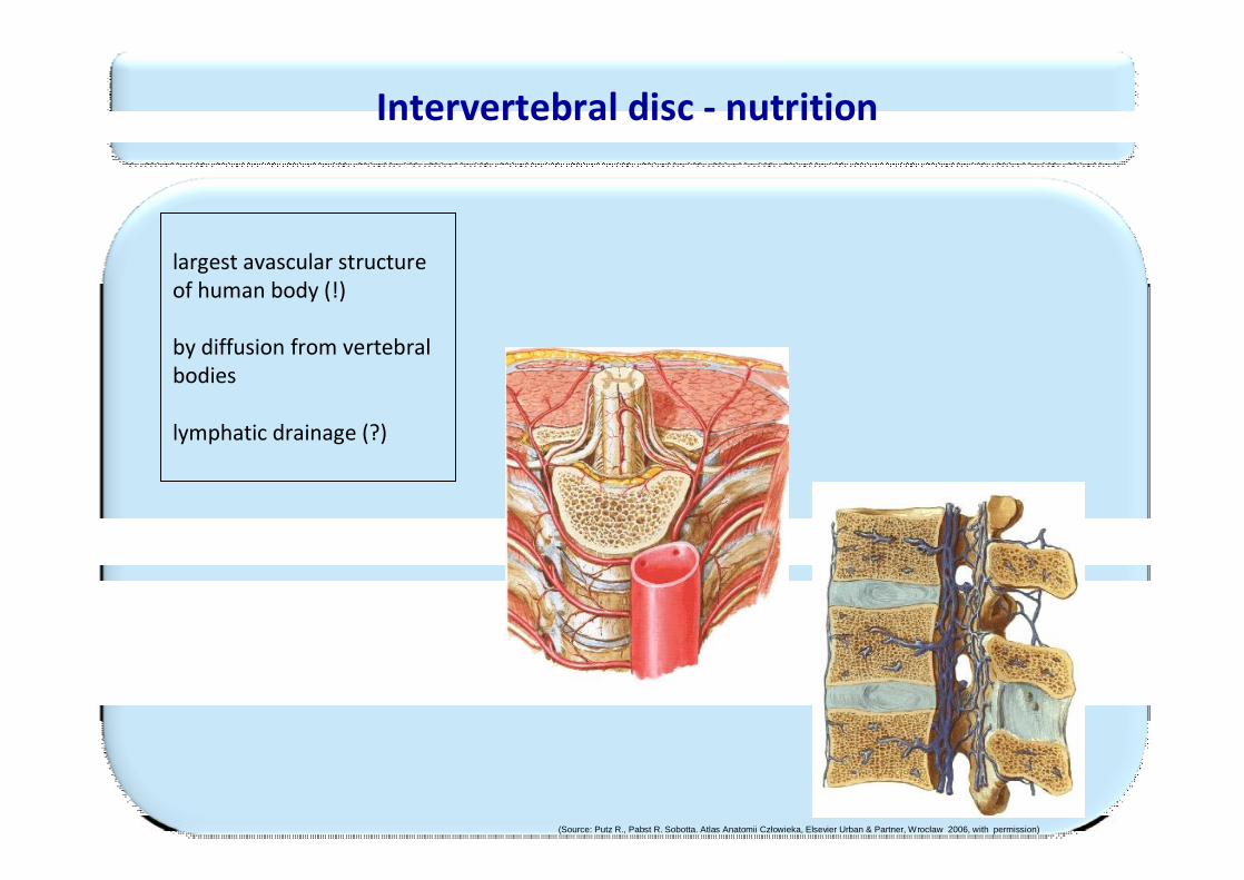
Intervertebral disc - nutrition
largest avascular structureof human body (!)
by diffusion from vertebral bodies
lymphatic drainage (?)
(Source: Putz R., Pabst R. Sobotta. Atlas Anatomii Człowieka, Elsevier Urban & Partner, Wrocław 2006, with permission)
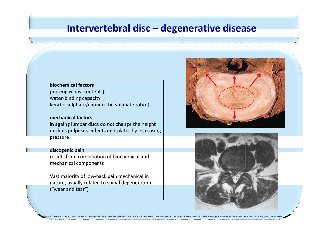
Intervertebral disc – degenerative disease
biochemical factorsproteoglycans content ↓water-binding capacity ↓keratin sulphate/chondroitin sulphate ratio ↑
mechanical factorsin ageing lumbar discs do not change the heightnucleus pulposus indents end-plates by increasing pressure
discogenic pain results from combination of biochemical and mechanical components
Vast majority of low-back pain mechanical in nature, usually related to spinal degeneration (“wear and tear”)
(Source: Drake R. L. et al. Gray. Anatomia. Podręcznik dla studentów. Elsevier Urban & Partner, Wrocław 2010 and Putz R., Pabst R. Sobotta. Atlas Anatomii Człowieka, Elsevier Urban & Partner, Wrocław 2006, with permission)
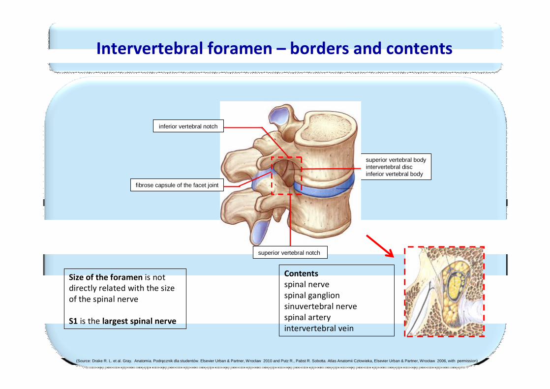
Intervertebral foramen – borders and contents
superior vertebral body intervertebral discinferior vertebral body
fibrose capsule of the facet joint
superior vertebral notch
Size of the foramen is not directly related with the sizeof the spinal nerve
S1 is the largest spinal nerve
Contentsspinal nervespinal ganglionsinuvertebral nervespinal arteryintervertebral vein
inferior vertebral notch
(Source: Drake R. L. et al. Gray. Anatomia. Podręcznik dla studentów. Elsevier Urban & Partner, Wrocław 2010 and Putz R., Pabst R. Sobotta. Atlas Anatomii Człowieka, Elsevier Urban & Partner, Wrocław 2006, with permission)
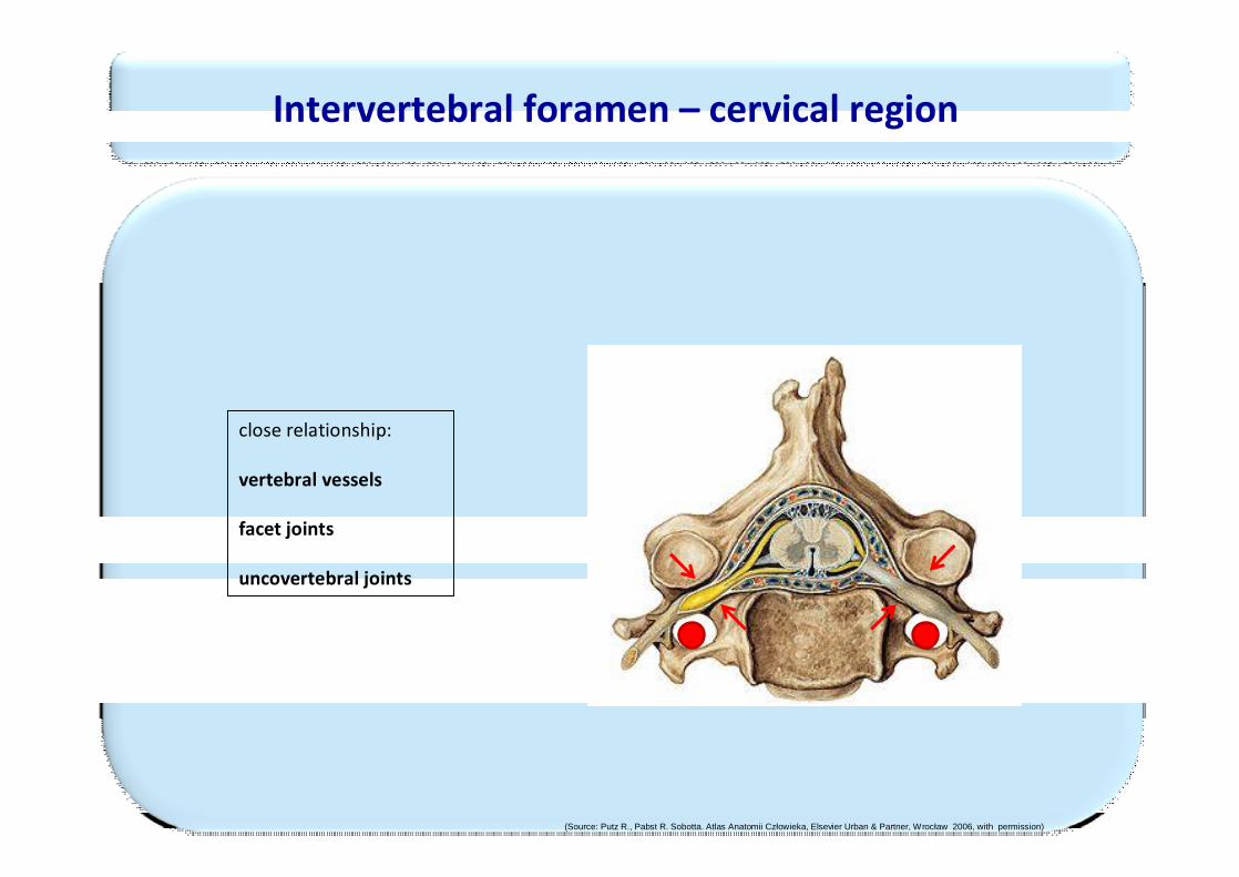
close relationship:
vertebral vessels
facet joints
uncovertebral joints
Intervertebral foramen – cervical region
(Source: Putz R., Pabst R. Sobotta. Atlas Anatomii Człowieka, Elsevier Urban & Partner, Wrocław 2006, with permission)
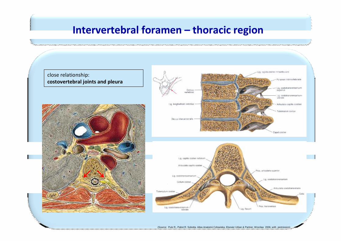
close relationship: costovertebral joints and pleura
Intervertebral foramen – thoracic region
(Source: Putz R., Pabst R. Sobotta. Atlas Anatomii Człowieka, Elsevier Urban & Partner, Wrocław 2006, with permission)
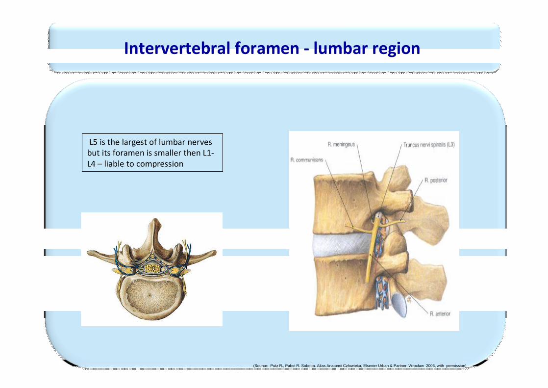
L5 is the largest of lumbar nerves but its foramen is smaller then L1-L4 – liable to compression
Intervertebral foramen - lumbar region
(Source: Putz R., Pabst R. Sobotta. Atlas Anatomii Człowieka, Elsevier Urban & Partner, Wrocław 2006, with permission)
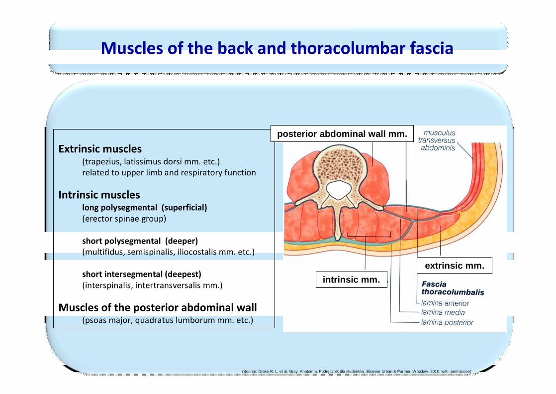
Muscles of the back and thoracolumbar fascia
Extrinsic muscles(trapezius, latissimus dorsi mm. etc.) related to upper limb and respiratory function
Intrinsic muscleslong polysegmental (superficial)(erector spinae group)
short polysegmental (deeper)(multifidus, semispinalis, iliocostalis mm. etc.)
short intersegmental (deepest)(interspinalis, intertransversalis mm.)
Muscles of the posterior abdominal wall(psoas major, quadratus lumborum mm. etc.)
extrinsic mm.intrinsic mm.
posterior abdominal wall mm.
(Source: Drake R. L. et al. Gray. Anatomia. Podręcznik dla studentów. Elsevier Urban & Partner, Wrocław 2010, with permission)
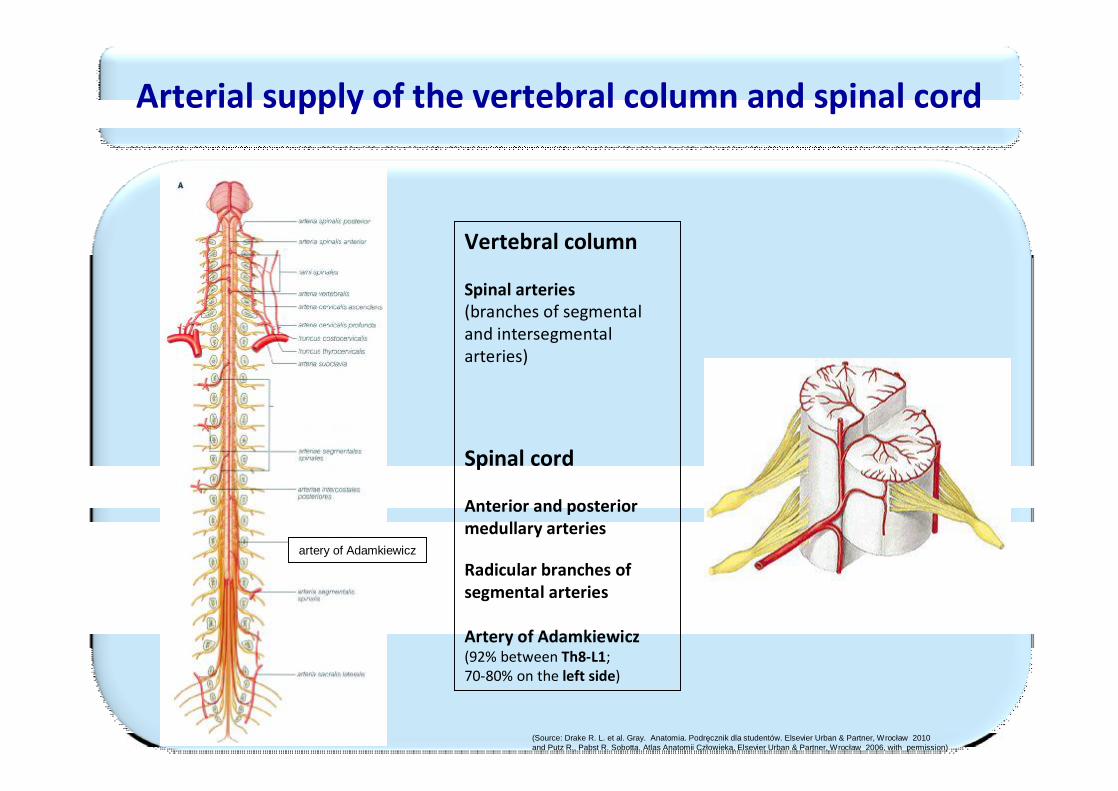
Arterial supply of the vertebral column and spinal cord
Vertebral column
Spinal arteries(branches of segmentaland intersegmentalarteries)
Spinal cord
Anterior and posteriormedullary arteries
Radicular branches ofsegmental arteries
Artery of Adamkiewicz(92% between Th8-L1;70-80% on the left side)
artery of Adamkiewicz
(Source: Drake R. L. et al. Gray. Anatomia. Podręcznik dla studentów. Elsevier Urban & Partner, Wrocław 2010 and Putz R., Pabst R. Sobotta. Atlas Anatomii Człowieka, Elsevier Urban & Partner, Wrocław 2006, with permission)
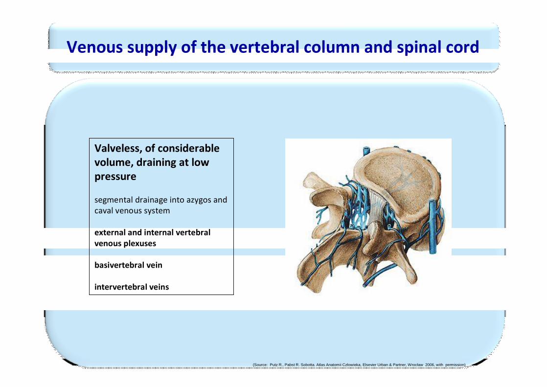
Valveless, of considerable volume, draining at low pressure
segmental drainage into azygos andcaval venous system
external and internal vertebral venous plexuses
basivertebral vein
intervertebral veins
Venous supply of the vertebral column and spinal cord
(Source: Putz R., Pabst R. Sobotta. Atlas Anatomii Człowieka, Elsevier Urban & Partner, Wrocław 2006, with permission)
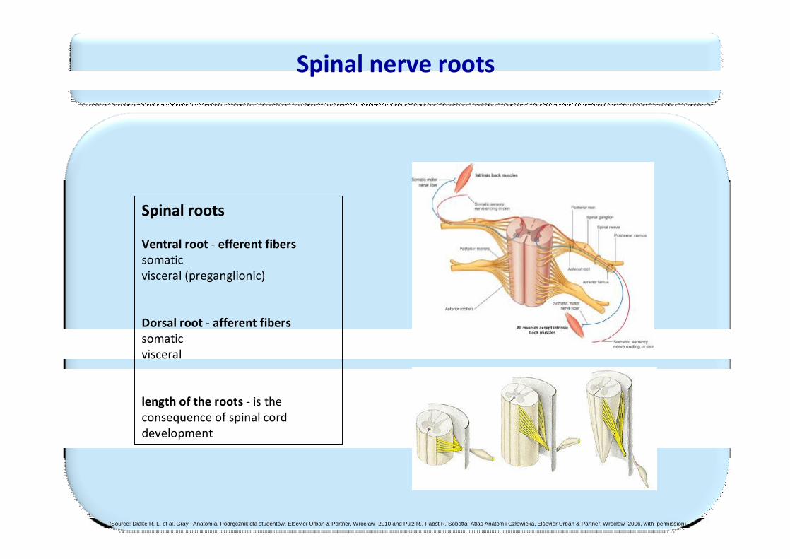
Spinal nerve roots
Spinal roots
Ventral root - efferent fiberssomaticvisceral (preganglionic)
Dorsal root - afferent fiberssomaticvisceral
length of the roots - is the consequence of spinal corddevelopment
(Source: Drake R. L. et al. Gray. Anatomia. Podręcznik dla studentów. Elsevier Urban & Partner, Wrocław 2010 and Putz R., Pabst R. Sobotta. Atlas Anatomii Człowieka, Elsevier Urban & Partner, Wrocław 2006, with permission)
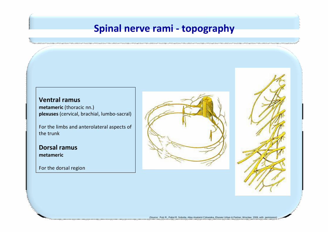
Spinal nerve rami - topography
Ventral ramusmetameric (thoracic nn.)plexuses (cervical, brachial, lumbo-sacral)
For the limbs and anterolateral aspects ofthe trunk
Dorsal ramusmetameric
For the dorsal region
(Source: Putz R., Pabst R. Sobotta. Atlas Anatomii Człowieka, Elsevier Urban & Partner, Wrocław 2006, with permission)
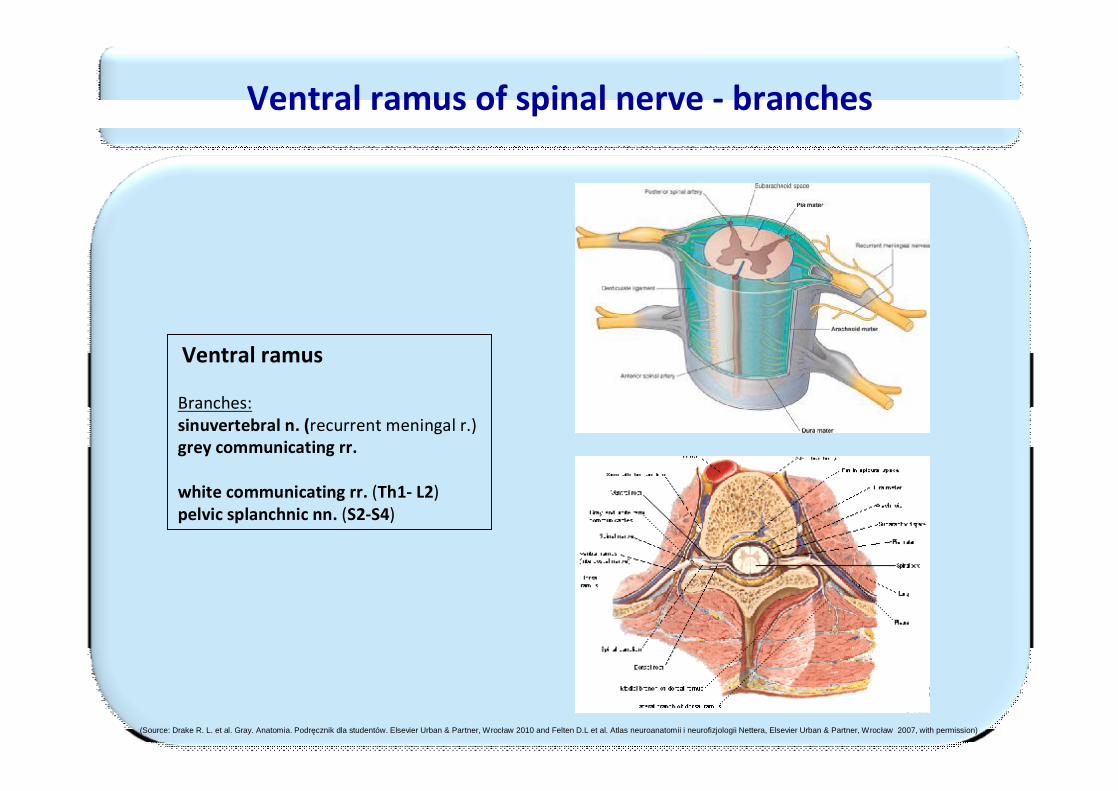
Ventral ramus of spinal nerve - branches
Ventral ramus
Branches:sinuvertebral n. (recurrent meningal r.)grey communicating rr.
white communicating rr. (Th1- L2)pelvic splanchnic nn. (S2-S4)
(Source: Drake R. L. et al. Gray. Anatomia. Podręcznik dla studentów. Elsevier Urban & Partner, Wrocław 2010 and Felten D.L et al. Atlas neuroanatomii i neurofizjologii Nettera, Elsevier Urban & Partner, Wrocław 2007, with permission)
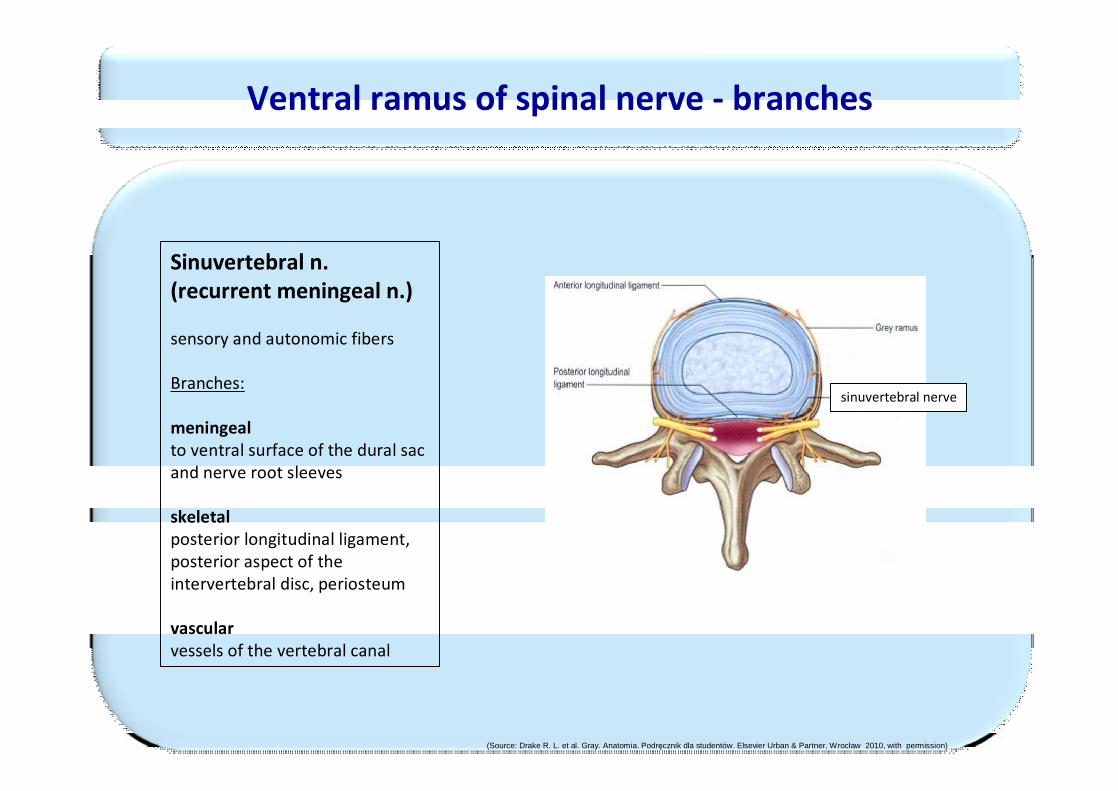
Sinuvertebral n. (recurrent meningeal n.)
sensory and autonomic fibers
Branches:
meningealto ventral surface of the dural sac and nerve root sleeves
skeletalposterior longitudinal ligament, posterior aspect of the intervertebral disc, periosteum
vascularvessels of the vertebral canal
sinuvertebral nerve
Ventral ramus of spinal nerve - branches
(Source: Drake R. L. et al. Gray. Anatomia. Podręcznik dla studentów. Elsevier Urban & Partner, Wrocław 2010, with permission)
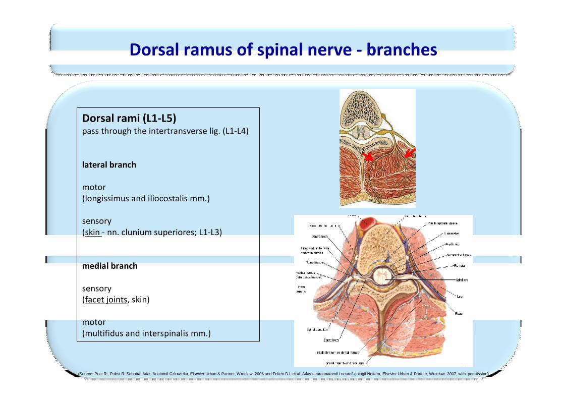
Dorsal rami (L1-L5) pass through the intertransverse lig. (L1-L4)
lateral branch
motor (longissimus and iliocostalis mm.)
sensory (skin - nn. clunium superiores; L1-L3)
medial branch
sensory(facet joints, skin)
motor(multifidus and interspinalis mm.)
Dorsal ramus of spinal nerve - branches
(Source: Putz R., Pabst R. Sobotta. Atlas Anatomii Człowieka, Elsevier Urban & Partner, Wrocław 2006 and Felten D.L et al. Atlas neuroanatomii i neurofizjologii Nettera, Elsevier Urban & Partner, Wrocław 2007, with permission)
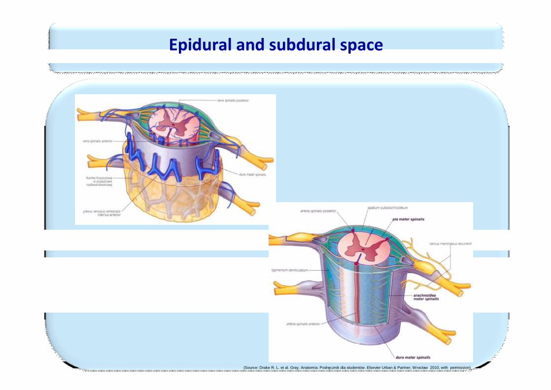
Epidural and subdural space
(Source: Drake R. L. et al. Gray. Anatomia. Podręcznik dla studentów. Elsevier Urban & Partner, Wrocław 2010, with permission)
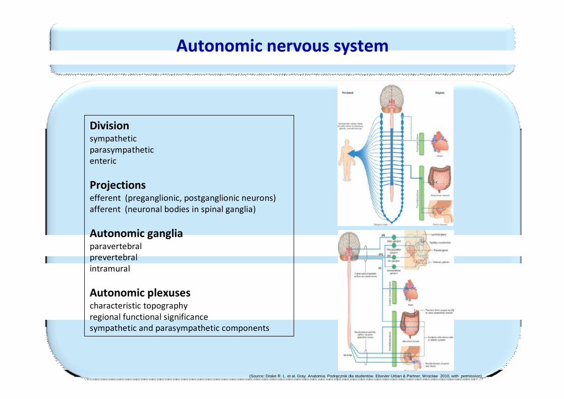
Autonomic nervous system
Divisionsympathetic parasympatheticenteric
Projectionsefferent (preganglionic, postganglionic neurons)afferent (neuronal bodies in spinal ganglia)
Autonomic gangliaparavertebral prevertebralintramural
Autonomic plexusescharacteristic topography regional functional significancesympathetic and parasympathetic components
(Source: Drake R. L. et al. Gray. Anatomia. Podręcznik dla studentów. Elsevier Urban & Partner, Wrocław 2010, with permission)
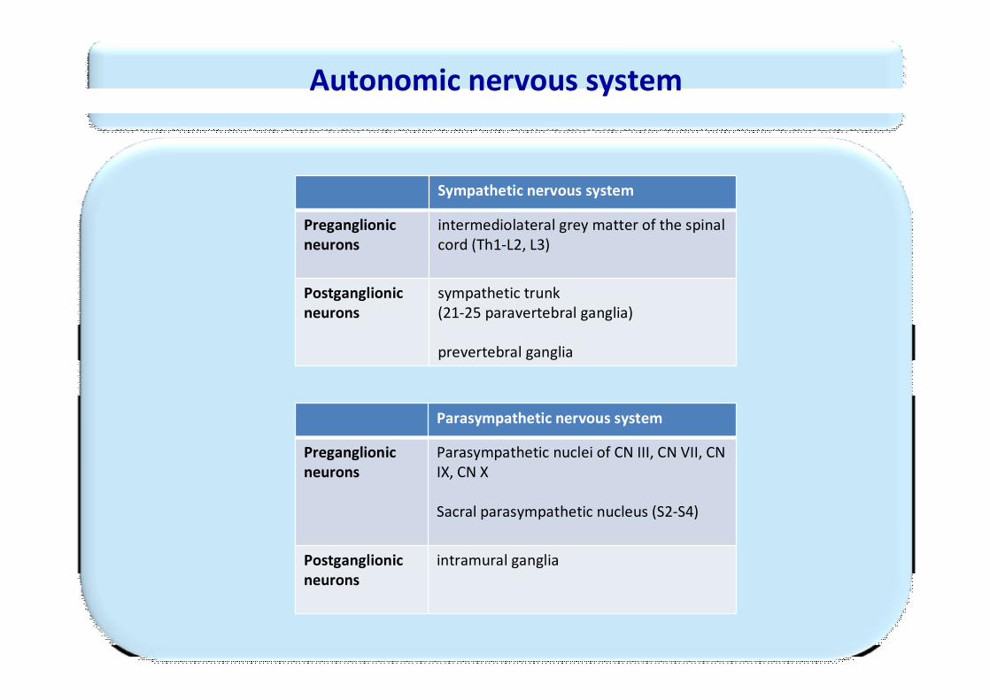
Sympathetic nervous system
Preganglionicneurons
intermediolateral grey matter of the spinal cord (Th1-L2, L3)
Postganglionicneurons
sympathetic trunk (21-25 paravertebral ganglia)
prevertebral ganglia
Autonomic nervous system
Parasympathetic nervous system
Preganglionicneurons
Parasympathetic nuclei of CN III, CN VII, CN IX, CN X
Sacral parasympathetic nucleus (S2-S4)
Postganglionicneurons
intramural ganglia
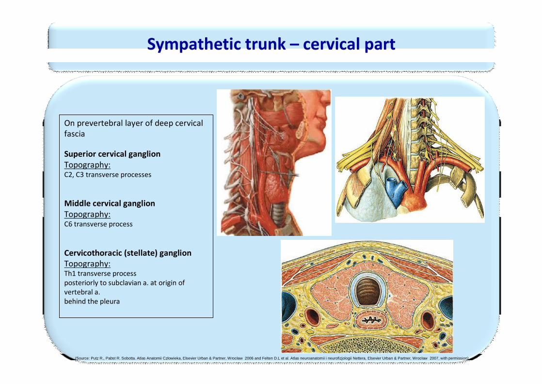
Sympathetic trunk – cervical part
On prevertebral layer of deep cervical fascia
Superior cervical ganglionTopography:C2, C3 transverse processes
Middle cervical ganglionTopography:C6 transverse process
Cervicothoracic (stellate) ganglion Topography:Th1 transverse processposteriorly to subclavian a. at origin of vertebral a.behind the pleura
(Source: Putz R., Pabst R. Sobotta. Atlas Anatomii Człowieka, Elsevier Urban & Partner, Wrocław 2006 and Felten D.L et al. Atlas neuroanatomii i neurofizjologii Nettera, Elsevier Urban & Partner, Wrocław 2007, with permission)
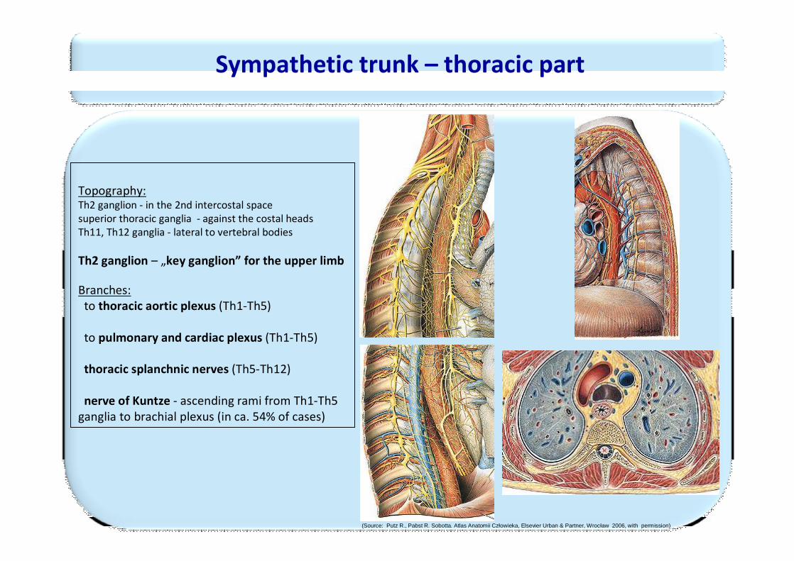
Topography:Th2 ganglion - in the 2nd intercostal spacesuperior thoracic ganglia - against the costal headsTh11, Th12 ganglia - lateral to vertebral bodies
Th2 ganglion – „key ganglion” for the upper limb
Branches:to thoracic aortic plexus (Th1-Th5)
to pulmonary and cardiac plexus (Th1-Th5)
thoracic splanchnic nerves (Th5-Th12)
nerve of Kuntze - ascending rami from Th1-Th5 ganglia to brachial plexus (in ca. 54% of cases)
Sympathetic trunk – thoracic part
(Source: Putz R., Pabst R. Sobotta. Atlas Anatomii Człowieka, Elsevier Urban & Partner, Wrocław 2006, with permission)
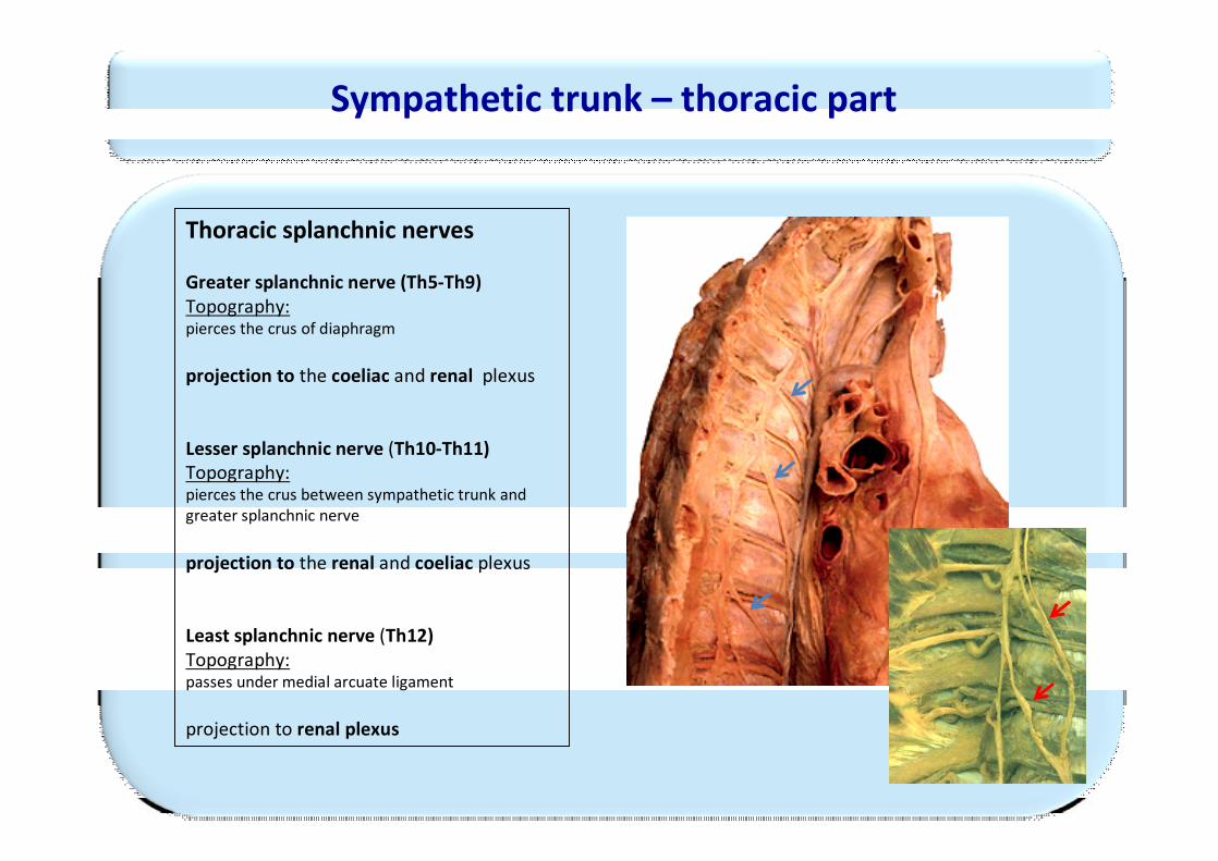
Thoracic splanchnic nerves
Greater splanchnic nerve (Th5-Th9)Topography:pierces the crus of diaphragm
projection to the coeliac and renal plexus
Lesser splanchnic nerve (Th10-Th11)Topography:pierces the crus between sympathetic trunk and greater splanchnic nerve
projection to the renal and coeliac plexus
Least splanchnic nerve (Th12)Topography:passes under medial arcuate ligament
projection to renal plexus
Sympathetic trunk – thoracic part
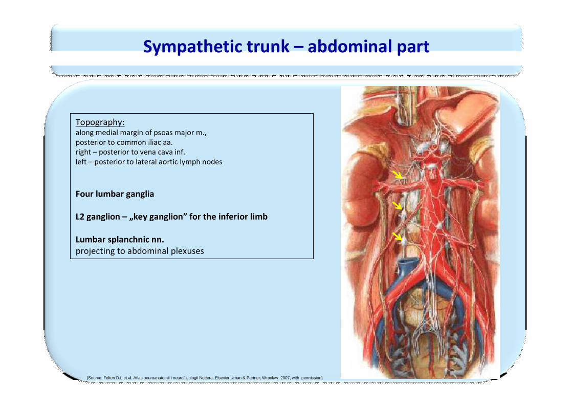
Topography:along medial margin of psoas major m., posterior to common iliac aa.right – posterior to vena cava inf. left – posterior to lateral aortic lymph nodes
Four lumbar ganglia
L2 ganglion – „key ganglion” for the inferior limb
Lumbar splanchnic nn.projecting to abdominal plexuses
Sympathetic trunk – abdominal part
(Source: Felten D.L et al. Atlas neuroanatomii i neurofizjologii Nettera, Elsevier Urban & Partner, Wrocław 2007, with permission)
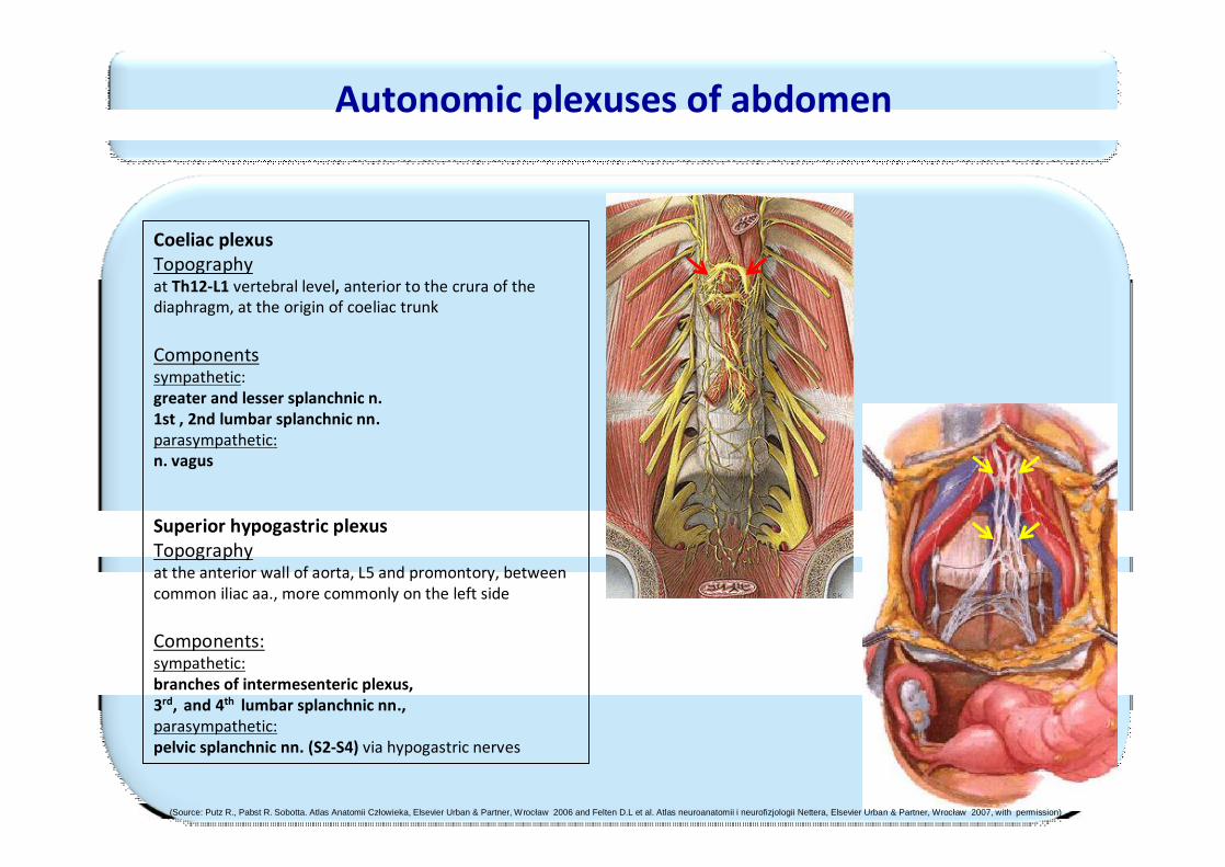
Autonomic plexuses of abdomen
Coeliac plexusTopographyat Th12-L1 vertebral level, anterior to the crura of thediaphragm, at the origin of coeliac trunk
Componentssympathetic:greater and lesser splanchnic n. 1st , 2nd lumbar splanchnic nn.parasympathetic:n. vagus
Superior hypogastric plexusTopographyat the anterior wall of aorta, L5 and promontory, between common iliac aa., more commonly on the left side
Components:sympathetic:branches of intermesenteric plexus, 3rd, and 4th lumbar splanchnic nn., parasympathetic:pelvic splanchnic nn. (S2-S4) via hypogastric nerves
(Source: Putz R., Pabst R. Sobotta. Atlas Anatomii Człowieka, Elsevier Urban & Partner, Wrocław 2006 and Felten D.L et al. Atlas neuroanatomii i neurofizjologii Nettera, Elsevier Urban & Partner, Wrocław 2007, with permission)
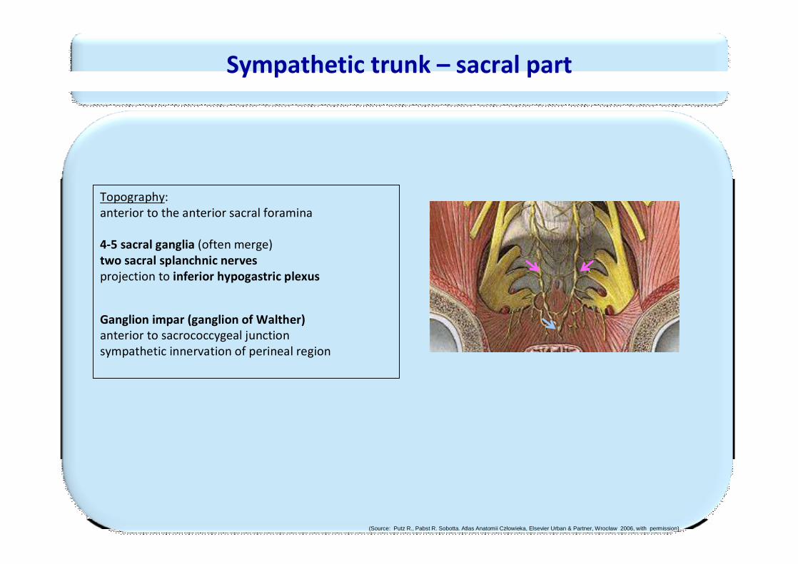
Sympathetic trunk – sacral part
Topography:anterior to the anterior sacral foramina
4-5 sacral ganglia (often merge)two sacral splanchnic nervesprojection to inferior hypogastric plexus
Ganglion impar (ganglion of Walther)anterior to sacrococcygeal junctionsympathetic innervation of perineal region
(Source: Putz R., Pabst R. Sobotta. Atlas Anatomii Człowieka, Elsevier Urban & Partner, Wrocław 2006, with permission)
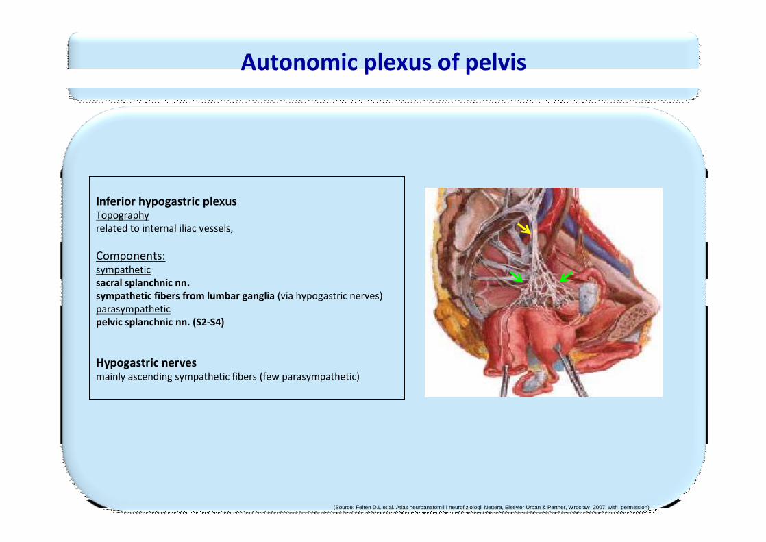
Inferior hypogastric plexusTopographyrelated to internal iliac vessels,
Components:sympatheticsacral splanchnic nn. sympathetic fibers from lumbar ganglia (via hypogastric nerves)parasympatheticpelvic splanchnic nn. (S2-S4)
Hypogastric nervesmainly ascending sympathetic fibers (few parasympathetic)
Autonomic plexus of pelvis
(Source: Felten D.L et al. Atlas neuroanatomii i neurofizjologii Nettera, Elsevier Urban & Partner, Wrocław 2007, with permission)