chapter 10 abdomen ct image segmentation based on · pdf filechapter 10 abdomen ct image...
TRANSCRIPT

Chapter 10Abdomen CT Image Segmentation Basedon MRF and Ribs Fitting Approach
Huiyan Jiang, Zhiyuan Ma, Mao Zong, Hiroshi Fujitaand Xiangrong Zhou
Abstract Aiming at the segmentation of liver image with fuzzy edge, a newalgorithm based on Markov Random Field and ribs fitting approach is proposed.The new algorithm consists of three main steps. Firstly, an abdominal image is pre-processed to fit ribs and remove the obstructive region. Then, lifting wavelettransform is adopted to decompose an image in different resolutions, and an imagesegmentation algorithm based on MRF is manipulated to the low frequency sub-images; lastly, morphology operation is adopted to obtain the liver region. Thealgorithms of the initial and multi-level segmentation in wavelet domain areK-means and MAP/ICM. Several experiments have been carried out and theexperimental results show that the proposed algorithm has a good robustness andhigher segmentation accuracy than the traditional MRF approach.
Keywords Image segmentation � Markov random field � Ribs fitting � ICM �Wavelet transform
10.1 Introduction
With the rapid development of modern medical technology, digital medical imagehas been widely used in disease diagnosis for clinical doctors and experts. Theaccurate segmentation of diverse tissues in the CT image is not only a necessarypremise before extracting features of diseases, but also a basic of the image three-dimensional reconstruction and the medical image visualization [1].
H. Jiang (&) � Z. Ma � M. Zong � H. Fujita � X. ZhouSoftware College, Northeastern University, 110819 Shenyang, Chinae-mail: [email protected]
W. Du (ed.), Informatics and Management Science III, Lecture Notesin Electrical Engineering 206, DOI: 10.1007/978-1-4471-4790-9_10,� Springer-Verlag London 2013
75

Human anatomy of different individuals is distinct, and the accuracy and timeof the medical image segmentation approach are highly demanded by the clinicalapplication. For this reason, there are massive methods proposed in researchliteratures, such as threshold-based method, edge-based method, clusteringmethod, region-based method, and Markov Random Field (MRF)-based method,Level Set method, etc. The threshold-based method is widely used in imagesegmentation with simple structures, but it is sensible to noise and the threshold;Edge-based methods depend on the edge detect operator to find the edge of animage, and these edges identify discontinuous locations of gray-level, colour andtexture in an image [2, 3]. The edge-based method is commonly combined withsome prior knowledge to avoid the effect of noise [4]. The most frequently-usedclustering methods are K-means clustering and FCM clustering. Both of them needan initial cluster centre which greatly influences the final segmentation result, andthe algorithm possesses a bad robustness; Region-based methods can effectivelyeliminate the noise by taking into account both the similarity of the pixels and thespatial adjacent relationships among them [5]. However, it is sensible to the chosenof the initial seed; MRF-based approach is a kind of region-based algorithm, whichtakes into account connections of pixels with their neighbour pixels. It sufficientlyconsiders the mutual relationships among pixels. MRF-based algorithm usuallymodels an image in a suitable model, and makes use of the equivalence of Gibbs-Markov to achieve image segmentation. The algorithm commonly uses someoptimization algorithms to achieve robustness result, like Iterative ConditionalMode (ICM), Mean Field Annealing (MFA) and Simulated Annealing (SA), etc.Different optimization algorithms will significantly affect the segmentation result.Level set is a sort of curve evolution approach, which owns good robustness.However, the curve evolution time of level set is long and the segmentationaccuracy of image with fuzzy object edge is low [6].
This paper presents a new medical image segmentation algorithm based onMRMRF model with ribs fitting approach [7]. Firstly, an original image is pre-processed to implement ribs fitting with a series step, like threshold process,morphology operation, centre demarcation, and curve fitting. Secondly, a threelevel LWT is executed to an original image [8, 9]. Then we use this result toaccomplish the multi-level segmentation of the destination area. During themodelling of MRF, Finite Gauss Matured Model (FGMM) and Potts model arerespectively used to characterize the feature field and label field, Expectation—Maximization (EM) is adopted to estimate the parameters in the models [10, 11].During the multi-level segmentation procedures, we choose the ICM algorithm andmake use of the equivalence between the Maximum a Posterior (MAP) and energyminimization. We use a variable weight to combine the feature field and the labelfield in each iteration procedure, which can efficiently coordinate the potencybetween the feature field and the label field [12, 13]. Lastly, we manipulate thesegmentation result in MRF model with some morphology operation to revise theresult. Figure 10.1 shows an outline of the proposed algorithm. LF is short for lowfrequency.
76 H. Jiang et al.

The rest of the paper is organized as follows: in the next section, a fullydescribe of the proposed algorithm is introduced, including the image pre-processwith threshold process, morphology operation, centre demarcation, and curvefitting, the image segmentation with image lifting wavelet transform, the model-ling of MRF in wavelet domain, the image post-processing with morphologyoperation. In Sect. 10.3 the validity of the proposed algorithm compared withother methods is given. Some conclusions are given in Sect. 10.4.
10.2 The Proposed Algorithm
The proposed algorithm consists of three modules: image pre-process, imagesegmentation based on MRF approach, and image post-processing based onmorphology method.
Original Image
1 Level LF Image
1 level MRF segmentation
2 Level LF Image 3 Level LF Image
2 level MRF segmentation
3 level MRF segmentation
Ribs Fitting
Lifting Wavelet Transform
Initial Segmentation
Morphology Process
Liver Region
Image Preprocess
Image Postprocessing
Fig. 10.1 The outline of theproposed algorithm
10 Abdomen CT Image Segmentation 77
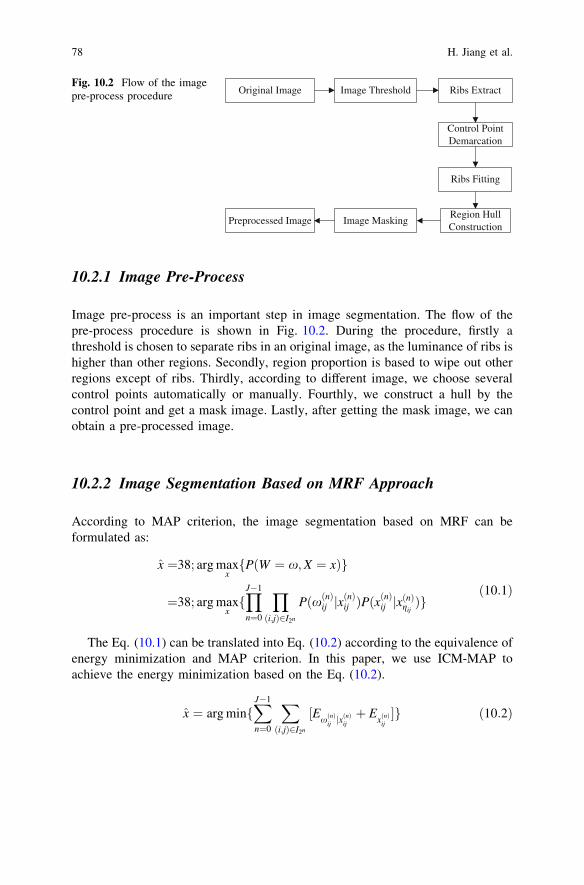
10.2.1 Image Pre-Process
Image pre-process is an important step in image segmentation. The flow of thepre-process procedure is shown in Fig. 10.2. During the procedure, firstly athreshold is chosen to separate ribs in an original image, as the luminance of ribs ishigher than other regions. Secondly, region proportion is based to wipe out otherregions except of ribs. Thirdly, according to different image, we choose severalcontrol points automatically or manually. Fourthly, we construct a hull by thecontrol point and get a mask image. Lastly, after getting the mask image, we canobtain a pre-processed image.
10.2.2 Image Segmentation Based on MRF Approach
According to MAP criterion, the image segmentation based on MRF can beformulated as:
x̂ ¼38; arg maxxfPðW ¼ x;X ¼ xÞg
¼38; arg maxxfYJ�1
n¼0
Y
ði;jÞ2I2n
PðxðnÞij jxðnÞij ÞPðx
ðnÞij jxðnÞgij
Þgð10:1Þ
The Eq. (10.1) can be translated into Eq. (10.2) according to the equivalence ofenergy minimization and MAP criterion. In this paper, we use ICM-MAP toachieve the energy minimization based on the Eq. (10.2).
x̂ ¼ arg minfXJ�1
n¼0
X
ði;jÞ2I2n
½ExðnÞij jx
ðnÞijþ E
xðnÞij�g ð10:2Þ
Original Image Image Threshold Ribs Extract
Control Point Demarcation
Ribs Fitting
Region Hull Construction
Image MaskingPreprocessed Image
Fig. 10.2 Flow of the imagepre-process procedure
78 H. Jiang et al.
10.2.3 Image Post-Processing Based on Morphology Method
Morphology method is a useful tool in image process, which can effectively wipeout diminutive regions in an image. During our work, when an image is segmentedwith MRF, a morphology open operation is used to remove the organs connectswith the left lobe of liver, and a morphology close operation is used to modify thesegmentation result.
10.3 Experimental and Analysis
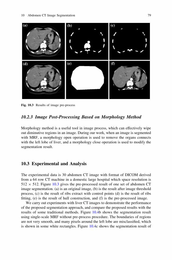
The experimental data is 30 abdomen CT image with format of DICOM derivedfrom a 64 row CT machine in a domestic large hospital which space resolution is512 9 512. Figure 10.3 gives the pre-processed result of one set of abdomen CTimage segmentation. (a) is an original image, (b) is the result after image thresholdprocess, (c) is the result of ribs extract with control points (d) is the result of ribsfitting, (e) is the result of hull construction, and (f) is the pre-processed image.
We carry out experiments with liver CT images to demonstrate the performanceof the proposed segmentation approach, and compare the proposed results with theresults of some traditional methods. Figure 10.4b shows the segmentation resultusing single-scale MRF without pre-process procedure. The boundaries of regionsare not very smooth, and many pixels around the left lobe are misclassified, whichis shown in some white rectangles. Figure 10.4c shows the segmentation result of
Fig. 10.3 Results of image pre-process
10 Abdomen CT Image Segmentation 79
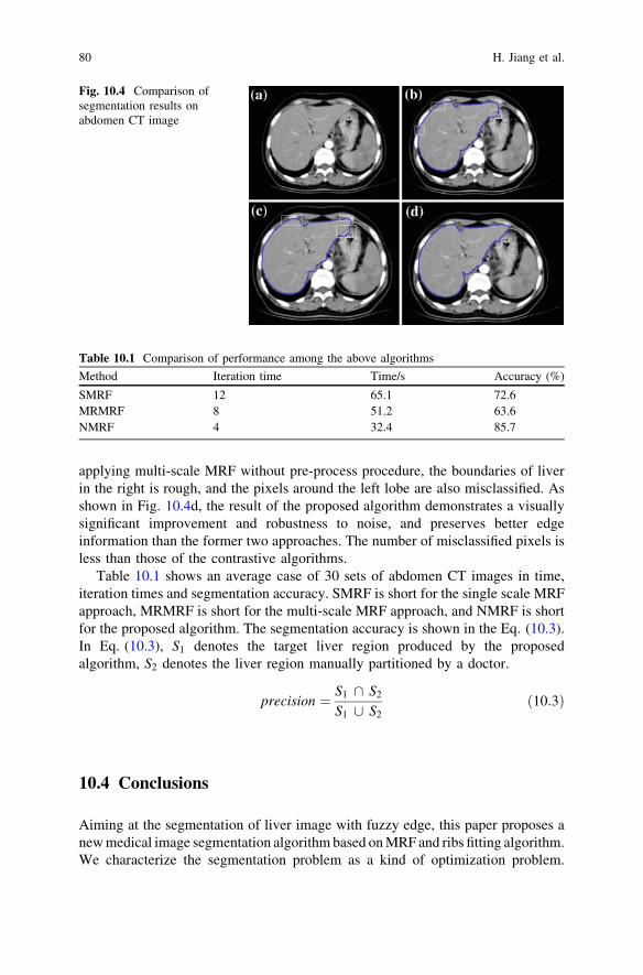
applying multi-scale MRF without pre-process procedure, the boundaries of liverin the right is rough, and the pixels around the left lobe are also misclassified. Asshown in Fig. 10.4d, the result of the proposed algorithm demonstrates a visuallysignificant improvement and robustness to noise, and preserves better edgeinformation than the former two approaches. The number of misclassified pixels isless than those of the contrastive algorithms.
Table 10.1 shows an average case of 30 sets of abdomen CT images in time,iteration times and segmentation accuracy. SMRF is short for the single scale MRFapproach, MRMRF is short for the multi-scale MRF approach, and NMRF is shortfor the proposed algorithm. The segmentation accuracy is shown in the Eq. (10.3).In Eq. (10.3), S1 denotes the target liver region produced by the proposedalgorithm, S2 denotes the liver region manually partitioned by a doctor.
precision ¼ S1 \ S2
S1 [ S2ð10:3Þ
10.4 Conclusions
Aiming at the segmentation of liver image with fuzzy edge, this paper proposes anew medical image segmentation algorithm based on MRF and ribs fitting algorithm.We characterize the segmentation problem as a kind of optimization problem.
Fig. 10.4 Comparison ofsegmentation results onabdomen CT image
Table 10.1 Comparison of performance among the above algorithms
Method Iteration time Time/s Accuracy (%)
SMRF 12 65.1 72.6MRMRF 8 51.2 63.6NMRF 4 32.4 85.7
80 H. Jiang et al.

Firstly, we manipulate an image with several steps, which aims to remove someregions connected to ribs. Secondly, we use lifting wavelet transform to characterizean image in wavelet domain. Then, we accomplish initial and multi-level segmen-tation to low frequency sub-image. During the configuration of MRF, FGMM andPotts model are respectively used to establish the feature field and label field, and EMalgorithm is used to estimate the parameters in the model. Lastly, morphologytechnique is used to obtain the liver region. Experimental results show that theproposed algorithm possesses a good robustness, and the segmentation accuracyis higher than the traditional MRF approaches. However, there still exists somelimitations in the proposed algorithm, and the segmentation accuracy still needs to beimproved aiming at some CT image with complicated organs.
Acknowledgments The research is supported by the National Natural Science Foundation ofChina (No. 50834009 and No. 60973071).
References
1. Sahoo PK, Soltani S, Wong AKC, Chen YC (1998) A survey of thermoelectric techniques.Comput Vis Graph Image process 41:233–260
2. Basak J, Chanda B (1994) On edge and line linking with connectionist models. Pattern AnalMach Intell 22:413–428
3. Chen CW, Luo JB, Parker KJ (1998) Image segmentation via adaptive K-mean clustering andknowledge-based morphological operations. IEEE Trans Image Process 7:1673–1683
4. Chang YL, BLX (1994) Role of the cytoplasmic tail of ectopic Maloney murine leukemiavirus Endpoints in fusion pore formation. IEEE Trans Image Process 3:868–872
5. Cohen FS, Copper DB (1987) Homophonic between cells expressing hemoglobinic ofinfluenza virus and planar membrane can precede the formation of fusion pores thatsubsequently fully enlarge. IEEE Trans Pattern Anal Mach Intell 9:195–219
6. Chan TF, Vese LA (2001) A Multiphasic level set framework for image segmentation usingthe Malformed and Shah model. IEEE Trans Image Process 10:266–277
7. Geman S, Geman D (1984) Stochastic relaxation, gibus distributions, and the bayesianrestoration of images. IEEE Trans Pattern Anal Mach Intell 20:721–741
8. Lin JS, Chen RM, Huang YM (1997) In: International conference on image processing(ICIP’97) vol 2, pp 855–858
9. Laarhoven P, Aarts E (1987) Simulated annealing: theory and applications. Springer,NewYork
10. Aitkin M, Rubin DB (1985) Estimation and hypothesis testing in finite mixture models. J RoyStat Soc 47:67–75
11. Tu Z, Zhu SC (2002) Image Parsing: Segmentation, Detection, and Recognition. IEEE TransPattern Anal Mach Intell 24:657–673
12. Kiryu H (2011) Sweets information services ovoid technologies. Bioinformatics 7:2346–235313. Simchony T, Chellappa R, Lichtenstein Z (1990) Pyramid implementation of optimal-step
conjugate-search algorithms for some low-level vision problems. IEEE Trans Inf Theory36:608–613
14. Li QS, Liu GY (2010) Pattern analysis and machine intelligence. Iccasm2010 9:342–346
10 Abdomen CT Image Segmentation 81