recent progress in optical biosensors for environmental applications
TRANSCRIPT

Chapter 1
Recent Progress in Optical Biosensors forEnvironmental Applications
Feng Long, Anna Zhu, Chunmei Gu andHanchang Shi
Additional information is available at the end of the chapter
http://dx.doi.org/10.5772/52252
1. Introduction
The rapid screening and sensitive monitoring of environmental pollutants, such as pesticides,persistent organic pollutants (POPs), endocrine disrupting chemicals (EDCs), explosives, andtoxins, is indeed essential to ensure environmental quality, and therefore, human health. Untilrecently, the quantification of most contaminants has been limited to the traditional chromato‐graphic and spectroscopic technologies. These methods, although accurate with low detectionlimits, are labor-intensive and require expensive and sophisticated instrumentation, as well ascomplicated and multistep sample preparation, which prohibits frequent and real-time on-site monitoring of contaminants in environment.[1] Considerable research interests, therefore,have risen for detecting low levels of environmental pollutants in biosensor development be‐cause of their simplicity, robustness, sensitivity, specificity and cost-effectiveness. [2]
A biosensor is an analytical device combined a biological sensing element with a physicaltransducer, in which the binding or reaction between the target and the recognition element istranslated into a measurable electrical signal. [3] Among them, optical biosensors are power‐ful alternative to conventional analytical techniques due to their cost-effective, fast and porta‐ble detection, which makes on-site and real-time monitoring possible without extensivesample preparation. Optical biosensors have vast potential applications in environmentalmonitoring, food safety, drug development, and medical diagnosis.[4] Although the use of op‐tical biosensors in water quality early-warning and pollution control is still in its infancy, re‐search on this topic is an active area and the remarkable technological progress has been made.
The present article gives an overview of the recent advances in optical biosensors and theirapplications in the environmental field. Functional biorecognition materials (e.g. enzyme,
© 2013 Long et al.; licensee InTech. This is an open access article distributed under the terms of the CreativeCommons Attribution License (http://creativecommons.org/licenses/by/3.0), which permits unrestricted use,distribution, and reproduction in any medium, provided the original work is properly cited.

antibody, aptamer, and DNAzyme), a key component of biosensor and specifically bindinga broad range of analytes including inorganic, organic, and biomolecules, will be first re‐viewed. Then, nanomaterials such as quantum dots, graphene, nanogold particles, carbonnanotubes, and magnetic nanoparticles will be introduced, which have been successfully in‐corporated into optical biosensors to improve the sensibility, sensitivity, and selectivity dueto their unique physicochemical properties. In addition, the recent significant improvementsin instrumentation will also be discussed, which have allowed a wider variety of pollutantsto be analysed in details, and led to the increasing application of optical biosensor technolo‐gy throughout the environmental detection field. Finally, recent developments of optical bi‐osensors for pollution control and early-warning will be highlighted.
2. Functional biorecognition materials
Functional biorecognition materials are key components of biosensors, and generally havehigh affinity (low detection limit), high specificity (low interference), wide dynamic range, fastresponse time, and long shelf life. The antibodies are most frequently used biorecognition mol‐ecules in the optical biosensor community. However, enzymes were the first recognition ele‐ments used in biosensors. Another frequently used recognition elements are nucleic acids suchas aptamer and DNAzyme for the monitoring of environmental pollutants.
2.1. Antibody
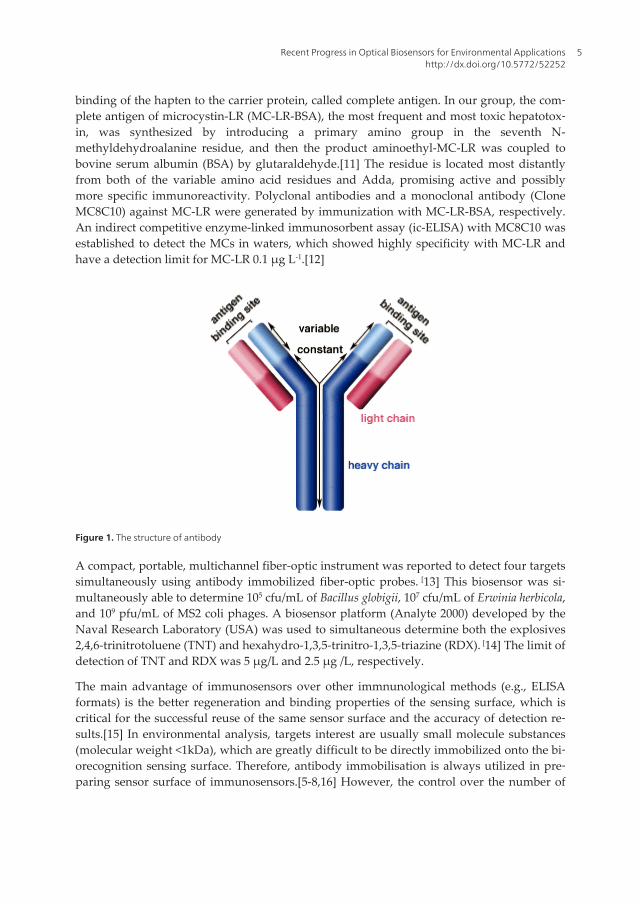
Immunosensors, based on specific antigen-antibody interactions, have become the gold-stand‐ard technique in clinical diagnostics and environmental monitoring. [4-9] Antibody is a large Y-shaped protein used by the immune system to identify and neutralize a unique part of foreigntarget, called an antigen, and is produced by white blood cell (a plasma cell). Antibodies aretypically made of basic structural units: each with two larger heavy chains and two shorterlight chains. The IgG molecule (see Figure 1) is the most used antibody type and is about 150-kDa protein composed of four polypeptide chains.[8] Antibodies are produced as monoclonaland polyclonal varieties, with monoclonal antibodies binding to a single epitope and polyclo‐nal antibodies being capable of binding to multiple epitopes.[9,10] In immunoassay, two anti‐gen binding sites of antibody have a highly specific interaction for one particular target, andthis immunochemical reaction can be detected by the transducer (e.g. optical, electronical).[5-9] Therefore, the immunosensor assay provides a highly repeatable and highly specific reac‐tion format, and the capacity for specific recognition of environmental contaminants.
Due to most of the environmental pollutants have the low molecular weight (<1 kDa) andare called haptens which are non-immunogenic, it has to be conjugated to carrier proteins tomake them immunogenic.[11] Preparation of antibodies against haptens, such as pesticides,persistent organic pollutants (POPs), and endocrine disrupting chemicals (EDCs), is basedon covalent binding of the hapten to a carrier protein and immunisation of animals by thesynthesised immunogens. The specificity of antibody is important for immunoassay, whilethe specificity and quality of antibody is mostly determined by the manner of chemical
State of the Art in Biosensors - Environmental and Medical Applications4
binding of the hapten to the carrier protein, called complete antigen. In our group, the com‐plete antigen of microcystin-LR (MC-LR-BSA), the most frequent and most toxic hepatotox‐in, was synthesized by introducing a primary amino group in the seventh N-methyldehydroalanine residue, and then the product aminoethyl-MC-LR was coupled tobovine serum albumin (BSA) by glutaraldehyde.[11] The residue is located most distantlyfrom both of the variable amino acid residues and Adda, promising active and possiblymore specific immunoreactivity. Polyclonal antibodies and a monoclonal antibody (CloneMC8C10) against MC-LR were generated by immunization with MC-LR-BSA, respectively.An indirect competitive enzyme-linked immunosorbent assay (ic-ELISA) with MC8C10 wasestablished to detect the MCs in waters, which showed highly specificity with MC-LR andhave a detection limit for MC-LR 0.1 µg L-1.[12]
Figure 1. The structure of antibody
A compact, portable, multichannel fiber-optic instrument was reported to detect four targetssimultaneously using antibody immobilized fiber-optic probes. [13] This biosensor was si‐multaneously able to determine 105 cfu/mL of Bacillus globigii, 107 cfu/mL of Erwinia herbicola,and 109 pfu/mL of MS2 coli phages. A biosensor platform (Analyte 2000) developed by theNaval Research Laboratory (USA) was used to simultaneous determine both the explosives2,4,6-trinitrotoluene (TNT) and hexahydro-1,3,5-trinitro-1,3,5-triazine (RDX). [14] The limit ofdetection of TNT and RDX was 5 µg/L and 2.5 µg /L, respectively.
The main advantage of immunosensors over other immnunological methods (e.g., ELISAformats) is the better regeneration and binding properties of the sensing surface, which iscritical for the successful reuse of the same sensor surface and the accuracy of detection re‐sults.[15] In environmental analysis, targets interest are usually small molecule substances(molecular weight <1kDa), which are greatly difficult to be directly immobilized onto the bi‐orecognition sensing surface. Therefore, antibody immobilisation is always utilized in pre‐paring sensor surface of immunosensors.[5-8,16] However, the control over the number of
Recent Progress in Optical Biosensors for Environmental Applicationshttp://dx.doi.org/10.5772/52252
5

antibodies and their orientation and position relative to the sensor surface is very difficult.Because of the possibility of inadvertently disrupting the binding site when conjugating an‐tibody with active surface of sensor, the activity loss of antibody is inevitable.[17,18] Mostimportant, due to strong acid being usually used in regeneration process, the recognitionability of antibodies immobilized may be lower after senor surface reuse, which will affectthe stability and reliability of the immunosensor. The cycles of regeneration are usually nomore than fifteen times and in each cycle, antibody activity decreased, which leads to inac‐curate detection results. Therefore, hapten-carrier-protein conjugates as bio-recognition mol‐ecules were immobilized onto the surface of immunosensor for obtaining the stable reusablesensor. For example, a reusable immunosurface is provided via the covalent attachment ofthe 2,4-D-BSA and MC-LR-OVA to a self-assembled monolayer formed onto the fiber opticsensor.[19] The regeneration of the sensor surface allows the performance of more than 100assay cycles.
2.2. Enzyme
Enzymes are biological molecules that catalyze (i.e., increase the rates of) chemical reactions,and are usually very specific as to which reactions they catalyze and the substrates that areinvolved in these reactions. Enzymes are historically the first molecular recognition ele‐ments included in biosensors and continue to be the basis for a significant number of publi‐cations reported for biosensors in general as well as for environmental applications.[4]Enzyme biosensors have several advantages, such as a stable source of material, the possibil‐ity of modifying the catalytic properties or substrate specificity by means of genetic engi‐neering, and catalytic amplification of the biosensor response by modulation of the enzymeactivity with respect to the target analyte. [20]
Most of enzyme biosensors normally use enzymes as the bioreceptors and achieved pollu‐tants detection based on the enzyme inhibition mechanism.[4][20] Due to ChE enzymes can beinhibited by several toxic chemicals such as organophosphate and pesticides, heavy metals,and toxins, the ChEs biosensors is of particular interest in the area of global toxicity monitor‐ing.[4,21,22]
Due to various pollutants that inhibit the activity of enzymes in a different ways, multi-ana‐lytes detection can be achieved by enzyme sensors. For example, simultaneous detectionboth pesticides and heavy metal ions in a sample solution is possible due to selective inhibi‐tion of butyrylcholine esterase by pesticides and urease by heavy metals ions.[4-6,23] Com‐paring with the inhibition level of urease or butyrylcholine esterase, respectively, thepesticides or heavy metal ions can be determined. The enzyme-inhibition based biosensorsarray could achieve the simultaneous determination of various pollutants in water samples.
Enzyme biosensors have some limitations for the detection of environmental pollutants,which include the limited number of substrates for which enzymes have been evolved, thelimited interaction between environmental pollutants and specific enzymes, and the lack ofspecificity in differentiating among compounds of similar classes.[6,23] However, artificialor synthetic enzymes could be a useful alternative to natural enzymes for the development
State of the Art in Biosensors - Environmental and Medical Applications6

of new biosensors, which are more robust, available, chemically malleable and cheap, in
comparison with their natural analogues.
No Target Aptamer type Sequence Reference
1 2,3',5,5'-
Tetrachlorobiphenyl;
2,3,3',4,5-
Pentachlorobiphenyl
DNA 9.1:CGCTACACCT,CGCCAGCAAA,TTGCCGCCCG,CAGCCCTCTA
9.2:GGGACTCGAG,ACCCGTTCCG,TTCTCCGCTT,GCCCCACAAT
[26]
2 4,4’-
methylenedianiline(MDA)
RNA M1:CUGCGAUCA,GGGGUAAAUU,UCCGCGCAGG,CUCCACGCCG,C
M2:CUCGA,GUCCUCUUGA,GCGGUUCCUA,CUUCCCUCUG,CUGUG
[27]
3 Organophosphorus
Pesticides:phorate,profenof
os, isocarbophos,
omethoateas
DNA 1: AAGCTTGCTTTATAGCCTGCAGCGAT TCTTGATCGGAAAAGG
CTGAGAGCTACGC
2:AAGCTTTTTTGACTGACTGCAGCGATTCTTGATCGCCACGGTCTGGAAAA
AGAG
[28]
4 bisphenol A DNA CCGGTGGGTGGTCAGGTGGGATAGCGTTCCGCGTATGGCCCAGCGCATC
ACGGGTTCGCACCA
[29]
5 17β-estradiol DNA GCTTCCAGCTTATTGAATTACACGCAGAGGGTAGCGGCTCTGCGCATTCA
ATTGCTGCGCGCTGAAGCGCGGAAGC
[30]
6 Chloramphenicol DNA 1:ACTTCAGTGA,GTTGTCCCAC,GGTCGGCGAG,TCGGTGGTAG
2:CACCAAGCGC,AGGGAATTAC,ATTGAAGTGT,GGGATTGGCT
[31]
7 Oxytetracycline DNA 1:CGACCGCAGGTGCACTGGGCGACGTCTCTGGTGTGGTGT
2:CGACGCGCGTTGGTGGTGGATGGTGTGTTACACGTGTTGT
[32]
8 Tetracycline DNA T7:GGGCAGCGGTGGTGTGGCGGGATCTGGGGT,TGTGCGGTGT
T15:GGAGGAACGGGTTCCAGTGTGGGGTCTATC,GGGGCGTGCG
[33]
9 Kanamycin DNA TGGGGGTTGAGGCTAAGCCGA [34]
10 ricin B chain DNA ACACCCACCGCAGGCAGACGCAACGCCTCGGAGACTAGCC [35]
11 Ochratoxin A DNA GATCGGGTGTGGGTGGCGTAAAGGGAGCATCGGACA [36]
12 E. coli DNA ATCCGTCACACCTGCTCTACGGCGCTCCCAACAGGCCTCTCCTTACGGCAT
ATTA TGGTGTTGGCTCCCGTAT
[37]
13 Staphylococcus aureus
Enterotoxin B
DNA GGTATTGAGGGTCGCATCCACTGGTCGTTG
TTGTCTGTTGTCTGTTATGTTGTTTCGTGATGG CTCTAACTCTCCTCT
[38]
14 Salmonella entericaserovars DNA 23:CCGCCTTTACTAAATTGACGAACATAGGAATCAATGAAGC
24:GGGAGTCAGAACGCCTGGGCAAGCATAGTACTCGCCGGAA
[39]
17 Ibuprofen DNA IBA2:ACAGTAGTGAGGGGTCCGTCGTGGGGTAGTTGGGTCGTGG
IBA8:GCGAACGACTTCATAAAATGCTATAAGGTTGCCCTCTGTC
[40]
18 Arsenic DNA TTACAGAACAACCAACGTCGCTCCGGGTACTTCTTCATCG [41]
Table 1. DNA/RNA aptamers
Recent Progress in Optical Biosensors for Environmental Applicationshttp://dx.doi.org/10.5772/52252
7

2.3. Aptamer
Aptamer is a single-stranded oligonucleotide that folds into complex three-dimensionalstructure and bind strongly and selectively to one certain kind of target or one class of tar‐gets. [4-6] The aptamer is selected using an in vitro process called Systematic Evolution of Li‐gands by EXponential enrichment (SELEX), which was first put forward by Ellington et al.and Tuerk et al. in 1990.[24,25] For the selection of DNA aptamer, the SELEX starts from theconstruction of a random pool of DNA sequences (~1015), and then the selection procedurecould take place, including: (a) binding between the library and the target; (b) separation ofthe unbound ssDNA and the ssDNA-target complex; (c) elution the bound ssDNA from thessDNA-target complex; (d) amplification of the bound ssDNA, usually using the PCR meth‐od; (e) generation of single strand DNA from the double strand PCR products. Generally, atraditional aptemer selection process need 10-12 cycles. The ssDNA pool from the first selec‐tion cycle is used as the starting library for the second selection cycle, then repeating proce‐dure (a), (b),(c),(d) and (e). After the last selection cycle, molecular cloning and DNAsequencing was applied, and the aptamer with high affinity and specifity is obtained.
Aptamers offer a useful alternative to antibodies as sensing molecules and have opened anew era in development of affinity biosensing due to their unique characterizations. In vitroselected aptamers could be produced for any targets such as proteins, peptides, amino acids,nucleotides, drugs, heavy metal ions, and other small organic and inorganic compounds.[26-41] Aptamers could be chemically synthesized without the complicated and expensivepurification steps by eliminating the batch-to-batch variation found when using antibodies.Furthermore, modifications in the aptamer through chemical synthesis can be introducedenhancing the stability, affinity and specificity of the molecules. In addition, aptamers aremore stable than antibodies and thus are more resistant to denaturation and degradation.Often the affinity parameters of aptamer-target complex can be changed for higher affinityor specificity. In addition, aptamers have the higher temperature stability and can recovertheir native active conformation after denaturation, whereas antibodies are large, tempera‐ture-sensitive proteins that can undergo irreversible denaturation. Recently, severalDNA/RNA aptamers, selected for POPs, EDCs, organophosphorus pesticides, antibiotics, bi‐otoxins, and pathogenic microorganisms, are listed in Table 1.
Aptamers have become increasingly important molecular tools for environmental bioassay.EDCs are contaminants of emerging concern and required routine monitoring in water sam‐ples, as posed by EPA Unregulated Contaminant Regulation (UCMR3). Gu et al. [42] report‐ed a reusable evanescent wave aptamer-based biosensor for rapid, sensitive and highlyselective detection of 17β-estradiol, frequently detected in environmental water samples. Inthis system, the capture molecular, β-estradiol 6-(O-carboxy-methyl) oxime-BSA, was cova‐lently immobilized onto the optical fiber sensor surface. With an indirect competitive detec‐tion mode, the limit of detection of 17β-estradiol was determined as 2.1 nM. Kim et al. [41]developed a high affinity DNA aptamer for arsenic that can bind to arsenate [(As(V)] andarsenite [As(III)] with a dissociation constant of 5 and 7 nM, respectively. Through the “sig‐nal-on” mode or the “signal-off” mode, reflecting the extent of the binding process therebyallowing for quantitative measurement of target concentration, several DNA aptamer fluo‐
State of the Art in Biosensors - Environmental and Medical Applications8

rescence-based sensors have been developed for the detection of Hg2+, Pb2+ and other tracepollutants.[43] Although a variety of aptamer has been successfully selected for environ‐mental contaminants, the detection of the real water samples using the right aptamer is stillin the cradle.
2.4. DNAzyme
DNAzymes are small single-stranded nucleic acids that fold into a well-defined three-di‐mensional structure with high specificity to various ligands, such as low-molecular-weightorganic or inorganic substrates or macromolecules or metal ions.[43] DNAzymes have apromising capacity to selectively identify charged organic and inorganic compounds at ul‐tratrace levels in environmental samples or biological systems. Furthermore, DNAzymescan perform chemical modifications on nucleic acids, while aptamers can bind a broad rangeof molecules. A combination of the two has generated a new class of functional nucleic acidsknown as allosteric DNAzymes or aptazymes. Combining the specificity of nano-biologicalrecognition probes and the sensitivity of laser-based optical detection, DNAzymes are capa‐ble of provide unambiguous identification and accurate quantification of environmental pol‐lutants, ranging from small ions to large molecules. RNA-cleaving DNAzymes are the mostwidely used due to their simple reaction conditions, fast turnover rates and significant mod‐ifications of their substrate lengths.
Using the in vitro selection of specifical DNAzymes, several fluorescence biosensors haveextensively been developed for the detection of various heavy metal ions, such as Pb2+, Cu2+,Mg2+, Ca2+, Zn2+, Co2+, Mn2+, UO2 2+, Hg2+ and Ag+, etc.[43,44] Moreover, DNAzymes and ap‐tazymes have already found many applications in almost every aspect of DNA nanotechnol‐ogy, which result to new materials and devices that may penetrate into many other fields forpractical applications, including environmental monitoring.
3. Nanomaterials in optical biosensors
Nanomaterials exhibit unique size-tunable and shape-dependent physicochemical proper‐ties that are different from those of bulk materials.[45-50] Specially, the interaction of nano‐materials and functional biomaterials opens a new door to develop various novel opticalbiosensors. NPs such as gold NPs (AuNPs), quantum dots (QDs), magnetic NPs (MNPs),graphene and carbon nanotubes have specific optical, fluorescence and magnetic properties,and interactions between these properties give NPs great potential for environmentalscreening.[45-51] The extremely high surface-to-volume ratios and exceptional nanoscaleproperties make NPs useful for next-generation environmental detection.
3.1. Quantum dots
Semiconductor quantum dots (QDs), nanocrystals of inorganic semiconductors, haveemerged as promising alternative bioanalytical tools because of their unique optical proper‐ties including high quantum yield, photostability, narrow emission spectrum, and broad ab‐
Recent Progress in Optical Biosensors for Environmental Applicationshttp://dx.doi.org/10.5772/52252
9
sorption.[52,53] QDs’ band gap depends on the size of the nanocrystal. That is to say, thesmaller the nanocrystal, the larger the difference between the energy levels and, therefore,the wider the energy gap and the shorter thewavelength of the fluorescence. [52,53]
The main application of QDs as sensors exploits the Forster resonance energy transfer effect(FRET) due to their narrow, size-tuned, and symmetric emission spectra, which has madethem excellent donors for fluorescence resonance energy transfer (FRET) sensors, and great‐ly reduces the overlap between the emission spectra of donor and acceptor and circumventsthe cross-talk in such FRET pairs.[52,53] Meanwhile, QDs have broad excitation spectra asdonor, and allow excitation at a single wavelength far removed (>100nm) from their respec‐tive emissions, which enables QDs to be used in multiplex assays without the need for mul‐tiple excitation sources. In addition, the high photobleaching threshold and good chemicalstability of QDs greatly improve the detection sensitivities and detection limits. Therefore,QD-based FRET biosensors have been widely used in environmental monitoring, medicalimaging, clinical/diagnostic assays, and biomolecular binding assay. Among available QDs,CdSe/ZnS core-shell quantum dots are most commonly used for biosensing applications.Antibody (or aptamer) bioconjugates of QDs, prepared using covalent or non-covalent link‐ing approaches, are the most developed and widespread detection bioprobes to integratingQDs into bioanalyses. The fluorescent detection of pathogens such as respiratory syncytialvirus,[54] E. coli O157:H7,[55] and Bacillus thurinigensis,[56] has been performed based onQDs-FRET.
However, the control over the number of antibodies (or aptamers) per QD and their orienta‐tion and position relative to the QD is very difficult. Due to the possibility of inadvertentlydisrupting the binding site when conjugating QD with antibody, the activity loss of anti‐body is inevitable.[52,57] Additionally, antibodies usually need to be cryopreserved but QDscannot be freezed, which makes the storage of QD-antibody a major obstacle for its practicalapplications. To effectively address these challenges, we have developed carrier-protein-haptens-coupled quantum-dot nanobioprobe protocols to perform rapid and sensitive detec‐tion of small targets in real water samples.[58] 2,4-Dicholrophenoxyacetic acid (2,4-D), oneof the most widely used pesticides worldwide, was selected as a model target. QD nano-im‐munoprobe were prepared through conjugating carboxyl quantum dots with 2,4-D-BSAconjugate, which regarded as the immunological recognition of anti-2,4-D antibody as wellas for optical transducer. With a competitive detection mode, samples containing differentconcentrations of 2,4-D were incubated with a given concentration of QD immunoprobe andfluorescence-labeled antibody, and then detected by an all-fiber microfluidic biosensingplatform developed by our group. A higher concentration of 2,4-D led to less fluorescence-labeled anti-2,4-D antibody bound to the QD immunoprobe surface, and thus to lower fluo‐rescence signal. The quantification of 2,4-D over concentration ranges from 0.5 nM to 3 µMwith a detection limit determined as 0.5 nM. The structure of multiplex-haptens/BSA conju‐gate coupling to QD greatly improves the FRET efficiency and nanosensor’s sensitivity.With the use of different QD immunoprobes modified by the conjugates of other haptens/carried protein, the methodology presented here has the potential to extend to toward the
State of the Art in Biosensors - Environmental and Medical Applications10

on-site monitoring other small analytes in a variety of application fields ranged from envi‐ronmental to biomedical areas.
3.2. Nanogold particles
Gold nanoparticles (AuNPs) typically have dimensions ranging from 1-100 nm and displaymany interesting electrical and optical properties. Nanogold particles based optical biosen‐sors commonly take use of the fluorescence quenching through fluorescence resonance ener‐gy transfer (FRET) or a visible color change due to the aggregation of AuNPs of appropriatesizes.[59] Over the past decades, AuNPs based optical sensors have an important role in thedetection of environmental pollutants such as toxins, heavy metals and other pollutants dueto their typically high signal-to-noise ratios.[51]
Uzawa et al.[60] developed sugar-coated GNPs for the detection of ricin with visual read-out using the naturally occurring infection mechanism and the strong affinity of the toxinricin to sugar. Many kinds of immunoassays using GNP-antibody conjugates have been de‐veloped for detection of ochratoxin A (OTA), zearalenone (ZEA), and aflatoxin B1 (AFB1).[51,61] AuNP-based biosensors have also been used to highly competitive assay technolo‐gies for the detection of oligonucleotide targets.[51]
Heavy metal contamination is an ongoing concern worldwide, and it is vital for rapid andsimple monitoring technologies of heavy metal ions in environment. Darbha et al.[62] devel‐oped a GNP-based sensor for rapid, easy and reliable detection of Hg2+ ions in aqueous solu‐tions, which had a detection limit of 5 ng/ml (ppb) through non-linear optical properties. Avisual detection methode of Cu2+ was reported by Lcysteine-functionalized GNPs in aque‐ous solution.[63] This colorimetric nanosensor allows rapid, quantitative detection of Cu2+
with a sensitivity of 10-5 M. Similarly, Xue et al.[64] developed a novel and practical systemfor room temperature colorimetric detection of mercury based on T-Hg2+-T structure with asensitivity of 3.0 ppb. Several AuNPs-based optical sensors have been developed on the ba‐sis of the principle of FRET. Based on modulating photoluminescent-quenching efficiencybetween a perylene bisimide chromophore and GNPs in the presence of Cu2+, a homogene‐ous assay to detect Cu2+ was reported.[65] Chen et al.[66] developed a GNP-rhodamine 6G-based fluorescent sensor for detecting Hg2+ in aqueous solution with a detection limit of0.012 ppb. Li et al.[67] used a T-Hg2+-T structure to develop a detection method of aqueousHg2+ with the limit of detection of 50 nM. Freeman et al.[68] showed a multiplex assay fordetecting Hg2+ and Ag+ using FRET.
Small molecules, such as hydrogen, carbon dioxide, TNT, and ammonium ions, can also bedetected by AuNPs. Dasary et al.[69] developed a cysteine-modified GNP-based label-freesurface enhanced Raman spectroscopy probe based on the reaction between TNT and cys‐teine on the GNP surface. An AuNPs color change is induced in the presence of TNT with adetection limit of 2 pM in water samples.
Recent Progress in Optical Biosensors for Environmental Applicationshttp://dx.doi.org/10.5772/52252
11
3.3. Graphene
Graphene, a true two-dimensional material, has received increasing interest due to itsunique physicochemical properties such as high surface area, fast electron transportation,high thermal conductivity, high mechanical strength, and excellent biocompatibility.[70]These properties of graphene give it potential applicability in biosensors, especially for elec‐trochemical biosensors. However, the optical properties of graphene have received consider‐able attention. Wen et al.[71] reported a fluorescence sensor for Ag(I) ions based on thetarget-induced conformational change of a silver-specific cytosine-rich oligonucleotide(SSO) and the interactions between the fluorogenic SSO probe and graphene oxide. He et al.[72] developed a SERS-based biosensor for DNA detection. The Raman signals of dye weredramatically enhanced by the substrate based on gold nanoparticles-decorated graphene.This platform showed extraordinarily high sensitivity and excellent specificity for DNA de‐tection with a detection limit as low as 10 pM.
Lee et al.[73] reported on a platform based on chemiluminescence resonance energy transfer(CRET) between graphene nanosheets and chemiluminescent donors. In contrast to FRET,CRET occurs via nonradiative dipole-dipole transfer of energy from a chemiluminescent do‐nor to a suitable acceptor molecule without an external excitation source. This graphene-based CRET platform was used for immunoassay of C-reactive protein (CRP) using aluminol/hydrogen peroxide chemiluminescence (CL) reaction catalyzed by horseradish per‐oxidase with a LOD of 1.6 ng mL-1.
Graphene oxide (GO), a promising precursor for graphene, has great potential for use in bio‐sensors due to its unique characteristics such as facile surface modification, high mechanicalstrength, good water dispersibility, and photoluminescence.[74,75] The GO has negativelycharged functional groups such as carboxylic acids, hydroxy groups, and epoxides, whichbenefit to the biomolecules bound to the GO sheets. A GO-based immuno-biosensor systemhas been developed for the detection of rotavirus.[76] The anti-rotavirus antibodies are im‐mobilized on the GO array, and captured the rotavirus cell by specific antigen-antibody in‐teraction. The capture of a target cell was verified by observing the fluorescence quenchingof GO by FRET between the GO and AuNPs. AuNP-linked antibodies were bridged with100-mer single stranded DNA molecules, which provide facile control of distance betweenAb and AuNPs. When the Ab-DNA-AuNP complexes were selectively bound to the targetcells that were attached to the GO arrays, the fluorescence emission of GO decreased byAuNP quenching, which enabled the identification of pathogenic target cells.
3.4. Carbon nanotubes
Carbon nanotubes (CNTs) are molecular-scale tubes of graphitic carbon with outstandingproperties such as high aspect ratios, high mechanical strength, high surface areas, excellentchemical and thermal stability, and rich electronic and optical properties.[77] Carbon nano‐tubes (CNTs) have been explored for highly sensitive biosensing assay of various types oftargets such as cells, proteins, DNA, heavy metal ions, small molecules, and so on.
State of the Art in Biosensors - Environmental and Medical Applications12

Compared with biosensors using CNTs’ electrochemical or electrical properties, the num‐ber of the CNTs’ biosensors that exploit the optical properties of CNTs is small. However,several CNTs-based optical biosensing platforms have been developed by the use of theability of CNTs to quench fluorescence or the near-infrared (NIR) photoluminescence ex‐hibited by semiconducting nanotubes. Due to NIR radiation is not absorbed by biologicalmaterials, luminescence of SWNTs is particularly interesting for biosensing, especiallywithin biological samples or organisms. With the ability of CNTs to quench fluorescence,Yang et al.[78] demonstrated a DNA detection system using the preference for singlestranded oligonucleotides to wrap around SWNTs compared with the related duplexes.Without the complementary DNA (cDNA), the oligonucleotides labeled with the fluoro‐phore 6-carboxyfluorescein wrap around the SWNTs and the fluorescence will bequenched. With the present of complementary DNA, the fluorescence labeled DNA probeshas hybridization with cDNA and forms a rigid duplex, which does not wrap around thenanotubes and hence a fluorescence signal will be observed. The similar methods have al‐so been used to detect the heavy metal ions.[79]
3.5. Magnetic nanoparticles
Magnetic nanoparticles (MNPs), consist of magnetic elements such as iron, nickel and cobaltand their chemical compounds, are a class of nanoparticle which can be manipulated usingmagnetic field.[80] MNPs are usually prepared in the form of superparamagnetic magnetite(Fe3O4), greigite (Fe3S4), and various types of ferrites (MeO Fe2O3). MNPs provide attractivepossibilities in environmental monitoring. On the one hand, MNPs bound to biorecognitivemolecules (e.g. DNA or antibody) can be used to enrich the analyte to substantially improvethe sensitivity of the biosensors. On the other hand, many of the MNPs are superparamag‐netic, which can immediately be magnetised with an external magnetic field and resuspend‐ed immediately once the magnet is removed. It is greatly useful for the separation of targetsfrom the complex matrix of environmental samples when developing sensitive and selec‐tively biosensors.
Chemla et al.[81] used MNPs labelled antibodies for detecting biological targets, in whichthe sensitive superconducting quantum interference device was used to only detect the anti‐gen-antibody magnetic NPs. A good relationship between the luminescence and the mouseIgG concentration was obtained in the 1-105 fg/cm3 range. Moreover, using magnetic NPssubstantially shortened the assay time. Tudorache et al.[82] reported a magnetic-beads-based immunoassay strategy for sensitive atrazine with a limit of detection of 3pg/L.
4. Optical biosensing platform for environmental applications
4.1. Optical waveguide based biosensors
Optical waveguide (e.g. fiber optic and planar waveguide) transmit light on the basis of theprinciple of total internal reflect (TIR). When the incident light is totally reflected, the evan‐escent wave that penetrates essentially into the surrounding cladding of lower refractive in‐dex, decays exponentially with distance[83] (Figure 3):
Recent Progress in Optical Biosensors for Environmental Applicationshttp://dx.doi.org/10.5772/52252
13

( ) ( )0 exp / pE z E dd= - (1)
Where δis the distance from the interface. For multimode waveguides, the penetrationdepth dp, is a function of the two refractive indices, the angle of incidence of the light, andthe wavelength, is given by:
( ) ( )2 1/2
22 1sin
2ex
pd n nl
ap
-é ù= -ê úë û
(2)
fluorescence labeled DNA probes has hybridization with cDNA and forms a rigid duplex, which does not wrap around the
nanotubes and hence a fluorescence signal will be observed. The similar methods have also been used to detect the heavy metal
ions.[79]
3.5. Magnetic nanoparticles
Magnetic nanoparticles (MNPs), consist of magnetic elements such as iron, nickel and cobalt and their chemical compounds, are a
class of nanoparticle which can be manipulated using magnetic field.[80] MNPs are usually prepared in the form of
superparamagnetic magnetite (Fe3O4), greigite (Fe3S4), and various types of ferrites (MeO·Fe2O3). MNPs provide attractive
possibilities in environmental monitoring. On the one hand, MNPs bound to biorecognitive molecules (e.g. DNA or antibody) can
be used to enrich the analyte to substantially improve the sensitivity of the biosensors. On the other hand, many of the MNPs are
superparamagnetic, which can immediately be magnetised with an external magnetic field and resuspended immediately once the
magnet is removed. It is greatly useful for the separation of targets from the complex matrix of environmental samples when
developing sensitive and selectively biosensors.
Chemla et al.[81] used MNPs labelled antibodies for detecting biological targets, in which the sensitive superconducting quantum
interference device was used to only detect the antigen-antibody magnetic NPs. A good relationship between the luminescence and
the mouse IgG concentration was obtained in the 1-105 fg/cm3 range. Moreover, using magnetic NPs substantially shortened the
assay time. Tudorache et al.[82] reported a magnetic-beads-based immunoassay strategy for sensitive atrazine with a limit of
detection of 3pg/L.
4. Optical biosensing platform for environmental applications
4.1. Optical waveguide based biosensors
Optical waveguide (e.g. fiber optic and planar waveguide) transmit light on the basis of the principle of total internal reflect (TIR).
When the incident light is totally reflected, the evanescent wave that penetrates essentially into the surrounding cladding of lower
refractive index, decays exponentially with distance[83] (Figure 3):
( ) ( )0 exp / pE z E dd= - (1)
Where d is the distance from the interface. For multimode waveguides, the penetration depth dp, is a function of the two refractive
indices, the angle of incidence of the light, and the wavelength, is given by:
2 1/2
22 1sin
2ex
pd n n
(2)
Figure 2. The principle of evanescent wave fluorescence biosensor
In evanescent wave fluorescence biosensor, the evanescent wave can excite fluorescence primarily from the fluorescently labelled
analyte complexes that have been bound to the surface through affinity recognition interactions. Not only does this decreases the
need for washing or separation procedures to avoid optical interference or contribution from free components, but the signal
obtained is directly related to the binding kinetics of the detection interaction.[84]
4.1.1. Evanescent wave fiber-optic biosensor
λex
E z
pdn1
n2
E(z)
n1
n2
dp
θ λex
Figure 2. The principle of evanescent wave fluorescence biosensor
In evanescent wave fluorescence biosensor, the evanescent wave can excite fluorescence pri‐marily from the fluorescently labelled analyte complexes that have been bound to the sur‐face through affinity recognition interactions. Not only does this decreases the need forwashing or separation procedures to avoid optical interference or contribution from freecomponents, but the signal obtained is directly related to the binding kinetics of the detec‐tion interaction.[84]
4.1.1. Evanescent wave fiber-optic biosensor
Evanescent wave fiber optic biosensors, one of the most promising detection technologies toachieve the rapid, specific, sensitive, cost-effective, and real-time on-site detection of the en‐vironmental pollutants. They have been applied to detect a wide variety of analytes such asTNT, 2,4-D, atrazine, Escherichia coli O157:H7, and Staphylococcal enterotoxin B, etc.[19]Despite the technological leaps made in the past decades, Evanescent wave fiber optic im‐munosensor, based on the selective interaction between antigen and antibody, has few ac‐tual applications to routine analysis. The following problems should be responsible for thissituation. The conventional evanescent wave fiber optic biosensor always have the largesize, number of optic components, such as chopper, off-axis parabolic reflector, biconvexsilica lens and so on. Such a conventional bulk optics arrangement is costly and requires cru‐cial optical alignment. Once the direction of any element appears inaccurate, the whole sys‐tem will be destroyed and be difficult to reconvert.
State of the Art in Biosensors - Environmental and Medical Applications14

Recently, we reported a portable evanescent wave all-fiber biosensor (EWAB) (Figure 3),[85]whose configuration is simple, compact and portable, has been developed.With a single–multi-fiber optic coupler, both the transmission of the excitation light and the collection andtransmission of the fluorescence are achieved by fiber optic in this system, which reducesoptical components required and does rarely need optical alignment. Meanwhile, the effi‐ciency of light transmission is higher, light loss lower, and the S/N ratio improved.
Figure 3. (a) Schematic of EWAB; (b) Photograph of EWAB.
Fast and sensitive detection of microcystin-LR (MC-LR) was conducted with this portableevanescent wave biosensor based on the principle of immunoassay and total internal reflec‐tion fluorescence. The reusable biosensing surface was produced by covalently immobiliz‐ing a MC-LR-ovalbumin (MC-LR-OVA) conjugate onto a self-assembled thiol-silanemonolayer of fiber optic sensor through a heterobifunctional reagent. The MC-LR-OVA im‐mobilized fiber optic probe is highly resistive to non-specific binding of proteins, and can bereused more than 150 times. The limit of detection (LOD) of MC-LR is 0.03 µg/L. The devel‐oped immunosensor method was applied to the monitoring of MC-LR in various types ofwater with the recovery ratio ranged from 80 to 110%. The sensitive and rapid detection ofthe herbicide 2,4-D has also been achieved with the EWAB. Under optimum conditions, cali‐bration curve obtained for 2,4-D had detection limits of 0.07µg/L. The portable biosensor iscommercially obtained from the company JQ-environ Co. Ltd. (China).
Ultrasensitive DNA detection was achieved by the EWAB based on quantum dots (QDs)and total internal reflection fluorescence (TIRF), which featured an exceptional detectionlimit of 3.2 amol of bound target DNA.[86] The ssDNA coated fiber probe was evaluated asa nucleic acid biosensor through a DNA-DNA hybridization assay for a 30-mer ssDNA, thesegments of the uidA gene of Escherichia coli, labeled by QDs using avidin/biotin interaction.Based on our proposed theory, the quantitative measurement of binding kinetics can be ach‐ieved with high accuracy, indicating 1.38×10⁶ M-1s-1 for association rate and 4.67×10⁻³ s-1 fordissociation rate.
Recent Progress in Optical Biosensors for Environmental Applicationshttp://dx.doi.org/10.5772/52252
15

Moreover, based on a direct structure-competitive detection mode, we report a rapid andhighly sensitive Hg2+ detection method using the EWAB.[87] In this system, a DNA probecovalently immobilized onto a fiber optic sensor contains a short common oligonucleotidesequences that can hybidize with a fluorescently labeled complementary DNA. The DNAprobe also comprises a sequence of T-T mismatch pairs that binds with Hg2+ to form a T-Hg2+-T complex by folding of the DNA segments into a hairpin structure. With a structure-competitive mode, higher concentration of Hg2+ lead to less fluorescence-labelled cDNAbound to the sensor surface and thus in lower fluorescence signal. The total analysis time fora single sample, including the measurement and surface regeneration, was <6 min with a de‐tection limit of 2.1 nM.
4.1.2. Surface Plasmon Resonance (SPR) biosensors
SPR is a surface sensitive optical technique for monitoring biomolecular interactions ex‐ploiting special electromagnetic waves due to fluctuations in the electron density at theboundary of two materials.[88] SPR has given it a great potential for the real-time and la‐bel-free study of the binding interactions between a biorecognition molecules immobilizedon sensor surface with its special receptors (analyte). The SPR biosensors have been usedto investigate protein binding, association/ dissociation kinetics, and affinity constants, andhave wide applications such as clinical diagnosis, drug discovery, food analysis, environ‐mental monitoring. [89]
The principle of SPR biosensor was shown in Figure 4. Using a Kretschmann configuration,SPR detects a small refractive index change at the metal/analyte interface, and the informa‐tion of the molecular interactions can be obtained by measuring the optical intensity (orphase/polarization) of light reflected from the optical instrument.[88,89] In SPR sensors,changes in the plasmonic resonance signals at a thin metal film are strongly dependent onthe refractive index (RI) of the medium. SPR biosensors containing a biorecognition mole‐cule layer can detect minute changes in RI on binding of the special receptors. The sensitivi‐ty of the SPR biosensor is limited by the magnitude of the refractive index change at themetal surface, and the minimum SPR shift is detectable by the instrument as a result of rec‐ognition events occurring between a surface-bound receptor and analyte of interest.
Recently, there is a growing interest to use indirect competitive SPR immunoassays for de‐tection of environmental contaminants including atrazine, dichloro diphenyl trichloroethane(DDT), 2,4-D, Benzo(a)pyrene (BaP), biphenyl derivatives, carbaryl, 2,3,7,8-tetrachlorodiben‐zop-dioxin (TCDD), TNT, and so on.[89] BaP, a potential marker of environmental pollution,is a carcinogenic endocrine disrupting chemical and its content well correlates with the totalamount of polycyclic aromatic hydrocarbons (PAHs) in the environment. An SPR immuno‐sensor for BaP was reported using the indirect competitive immunoreaction principle with adetection limit of 10 ppt.[90] Svitel et al.[91] showed the sensitive detection of 2,4-D by ex‐ploring the binding interaction of dextran matrix with D-glucose and concanavalin A. Shi‐momura et al.[92] developed an immunosensor for the detection of TCDD,polychlorobiphenyl (PCB) and atrazine, and found a higher sensitivity with the indirectcompetitive assay than the direct assay. A possibility of ultra-highly sensitive detection of
State of the Art in Biosensors - Environmental and Medical Applications16

TNT has been shown by an indirect competitive SPR immunoassay using commercial andhome-made antibodies. Mauriz et al.[93] showed the detection of carbaryl, DDT and chlor‐pyrifos using a portable SPR instrument, where the immunosensor fabricated by a self-as‐sembly method is highly stable and regenerable for more than 250 cycles. Despite theprogress has been made, the complex matrix of environmental water samples will still be agreat challenge for the practical applications of SPR biosensor.
Detector
Laser
Splitter
Metal nano-membrane
Lens
Ab-Ag
Figure 4. The schematic of SPR biosensor
4.2. Optical biosensor arrays
Analytical microarrays have emerged as powerful tools for high-throughput and rapid anal‐ysis of multiple analytes.[94] Antibody and hapten arrays are specific quantitative analyticaltechniques using antibodies/antigens as highly specific biological recognition elements.They possess the capability to simultaneously detect numerous analytes in low sample vol‐umes. Because antibodies have been generated which specifically bind to individual com‐pounds or groups of structurally related compounds with a wide range of affinities,immunosensors are inherently more versatile than enzyme-based biosensors. Recent advan‐ces reported for immunosensor arrays for environmental applications have primarily beenfocused on using analyte derivatives as immobilized recognition molecules. For example, Jinet al.[95] have developed a fluorescent immunosensor system for the detection of bioterror‐ism agents with high sensitivity, specificity, and reproducibility.
We developed a proof-of-concept development of a novel optic fiber-based immunoarray bi‐osensor for the detection of multiple small analytes.[96] This was developed through immo‐bilization of two kinds of hapten conjugates, MC-LR-OVA and NB-OVA, onto the samefiber optic probe. The technique is significantly different from conventional immunoarraysensors. Microcystin-LR and trinitrotoluene (TNT) could be detected simultaneously andspecifically within an analysis time of about 10 min for each assay cycle. The limits of detec‐tion for MC-LR and TNT were 0.04 µg/L and 0.09mg/L, respectively. Good regeneration per‐formance, binding properties, and robustness of the sensor surface of the proposedimmunoarray biosensor ensure the cost-effective and accurate measurement of small analy‐
Recent Progress in Optical Biosensors for Environmental Applicationshttp://dx.doi.org/10.5772/52252
17

tes. This compact and portable quantitative immunoarray provides an excellent multiple-as‐say platform for clinical and environmental samples.
There are, however, several limitations in the use of immunosensors for environmentalmonitoring applications. For example, the complexity of assay formats; and the number ofspecialized reagents (e.g., antibodies, antigens, tracers, etc.) that must be developed andcharacterized for each compound; and the limited number of compounds typically deter‐mined in an individual assay as compared to the multiple compounds that contaminate en‐vironmental samples.[4-8]
4.3. Emerging optical biosensors
Label-free optical biosensing is a rapidly emerging research area with potential applicationsranging from medical and clinical diagnostics to food safety and environmental detection,especially for portable, easy-to-use devices.[88,89] Without the use of radioactive or fluores‐cent labels, the complexity in the detection and screening process significantly reduces andthe intrinsic properties of the target molecules have been few influenced.[88,89] To dated,the most well established technique for label-free optical biosensing is surface plasmon reso‐nance (SPR) based biosensor. Recently, several novel optical biosenors such as optical ringresonator based biosensor, photonic crystal biosensors, and optical nano-biosensors, havebeen developed in very small dimensions and allowed to fabricate with standard CMOStechniques. Although they have little applications in the monitoring of environmental pollu‐tants, these biosensors have great potential posibilities for on-site real-time detection of mi‐cro-environment.
4.3.1. Optical ring resonator based biosensors
Optical ring resonator is an emerging sensing technology, in which at least one is a closedloop coupled some sort of light input and output (see Figure 5).[97] In a ring resonator, thelight propagates in the form of whispering gallery modes (WGMs) or circulating waveguidemodes. When light of the resonant wavelength transports through the loop from inputwaveguide, it builds up in intensity over multiple round-trips due to constructive interfer‐ence and is output to the output bus waveguide which serves as a detector waveguide. [97]
The WGM spectral position is related to the refractive index (RI) through the resonant con‐dition:θ=2πrneff / m, where r is the ring outer radius, neff the effective RI experienced by theWGM, and m is an integer. neff changes when the RI near the ring resonator surface is modi‐fied due to the capture of target molecules on the surface, which in turn leads to a shift inthe WGM spectral position.[98] Thus, by directly or indirectly detecting the WGM spectralshift, the quantitative detection of targets will be achieved.
4.3.2. Photonic crystal biosensors
Photonic crystal fibres have wavelength-scale morphological microstructures that run alongthe entire fiber length by corralling it within a periodic array of microscopic air holes.[99]Overcoming the limitations of conventional fiber optics, photonic crystal fibers are proving
State of the Art in Biosensors - Environmental and Medical Applications18

to have a multitude of important technological and scientific applications including biosen‐sors. Due to their well-defined physical properties such as reflectance/ transmittance, pho‐tonic crystal biosensors are enabled superior levels of sensitivity resulting in precisedetection limits. Photonic crystal biosensors are very small and are possible through cou‐pling the incident and reflected/transmitted light to optical fibers and analyzing them in re‐mote locations.
Figure 5. Various ring resonator biosensors
To design a photonic crystal biosensor, some portion of the resonant electric field must be incontact with liquid media that contains the analyte, providing a surface on which biorecog‐nition molecules may be adsorbed. Label-free photonic crystal biosensors generally detectshifts in resonant wavelength or coupling angle caused by the interaction between the targetmolecule and the evanescent wave.[99] The narrow spectral linewidth achieved by usinghigh Q factor passive optical resonators enables sensor systems to resolve smaller wave‐length shifts associated with the detection of analytes at low concentration, such as environ‐mental pollutants. Photonic crystals biosensors have been applied for sensing the pH andionic strength of solutions, metal ions and trace orgnic pollutants. [100]
4.3.3. Optical nano-biosensors
Recent developments have greatly improved the sensitivity of optical sensors based onnano-structures and nanoparticles.[101] Optical biosensors have been used to provide a reli‐able method of monitoring various chemicals in microscopic environments and to detect dif‐ferent entities within single cells.
Vo-Dinh et al.[102] have designed fiber-optic nanosensors for environmental and biochemi‐cal monitoring. The nanosensors were fabricated with tapered optical fibers with distal endswith a 20~500m diameter. Biorecognition molecules, such as antibody, peptides, and nucleicacids, are immobilized on the fiber tips and designed to be selective to bind target molecules
Recent Progress in Optical Biosensors for Environmental Applicationshttp://dx.doi.org/10.5772/52252
19

(analyte) of interest. This fiber-optic nanosensor has become a powerful tool for measure‐ments in submicron environments and for probing individual chemical species in specificlocations throughout a living cell due to their small nanoscale sizes. In their previous work,[103] the nanosensors have been developed for in situ measurements of the carcinogen BaPinside single cells using the antibody probe. In another study,[102] nanosensors have beenused for the measurement of intracellular concentrations of benzopyrene tetrol (BPT) in thecytoplasm of human mammary carcinoma cells and rat liver epithelial cells. They performedcalibration measurements of solutions containing different BPT concentrations ranging from1.56 × 10−10 to 1.56 ×10⁻⁸ M. Fiber-optic nanosensors for monitoring single cells have openedup new applications due to their small sizes, which provide important tools for minimal in‐vasive analysis at single cellular or subcellular level.
5. Optical biosensors for pollution control and early-warning
The new technologies for environment pollutants, which are rapid, specific, sensitive, cost-effective, and suitable for real-time on-site detection, have a strong demand due to a largenumber of pollutants and their derivates present in surface and ground waters and stricterregulations for the detection of these pollutants set out by the legislative bodies.[104] Exist‐ing analysis methods, such as HPLC or GC/MS, are very sensitive at detecting these toxictargets, however, the analytical procedure are rather complicated and therefore labour-in‐tensive and time-consuming. Moreover, contaminant concentrations in water courses aredynamic, changing both as a result of inputs and changes in water flow. With monthly sam‐pling and analysis, it is extremely unlikely that the maximum concentration for a period oftime can be detected. The need for cheap and general network system (multiple autonomousanalytical stations that extensively control the sites of interest in rivers and lakes) for pollu‐tion control and early warning has generated great interest.
The EWS is an integrated system for monitoring, analyzing, interpreting, and communicat‐ing monitoring data, which identify low probability/high-impact contamination events insufficient time to be able to safeguard the public health. The ideal integrated EWS shoulddemonstrate a number of characteristics as following: [104]
• provide a rapid response and warning in sufficient time for action
• covers all potential threats
• exhibit a significant degree of automation, including automatic sampling
• allows acquisition, maintenance, and upgrades at an affordable cost
• require low skill and training
• identify the source of the contaminant
• demonstrate sufficient sensitivity
• give minimal false-positives/false-negatives
State of the Art in Biosensors - Environmental and Medical Applications20

• exhibit robustness and ruggedness in long-term monitoring
• reproducible and verifiable
• allow remote operation
Single device alone may not satisfy all of these requirements. Therefore, an early warningsystem network (EWSN) including various detection technologies will be usesful for homo‐geneous environments such as for rivers and coastal areas. A new generation of monitoringtools based on sensor technology has emerged in the last decades. Optical biosensors haveproven advantages over other types of sensors for multitarget sensing and continuous real-time on-site monitoring. Optical biosensors have been integrated into many early warningsystems (EWS) that can provide easy, rapid and on-site measurements. These EWS are use‐ful for mapping of contamination such as after accidental spills or pollution events.
A number of early warning systems have been developed. For example, J-Mar BiosentryTM
can perform low density microbial suspension detection in drinking water using eight on‐line sensors and instruments.[105] This system was able to indicate significant visual re‐sponses to the introduction of E. coli and B. globigii down to concentrations of 600 cfu/mL.The YSI SondeTM system can simultaneously achieve measurement of conductivity, salini‐ty, temp, depth, pH, dissolved oxygen, turbidity, chlorophyll and blue-green algae, whichis ideal as early warning of algae blooms with good sensitivity at natural levels. [106]
TOXcontrolTM uses freshly cultivated light emitting bacteria (Vibrio fischeri) as a biologicalsensor,[107] which combines the advantages of whole organism toxicity testing and instru‐mental precision. The luminescence is measured before and after exposition to calculate theinhibition in percent. The more toxic the sample, the greater the percent light loss from thetest suspension of luminescent bacteria.
The DaphTox II[108] is a new sensitive system to detect hazardous compounds in waterfrom rivers (source-water protection) based on the Extended Dynamic Daphnia Test. Sam‐ple water (0.5-2 L/h) continuously runs through the measuring chamber containing thedaphnia. The live images obtained using a CCD-camera are evaluated online with an inte‐grated PC to analyse changes in the behaviour of the daphnia. If the change is statisticallysignificant, an alarm is triggered. The method of image analysis enables a series of meas‐urement methods and plausibility tests to assess the daphnia's behaviour using differentcriteria.
Supported by the Water Framework Directive (WFD), the automated water analyser com‐puter-supported system (AWACSS)[109], based on an optical immunosensor, was the es‐tablishment of an early-warning system by means of a network of measurement andcontrol stations. The AWACSS system included four major components: the AWACSS in‐strument with fluidics control and optical transducer chip, the HTC PAL autosampler forsample preparation, the personal computer at the sampling site and the server with data‐base and web site. The sampling site software allows for bi-directional autosampler con‐trol. Using fluorescence-based immunoassay technology, this system can measure severalorganic pollutants at low ng/L in a single few-minutes analysis without any prior sample
Recent Progress in Optical Biosensors for Environmental Applicationshttp://dx.doi.org/10.5772/52252
21
neither pre-concentration nor pre-treatment steps. A web-based AWACSS system allowsfor the internet-based networking between the measurement and control stations, globalmanagement, trend analysis, and early-warning applications.
6. Key trends and perspectives
There is no doubt that the progress of biosensor technology in recent years makes an im‐portant contribution to protect human health and local ecosystems.[2-5] However, biosen‐sors are not as successful as was expected initially, and there is a challenge to creatingimproved, cost-effective, and more reliable instruments. There are many reasons for this,and a few of them are mentioned here. First, most of the biosensor systems commerciallyavailable today are either prohibitively costly or highly inflexible. Second, the content ofthe environmental samples is complex and vary, which is not like that of clinical sam‐ples. It is essential to reduce the effect of matrix of environmental samples on the bioas‐say. Third, the storage of bio-reagents is one of the key issues to be resolved in long-termmonitoring. Finally, the stability and reliability of biosensors should be improved to satis‐fy the practical applications.
Optical biosensors have proven advantages over other types of sensors for multi-targetsensing and continuous monitoring.[4,6-8] Development of new functional materials al‐lows the optical biosensor to have more practical applications. The unique properties ofnano-materials offer excellent prospects for interfacing biological recognition events withoptical transductor and for designing next-generation of biosensors exhibiting novel func‐tions. Recent technological developments in miniaturizing optical biorecognition elementsand wireless-communication technology have led to the emergence of environmental sen‐sor networks, which will greatly enhance on-site long-term monitoring ability of the natu‐ral environment and provide more effective way to deal with the pollution incidents withless effort and cost. The trend toward multianalyte sensing and toward biosensor arraysallow optical biosensors become more compact, robust, smaller and adaptable for rapidtoxicity screening, multianalyte testing, and continuous on-site monitoring of environmen‐tal pollutants. Moreover, biosensor will offer strong potential for researchers to more ef‐fectively investigate and understand diverse environmental phenomena, including the fateand transport of contaminants, which provide novel insights into the mechanisms of re‐mediation. The number of opportunities to incorporate new science and technology intooptical biosensor systems is almost overwhelming. In the near future, we believe that op‐tical biosensor will provide the most productive paths to solve real problems in everydaylife.
State of the Art in Biosensors - Environmental and Medical Applications22

Acknowledgement
This research was financially supported by the National Natural Science Foundation ofChina (21077063), the 863 National High Science and Technology Development Programsof China (2009AA06A417-07), and the Supervisor’s Project of Outstanding Doctoral Dis‐sertation Award of Beijing (YB20091000302).
Author details
Feng Long1, Anna Zhu2, Chunmei Gu3 and Hanchang Shi3
1 School of Environment and Natural Resources, People’s University of China; State KeyJoint Laboratory of ESPC, School of Environment, Tsinghua University, Beijing, China
2 Research Institute of Chemical Defence, Beijing, China
3 State Key Joint Laboratory of ESPC, School of Environment, Tsinghua University, Beijing,China
References
[1] A. Jang, Z. Zou, K.K. Lee, C.H. Ahn, P.L. Bishop, Meas. Sci. Technol. 22(2011)1-18.
[2] K.R. Rogers, Anal. Chim. Acat, 568(2006)222-231.
[3] D.R.Thevenot, K.Toth, R.A. Durst, G.S.Wilson, Biosens. Bioelectron. 16(2001) 121-131.
[4] S.M. Borisov and O.S. Wolfbeis, Chem. Rev. 108(2008)423-461.
[5] N.J. Ronkainen, H.B. Halsall, W.R. Heineman. Chem. Soc. Rev. 39(2010)1747-1763.
[6] M.M.F. Choi, Microchim. Acta, 148(2004)107-132.
[7] E.M.D. Barcelo, C.B.G. Gauglitz, R.Abuknesha, Trends. Anal. Chem. 20(2001)124-132.
[8] B. Byrne, E. Stack, N. Gilmartin, R. O’Kennedy, Sensors, 9(2009) 4407-4445.
[9] S. Tokonami, H. Shiigi, T. Nagaoka, Anal. Chim. Acta, 641(2009)7-13.
[10] J. Woof, D. Burton. Nat. Rev. Immunol.4 (2004) 89–99.
[11] J.W. Sheng, M. He, H.C. Shi, Y. Qian, Anal. Chim. Acta, 572(2006)309-315.
[12] J.W. Sheng, M. He, H.C. Shi, Anal. Chim. Acta, 603(2007)111-118.
Recent Progress in Optical Biosensors for Environmental Applicationshttp://dx.doi.org/10.5772/52252
23

[13] F.P. Anderson, K.D. King, K.L. Gaffney, L.H. Johnson, Biosens.Bioelectron.14(2000)771-777.
[14] I.B. Bakaltcheva, F.S. Ligler, C.H. Patterson, L.C. Shriver-Lake, Anal. Chim. Acta, 399(1999) 13-30.
[15] O. Hofstetter, H. Hofstetter, M. Wilchek, V. Schurig, B.S. Green, Nat. Biotechnol.17(1999) 371-374.
[16] L. Tedeschi, C. Domenici, A. Ahluwalia, F. Baldini, A. Mencaglia, Biosens. Bioelec‐tron. 19(2003) 85-93.
[17] W.R. Algar, A.J. Tavares, U.J. Krull, Anal. Chimi. Acta, 673(2010)1-25.
[18] S.J. Rosenthal, J.C. Chang, O. Kovtun, J.R. McBride, I.D. Tomlinson, Chemistry & Bi‐ology, 18(2011)10-24.
[19] F. Long, M. He, H.C. Shi, A.N. Zhu, Biosens. Bioelectron. 23(2008)952-958.
[20] L. Vial, P. Dumy, New J. Chem. 33(2009)939-946.
[21] R.E. Luckham, J. D. Brennan. Analyst 135(2010)2028-2035.
[22] C.R. Ispas, G. Crivat, S. Andreescu. Analytical Letters, 45(2012)168-186.
[23] C. Malitest, M. R. Guascito, Biosens. Bioelectron. 20(2005)1643-1647.
[24] C. Tuerk, L. Gold, Science, 249 (1990) 505-510.
[25] A.D. Ellington, J.W. Szostak, Nature, 346 (1990) 818-822.
[26] J. Mehta, E. Rouah-Martin. Anal. Chem. 84(2011)1669-1676.
[27] U. Brockstedt, A. Uzarowska. A. Montpetit, W. Pfau, D. Lauda. Biochem. Biophy.Res. Commun. 313(2004) 1004-1008.
[28] L. Wang, X. Liu, Q. Zhang, C. Zhang, Y. Liu, K. Tu, J. Tu. Biotechnol. Lett.34(2012)869-874.
[29] M. Jo, J.Y.Ahn. Oligonuleotide, 21(2011)85-92.
[30] Y.S. Kima, H.S. Jung, T. Matsuura, H.Y. Lee, T. Kawai, M.B. Gu. Biosens. Bioelectron.22(2007)2525-2531.
[31] J. Mehta, B. Van Dorst, E. Rouah-Martin, W. Herrebout, M.L. Scippo, R. Blust, J. Rob‐bens. J. Biotechnol. 155(2011)361-369.
[32] J.H. Niazi, S.J. Lee. Bioorgnic, 16 (2008).1254-1261.
[33] J.H. Niazi, S.J. Lee, M.B. Gu. Bioorgan. Med. Chem. 16(2008)7245-7253.
[34] K.M. Song, M. Cho, H. Jo, K. Min. Anal. Biochem. 415(2011)175-181.
[35] E.A. Lamont, L. He, K. Warriner, T.p. Labuza, S.A. Sreevatsan. Analyst, 136(2011)3884-3895.
State of the Art in Biosensors - Environmental and Medical Applications24

[36] J.A. Cruz-Aguado, G. Penner. Agric. Food Chem. 56 (2008)10456-10461.
[37] J.G. Bruno, M.P. Carrillo, T. Phillips, C.J. Andrews. J. Fluoresc 20(2010)1211-1223.
[38] J.A. DeGrasse. PLoS ONE 7(2012): e33410.
[39] R. Joshi, H. Janagama, et al. Molecular and Cellular Probes 23(2009)20-28.
[40] Y.S. Kim, C.J. Hyun, et al. Bioorgan. Med. Chem. 18(2010) 3467-3473.
[41] M. Kim, H. Um, et al. Environ. Sci. Technol. 43(2009) 9335-9340.
[42] N. Yildirim, F. Long, C. Gao, M. He, H.C. Shi, A.Z. Gu, Environ. Sci. Technol.46(2012)3288-3294.
[43] X.B. Zhang, R.M. Kong, Y. Lu, Annu. Rev. Anal. Chem, 4(2011)105-128.
[44] M. Hollenstein, C. Hipolito, C. Lam, D. Dietrich, D.M. Perrin, Angew. Chem. Int. Ed.47(2008)4346-4350.
[45] A. Gómez-Hens, J.M. Fernández-Romero, M.P. Aguilar-CaballosL. Trends in Anal.Chem. 27(2008)394-406.
[46] C.Y. Zhang, H.C. Yeh, M.T. Kuroki, T.H. Wang, Nat. Mat. 4(2005)826-831.
[47] S.S. Agasti, S. Rana, M. Park, C.K. Kim, C. You, V.M. Rotello. Adv. Drug Deliv. Rev.62(2010)316-328.
[48] W.R. Algar, A.J. Tavares, U.J. Krull. Anal. Chimi. Acta, 673(2010)1-25.
[49] F. Amaro, A.P. Turkewitz , A. Martín-González , J.C. Gutiérrez. Microb. Biotechnol.4(2011)513-522.
[50] Y. Cui, Q. Wei, H. Park, C.M. Lieber, Science, 293(2001)1289-1292.
[51] L. Wang, W. Ma, L. Xu, W. Chen, Y. Zhu, C. Xu, N.A. Kotov, Mater. Sci. Eng. R,70(2010)265-274.
[52] I.L. Medintz, A.R. Clapp, H. Mattoussi, E.R. Goldman, B. Fisher, J.M. Nat. Mat.2(2003)630-638.
[53] W.R. Algar, A.J. Tavares, U.J. Krull, Anal. Chimi. Acta, 673(2010)1-25.
[54] E.L. Bentzen, F. House, T.J. Utley, J.E. Crowe, D.W. Wright, Nano Lett. 5(2005)591-595.
[55] M.A. Hahn, P.C. Keng, T.D. Krauss, Anal. Chem. 80(2008)864-872.
[56] M. Ikanovic, W.E. Rudzinski, J.G. Bruno, A. Allman, M.P. Carrillo, S. Dwarakanath,S. Bhahdigadi, P. Rao, J.L. Kiel, C.J. Andrews, J. Fluoresc. 17(2007)193-199.
[57] S.J. Rosenthal, J.C. Chang, O. Kovtun, J.R. McBride, I.D. Tomlinson, Chemistry & Bi‐ology 18 (2011)10-24.
[58] F. Long, C.M. Gu, A.Z. Gu, H.C. Shi, Anal.Chem. 84(2012)3646-3653.
Recent Progress in Optical Biosensors for Environmental Applicationshttp://dx.doi.org/10.5772/52252
25

[59] K. Saha, S.S. Agasti, C. Kim, X. Li, V.M. Rotello, Chem. Rev. 112(2012)2739-2779.
[60] H. Uzawa, K. Ohga, Y. Shinozaki, I. Ohsaw, T. Nagatsukac, Y. Setob, Y. Nishidad,,Bi‐osens. Bioelectron. 24 (2008) 923-927.
[61] W.B. Shim, K.Y. Kim, D.H. Chung, J. Agric. Food Chem. 57 (2009) 4035-4041.
[62] G.K. Darbha, A.K. Singh, U.S. Rai, E. Yu, H.T. Yu, P.C. Ray, J. Am. Chem. Soc. 130(2008) 8038-8043.
[63] W.R. Yang, J.J. Gooding, Z.C. He, Q. Li, G.N. Chen, J. Nanosci. Nanotechnol. 7(2007)712-716.
[64] X.J. Xue, F. Wang, X.G. Liu, J. Am. Chem. Soc. 130 (2008) 3244-3245.
[65] X.R. He, H.B. Liu, Y.L. Li, S. Wang, Y.J. Li, N. Wang, J.C. Xiao, X.H. Xu, D.B. Zhu,Adv.
[66] Mater. 17 (2005) 2811-2815.
[67] J.L. Chen, A.F. Zheng, A.H. Chen, Y.C. Gao, C.Y. He, X.M. Kai, G.H. Wu, Y.C. Chen,Anal. Chim. Acta 599 (2007) 134-142.
[68] T. Li, S.J. Dong, E. Wang, Anal. Chem. 81 (2009) 2144-2149.
[69] R. Freeman, T.L. Finder, I. Willner, Angew. Chem. Int. Ed. 48 (2009) 7818-7821
[70] S.S.R. Dasary, A.K. Singh, D. Senapati, H.T. Yu, P.C. Ray, J. Am. Chem. Soc.131(2009) 13806-13812.
[71] C.N.R. Rao, A.K. Sood, K.S. Subrahmanyam, A. Govindaraj, Angew. Chem. Int. Ed.48(2009), 7752-7777.
[72] Y.Q. Wen, F.F. Xing, S.J. He, S.P. Song, L.H. Wang, Y.T. Long, D. Li, C.H. Fan, Chem.Commun. 46(2010)2596-2598.
[73] S.J. He, B. Song, D.Li, C.F. Zhu, W.P. Qi, Y.Q. Wen, L.H. Wang, S.P. Song, H.P. Fang,C.H. Fan, Adv. Funct. Mater. 20(2010)453-459.
[74] J.S. Lee, H. Joung, M. Kim, C.B. Park, ACS Nano, 6(2012)2978-2983.
[75] Y. Liu, X. Dong, P. Chen, Chem. Soc. Rev, 41(2012)2283-2307.
[76] G. Eda, Y.Y. Lin, C. Mattevi, H. Yamaguchi, H. A. Chen, I.S. Chen, C.W. Chen, M.Chhowalla, Adv. Mater. 22(2010)505-509.
[77] J.H. Jung, D.S. Cheon, F. Liu, K.B. Lee, T.S. Seo, Angew. Chem. Int. Ed.49(2010)5708-5711.
[78] W. Yang, K.R. Ratinac, S.P. Ringer, P. Thordarson, J.J. Gooding, F. Braet, Angew.Chem. Int. Ed. 49(2010)2114-2138.
[79] R.H. Yang, Z.W. Tang, J.L. Yan, H.Z. Kang, Y.M. Kim, Z. Zhu, W.H. Tan, Anal.Chem. 80(2008)7408.
State of the Art in Biosensors - Environmental and Medical Applications26

[80] D.A. Heller, E.S. Jeng, T.K. Yeung, B.M. Martinez, A.E. Moll, J.B. Gastala, M.S. Stra‐no, Science 311(2006)508-511.
[81] S. Andreescu, J. Njagi, C. Ispas, M.T. Ravalli, J. Environ. Monit. 11(2009)27-40.
[82] Y.R. Chemla, H.L. Grossman, Y. Poon, R. McDermott, R. Stevens, M.D. Alper, J.Clarke. Proc. Natl. Acad. Sci. 97(2000)14268-14272.
[83] M. Tudorache, A. Tencaliec, C. Bala, Talanta, 77(2008)839-843.
[84] J.D. Andrade, R.A. Vanwagenen, D.E. Gregonis, IEEE Trans. Electron Devices,32(1985)1175-1179
[85] J.P. Golden, E.W. Saaski, L.C. Shriver-Lake, G.P. Anderson, F.S. Ligle, Opt. Eng.36(1997)1008-1013.
[86] F. Long, M. He, A.N. Zhu, H.C. Shi, Biosens. Bioelectron. 24(2009)2346-2351.
[87] F. Long, S. Wu, M. He, T. Tong, H. Shi, Biosens. Bioelectron. 26(2011)2390-2395.
[88] F. Long, C. Gao, H.C. Shi, M. He, A.N. Zhu, A.M. Klibanov, A.Z. Gu, Biosens. Bioe‐lectron. 26(2011)4018-4023.
[89] M.A. Cooper, Optical biosensors in drug discovery, Nat. Rev. 1 (2002)515-528.
[90] D.R. Shankaran, K.V. Gobi, N. Miura, Sensors and Actuators B, 121(2007)158-177.
[91] N. Miura, M. Sasaki, K.V. Gobi, C. Kataoka, Y. Shoyama, Biosens. Bioelectron. 18(2003) 953-959.
[92] J. Svitel, A. Dzgoev, K. Ramanathan, B. Danielsson, Biosens. Bioelectron. 15 (2000)411–415.
[93] E. Mauriz, A. Calle, A. Abad, A. Montoya, A. Hildebrandt, D. Barcelo, L.M. Lechuga,Biosens. Bioelectron. 21 (2006) 2129-2136.
[94] T.M. Blicharz, W.L. Siqueira, E.J. Helmerhorst, F.G. Oppenheim, P.J. Wexler, F.F. Lit‐tle, D.R. Walt, Anal. Chem. 81(2009)2106-2114.
[95] W. Lian, D. Wu, D.V. Lim, S. Jin, Anal Biochem, 401 (2010) 271-279.
[96] F. Long, M. He, A. Zhu, B. Song, J. Sheng, H. Shi, Biosens. Bioelectron. 26(2010)16-22.
[97] I.M. White, X. Fan, Opt. Express 16 (2008) 1020-1028.
[98] X. Fan, I.M. White, S.I. Shopova, H. Zhu, J.D. Suter, Y. Sun, Anal. Chim. Acta,620(2008)8-26.
[99] J.C. Knight, Nature, 424(2003)847-851
[100] R.V. Nair, R. Vijaya, Progress in Quantum Electronics, 34(2010)89-134.
[101] B. Lee, S. Roh, J. Park, Optical Fiber Technology, 15(2009)209-221.
[102] T. Vo-Dinh, Spectrochim Acta Part B 63(2008)95-103.
Recent Progress in Optical Biosensors for Environmental Applicationshttp://dx.doi.org/10.5772/52252
27

[103] T. Vo-Dinh, J.P. Alarie, B.M. Cullum, G.D. Griffin. Nat Biotechnol. 18(2000)764-767.
[104] USEPA, 2005. Technologies and Techniques for Early Warning Systems to Monitorand Evaluate Drinking Water Quality: A State-of-the-art Review, EPA-600-R-05e156.U.S. Environmental Protection Agency, Office of Research and Development, Nation‐al Homeland Security Research Center, Cincinnati, OH.
[105] USEPA, 2010. Detection of Biological Suspensions Using Online Detectors in a Drink‐ing Water Distribution System Simulator. EPA/600/R-10/005, Washington, DC.
[106] S.F. Atkinson, J.A. Mabe, Environ. Monit. Assess. 120(2006)449-460.
[107] J.L. Zurita, A. Jos, A.M. Cameán, M. Salguero, M. López-Artíguez, G.Repetto, Che‐mosphere, 67(2007)1-12.
[108] C.J. de Hoogh, A.J. Wagenvoort, F. Jonker, J.A. Van Leerdam, A.C. Hogenboom, , En‐viron. Sci. Technol. 40(2006)2678-2685.
[109] J. Tschmelak, G. Proll, J. Riedt, J. Kaiser, P. Kraemmer, J.S. Wilkinson, Biosens. Bioe‐lectron. 20(2005)1499-1508.
State of the Art in Biosensors - Environmental and Medical Applications28