recombinant expression and immunological characterisation of proteins derived from human...
TRANSCRIPT
Rf
La
b
c
a
ARRA
KHHBHE
1
ahprndH
s
1d
Journal of Clinical Virology 52 (2011) 236– 243
Contents lists available at ScienceDirect
Journal of Clinical Virology
jo u r n al hom epage: www.elsev ier .com/ locate / j cv
ecombinant expression and immunological characterisation of proteins derivedrom human metapneumovirus
uke O’Shaughnessya,∗, Michael Carrb, Brendan Crowleyb,c, Stephen Carberrya, Sean Doylea,∗
National Institute for Cellular Biotechnology, Department of Biology, National University of Ireland, Maynooth, Co. Kildare, IrelandNational Virus Reference Laboratory, University College Dublin, Dublin 4, IrelandMolecular Microbiology/Virology Diagnostic Laboratory, St James’s Hospital, Dublin 8, Ireland
r t i c l e i n f o
rticle history:eceived 2 March 2011eceived in revised form 26 July 2011ccepted 29 July 2011
eywords:uman metapneumovirusMPVaculovirus expressionMPV recombinant antigensLISA and B cell ELISpot assay
a b s t r a c t
Background: Human metapneumovirus (HMPV) has been shown to cause respiratory infection, account-ing for approximately 7% of all such disease, and contributes to the development of asthma in humans.HMPV has a worldwide distribution with infectivity rates approaching 100%, and immunocompromisedpatients are particularly at risk from viral exposure. No anti-HMPV vaccine is available and diagnosis is pri-marily based on in-house molecular or serological tests, in part due to limited availability of recombinantHMPV antigens.Objective: To generate a panel of HMPV-derived recombinant antigens, develop standardised ELISA sys-tems for HMPV IgG detection and explore the nature of B cell memory against HMPV to underpin futurevaccine studies.Study design: HMPV viral RNA was isolated from a clinical specimen and RT-PCR was conducted. TheHMPV M and P genes were cloned and expressed in Escherichia coli. The HMPV N gene was cloned andexpressed in insect cells using the baculovirus expression system. Each purified recombinant antigenswas subsequently employed in HMPV-specific ELISA.Results: High-level expression, and purification, of both HMPV matrix (M) (10 mg/g cells) and phospho-protein (P) (3.82 mg/g cells) were achieved in an E. coli expression system. Recombinant HMPV (N) wassuccessfully expressed in, and purified from the baculovirus expression system. Overall, a 99% HMPV IgGseroprevalence was observed (n = 96) using HMPV M-, N- and P-ELISA, respectively. The M antigen provedto be the most diagnostically useful with 99% of specimens tested exhibiting anti-M protein reactivity. A
high correlation was observed between anti-M and N IgG reactivity (r = 0.96), with significant correlationalso evident for anti-N and P IgG reactivity (r = 0.74). Lowest correlation was evident for anti-M and P IgGreactivity (r = 0.57). Finally, the first demonstration of HMPV-specific B cell memory (ranging 1–15 spotforming cells (SFC)/million cells) was achieved against M and P antigens in 40% of individuals tested.Conclusion: This work describes robust diagnostic systems for HMPV and new insight into antigen-specificB cell memory against HMPV.. Background
Viral respiratory diseases are a major health problem. Theyffect people of all ages and exert a great economic impact on theealth care system. The viruses most often associated with res-iratory tract illness include influenza virus, parainfluenza virus,espiratory syncytial virus (RSV), adenovirus, rhinovirus and coro-
avirus. In 2001, Van den Hoogen and colleagues first reported theiscovery of a new respiratory virus (human metapneumovirus,MPV) in the Netherlands.1 Serological studies have revealed that∗ Corresponding authors. Tel.: +353 1 7083858.E-mail addresses: [email protected] (L. O’Shaughnessy),
[email protected] (S. Doyle).
386-6532/$ – see front matter © 2011 Elsevier B.V. All rights reserved.oi:10.1016/j.jcv.2011.07.018
© 2011 Elsevier B.V. All rights reserved.
virtually all children have been exposed to HMPV by the age of 5years.1–5 Similar studies seem to support this hypothesis.6–10 How-ever, in other case studies HMPV infections have been reported inthe elderly11–14 and immunocompromised individuals,15–19 whichsuggests that the virus is not strictly limited to infecting infantsor children. Since its discovery, the occurrence of HMPV has beenreported in many countries, such as Australia, Canada, Finland,United States, United Kingdom, Spain, Ireland, Israel and Japan. Itis now thought to be prevalent worldwide, indicating that it is acommon and ubiquitous human pathogen.20–22
The clinical manifestations of HMPV include tachypnea; rhi-
norrhea; nasal congestion; cough; fever; hypoxia; pharyngitis;hyperinflation; peribronchial cuffing; wheezing; bronchiolitis;pneumonia and respiratory failure.23–25 Earlier recognition of thisvirus was delayed because it had been difficult to detect in cell
f Clin
ct(
oooaatlcIims
2
dev
3
3
fwRpa
3
firGGCupc1o2wa
3
itaaattb(d
L. O’Shaughnessy et al. / Journal o
ulture due to its slow growth and mild cytopathic effect, andherefore awaited the development of reverse transcriptase-PCRRT-PCR).
Considerable effort has been directed towards the elucidationf the nature of the T cell response to HMPV,26–30 yet the naturef B cell memory directed against HMPV remains unclear. Mem-ry B cells make a significant contribution to protective immunitynd are characterised in terms of (i) a rapid proliferative response,ccompanied by cellular differentiation upon antigen re-exposure,o produce affinity-matured, antibody-secreting plasma cells, (ii) aower activation threshold relative to naïve B cells, in response toytokine and antigen presence and (iii) an absence of spontaneousg secretion.31 Evaluating B cell memory may have considerablemportance with respect to the investigation of immunological
emory to HMPV and prove useful in the elucidation of virus-pecific B cell mediated immunity to HMPV.
. Objectives
To generate a panel of HMPV-derived recombinant antigens,evelop standardised ELISA systems for HMPV IgG detection andxplore the nature of B cell memory against HMPV to underpinaccine studies.
. Study design
.1. Isolation of RNA from a clinical specimen and RT-PCR
Briefly, RNA was isolated from a bronchoalveolar-lavage (BAL)rom a 48 year old female patient (ROI135). This HMPV isolateas genotype A2. RT-PCR was conducted using a Qiagen one stepT-PCR kit. Three genes the HMPV Matrix (M) 0.8 kb; HMPV phos-hoprotein (P) 0.9 kb and the HMPV nucleoprotein (N) 1.2 kb weremplified.
.2. Cloning of HMPV M and P genes in Escherichia coli
The HMPV M and P gene sequences were ampli-ed using oligonucleotide primers for the selectedegions (M-For: GAGAAGGCCTATG GAGTCCTACCTAGTA-AC and M-Rev: GAGACTCGAGTCTGGACTTCAGCAC; P-For:AGAAGGCCTATGTCGTTCCCTGAAGGA and P-Rev: GAGACTCGAGATAATTAACTGGTAGATGTC, restriction sites StuI and XhoI arenderlined), ensuring that optimal directional cloning into theProEXTM HTb expression vector (Fig. 1). The HMPV M PCR cyclingonditions were 95 ◦C for 2 min followed by 30 cycles of 94 ◦C for
min, 65 ◦C for 1 min, 72 ◦C for 1 min, and a final extension stepf 72 ◦C for 10 min in a Perkin-Elmer (Warrington, Cheshire, U.K.)400 model thermal cycler. The HMPV P PCR cycling conditionsere identical to that of the M gene with the exception of an
nnealing temp of 63.5 ◦C.
.3. Expression and purification of HMPV M
Expression of HMPV M protein was induced by the addition ofsopropyl �-d-thiogalactoside (IPTG; 0.6 mM final) under the con-rol of the lac promoter (Fig. 1). The M protein was highly insolublend was present in the cell pellet as determined by SDS-PAGEnd Western blot analysis using monoclonal antibody reactivitygainst a His6 tag present on recombinant M protein and washerefore purified from inclusion bodies using a differential pro-
ein extraction method. Briefly, 3 h post-induction, cells were lysedy incubation with lysozyme (90 �g/ml) and sodium deoxycholate0.04% (w/v)), in the presence of protease inhibitor cocktail. Cellebris was removed by centrifugation at 10,000 × g for 10 min.ical Virology 52 (2011) 236– 243 237
Inclusion bodies were washed twice in 25 mM Tris, 1 mM EDTA(containing Triton X-100) pH 8.0, followed by a third 25 mM Tris,2 M urea pH 8.0 wash. Centrifugation was performed as describedabove. The final protein pellet was solubilised by the additionTris (25 mM; pH 8.0) containing 8 M urea, 1 mM EDTA and 2 mMdithiothreitol (DTT) with agitation for 30 min at room temperature.Aliquots of purified HMPV M protein were stored at −20 ◦C. Recom-binant HMPV M (250 �g/ml) was serially dialysed from the 8 M ureabuffer to 50 mM sodium carbonate, pH 9.4.
3.4. Expression and purification of HMPV P
Expression of HMPV P protein was induced similar to that of theM protein. The HMPV P protein was expressed with an N-terminalHis6-tag to aid protein purification (Fig. 1). The recombinant HMPVP protein was purified by Ni-NTA chromatography (Qiagen, WestSussex, U.K.) under denaturing conditions.
3.5. Cloning and expression of HMPV N in Spodoptera frugiperda9 (Sf9) insect cells
It was necessary to design primers to amplify these regionsfor molecular cloning into the pBlueBac 4.5 V5-His vec-tor. Oligonucleotide primers were designed for the selectedregion (N-For: ACAGGATCCGATGTCTCTTCAAGGGATTCAC, N-Rev:TATGAATTCGCCTCATAATCATTTTGACTG, and the PCR product wasdigested and ligated into the BamHI/EcoRI sites (restriction sites areunderlined) of the pBlueBac 4.5/V5-His vector (Fig. 1). The HMPV NPCR cycling conditions were 95 ◦C for 2 min followed by 30 cyclesof 94 ◦C for 1 min, 54.2 ◦C for 1 min, 72 ◦C for 2 min 30 s, and a finalextension step of 72 ◦C for 10 min.
Recombinant HMPV N protein was purified from whole cell sus-pensions (2 × 10⁸ cells), lysed in 16 ml lysis buffer (20 mM Tris–HCl,8 M urea, 300 mM NaCl, pH 8.0), by His6-affinity chromatographyunder denaturing conditions.
3.6. MALDI–ToF mass spectrometry (MS)
MALDI–ToF MS was carried out using an Ettan MALDI–ToF massspectrometer (Amersham Biosciences (Europe) GmbH, Freiburg,Germany). Protein samples were excised from the gel and pro-cessed as described.32
3.7. Immunoblot and immunosorbent assay (ELISA) analysis
Immunoblots were conducted to assess IgG reactivity to thedenatured form of each HMPV antigen. Briefly, each recombinantantigen was solubilised in SDS sample buffer (0.15 M Tris–Cl, pH6.8, 4.6% SDS, 23% glycerol, and 0.2 M DTT in 0.1% (w/v) bro-mophenol blue) and heated at 100 ◦C for 5 min, prior to layeringonto a SDS-PAGE gel (12.5%). After SDS-PAGE, proteins were elec-trophoretically transferred to nitrocellulose (NCP, Schleicher &Schuell, 0.45 �m pore size). Proteins were transferred at 120 mAin a transblotting chamber (Bio-Rad Instruments), for 1 h at 4 ◦C,using 25 mM Tris–HCl, 150 mM glycine, 20% (v/v) methanol. Aftertransfer, the blots were blocked by incubation with 5% (w/v) non-fatmilk powder in PBS for 1 h at room temperature. The membraneswere then cut into strips and incubated for 1 h with human serum(1/100). Anti-His6 monoclonal antibody was used as positive con-trol for protein presence. The immuno-strips were washed 3 timesin PBS, containing 0.05% v/v Tween-20 (PBST) and then incubated
with horseradish peroxidase (HRP)-conjugated anti-human IgG(Dako A/S, Glostrup, Denmark) for 1 h. Following a wash step (4times with PBST), immuno-reactive strips were visualized using 3,3′-diaminobenzidine (DAB).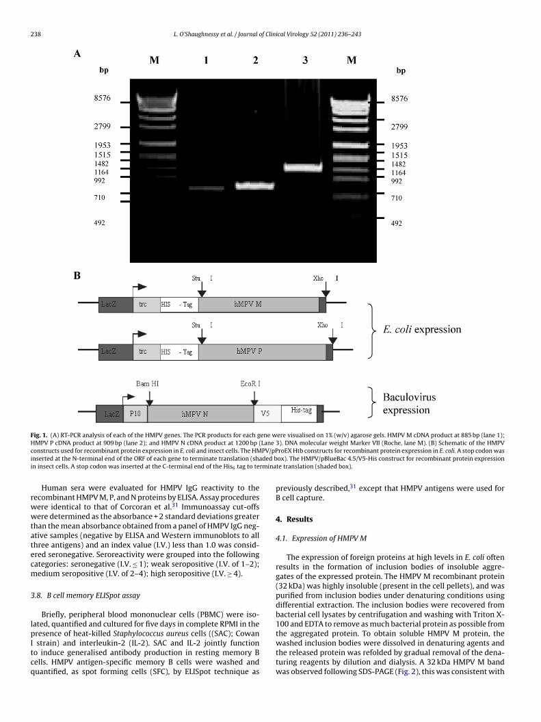
238 L. O’Shaughnessy et al. / Journal of Clinical Virology 52 (2011) 236– 243
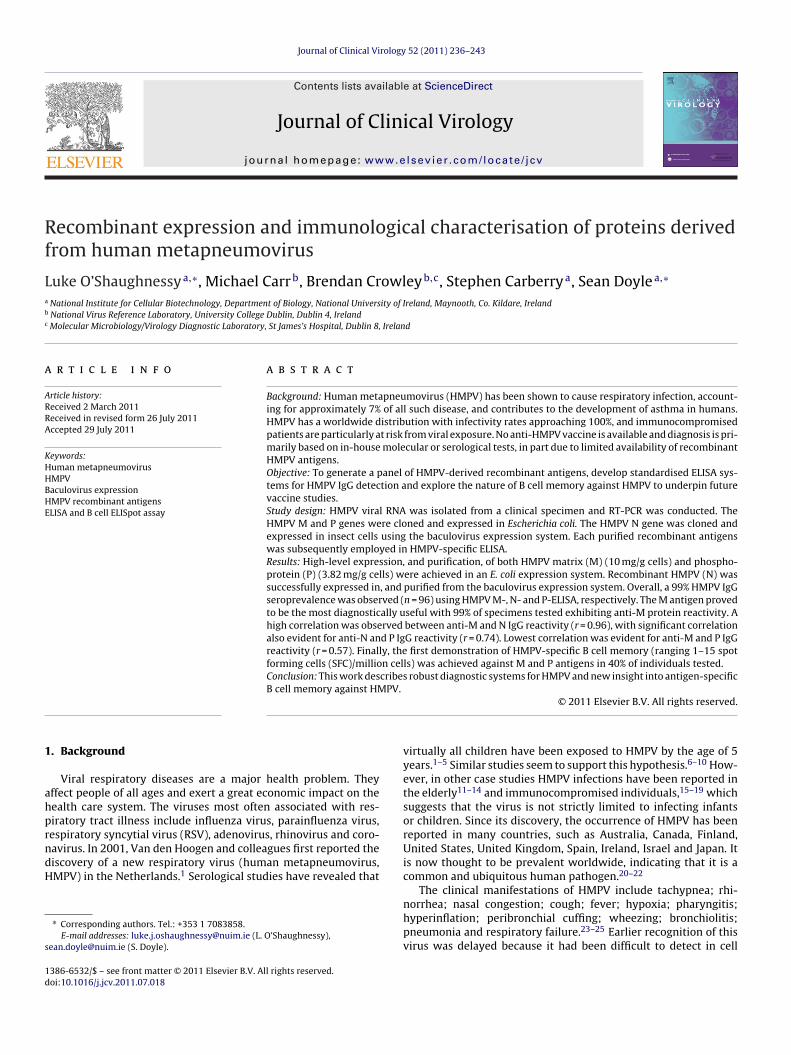
Fig. 1. (A) RT-PCR analysis of each of the HMPV genes. The PCR products for each gene were visualised on 1% (w/v) agarose gels. HMPV M cDNA product at 885 bp (lane 1);HMPV P cDNA product at 909 bp (lane 2); and HMPV N cDNA product at 1200 bp (Lane 3). DNA molecular weight Marker VII (Roche, lane M). (B) Schematic of the HMPVc PV/pPi ded bi rmina
rwwtatecm
3
lpItcq
onstructs used for recombinant protein expression in E. coli and insect cells. The HMnserted at the N-terminal end of the ORF of each gene to terminate translation (shan insect cells. A stop codon was inserted at the C-terminal end of the His6 tag to te
Human sera were evaluated for HMPV IgG reactivity to theecombinant HMPV M, P, and N proteins by ELISA. Assay proceduresere identical to that of Corcoran et al.31 Immunoassay cut-offsere determined as the absorbance + 2 standard deviations greater
han the mean absorbance obtained from a panel of HMPV IgG neg-tive samples (negative by ELISA and Western immunoblots to allhree antigens) and an index value (I.V.) less than 1.0 was consid-red seronegative. Seroreactivity were grouped into the followingategories: seronegative (I.V. ≤ 1); weak seropositive (I.V. of 1–2);edium seropositive (I.V. of 2–4); high seropositive (I.V. ≥ 4).
.8. B cell memory ELISpot assay
Briefly, peripheral blood mononuclear cells (PBMC) were iso-ated, quantified and cultured for five days in complete RPMI in theresence of heat-killed Staphylococcus aureus cells ((SAC); Cowan
strain) and interleukin-2 (IL-2). SAC and IL-2 jointly functiono induce generalised antibody production in resting memory Bells. HMPV antigen-specific memory B cells were washed anduantified, as spot forming cells (SFC), by ELISpot technique as
roEX Htb constructs for recombinant protein expression in E. coli. A stop codon wasox). The HMPV/pBlueBac 4.5/V5-His construct for recombinant protein expressionte translation (shaded box).
previously described,31 except that HMPV antigens were used forB cell capture.
4. Results
4.1. Expression of HMPV M
The expression of foreign proteins at high levels in E. coli oftenresults in the formation of inclusion bodies of insoluble aggre-gates of the expressed protein. The HMPV M recombinant protein(32 kDa) was highly insoluble (present in the cell pellets), and waspurified from inclusion bodies under denaturing conditions usingdifferential extraction. The inclusion bodies were recovered frombacterial cell lysates by centrifugation and washing with Triton X-100 and EDTA to remove as much bacterial protein as possible fromthe aggregated protein. To obtain soluble HMPV M protein, the
washed inclusion bodies were dissolved in denaturing agents andthe released protein was refolded by gradual removal of the dena-turing reagents by dilution and dialysis. A 32 kDa HMPV M bandwas observed following SDS-PAGE (Fig. 2), this was consistent with
L. O’Shaughnessy et al. / Journal of Clinical Virology 52 (2011) 236– 243 239
Fig. 2. SDS-PAGE analysis of each recombinant HMPV antigen. (A) HMPV M recombinant protein. Clarified cell lysate (lane 1), purified HMPV M at 32 kDa (lane 2). (B) HMPV Pr ). (C) HN rker (NB ng con
t3tMnp
TTtdB
TMe
ecombinant protein. Clarified cell lysate (lane 1), purified HMPV P at 29 kDa (lane 2 (lane 1), Ni-NTA affinity-purified HMPV N (47 kDa) (lane 2). Molecular Mass Mariefly, samples were electrophoresed on 12% SDS-PAGE gels at 120 V under reduci
he predicted Mr of the M protein including the His6 tag region at1.6 kDa (Table 1). The purification process yielded 10 mg M pro-ein/gram of cells. MALDI–ToF MS analysis of recombinant HMPV
following trypsin digestion confirmed the identity of recombi-ant HMPV M (GenBank accession no. gi|24429832) whereby 6/15eptides (36% sequence coverage) was observed (Table 2).
able 1he predicted, versus actual Mr by SDS-PAGE, of each recombinant HMPV pro-ein. The computation of the theoretical pI (isoelectric point) and Mr wasetermined using http://expasy.org/tools/pi tool.html from the Swiss Institute ofioinformatics.
Protein ID PredictedpI
PredictedMr (kDa)
PredictedMr + His tag(kDa)
SDS-PAGEMr (kDa)
M 8.2 27.6 31.6 32P 4.81 32.71 36.7 29N 6.73 43.51 47.5 47
able 2ass spectrometry analysis of purified HMPV recombinant antigens. Sequence cov-
rage of 35–56% confirmed the identity of each HMPV recombinant antigen.
Protein Peptides identified % Sequencecoverage
Protein ID
HMPV M K. TLTITTLYAASQSGPILK.VK.VNASAQGAAMSVLPK.KK.FEVNATVALDEYSK.LK.NTPVTIPAFIK.SK.ESESATVEAAISSEADQALTQAK.IK.TWSHQGTRYVLK.S
(6/15) 36% gi|24429832
HMPV P K. DILFMGNEAAKK.LAEAFQKK.VNTVSETLELPTISRPTKPTILSEPKK.KLAWTDKK.LKPSTNTKKK.LKPSTNTKKKK.KVSFTPNEPGKK.DALDLLSDNEEEDAESSILTFEERR.DTSSLSIEARLESIEEKR.LESIEEKK.LSMILGLLR
(12/28) 56% gi|45388092
HMPV N K.YAAEIGIQYISTALGSERR.VQQILRK.GEDLQMLDIHGVEKK.LASTIEVGLETTVRR.VLSDALKRR.SFYDLFEQKR.SLFIEYGKK.AESLFVNIFMQAYGAGQTMLRK.AESLFVNIFMQAYGAGQTMLRR.WGVIARR.GRVPNTELFSAAESYAKR.VPNTELFSAAESYAK
(12/25) 35% gi|38327234
MPV N recombinant protein. Clarified supernatant from Sf9 cells expressing HMPVew England; P7703 (lane M)). SDS-PAGE was conducted according to Laemmli.35
dition for 1.5 h. Gels were stained with Coomassie Brilliant Blue R-250 (Sigma).
4.2. Expression of HMPV P
Recombinant P protein was observed at 29 kDa following SDS-PAGE analysis (Fig. 2). The predicted Mr of the P protein plus theHis6 tag region is 36.7 kDa (Table 1). The discrepancy between theobserved and predicted Mr may be attributed to the fact that the Pprotein has a low isoelectric point (pH 4.8) and therefore migratesfurther upon SDS-PAGE. The P Protein was purified by His6 affinitychromatography under denaturing conditions. The relative yield ofpurified P protein was 3.82 mg/g of lysed E. coli (Fig. 2). MALDI–ToFMS analysis of recombinant HMPV P following trypsin digestionconfirmed the identity of recombinant HMPV P (GenBank accessionno. gi|4588092) with 56% sequence coverage (Table 2).
4.3. Expression of HMPV N in Sf9 insect cells
Insect cells were infected with recombinant baculovirus-encoding HMPV N at multiplicity of infection (MOI) = 10. The HMPVN recombinant protein was successfully purified by His6 affinitychromatography under denaturing conditions using an AKTA chro-matography system (Amersham). Purification resulted in a singlemajor protein band a molecular mass of 47 kDa (Fig. 2). The puri-fied protein yield was 1.5 mg/1 × 108 cells. MALDI–ToF MS analysisof the purified HMPV N protein confirmed identity with a 35%sequence coverage (Table 2).
4.4. Immunological analysis
To define the seroepidemiology of HMPV in an Irish popu-lation, ELISA systems were developed using three recombinantHMPV proteins as detection antigens. To investigate the seropreva-lence of HMPV, a total of 96 human sera were screened by ELISA.Sera from this blood donor cohort (n = 96) were analysed in dupli-cate by ELISA against E. coli-expressed HMPV M and P antigensand the baculovirus-expressed HMPV N protein. To establish theimmunoassay cut-offs for each of the HMPV ELISAs, a panel IgGnegative sera from healthy individuals were identified by ELISAand Western immunoblots (Fig. 3). The cut-off was establishedas the absorbance + 2 standard deviations greater than the meanabsorbance obtained from a panel of HMPV IgG negative samples(negative by ELISA and Western immunoblots to all three antigens)and an I.V. less than 1 was considered seronegative.
Of the total number of seropositive serum specimens (99%);58% were weakly seropositive, 30% were showed medium serore-activity and only 11% had high human IgG reactivity to theE. coli-expressed M antigen (Table 3). This extensive seroreactiv-
ity suggests that the M antigen binds IgG with high affinity or thatthe occurrence of specific anti-HMPV M IgG is common.For the recombinant P ELISA, the overall total percentageseropositivity was 74% (70/96) and 26% were seronegative (Table 3).
240 L. O’Shaughnessy et al. / Journal of Clinical Virology 52 (2011) 236– 243
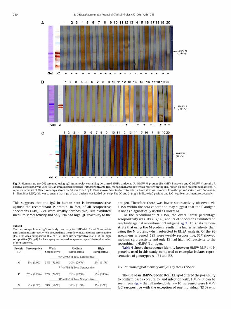
Fig. 3. Human sera (n = 20) screened using IgG immunoblot containing denatured HMPV antigens. (A) HMPV M protein, (B) HMPV P protein and C. HMPV N protein. Ap monor ior to
B p. The
Tasm
TTn(so
ositive control (C) was used (i.e., an immunostrip probed (1/1000)) with anti-His6
epresentative set of 20 serum samples from the 96 sera tested by ELISA is shown. Prrilliant Blue R250, this was to ensure that 1 �g of each antigen was loaded per stri
his suggests that the IgG in human sera is immunoreactive
gainst the recombinant P protein. In fact, of all seropositivepecimens (74%), 27% were weakly seropositive, 28% exhibitededium seroreactivity and only 19% had high IgG reactivity to theable 3he percentage human IgG antibody reactivity to HMPV-M, P and N recombi-ant antigens. Seroreactivity is grouped into the following categories: seronegativeI.V. ≤ 1); weak seropositive (I.V. of 1–2); medium seropositive (I.V. of 2–4); higheropositive (I.V. ≥ 4). Each category was scored as a percentage of the total numberf sera screened.
Protein ID
Seronegative Weak Seropositive
Medium Seropositive
High Seropositive
99% (95/96) Total Seropositive
M 1% (1/96) 58% (55/96) 30% (29/96) 11% (11/96)
74% (71/96) Total Seropositive
P 26% (25/96) 27% (26/96) 28% (27/96) 19% (18/96)
91% (88/96) Total Seropositive
N 9% (8/96) 58% (56/96) 32% (31/96) 1% (1/96)
clonal antibody which reacts with the His6 region on each recombinant antigen. Aelectrotransfer, a 1 mm strip was removed from the gel and stained with Coomassie
(+) and (−) signs indicate IgG positive and IgG negative specimens, respectively.
antigen. Therefore there was lower seroreactivity observed viaELISA within the sera cohort and may suggest that the P antigenis not as diagnostically useful as HMPV M.
For the recombinant N ELISA, the overall total percentageseropositivity was 91% (87/96), and 9% of specimens exhibited noreactivity against recombinant N antigen (Fig. 3). This data demon-strate that using the M protein results in a higher sensitivity thanusing the N protein, when subjected to ELISA analysis. Of the 96specimens screened, 58% were weakly seropositive, 32% showedmedium seroreactivity and only 1% had high IgG reactivity to therecombinant HMPV N antigen.
Table 4 shows the sequence identity between HMPV M, P and Nproteins used in this study, compared to exemplar isolates repre-sentative of genotypes A1, B1 and B2.
4.5. Immunological memory analysis by B cell ELISpot
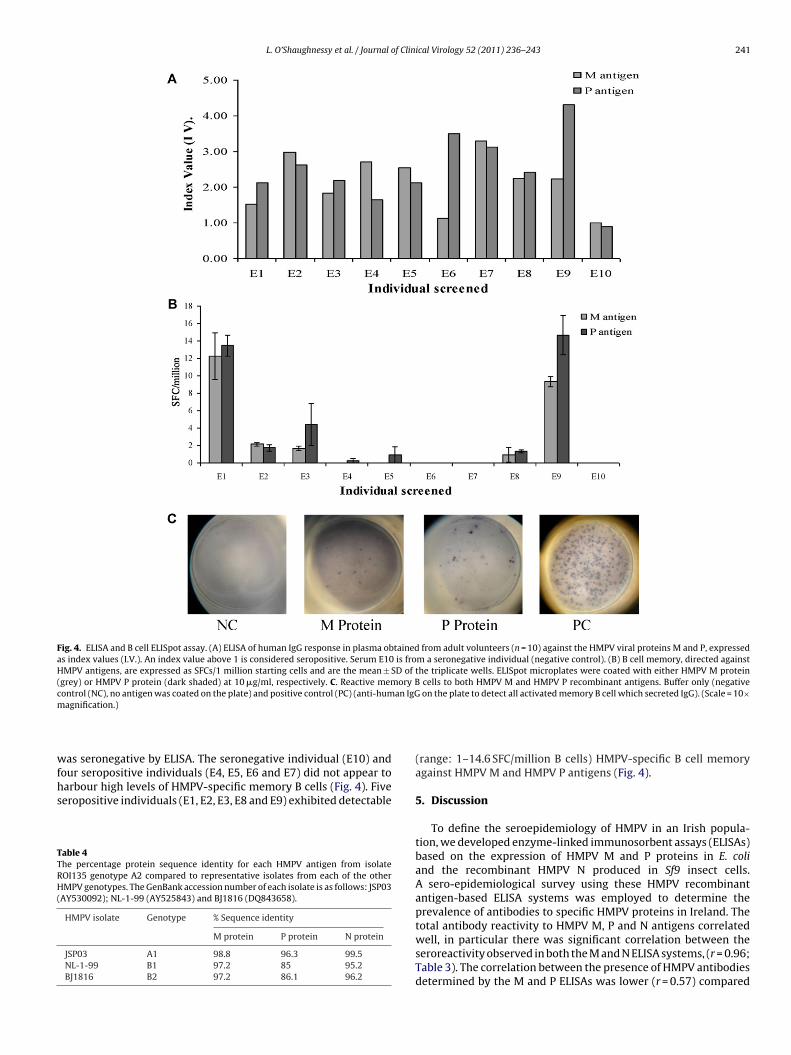
The use of an HMPV-specific B cell ELISpot offered the possibilityto confirm past exposure to, and infection with, HMPV. It can beseen from Fig. 4 that all individuals (n = 10) screened were HMPVIgG seropositive with the exception of one individual (E10) who
L. O’Shaughnessy et al. / Journal of Clinical Virology 52 (2011) 236– 243 241
Fig. 4. ELISA and B cell ELISpot assay. (A) ELISA of human IgG response in plasma obtained from adult volunteers (n = 10) against the HMPV viral proteins M and P, expressedas index values (I.V.). An index value above 1 is considered seropositive. Serum E10 is from a seronegative individual (negative control). (B) B cell memory, directed againstHMPV antigens, are expressed as SFCs/1 million starting cells and are the mean ± SD of the triplicate wells. ELISpot microplates were coated with either HMPV M protein(grey) or HMPV P protein (dark shaded) at 10 �g/ml, respectively. C. Reactive memory B cells to both HMPV M and HMPV P recombinant antigens. Buffer only (negativecontrol (NC), no antigen was coated on the plate) and positive control (PC) (anti-human IgG on the plate to detect all activated memory B cell which secreted IgG). (Scale = 10×m
wfhs
TTRH(
agnification.)
as seronegative by ELISA. The seronegative individual (E10) andour seropositive individuals (E4, E5, E6 and E7) did not appear to
arbour high levels of HMPV-specific memory B cells (Fig. 4). Fiveeropositive individuals (E1, E2, E3, E8 and E9) exhibited detectableable 4he percentage protein sequence identity for each HMPV antigen from isolateOI135 genotype A2 compared to representative isolates from each of the otherMPV genotypes. The GenBank accession number of each isolate is as follows: JSP03
AY530092); NL-1-99 (AY525843) and BJ1816 (DQ843658).
HMPV isolate Genotype % Sequence identity
M protein P protein N protein
JSP03 A1 98.8 96.3 99.5NL-1-99 B1 97.2 85 95.2BJ1816 B2 97.2 86.1 96.2
(range: 1–14.6 SFC/million B cells) HMPV-specific B cell memoryagainst HMPV M and HMPV P antigens (Fig. 4).
5. Discussion
To define the seroepidemiology of HMPV in an Irish popula-tion, we developed enzyme-linked immunosorbent assays (ELISAs)based on the expression of HMPV M and P proteins in E. coliand the recombinant HMPV N produced in Sf9 insect cells.A sero-epidemiological survey using these HMPV recombinantantigen-based ELISA systems was employed to determine theprevalence of antibodies to specific HMPV proteins in Ireland. Thetotal antibody reactivity to HMPV M, P and N antigens correlated
well, in particular there was significant correlation between theseroreactivity observed in both the M and N ELISA systems, (r = 0.96;Table 3). The correlation between the presence of HMPV antibodiesdetermined by the M and P ELISAs was lower (r = 0.57) compared
2 f Clin
trTsafpppHpMpHaHoag(8otcHorfrtps
iidtovesimo(t1iw
Hcw(EfHa
MttsliI
42 L. O’Shaughnessy et al. / Journal o
o the correlation between HMPV M and N ELISAs. Significant cor-elation was also observed between the N and P ELISAs (r = 0.74).he lack of correlation between the M and P protein-based ELISAystems may be due to variable levels of IgG to each recombinantntigen within the selected donor cohort. Ishiguro et al. testedor specific antibodies against nucleocapsid (N) and matrix (M)roteins in 97 sera by Western blot using recombinant N and Mroteins of HMPV expressed in E. coli.33 The results were com-ared with those of immunofluorescence assays (IFAs) based onMPV-infected LLC-MK2 cells, which expressed the whole HMPVroteome. Their results indicated that the antibodies against N and
proteins are highly specific (100%) but less sensitive (42.1%, Nrotein; 40.8%, M protein) than those against whole proteins ofMPV detected by IFA. This would also suggest that a multi-HMPVntigen ELISA would increase the sensitivity of the detection ofMPV antibodies within human sera. However, the lower detectionf P antigen-specific IgG may be due to the relatively low percent-ge sequence identity between HPMV P (from the A2 genotype) andenotypes A1 B1/2, respectively.34 The HMPV genotype A2 proteinsM, P and N from ROI135) used in this study exhibited 97.2–98.8,5–96.3, and 95.2–99.5% sequence identify to selected examplesf genotypes A1 and B1/2, respectively (Table 4). Extensive valida-ion of the HMPV ELISA systems described herein, whereby antigenoating on microtitre plates was optimised (2, 3 and 5 �g/ml forMPV M, P and N, respectively) and assay reproducibility, in termsf % coefficient of variation, was determined to be 5.5, 8 and 8%,espectively, preclude sub-optimal assay performance as a reasonor the observed differences in antibody detection. In summary,ecombinant M, P and N proteins of HMPV were antigenic, andhe responses to M, P, and N proteins differed across the studyopulation. Assays based on multiple antigens provided higher sen-itivities than assays based on single antigens.
The defining feature of the acquired immune system is its abil-ty to generate immunological memory to a particular pathogenn defence of re-infection. Recently, considerable efforts have beenirected towards the elucidation of the nature of the T cell responseo HMPV and the definition of T cell epitopes,26–30 yet the naturef B cells memory directed against HMPV remains unclear. All indi-iduals (n = 10) screened were HMPV IgG seropositive with thexception of one individual E10, who was seronegative for HMPVpecific IgG (Fig. 4). Five individuals (E1, E2, E3, E8 and E9) exhib-ted low, but detectable (range: 1–14.6 SFC/million B cells) B cell
emory. There was a significant correlation between the numberf memory B cells directed against HMPV M and HMPV P antigensr = 0.655). However, the greatest numbers of SFCs were evident inwo individuals (E1, mean ± SD: 13.5 ± 1.2 SFC/million cells and E9,4.7 ± 2.2 SFC/million cells). B cell memory could not be detected
n the absence of IL-2 and SAC stimulation prior to ELISpot analysis,hich was an unstimulated B cell control (Fig. 4).
When the IgG reactivity of the ten specimens (E1–E10) againstMPV M was analysed by ELISA and compared against the Bell ELISpot data, no statistically significant positive correlationas observed between SFC/million cells and antibody reactivity
r = 0.022). Conversely, a correlation was observed between theLISpot results and IgG reactivity to the HMPV P antigen (r = 0.655)or individuals exhibiting IgG specific antibody responses to theMPV P antigen. This confirms that previous exposure to HMPVlso results in B cell memory (mainly individuals E1 and E9).
The lack of correlation between the ELISpot data obtained for the protein and that obtained for ELISA data is not unusual; the detec-
ion of specific serum antibodies is the most widely applied methodo investigate immunity against diseases like HMPV, although a
pecific correlate of protection against HMPV has not been estab-ished yet. Besides antibodies, long-term memory B and T cellmmunity might play an important role in protection against HMPV.n some individuals, high antibody levels with low or no numbersical Virology 52 (2011) 236– 243
of memory B cells were found. This strengthens the idea that long-lived plasma cells maintain antibody levels and that memory B cellsare a distinct population of cells. Memory B cells do not secreteantibodies prior to activation. Upon renewed antigen stimulation,memory B cells may rapidly respond, proliferate and differentiatein antibody secreting cells. The ongoing circulation of HMPV amongthe Irish population causing antigenic re-challenge might explainthe low correlation between circulating HMPV-specific memory Bcells and antibody levels measured in our study. Importantly, insome individuals no HMPV-specific memory B cells, as well as lowHMPV-specific antibody levels, were detectable which might resultin a higher susceptibility for infection with HMPV.
In conclusion, this work has provided multiple new detectionsystems for, and establishes the seroprevalence of, HMPV IgG in anIrish population. We also report the first demonstration of HMPV-specific B cell memory against HMPV M and P antigens whichfurthers our knowledge on the antigen-specific B cell memoryagainst the HMPV viral proteins. Determination of antigen-specificB cell memory status may enhance the serological and molecularanalyses of persistent HMPV infection. Further studies are war-ranted to elucidate the nature of differential seroreactivity to eachviral antigen.
Competing interests
The authors declare that they have no conflict of interest.
Funding
Serum specimens were obtained from the Irish Blood Transfu-sion Service ((IBTS) Dublin, Ireland). Project funders had no inputinto either project design or data analysis.
Ethical approval
Ethics permission to obtain and use material of human originwas obtained from the NUI Maynooth Ethics Committee.
Acknowledgments
The work was funded by the HEA-funded Programme forResearch in Third Level Institutions (Cycles 3 & 4). Mass spectrom-etry facilitates were supported by the Health Research Board.
References
1. Van den Hoogen BG, de Jong JC, Groen J, Kuiken T, de Groot R, Fouchier RA,et al. A newly discovered human pneumovirus isolated from young children withrespiratory tract disease. Nat Med 2001;7:719–24.
2. Ebihara T, Endo R, Kikuta H, Ishiguro N, Ishiko H, Hara M, et al. Human metap-neumovirus infection in Japanese children. J Clin Microbiol 2004;42:126–32.
3. Leung J, Esper F, Weibel C, Kahn JS. Seroepidemiology of human metapneumovirus(HMPV) on the basis of a novel enzyme-linked immunosorbent assay utilizingHMPV fusion protein expressed in recombinant vesicular stomatitis virus. J ClinMicrobiol 2005;43:1213–9.
4. Hamelin M, Boivin G. Development and validation of an enzyme-linkedimmunosorbent assay for human metapneumovirus serology based on a recom-binant viral protein. J Clin Lab Immunol 2005;12:249–53.
5. Pavlin JA, Andrew C, Hickey AC, Ulbrandt N, Chan YP, Endy TP, et al. Humanmetapneumovirus reinfection among children in Thailand determined by anenzyme-linked immunosorbent assay using purified soluble fusion protein. J InfectDis 2008;198:836–42.
6. Fabbiani M, Terrosi C, Martorelli B, Valentini M, Bernini L, Cellesi C, et al. Epi-demiological and clinical study of viral respiratory tract infections in children fromItaly. J Med Virol 2009;81:750–6.
7. Heininger U, Kruker AT, Bonhoeffer J, Schaad UB. Human metapneumovirus
infections—biannual epidemics and clinical findings in children in the region ofBasel, Switzerland. Eur J Pediatr 2009;168:1455–60.8. Williams JV, Harris PA, Tollefson SJ, Halburnt-Rush LL, Pingsterhaus JM,Edwards KM, et al. Human metapneumovirus and lower respiratory tract diseasein otherwise healthy infants and children. N Engl J Med 2004;350:443–50.
f Clin
L. O’Shaughnessy et al. / Journal o9. Mullins JA, Erdman DD, Weinberg GA, Edwards K, Hall CB, Walker FJ, et al.Human metapneumovirus among children hospitalized for acute respiratory ill-ness. Emerg Infect Dis 2004;10:700–5.
10. Peiris JS, Tang WH, Chan KH, Khong PL, Guan Y, Lau YL, et al. Children withrespiratory disease associated with metapneumovirus in Hong Kong. Emerg InfectDis 2003;9:628–33.
11. Ren L, Gonzalez R, Wang Z, Xiang Z, Wang Y, Zhou H, et al. Prevalence of humanrespiratory viruses in adults with acute respiratory tract infections in Beijing,2005–2007. Clin Microbiol Infect 2009;15:1146–53.
12. Falsey AR. Human metapneumovirus infection in adults. Pediatr Infect Dis J2008;27(10 Suppl.):S80–3.
13. Falsey AR, Erdman D, Anderson LJ, Walsh EE. Human metapneumovirus infectionsin young and elderly adults. J Infect Dis 2003;187:785–90.
14. O’Gorman C, McHenry E, Coyle PV. Human metapneumovirus in adults: a shortcase series. Eur J Clin Microbiol Infect Dis 2006;25:190–2.
15. Muller A, Klinkenberg D, Vehreschild J, Cornely O, Tillmann RL, Franzen C,et al. Low prevalence of human metapneumovirus and human bocavirus in adultimmunocompromised high risk patients suspected to suffer from Pneumocystispneumonia. J Infect 2009;58:227–31.
16. Oliveira R, Machado A, Tateno A, Boas LV, Pannuti C, Machado C. Frequencyof human metapneumovirus infection in hematopoietic SCT recipients during 3consecutive years. Bone Marrow Transplant 2008;42:265–9.
17. Dare R, Sanghavi S, Bullotta A, Keightley MC, George KS, Wadowsky RM,et al. Diagnosis of human metapneumovirus infection in immunosuppressedlung transplant recipients and children evaluated for pertussis. J Clin Microbiol2007;45:548–52.
18. Englund JA, Boeckh M, Kuypers J, Nichols WG, Hackman RC, Morrow RA, et al.Brief communication: fatal human metapneumovirus infection in stem-cell trans-plant recipients. Ann Intern Med 2006;144:344–9.
19. Debiaggi M, Canducci F, Sampaolo M, Marinozzi MC, Parea M, Terulla C, et al.Persistent symptomless human metapneumovirus infection in hematopoietic stemcell transplant recipients. J Infect Dis 2006;194:474–8.
20. Kahn JS. Epidemiology of human metapneumovirus. Clin Microbiol Rev2006;19:546–57.
21. Dong J, Olano JP, McBride JW, Walker DH. Emerging pathogens: challenges andsuccesses of molecular diagnostics. J Mol Diagn 2008;10:185–97.
22. Principi N, Bosis S, Esposito S. Human metapneumovirus in paediatric patients.Clin Microbiol Infect 2006;12:301–8.
23. Wilkesmann A, Schildgen O, Eis-Hubinger AM, Geikowski T, Glatzel T, LentzeMJ, et al. Human metapneumovirus infections cause similar symptoms and clinicalseverity as respiratory syncytial virus infections. Eur J Pediatr 2006;165:467–75.
ical Virology 52 (2011) 236– 243 243
24. Baer G, Schaad UB, Heininger U. Clinical findings and unusual epidemiologic char-acteristics of human metapneumovirus infections in children in the region of Basel,Switzerland. Eur J Pediatr 2008;167:63–9.
25. Boivin G, Abed Y, Pelletier G, Ruel L, Moisan D, Cote S, et al. Virological fea-tures and clinical manifestations associated with human metapneumovirus: a newparamyxovirus responsible for acute respiratory-tract infections in all age groups.J Infect Dis 2002;186:1330–4.
26. Herd KA, Nelson M, Mahalingam S, Tindle RW. Pulmonary infection of mice withhuman metapneumovirus induces local cytotoxic T-cell and immunoregulatorycytokine responses similar to those seen with human respiratory syncytial virus. JGen Virol 2010;91:1302–10.
27. Herd KA, Nissen MD, Hopkins PM, Sloots TP, Tindle RW. Major histocompatibil-ity complex class I cytotoxic T lymphocyte immunity to human metapneumovirus(HMPV) in individuals with previous HMPV infection and respiratory disease. JInfect Dis 2008;197:584–92.
28. Kolli D, Bataki EL, Spetch L, Guerrero-Plata A, Jewell AM, Piedra PA, et al. Tlymphocytes contribute to antiviral immunity and pathogenesis in experimentalhuman metapneumovirus infection. J Virol 2008;82:8560–9.
29. Melendi GA, Zavala F, Buchholz UJ, Boivin G, Collins PL, Kleeberger SR, et al.Mapping and characterization of the primary and anamnestic H-2d-restrictedcytotoxic T-lymphocyte response in mice against human metapneumovirus. J Virol2007;81:11461–7.
30. Le Nouen C, Hillyer P, Munir S, Winter CC, McCarty T, Bukreyev A, et al. Effectsof human respiratory syncytial virus, metapneumovirus, parainfluenza virus 3and influenza virus on CD4+ T cell activation by dendritic cells. PLoS One 2010;5:15017.
31. Corcoran A, Mahon BP, Doyle S. B cell memory is directed toward conformationalepitopes of parvovirus B19 capsid proteins and the unique region of VP1. J InfectDis 2004;189:1873–80.
32. Carberry S, Neville CM, Kavanagh KA, Doyle S. Analysis of major intracellularproteins of Aspergillus fumigatus by MALDI mass spectrometry: identification andcharacterisation of an elongation factor 1B protein with glutathione transferaseactivity. Biochem Biophys Res Commun 2006;341:1096–104.
33. Ishiguro N, Ebihara T, Endo R, Ma X, Kawai E, Ishiko H, et al. Detection ofantibodies against human metapneumovirus by Western blot using recombinantnucleocapsid and matrix proteins. J Med Virol 2006;78:1091–5.
34. Bastien N, Normand S, Taylor T, Ward D, Peret TC, Boivin G, et al. Sequenceanalysis of the N, P, M and F genes of Canadian human metapneumovirus strains.Virus Res 2003;93:51–62.
35. Laemmli UK. Cleavage of structural proteins during the assembly of the head ofbacteriophage T4. Nature 1970;227:680–5.