reconstitution of a human dna damage checkpoint response in
TRANSCRIPT
Coupling DNA excision repair and the ATR-mediated checkpoint
1
Coupling of Human DNA Excision Repair and ATR-mediated DNA Damage Checkpoint
in a Defined In Vitro System*
Laura A. Lindsey-Boltz1, Michael G. Kemp
1, Joyce T. Reardon
1, Vanessa DeRocco
1, Ravi R. Iyer
2,#,
Paul Modrich2,3
, and Aziz Sancar1, +
1From the Department of Biochemistry and Biophysics, University of North Carolina School of Medicine,
Chapel Hill, NC 27599-7260 2Department of Biochemistry and
3Howard Hughes Medical Institute, Duke University Medical Center,
Durham, NC 27710
*Running title: Coupling DNA excision repair and the ATR-mediated checkpoint
To whom correspondence should be addressed: Aziz Sancar, Department of Biochemistry and
Biophysics, University of North Carolina School of Medicine, CB#7260, Chapel Hill, NC USA 27599-
7260, Tel.: 919-962-0115; E-mail: [email protected]
Keywords: TopBP1, RPA, p53, EXO1, kinase, UV repair
Background: Nucleotide excision repair and the
ATR-mediated DNA damage checkpoint
responses are genetically coupled.
Results: We have analyzed the basic steps of ATR
activation in a biochemically defined system.
Conclusion: ATR signaling requires enlargement
of the DNA excision gap by EXO1.
Significance: The six excision repair factors,
ATR-ATRIP, TopBP1, and EXO1 constitute the
minimum essential set of proteins for ATR-
activation upon UV-induced DNA damage.
SUMMARY
DNA repair and DNA damage checkpoints
work in concert to help maintain genomic
integrity. In vivo data suggest that these two
global responses to DNA damage are coupled.
It has been proposed that the canonical 30
nucleotide single-stranded DNA gap generated
by nucleotide excision repair is the signal that
activates the ATR-mediated DNA damage
checkpoint response and that the signal is
enhanced by gap enlargement by EXO1
(exonuclease 1) 5’ to 3’ exonuclease activity.
Here we have used purified core nucleotide
excision repair factors (RPA, XPA, XPC,
TFIIH, XPG, and XPF-ERCC1), core DNA
damage checkpoint proteins (ATR-ATRIP,
TopBP1, RPA), and DNA damaged by a UV-
mimetic agent to analyze the basic steps of DNA
damage checkpoint response in a biochemically
defined system. We find that checkpoint
signaling as measured by phosphorylation of
target proteins by the ATR kinase requires
enlargement of the excision gap generated by
the excision repair system by the 5’ to 3’
exonuclease activity of EXO1. We conclude
that, in addition to damaged DNA, RPA, XPA,
XPC, TFIIH, XPG, XPF-ERCC1, ATR-ATRIP,
TopBP1, and EXO1 constitute the minimum
essential set of factors for ATR-mediated DNA
damage checkpoint response.
DNA damage activates three major
biochemical pathways in eukaryotic cells: DNA
repair, DNA damage checkpoints, and apoptosis
(1). The DNA damage checkpoint response delays
or arrests cell cycle progression which helps
prevent the mutagenic or lethal consequences of
damage to the cell. In mammalian organisms two
main DNA damage checkpoint pathways/networks
have been defined based on the damage sensing
kinases that initiate the signal: the ATM pathway
and the ATR pathway (2). Although there is some
overlap and crosstalk between the two signaling
pathways, in general, most studies support the
view that ionizing radiation and other agents that
produce double-strand breaks in DNA activate the
ATM pathway, while ultraviolet (UV)4 light and
other genotoxic agents that generate bulky base
adducts activate the ATR-mediated checkpoint
signaling pathway (3). The ATM kinase is
http://www.jbc.org/cgi/doi/10.1074/jbc.M113.542787The latest version is at JBC Papers in Press. Published on January 8, 2014 as Manuscript M113.542787
Copyright 2014 by The American Society for Biochemistry and Molecular Biology, Inc.
by guest on January 15, 2019http://w
ww
.jbc.org/D
ownloaded from

Coupling DNA excision repair and the ATR-mediated checkpoint
2
activated by recruitment by the MRN complex to
duplex termini generated by double-strand breaks
and dimer to monomer transition or during
oxidative stress by formation of disulfide
crosslinks between the two subunits of the ATM
homodimer (4). In the case of ATR, the signal
could be the bulky adduct itself (5,6), the stalled
replication fork (7,8), the transcription elongation
complex stalled at the site of damage (9), the
repair complex assembled at the site of damage
(10), or the canonical 30 nucleotide gap generated
by nucleotide excision repair (11-13). Although
overwhelming evidence indicates that in S phase
the stalled replication fork and the long stretches
of ssDNA that result from replication fork stalling
are the primary signals for the ATR-mediated
checkpoint (14), the contributions of other factors
to checkpoint activation in G1 and G2/M are not as
well-defined (3). However, several studies have
strongly supported a model whereby the canonical
30-nucleotide gap generated by nucleotide
excision repair is enlarged by EXO1, and the
enlarged single-stranded gap (presumably
occupied by RPA protein) constitutes the major
signal for the ATR-mediated checkpoint response
outside of S phase (15-17).
While the in vivo data is compelling in
support of the model, there are alternative
explanations for some key observations upon
which the model is based because transient
knockdown of many gene products outside the
core constituents of nucleotide excision repair
have been reported to interfere with ATR-
mediated checkpoint signaling (14). Thus, the
basic model can be evaluated only in vitro in a
system that contains components with precisely
defined function which would eliminate the in vivo
artifacts arising from mutations that affect the
ATR pathway through secondary effects on
cellular homeostasis. In this study, using highly
purified minimal essential sets of both the human
nucleotide excision repair system and the ATR
checkpoint signaling pathway, we have
reconstituted the ATR checkpoint system in vitro.
We find that the nucleotide excision repair
canonical 30 nucleotide gap enlarged by EXO1 is
necessary and sufficient to activate ATR
checkpoint signaling in the presence of the ATR
co-activator TopBP1 protein. This is the first in
vitro system that couples nucleotide excision
repair and the ATR mediated DNA damage
checkpoint.
EXPERIMENTAL PROCEDURES
Protein Purification - The excision repair
proteins His-XPA, XPC-HR23B, XPG, and XPF-
ERCC1 were purified as recombinant proteins
using the Sf21/baculovirus insect cell/vector
system as previously described (18). The multi-
subunit TFIIH complex was purified from HeLa
Flp-In T-REx cells (19,20) expressing
Tetracycline-inducible FLAG-p62 as described in
the manufacturer’s directions (Invitrogen), and
purified with P11 chromatography and affinity
chromatography with anti-FLAG-M2 agarose
(Sigma) as previously described (21). The ATR-
ATRIP complex was similarly purified from HeLa
Flp-In T-REx cells containing a tetracycline-
inducible Flag epitope-tagged ATRIP subunit by
anti-FLAG-M2 affinity chromatography as
previously described (22). The following proteins
were purified as recombinant proteins expressed in
E. coli as previously described: GST-TopBP1-His
(23), EXO1 (amino acids 1-450) (24), GST-p53
(Addgene plasmid 10852) (25), and RPA (26).
The purified proteins were separated on 4-15%
TGX-PAGE and analyzed by silver staining.
Cell Lines and Antibodies- Immortalized wild-
type (WT) and Exo1-/-
mouse embryonic
fibroblasts (MEFs) were cultured in DMEM
containing 10% fetal bovine serum (FBS) and
penicillin/streptomycin. The Exo1-/-
MEFs were
obtained from Winfried Edelmann (Albert
Einstein College of Medicine) (27). To drive cells
into quiescence, cells were grown in DMEM
containing 0.5% FBS for 3-4 days. Irradiation of
cells with UV light involved the removal of the
medium from the cells, exposure to a UV-C light
source (254 nm), and replacement of the medium.
Following a 1 hr incubation, cells were washed
with cold PBS, scraped from the plate into cold
PBS, and then lysed in a buffer containing 25 mM
HEPES-KOH pH 7.9, 100 mM KCl, 12 mM
MgCl2, 0.5 mM EDTA, 12.5% glycerol, 1 mM
DTT, and 0.5% NP-40. Cell lysates were
fractionated by SDS-PAGE, transferred to
nitrocellulose, and analyzed by immunoblotting.
The following primary antibodies were
obtained from the indicated companies and used at
the indicated dilution: from Cell Signaling
Technology, phospho-Chk1-Ser345
(catalog no.
by guest on January 15, 2019http://w
ww
.jbc.org/D
ownloaded from

Coupling DNA excision repair and the ATR-mediated checkpoint
3
2348, 1:10,000), phospho-p53-Ser15
(catalog no.
9284, 1:10,000); from Bethyl Laboratories, RPA1
(catalog no. A300-241A, 1:2,000) and phospho-
RPA2-Ser33
(catalog no. A300-246A, 1:10,000);
from Santa Cruz Biotechnology, Inc., Chk1
(catalog no. sc-8408, 1:2,000), GST (catalog no.
sc-138, 1:1,000), and from Leica Biosystems, p53
(NCL-p53-505, 1:1,000).
Preparation of DNA Substrates - Gapped
plasmid was generated by treating pBC-KS.nick
(28) with Nt.BbvCI endonuclease which cuts only
one strand of the plasmid 43 nucleotides apart.
The excised oligomer was released by heat
denaturation in the presence of excess
complementary oligo. The X174 ssDNA was
purchased from New England Biolabs (N3023).
N-acetoxy-2-acetylaminofluorene (AAF) was
obtained from the NCI Chemical Carcinogen
Repository (Midwest Research Institute, Kansas
City, MO). AAF-damaged plasmid DNA (pBC-
KS.nick) was prepared as described previously
(23). The concentration of AAF was empirically
determined to generate approximately 3 adducts
per plasmid.
Excision Repair Assay – The repair assay was
performed as previously described (18).
Unmodified or AAF-damaged plasmid DNA (100
ng) was incubated in a 12.5 μl reaction containing
the core excision repair factors [XPA (86 ng),
XPC-hR23B (17.5 ng), XPF-ERCC1 (7.5 ng),
XPG (4 ng), TFIIH (100 ng) and 170 ng of RPA].
The final reactions contained 23 mM Hepes-KOH
(pH 7.9), 44 mM KCl, 2.5 mM MgCl2, 2 mM
ATP, 2.5% glycerol, 0.04 mM EDTA, and 0.2 mM
DTT. After 90 min at 30°C, 2.5 µl of the reaction
was diluted 1:4 with TE buffer and reserved for
kinase assays. To the remaining 10 µl, 2 µl of
phenol and 12 µl of agarose gel loading buffer
containing TBE (0.1M Tris, 0.1M Boric Acid,
0.002M EDTA), 1% SDS, 0.05% bromophenol
blue, and 10% glycerol was added and then
separated on an ethidium bromide-containing 1%
agarose gel which was then analyzed using a
BioRad Molecular Imager ChemiDoc XRS+
system.
Checkpoint assay - The procedure was
essentially as previously described (29). Briefly,
kinase assay reactions contained 14 mM Hepes,
pH 7.9, 30 mM KCl, 1 mM MgCl2, 1 mM ATP,
0.5 mM DTT, 2% glycerol, and 1 μM microcystin
in a 12 μl final volume. Purified ATR-ATRIP (0.2
nM), TopBP1 (2.5 nM), RPA (100 nM), p53 (50
nM), and EXO1 (8 nM), where indicated, were
incubated in reaction buffer for 20 min at 30°C
with DNA (2 ng) as indicated. The reactions were
terminated by the addition of 3 μl of 5X SDS-
PAGE loading buffer (100 mM Tris, pH 6.8, 10%
(v/v) glycerol, 200 mM DTT, 2% (w/v) SDS,
0.01% (w/v) bromphenol blue) and then boiled
and separated by 15% SDS-PAGE.
Phosphorylation of p53 and RPA2 were detected
by immunoblotting using the indicated phospho-
specific antibodies, and the level of total protein
was subsequently detected by immunoblotting the
same membrane with the indicated antibodies.
Chemiluminescent signals were visualized with
Clarity Western blotting detection reagent
(BioRad) and analyzed with the Molecular Imager
ChemiDoc XRS+ system (BioRad). The highest
phosphorylation signal on each blot was set to
100%, and the levels of phosphorylation of other
samples were expressed relative to this value.
Graphed values are the average and S.D. from at
least two independent experiments.
RESULTS
Purification of nucleotide excision repair and ATR
checkpoint signaling proteins. In vitro assays with
cell-free extract to test various models for ATR-
mediated checkpoint signaling are hampered by
the fact that, in humans, DNA-PK is the most
abundant member of the PIKK family kinases
(ATM, ATR, DNA-PK) and has the most robust
activity of the three kinases (30-33). As a
consequence, it dominates the kinase activity in
cell-free extracts with any putative ATM or ATR
substrates, as there is considerable overlap among
substrates of the PIKK family (28,34). Use of
kinase inhibitors only partially alleviates the
problem (28,34,35). Perhaps most importantly, by
using cell-free extracts, it is not possible to define
the necessary and sufficient components of a
biochemical pathway. For these reasons, we have
not been able to test the various models for ATR
checkpoint in cell-free extracts and found it
necessary to purify the nucleotide excision repair
and checkpoint proteins that are known to be
essential for ATR-mediated checkpoint signaling.
Figure 1 shows our highly purified nucleotide
excision repair and DNA damage checkpoint
proteins. The excision repair proteins XPA, XPC-
HR23B, XPG, and XPF-ERCC1 were purified as
by guest on January 15, 2019http://w
ww
.jbc.org/D
ownloaded from

Coupling DNA excision repair and the ATR-mediated checkpoint
4
recombinant proteins using the Sf21/baculovirus
insect cell/vector system. The multisubunit TFIIH
was purified from HeLa cells containing an
inducible FLAG epitope-tagged p62 subunit
through conventional chromatographical steps and
contained some minor high molecular weight
contaminants. The identities of the main bands
seen by silver staining as those corresponding to
the known TFIIH subunits were confirmed by
immunoblotting. The ATR-ATRIP complex was
similarly purified from HeLa cells containing an
inducible FLAG epitope-tagged ATRIP subunit by
affinity chromatography, yielding a preparation in
which the major protein bands on SDS-PAGE are
ATR and ATRIP as confirmed by
immunoblotting. The ATR co-activator, TopBP1,
was purified as a recombinant protein expressed in
E. coli, as were EXO1 nuclease, p53, and RPA.
A model system for excision repair-checkpoint
coupling. Two general models have been
proposed for coupling of repair to the DNA
damage checkpoint. In one, it is suggested that
either the mismatch repair protein MutS (36) or
the nucleotide excision repair protein XPA (10)
binds to a mismatch or to a bulky base adduct,
respectively, and by some ill-defined mechanism
recruits ATR to damage sites and stimulates its
kinase activity. While these mechanisms may play
some minor roles in ATR activation, attempts to
demonstrate such effects in defined systems have
not been successful. In the alternative mechanism,
it is proposed that the canonical 30-nt-long
excision gap generated by nucleotide excision
repair either as such or after enlargement by
exonucleases constitutes the structure/signal that
couples nucleotide excision repair to ATR-
initiated DNA damage checkpoint (11-13).
To test the model that the 30 nt-long
nucleotide excision repair-gap either as is, or after
processing by EXO1 exonuclease, constitutes the
signal for ATR checkpoint, we first used a model
DNA substrate. A plasmid DNA containing a 43
nucleotide gap was generated by treating the
plasmid with Nt.BbvCI endonuclease which cuts
at two sites 43 nucleotides apart in only one strand
of the plasmid. The gap generated by nicking with
this enzyme followed by release of the excised
oligomer by heat denaturation was used in our
reconstituted ATR-ATRIP + TopBP1 kinase
system with RPA2 (RPA32 subunit of RPA) as a
substrate for ATR kinase, and the results are
shown in Figure 2A. The unprocessed gap was
insufficient to activate ATR (lane 5). However,
upon addition of human EXO1, which enlarges the
gap by digesting DNA in the 5’ to 3’ direction,
resulted in ATR activation as efficiently as ssDNA
(compare lanes 6 and 4). Addition of EXO1 to
reactions containing circular dsDNA had no
significant effect (lane 8). Thus, we conclude that
the canonical 30-nt excision gap would constitute
a signal for ATR kinase after enlargement with
EXO1.
We and others have previously reported that
Ser33
of RPA2 is phosphorylated by ATR in a
manner dependent on ssDNA in in vitro kinase
assays (37,38). This residue of RPA2 is known to
be phosphorylated by ATR in cells treated with
UV light (39). However, it has not been reported
whether RPA phosphorylation at this site occurs in
a manner dependent on excision repair and EXO1.
Therefore, we examined phosphorylation of RPA2
after UV in WT and Exo1-/-
mouse embryonic
fibroblast (MEF) cells, and the results are shown
in Figure 2B. We find that in quiescent cells,
where UV-induced ATR activation is known to be
dependent on nucleotide excision repair (11-
13,40), that indeed, RPA2 is phosphorylated on
Ser33
and the phosphorylation is dependent on the
presence of EXO1 (compare lanes 2 and 4). As
was previously shown (15), p53 phosphorylation
on Ser18
(equivalent to Ser15
in human p53) is also
dependent on EXO1 under these conditions. Also
as previously reported (13), in quiescent cells
Chk1 protein levels are low and there is no
detectable phosphorylation at Ser345
. In contrast,
as expected, in asynchronous cells where ATR
activation after UV is largely the result of
replication fork stalling, phosphorylation of RPA2,
p53, and Chk1 is not dependent on EXO1 (lane 8).
Thus, we conclude that RPA2 phosphorylation on
Ser33
and p53 phosphorylation on Ser15
are the
physiologically relevant readouts for ATR
activation dependent on excision gap enlargement
by EXO1 after UV-induced DNA damage in
quiescent cells, and we set out to test this model in
our defined system in vitro.
Human Nucleotide Excision Repair In Vitro and
Excision Gap Enlargement with EXO1. N-
acetoxy-2-acetylaminofluorene (N-Aco-AAF)-
damaged DNA is one of the best substrates for
by guest on January 15, 2019http://w
ww
.jbc.org/D
ownloaded from

Coupling DNA excision repair and the ATR-mediated checkpoint
5
nucleotide excision repair (41), and is considered
to be a UV-mimetic (42). Therefore, for our
checkpoint assay we used AAF-damaged plasmid
as a substrate for the reconstituted human excision
nuclease system to generate the excision gaps that
have been proposed to initiate checkpoint
signaling. First, we tested the specificity of our
reconstituted excision nuclease system by using
undamaged and AAF-modified plasmids with the
6 core repair factors in a nicking assay for excision
repair. As seen in Figure 3A, the purified repair
factors are virtually free of non-specific
endonucleases as evidenced by the lack of nicking
activity on undamaged DNA under conditions
where on average one gap per plasmid is produced
in damaged DNA (Fig. 3A lane 2 vs. lane 4).
Because it has been proposed that the enlargement
of the excision gap significantly amplifies the
checkpoint signal we tested the effect of EXO1 on
the gapped plasmid. As seen in Figure 3B, with
increasing concentration of EXO1, the gapped
plasmid band becomes more diffuse in the agarose
gel, while the band corresponding to covalently
closed DNA remains unchanged, consistent with
the prediction that EXO1 enlarges the excision
repair gap. Because the excision gap is enlarged
in individual gapped molecules to varying degrees,
the “open circular” plasmid band on the agarose
gel has a diffuse appearance.
Coupling of Excision Repair with ATR
Checkpoint. To test the model of the checkpoint
response to UV and UV-mimetic agents, we
treated undamaged and AAF-damaged plasmid
DNAs with various combinations of repair and
checkpoint factors in the absence and presence of
EXO1 and tested for ATR signaling using RPA2
phosphorylation as a readout. The results are
shown in Figure 4. As is clear from the figure,
even though a low level signal is seen with
undamaged DNA, in agreement with earlier data
(23), only the combination of AAF-DNA + Repair
Factors + ATR-ATRIP + TopBP1 resulted in
strong checkpoint signaling well above all other
combinations including signal with undamaged
DNA (lane 7 vs. lane 1, p <0.01), with damaged
DNA in the absence of repair factors (lane 7 vs.
lane 9, p<0.01), or with both damaged DNA and
repair factors but in the absence of EXO1 (lane 7
vs. lane 12, p<0.01). The low level of ATR kinase
activity observed in the presence of TopBP1 and
undamaged DNA (lanes 1 and 3) and with
damaged DNA in the absence of repair factors
(lane 9) is consistent with previous observations
that under certain experimental conditions DNA +
TopBP1 are sufficient to cause moderate
checkpoint activation both in vitro (23) and in vivo
(43).
DNA Concentration Effect and Kinetics of ATR
Checkpoint Signaling. Because DNA is a key
component of the ATR checkpoint pathway, we
next determined the effect of DNA concentration
on excision repair + EXO1 enlargement-dependent
ATR checkpoint signaling. We find that under our
reaction conditions ~4 ng DNA per reaction yields
the best signal-to-noise ratio (Figure 5A). Next,
using this amount of DNA we carried out a time
course experiment. As apparent in Figure 5B, at
all time points the complete reaction is
significantly more effective in promoting RPA2
phosphorylation by ATR than partial reactions in
which the repair factors or damaged DNA were
omitted. Taken together, these data indicate that
our in vitro system is a faithful representation of
the ATR signaling system defined genetically in
yeast (3) and mammalian cells or with partial
reactions in Xenopus extracts (43).
Repair-Checkpoint Coupling as Measured by p53
Phosphorylation. Although RPA2 phosphorylation
is a commonly used readout for ATR checkpoint
signaling in vivo and in some studies with cell-free
extracts (34,35,38), its significance in delaying or
arresting cell cycle progression (checkpoint
response) in G1/quiescent cells is not known. In
contrast, the phosphorylation of p53 by ATR is
known to be an important step in ATR signaling
during G1 (44). Therefore, we wished to ascertain
that our in vitro repair checkpoint coupling system
was operative on p53 as well. Data shown in
Figure 6 indicate that phosphorylation of p53 at
Ser15
by ATR is dependent on damaged DNA +
repair factors (lane 2 vs. lane 4, p <0.05). This
indicates that our in vitro system is a true
representative of the ATR checkpoint signaling
pathway.
DISCUSSION ATM and ATR Checkpoints. In humans, the two
main DNA damage checkpoint pathways are the
ATM- and ATR-mediated checkpoints (2). To a
by guest on January 15, 2019http://w
ww
.jbc.org/D
ownloaded from
Coupling DNA excision repair and the ATR-mediated checkpoint
6
first approximation, the ATM checkpoint response
is activated by DNA double strand breaks and the
ATR checkpoint response is activated by
inhibition of replication in S-phase and by UV and
UV-mimetic agents that introduce bulky base
adducts in G1 and G2/M phases (1). Substantial
progress has been made in mechanistic
understanding of the ATM signaling pathway. It
appears that ATM is activated by two mechanisms
(4). In one, activation is initiated by double
strand breaks: The MRN complex
(Mre11/Rad50/Nbs1) binds to duplex DNA ends
and unwinds the duplex by the Rad50 helicase
activity to generate long stretches of single-
stranded DNA (~2,000 nucleotides is optimal for
activity) to which ATM binds and undergoes
dimer-to-monomer transition concomitant with
unmasking of the ATM kinase activity on MRN
and signal transducing- and effector proteins such
as the Chk2 kinase and the p53 transcription
factor. In the second mode of activation, it was
reported that oxidative stress, independent of its
genotoxic effect, causes disulfide bond formation
between the ATM monomers, producing a stable
dimer and in the process induces a conformational
change that activates the ATM kinase (4).
In the case of ATR, early on it was realized
that inhibition of replication by genotoxic agents
or by depletion of the dNTP pools, both of which
uncouple the activities of the replication helicases
and polymerases and result in the formation of
long stretches of ssDNA, is a potent signal for
ATR activation and therefore it was concluded
that ssDNA-RPA filaments constituted the
primary structure for ATR signaling (45,46).
However, other studies indicated that the ATR-
mediated checkpoint can be activated in G1 and
G2/M phases in cells by UV damage or by base
pair mismatches, and models were proposed for
mechanisms of ATR activation in the absence of
DNA replication (12,13,36).
Experiments in yeast, Xenopus egg extracts,
and human cell lines and cell-free systems have
led to 3 general models for checkpoint activation
by ATR outside of S-phase: (1) Direct
Recruitment by DNA Damage. Evidence has been
presented that ATR and the 9-1-1 checkpoint
clamp assemble at the site of bulky base damage
or DNA mismatches and that this assembly of
ATR-ATRIP/Rad17-RFC/9-1-1 complex on DNA
activate the ATR kinase (6). (2) Recruitment by
Repair Proteins. It has been reported that the
nucleotide excision repair protein XPA and the
mismatch repair protein MSH2 bind to the
respective damage/mismatch sites and recruit ATR
(Mec1 in budding yeast) to chromatin, leading to
its activation (36,47,48). (3) Recruitment by the
Repair Gap. Nucleotide excision repair generates
a canonical 30 nucleotide gap which acts as a
signal for ATR checkpoint. There are several
variants of this model. In one, the 9-1-1 (Ddc1-
Rad17-Mec3 in budding yeast) checkpoint clamp
is loaded onto the 5’ terminus of the gap occupied
by RPA, and ATR-ATRIP (Mec1-Ddc2) is
recruited to the gap occupied by RPA through
RPA-ATRIP interaction, placing ATR in
proximity of 9-1-1 and RPA-coated DNA,
resulting in ATR kinase activation (49).
Presumably, TopBP1 is not required for this mode
of activation. In the second model, the MRN
complex binds to the 5’ terminus of the RPA-
coated excision gap; and, independent of MRN,
the 9-1-1 complex is also loaded at the 5’ end of
the gap; ATR-ATRIP is recruited to the gap
through ATRIP-RPA interaction (50). MRN
recruits TopBP1 to the 5’ end through direct
protein-protein interaction. Then, TopBP1 binds
to the tail of Rad9 in the 9-1-1 complex causing a
conformational change in TopBP1, exposing its
AAD (ATR-activating domain) which then
interacts with ATR and activates its kinase
function (51,52). In a third model, it is proposed
that enlargement of the canonical 30-nt gap by the
5’ to 3’ exonuclease, EXO1, both in yeast and in
humans, is necessary for optimal activation of
ATR/Mec1 checkpoint (15-17). In support of this
model it was reported that the ATR/Mec1
checkpoint signaling was severely attenuated in
EXO1 mutant yeast or EXO1 knockdown in
human cell lines. In further support of this model,
it was found that in Xenopus egg extract, a 35-nt
gap was only marginally capable of activating the
ATR checkpoint and that larger gaps in the range
of 2000-5000-nt were optimal for activation (53).
In support of the notion that gaps of relatively
large size are required for ATR/Mec1 checkpoint
activation, it has been reported that DNA damage
by agents that produce non-bulky base lesions that
are mainly repaired by base excision repair do not
activate ATR/Mec1 checkpoint in G1 phase; but,
when cells are defective in base excision repair the
damage is primarily repaired by nucleotide
by guest on January 15, 2019http://w
ww
.jbc.org/D
ownloaded from

Coupling DNA excision repair and the ATR-mediated checkpoint
7
excision repair which generates larger gaps
(possibly after processing by EXO1) activate
Mec1/ATR checkpoint (54).
Minimal Essential Set of Factors for ATR
Activation in the Absence of DNA Replication. In
addition to the DNA and protein components
discussed here, numerous other genes have been
implicated in the ATR checkpoint response. It is
beyond the scope of this discussion to critique the
data on which these conclusions were based and
what might be direct and indirect effects of
mutations affecting DNA dynamics and
metabolism on ATR activation. In vitro
reconstitution experiments are necessary to
differentiate direct from indirect effects and to
define the ATR checkpoint at a mechanistic level.
In yeast, experiments with purified proteins
led to reconstitution of an in vitro system
consisting of primed-DNA + RPA + Mec1/Ddc2 +
Rad24-RFC/Ddc1-Rad17-Mec3 combination with
RPA and Rad53 (Chk1/Chk2 ortholog) as
substrates (49). This system closely recapitulated
the Mec1 signaling pathway, but was independent
of Dpb11 (TopBP1 ortholog). Dpb11 does
activate Mec1/Ddc2 in yeast (55,56), but does not
appear to play the essential role that human
TopBP1 has in ATR activation (43,57) as it is
functionally redundant with Ddc1 and Dna2 (58-
60).
In humans, partial reconstitution reactions have
been reported with ATR-ATRIP + DNA (5);
ATR-ATRIP + ssDNA + RPA (22); and ATR-
ATRIP + TopBP1 + ssDNA + RPA +/- Claspin
with substrates that included RPA, Chk1, and p53
(23,29,37,61-64). In addition to these systems
with purified proteins, a number of other in vitro
systems with cell-free extracts have been reported
(28,34-36,65). However, because of the
limitations of cell-free extracts to unambiguously
assign functions to specific proteins, their utility in
defining the ATR checkpoint is also limited and
therefore those systems will not be taken into
consideration in formulating a mechanistic model
for ATR checkpoint. In this paper we have
described a system encompassing purified
nucleotide excision repair factors, purified
checkpoint proteins, an exonuclease (EXO1) that
couples the two pathways along with DNA
damaged by a UV-mimetic agent and appropriate
substrates for checkpoint signaling. We have
demonstrated that the activation of the signaling
pathway is dependent on all of these components.
Note, that while other nucleases, such as E. coli
EXOIII, can substitute for EXO1 (data not shown)
to generate the ssDNA to activate ATR in vitro as
in Fig. 2A, the in vivo data in Fig. 2B indicate that
EXO1 is the most physiologically relevant
nuclease for gap enlargement in the cell.
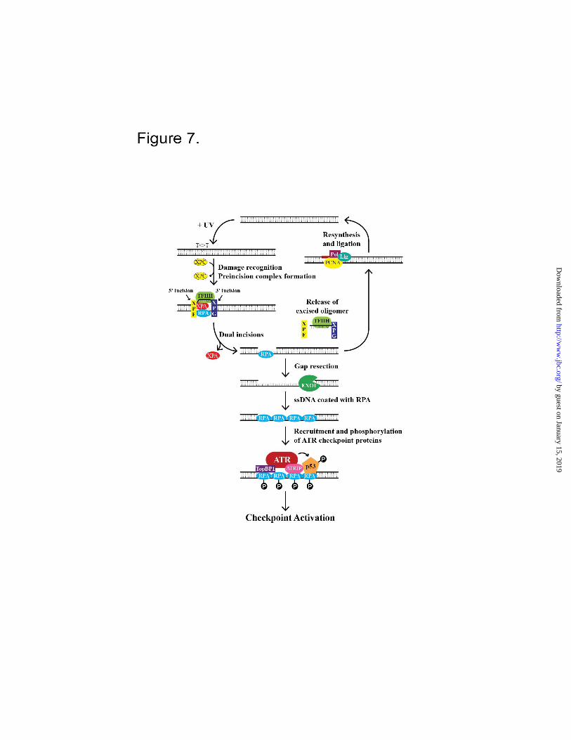
Therefore, we propose that the following
constitutes the minimal essential set of factors for
ATR checkpoint signaling (Figure 7): Signal:
DNA damaged by UV or a UV-mimetic agent;
Core Excision Repair Factors: RPA, XPA, XPC,
TFIIH, XPG, XPF-ERCC1; Core ATR Checkpoint
Factors: ATR-ATRIP, TopBP1, RPA; Excision
Repair-Checkpoint Coupling Factor: EXO1. This
minimal set of factors is sufficient to enable ATR
to phosphorylate RPA and p53 without the need
for additional proteins.
REFERENCES
1. Sancar, A., Lindsey-Boltz, L. A., Unsal-Kacmaz, K., and Linn, S. (2004) Molecular mechanisms of
mammalian DNA repair and the DNA damage checkpoints. Annu. Rev. Biochem. 73, 39-85
2. Abraham, R. T. (2001) Cell cycle checkpoint signaling through the ATM and ATR kinases. Genes
Dev. 15, 2177-2196
3. Nam, E. A., and Cortez, D. (2011) ATR signalling: more than meeting at the fork. The Biochemical
J. 436, 527-536
4. Ditch, S., and Paull, T. T. (2012) The ATM protein kinase and cellular redox signaling: beyond the
DNA damage response. Trends in Biochemical Sciences 37, 15-22
5. Unsal-Kacmaz, K., Makhov, A. M., Griffith, J. D., and Sancar, A. (2002) Preferential binding of
ATR protein to UV-damaged DNA. Proc. Natl. Acad. Sci. U.S.A. 99, 6673-6678
6. Jiang, G., and Sancar, A. (2006) Recruitment of DNA damage checkpoint proteins to damage in
transcribed and nontranscribed sequences. Mol. Cell Biol. 26, 39-49
by guest on January 15, 2019http://w
ww
.jbc.org/D
ownloaded from

Coupling DNA excision repair and the ATR-mediated checkpoint
8
7. Lupardus, P. J., Byun, T., Yee, M. C., Hekmat-Nejad, M., and Cimprich, K. A. (2002) A
requirement for replication in activation of the ATR-dependent DNA damage checkpoint. Genes
Dev. 16, 2327-2332
8. Ward, I. M., Minn, K., and Chen, J. (2004) UV-induced ataxia-telangiectasia-mutated and Rad3-
related (ATR) activation requires replication stress. J. Biol. Chem. 279, 9677-9680
9. Derheimer, F. A., O'Hagan, H. M., Krueger, H. M., Hanasoge, S., Paulsen, M. T., and Ljungman,
M. (2007) RPA and ATR link transcriptional stress to p53. Proc. Natl. Acad. Sci. U.S.A. 104,
12778-12783
10. Bomgarden, R. D., Lupardus, P. J., Soni, D. V., Yee, M. C., Ford, J. M., and Cimprich, K. A.
(2006) Opposing effects of the UV lesion repair protein XPA and UV bypass polymerase eta on
ATR checkpoint signaling. EMBO J. 25, 2605-2614
11. Hanasoge, S., and Ljungman, M. (2007) H2AX phosphorylation after UV irradiation is triggered by
DNA repair intermediates and is mediated by the ATR kinase. Carcinogenesis 28, 2298-2304
12. Marini, F., Nardo, T., Giannattasio, M., Minuzzo, M., Stefanini, M., Plevani, P., and Muzi-Falconi,
M. (2006) DNA nucleotide excision repair-dependent signaling to checkpoint activation. Proc.
Natl. Acad. Sci. U.S.A. 103, 17325-17330
13. Matsumoto, M., Yaginuma, K., Igarashi, A., Imura, M., Hasegawa, M., Iwabuchi, K., Date, T.,
Mori, T., Ishizaki, K., Yamashita, K., Inobe, M., and Matsunaga, T. (2007) Perturbed gap-filling
synthesis in nucleotide excision repair causes histone H2AX phosphorylation in human quiescent
cells. J. Cell Sci. 120, 1104-1112
14. Cimprich, K. A., and Cortez, D. (2008) ATR: an essential regulator of genome integrity. Nat Rev
Mol. Cell Biol. 9, 616-627
15. Sertic, S., Pizzi, S., Cloney, R., Lehmann, A. R., Marini, F., Plevani, P., and Muzi-Falconi, M.
(2011) Human exonuclease 1 connects nucleotide excision repair (NER) processing with
checkpoint activation in response to UV irradiation. Proc. Natl. Acad. Sci. U.S.A. 108, 13647-
13652
16. Novarina, D., Amara, F., Lazzaro, F., Plevani, P., and Muzi-Falconi, M. (2011) Mind the gap:
keeping UV lesions in check. DNA Repair (Amst) 10, 751-759
17. Giannattasio, M., Follonier, C., Tourriere, H., Puddu, F., Lazzaro, F., Pasero, P., Lopes, M.,
Plevani, P., and Muzi-Falconi, M. (2010) Exo1 competes with repair synthesis, converts NER
intermediates to long ssDNA gaps, and promotes checkpoint activation. Mol. Cell 40, 50-62
18. Reardon, J. T., and Sancar, A. (2006) Purification and characterization of Escherichia coli and
human nucleotide excision repair enzyme systems. Methods Enzymol. 408, 189-213
19. Ling, S. C., Albuquerque, C. P., Han, J. S., Lagier-Tourenne, C., Tokunaga, S., Zhou, H., and
Cleveland, D. W. (2010) ALS-associated mutations in TDP-43 increase its stability and promote
TDP-43 complexes with FUS/TLS. Proc. Natl. Acad. Sci. U.S.A. 107, 13318-1332320.
20. Tighe, A., Staples, O., and Taylor, S. (2008) Mps1 kinase activity restrains anaphase during an
unperturbed mitosis and targets Mad2 to kinetochores. J. Cell Biol. 181, 893-901
21. Kershnar, E., Wu, S. Y., and Chiang, C. M. (1998) Immunoaffinity purification and functional
characterization of human transcription factor IIH and RNA polymerase II from clonal cell lines
that conditionally express epitope-tagged subunits of the multiprotein complexes. J. Biol. Chem.
273, 34444-34453
22. Unsal-Kacmaz, K., and Sancar, A. (2004) Quaternary structure of ATR and effects of ATRIP and
replication protein A on its DNA binding and kinase activities. Mol. Cell Biol. 24, 1292-1300
23. Choi, J. H., Lindsey-Boltz, L. A., and Sancar, A. (2007) Reconstitution of a human ATR-mediated
checkpoint response to damaged DNA. Proc. Natl. Acad. Sci. U.S.A. 104, 13301-13306
24. Orans, J., McSweeney, E. A., Iyer, R. R., Hast, M. A., Hellinga, H. W., Modrich, P., and Beese, L.
S. (2011) Structures of human exonuclease 1 DNA complexes suggest a unified mechanism for
nuclease family. Cell 145, 212-223
25. Huibregtse, J. M., Scheffner, M., and Howley, P. M. (1991) A cellular protein mediates association
of p53 with the E6 oncoprotein of human papillomavirus types 16 or 18. Embo J 10, 4129-4135
by guest on January 15, 2019http://w
ww
.jbc.org/D
ownloaded from

Coupling DNA excision repair and the ATR-mediated checkpoint
9
26. Henricksen, L. A., Umbricht, C. B., and Wold, M. S. (1994) Recombinant replication protein A:
expression, complex formation, and functional characterization. J. Biol. Chem. 269, 11121-11132
27. Schaetzlein, S., Chahwan, R., Avdievich, E., Roa, S., Wei, K., Eoff, R. L., Sellers, R. S., Clark, A.
B., Kunkel, T. A., Scharff, M. D., and Edelmann, W. (2013) Mammalian Exo1 encodes both
structural and catalytic functions that play distinct roles in essential biological processes. Proc.
Natl. Acad. Sci. U.S.A. 110, E2470-2479
28. Kemp, M. G., Lindsey-Boltz, L. A., and Sancar, A. (2011) The DNA damage response kinases
DNA-dependent protein kinase (DNA-PK) and ataxia telangiectasia mutated (ATM) Are stimulated
by bulky adduct-containing DNA. J. Biol. Chem. 286, 19237-19246
29. Choi, J. H., Lindsey-Boltz, L. A., Kemp, M., Mason, A. C., Wold, M. S., and Sancar, A. (2010)
Reconstitution of RPA-covered single-stranded DNA-activated ATR-Chk1 signaling. Proc. Natl.
Acad. Sci. U.S.A. 107, 13660-13665
30. Carter, T., Vancurova, I., Sun, I., Lou, W., and DeLeon, S. (1990) A DNA-activated protein kinase
from HeLa cell nuclei. Mol. Cell Biol. 10, 6460-6471
31. Anderson, C. W., and Lees-Miller, S. P. (1992) The nuclear serine/threonine protein kinase DNA-
PK. Critical Reviews in Eukaryotic Gene Expression 2, 283-314
32. Yang, J., Yu, Y., Hamrick, H. E., and Duerksen-Hughes, P. J. (2003) ATM, ATR and DNA-PK:
initiators of the cellular genotoxic stress responses. Carcinogenesis 24, 1571-1580
33. Munoz, D. P., Kawahara, M., and Yannone, S. M. (2013) An autonomous chromatin/DNA-PK
mechanism for localized DNA damage signaling in mammalian cells. Nucleic Acids Res. 41, 2894-
2906
34. Vidal-Eychenie, S., Decaillet, C., Basbous, J., and Constantinou, A. (2013) DNA structure-specific
priming of ATR activation by DNA-PKcs. J. Cell Biol. 202, 421-429
35. Shiotani, B., and Zou, L. (2009) Single-stranded DNA orchestrates an ATM-to-ATR switch at
DNA breaks. Mol. Cell 33, 547-558
36. Yoshioka, K., Yoshioka, Y., and Hsieh, P. (2006) ATR kinase activation mediated by MutSalpha
and MutLalpha in response to cytotoxic O6-methylguanine adducts. Mol. Cell 22, 501-510
37. Lindsey-Boltz, L. A., Reardon, J. T., Wold, M. S., and Sancar, A. (2012) In vitro analysis of the
role of replication protein A (RPA) and RPA phosphorylation in ATR-mediated checkpoint
signaling. J. Biol. Chem. 287, 36123-36131
38. Shiotani, B., Nguyen, H. D., Hakansson, P., Marechal, A., Tse, A., Tahara, H., and Zou, L. (2013)
Two distinct modes of ATR activation orchestrated by Rad17 and Nbs1. Cell Reports 3, 1651-1662
39. Olson, E., Nievera, C. J., Klimovich, V., Fanning, E., and Wu, X. (2006) RPA2 is a direct
downstream target for ATR to regulate the S-phase checkpoint. J. Biol. Chem. 281, 39517-39533
40. O'Driscoll, M., Ruiz-Perez, V. L., Woods, C. G., Jeggo, P. A., and Goodship, J. A. (2003) A
splicing mutation affecting expression of ataxia-telangiectasia and Rad3-related protein (ATR)
results in Seckel syndrome. Nat. Genet. 33, 497-501
41. Hara, R., and Sancar, A. (2003) Effect of damage type on stimulation of human excision nuclease
by SWI/SNF chromatin remodeling factor. Mol. Cell Biol. 23, 4121-4125
42. van Oosterwijk, M. F., Versteeg, A., Filon, R., van Zeeland, A. A., and Mullenders, L. H. (1996)
The sensitivity of Cockayne's syndrome cells to DNA-damaging agents is not due to defective
transcription-coupled repair of active genes. Mol. Cell Biol. 16, 4436-4444
43. Kumagai, A., Lee, J., Yoo, H. Y., and Dunphy, W. G. (2006) TopBP1 activates the ATR-ATRIP
complex. Cell 124, 943-955
44. Tibbetts, R. S., Brumbaugh, K. M., Williams, J. M., Sarkaria, J. N., Cliby, W. A., Shieh, S. Y.,
Taya, Y., Prives, C., and Abraham, R. T. (1999) A role for ATR in the DNA damage-induced
phosphorylation of p53. Genes Dev. 13, 152-157
45. Zou, L., and Elledge, S. J. (2003) Sensing DNA damage through ATRIP recognition of RPA-
ssDNA complexes. Science 300, 1542-1548
by guest on January 15, 2019http://w
ww
.jbc.org/D
ownloaded from
Coupling DNA excision repair and the ATR-mediated checkpoint
10
46. Byun, T. S., Pacek, M., Yee, M. C., Walter, J. C., and Cimprich, K. A. (2005) Functional
uncoupling of MCM helicase and DNA polymerase activities activates the ATR-dependent
checkpoint. Genes Dev. 19, 1040-1052
47. Wang, Y., and Qin, J. (2003) MSH2 and ATR form a signaling module and regulate two branches
of the damage response to DNA methylation. Proc. Natl. Acad. Sci. U.S.A. 100, 15387-15392
48. Liu, Y., Fang, Y., Shao, H., Lindsey-Boltz, L., Sancar, A., and Modrich, P. (2010) Interactions of
human mismatch repair proteins MutSalpha and MutLalpha with proteins of the ATR-Chk1
pathway. J. Biol. Chem. 285, 5974-5982
49. Majka, J., Niedziela-Majka, A., and Burgers, P. M. (2006) The checkpoint clamp activates Mec1
kinase during initiation of the DNA damage checkpoint. Mol. Cell 24, 891-901
50. Duursma, A. M., Driscoll, R., Elias, J. E., and Cimprich, K. A. (2013) A role for the MRN complex
in ATR activation via TOPBP1 recruitment. Mol. Cell 50, 116-122
51. Delacroix, S., Wagner, J. M., Kobayashi, M., Yamamoto, K., and Karnitz, L. M. (2007) The Rad9-
Hus1-Rad1 (9-1-1) clamp activates checkpoint signaling via TopBP1. Genes Dev. 21, 1472-1477
52. Lee, J., Kumagai, A., and Dunphy, W. G. (2007) The Rad9-Hus1-Rad1 checkpoint clamp regulates
interaction of TopBP1 with ATR. J. Biol. Chem. 282, 28036-28044
53. MacDougall, C. A., Byun, T. S., Van, C., Yee, M. C., and Cimprich, K. A. (2007) The structural
determinants of checkpoint activation. Genes Dev. 21, 898-903
54. Leroy, C., Mann, C., and Marsolier, M. C. (2001) Silent repair accounts for cell cycle specificity in
the signaling of oxidative DNA lesions. EMBO J. 20, 2896-2906
55. Mordes, D. A., Nam, E. A., and Cortez, D. (2008) Dpb11 activates the Mec1-Ddc2 complex. Proc.
Natl. Acad. Sci. U.S.A. 105, 18730-18734
56. Navadgi-Patil, V. M., and Burgers, P. M. (2008) Yeast DNA Replication Protein Dpb11 Activates
the Mec1/ATR Checkpoint Kinase. J. Biol. Chem. 283, 35853-35859
57. Mordes, D. A., Glick, G. G., Zhao, R., and Cortez, D. (2008) TopBP1 activates ATR through
ATRIP and a PIKK regulatory domain. Genes Dev. 22, 1478-1489
58. Kumar, S., and Burgers, P. M. (2013) Lagging strand maturation factor Dna2 is a component of the
replication checkpoint initiation machinery. Genes Dev. 27, 313-321
59. Navadgi-Patil, V. M., and Burgers, P. M. (2011) Cell-cycle-specific activators of the Mec1/ATR
checkpoint kinase. Biochemical Society Transactions 39, 600-605
60. Navadgi-Patil, V. M., and Burgers, P. M. (2009) The unstructured C-terminal tail of the 9-1-1
clamp subunit Ddc1 activates Mec1/ATR via two distinct mechanisms. Mol. Cell 36, 743-753
61. Choi, J. H., Lindsey-Boltz, L. A., and Sancar, A. (2009) Cooperative activation of the ATR
checkpoint kinase by TopBP1 and damaged DNA. Nucleic Acids Res. 37, 1501-1509
62. Choi, J. H., Sancar, A., and Lindsey-Boltz, L. A. (2009) The human ATR-mediated DNA damage
checkpoint in a reconstituted system. Methods 48, 3-7
63. Lindsey-Boltz, L. A., Sercin, O., Choi, J. H., and Sancar, A. (2009) Reconstitution of human
claspin-mediated phosphorylation of Chk1 by the ATR (ataxia telangiectasia-mutated and rad3-
related) checkpoint kinase. J. Biol. Chem. 284, 33107-33114
64. Hassan, B. H., Lindsey-Boltz, L. A., Kemp, M. G., and Sancar, A. (2013) Direct role for the
replication protein treslin (Ticrr) in the ATR kinase-mediated checkpoint response. J. Biol. Chem.
288, 18903-18910
65. Clarke, C. A., and Clarke, P. R. (2005) DNA-dependent phosphorylation of Chk1 and Claspin in a
human cell-free system. The Biochemical J. 388, 705-712
by guest on January 15, 2019http://w
ww
.jbc.org/D
ownloaded from

Coupling DNA excision repair and the ATR-mediated checkpoint
11
FOOTNOTES *This work was supported by National Institutes of Health grant GM32833 (to A.S.) and GM45190 (to
P.M.). P.M. is an Investigator of the Howard Hughes Medical Institute. #Present address: Teva Pharmaceuticals, Inc, 145 Brandywine Parkway, West Chester, PA 19380
+To whom correspondence may be addressed: Aziz Sancar, Department of Biochemistry and Biophysics,
University of North Carolina School of Medicine, CB#7260, Chapel Hill, NC 27599-7260, Tel: 919-
962-0115 E-mail: [email protected]. 4The Abbreviations used are: UV, ultraviolet; RPA, Replication Protein A; ssDNA, single-stranded
DNA; AAF, N-acetoxy-2-acetylaminofluorene.
FIGURE LEGENDS
FIGURE 1. Purified Repair and Checkpoint Factors. Analysis of the 10 excision repair and checkpoint
factors by silver staining. Approximately 5-20 ng of each factor was subjected to 4-15% TGX-PAGE
analysis. The different subunits in the complexes are indicated by asterisks.
FIGURE 2. EXO1-dependent ATR Activation. A. A Model System for Excision Repair-Checkpoint
Coupling. ATR kinase reactions were carried out with ATR-ATRIP, TopBP1, RPA, and EXO1 as
indicated. 0.6 ng (27 pM) single-stranded X174 DNA (ssDNA), plasmid DNA (dsDNA), or gapped
DNA was added to the reaction as indicated and incubated 20 minutes at 30°C. Reactions were analyzed
by immunoblotting for phospho-RPA2 (Ser33
) and RPA1. B. EXO1-dependent phosphorylation of RPA2
(Ser33
) and p53 (Ser18
) in quiescent cells after UV damage. Serum-starved (quiescent) or asynchronously
growing (proliferating) wild-type (WT) and Exo1-/-
MEFs were exposed to 20 J/m2 of UVC light and
harvested 1 hr later. Cell lysates were analyzed by SDS-PAGE and immunoblotting with the indicated
antibodies.
FIGURE 3. Repair factor- and damage-dependent gap generation and resection by EXO1. A. Repair
Factor (RF)-dependent generation of gapped-DNA. Excision reactions were performed with unmodified
DNA (lanes 1 and 2) or AAF-damaged DNA (lanes 3 and 4) in the presence (lanes 2 and 4) or absence
(lanes 1 and 3) of the 6 core excision repair factors (RF). The percentage of gapped DNA was quantified
from identical repeats of the experiment and presented as mean ± SD (n≥3). B. EXO1 specifically digests
the gapped DNA generated by repair factors. After 90 minute excision reactions with AAF-damaged
DNA without (lanes 1-3) or with repair factors (lanes 4-6), EXO1 was added at the indicated
concentrations for an additional 10 minutes before analysis by agarose gel electrophoresis (0 lanes 1 and
4, 4 nM lanes 2 and 5, 8 nM lanes 3 and 6).
FIGURE 4. Repair-Checkpoint coupling as measured by RPA phosphorylation by ATR. Kinase
reactions containing 100 nM RPA as a substrate were incubated 20 minutes at 30°C. 0.2 nM ATR-
ATRIP, 2.5 nM TopBP1, and 8 nM EXO1 were added, as indicated, to kinase reactions containing 4 ng
unmodified (UM) or AAF DNA from excision reactions with or without repair factors (RF) as indicated.
All six repair factors were required, as determined by omission studies (data not shown). Reactions were
analyzed by immunoblotting for phospho-RPA2 (Ser33
). The blots were also analyzed for RPA1 to
control for loading. The relative levels of phosphorylated RPA2 from identical repeats of the experiment
were quantified and presented as mean ± SD (n≥3).
FIGURE 5. DNA concentration effect and kinetics of ATR checkpoint signaling. A. Titration of
unmodified (UM) or AAF DNA (2, 4, or 8 ng) from excision reactions with or without repair factors (RF)
into kinase reactions containing ATR-ATRIP, TopBP1, RPA, and EXO1 as in Figure 4. B. Time course
analysis of RPA2 phosphorylation in kinase reactions with 4 ng of DNA as in panel A. The graphs below
by guest on January 15, 2019http://w
ww
.jbc.org/D
ownloaded from

Coupling DNA excision repair and the ATR-mediated checkpoint
12
show the relative levels of phosphorylated RPA2 from identical repeats of the experiments quantified and
presented as mean ± SD (n≥3).
FIGURE 6. Repair-Checkpoint coupling as measured by p53 phosphorylation. Kinase reactions
containing p53, ATR-ATRIP, TopBP1, RPA, EXO1, and the indicated DNA were performed as in Figure
4. Reactions were analyzed by immunoblotting for phospho-p53 (Ser15
). The blots were also analyzed
for GST-p53 to control for loading. The relative levels of phosphorylated p53 from identical repeats of
the experiment were quantified and presented as mean ± SD (n≥2).
FIGURE 7. Model of activation of the ATR-signaling pathway during G1/G0 or G2-M phases of the cell
cycle. When DNA is damaged by UV or a UV-mimetic agent, the core excision repair factors (RPA,
XPA, XPC, TFIIH, XPG, XPF-ERCC1) excise a ~30 nt oligomer containing the damage. (Note that
XPC, after playing an essential role in damage recognition, dissociates from the repair complex prior to
the dual incisions (1) (18), and hence it is not shown in the incision complex). The resulting gap is either
filled in by polymerases or the gap is enlarged by EXO1. The enlarged ssDNA gap is coated with RPA
which recruits ATR-ATRIP, TopBP1, and substrates including p53. The close proximity of the ATR
kinase with its activator, TopBP1, and substrate, p53, results in phosphorylation and checkpoint
activation.
by guest on January 15, 2019http://w
ww
.jbc.org/D
ownloaded from


Iyer, Paul Modrich and Aziz SancarLaura A. Lindsey-Boltz, Michael G. Kemp, Joyce T. Reardon, Vanessa DeRocco, Ravi R.
Checkpoint in a Defined In Vitro SystemCoupling of Human DNA Excision Repair and ATR-mediated DNA Damage
published online January 8, 2014J. Biol. Chem.
10.1074/jbc.M113.542787Access the most updated version of this article at doi:
Alerts:
When a correction for this article is posted•
When this article is cited•
to choose from all of JBC's e-mail alertsClick here
by guest on January 15, 2019http://w
ww
.jbc.org/D
ownloaded from