reconstruction of highly proliferative auto-tissue ... · reconstruction of highly proliferative...
TRANSCRIPT

Reconstruction of Highly ProliferativeAuto-Tissue-Engineered Lamellar Cornea
Enhanced by Embryonic Stem Cell
Qiang Zhou, PhD,* Zhao Liu, PhD,* Zheng Wu, PhD, Xiaoran Wang, PhD,Bowen Wang, PhD, Chaoyang Li, MD, Ying Liu, PhD, Liangliang Li, PhD,Pengxia Wan, PhD, Zheqian Huang, PhD, and Zhichong Wang, MD, PhD
To increase the epithelial proliferation of an auto-tissue-engineered lamellar cornea, 3.0 · 106 corneal epithelialcells (CECs) were combined with 3 · 105 mouse embryonic stem cells (ESCs) pretransfected with the HSV-tkgene (CECs + ESCs-TK group), and 3.3 · 106 corneal epithelial cells (CECs group) were seeded between theacellular porcine corneal stroma and the amniotic membrane using the centrifugal cell seeding method. After 4days of perfusion culture (treatment with ganciclovir starting on day 2), a thicker corneal epithelium (four tofive layers) formed in the CECs + ESCs-TK group compared with that observed in the CECs group (two to threelayers). More stem/progenitor cell (K3 - , p63 + , ABCG2 + , and integrin-b1 + ) and proliferation phenotypes(Ki67 + ) were measured in the CECs + ESCs-TK group compared with the CECs group using immunofluo-rescence staining, real-time quantitative reverse transcription polymerase chain reaction, and flow cytometry.Consistent with these findings, the colony-forming efficiency and cellular doubling time were significantlydifferent between the CECs + ESCs-TK group (16.18% – 3.98%, 28.45 – 2.03 h) and CECs group (11.96% –2.60%, 36.3 – 1.15 h). In a rabbit lamellar transplantation model, the CECs + ESCs-TK group had better epi-thelial barrier functions and wound healing abilities compared with the CECs group. Furthermore, ESCs-TKcould be completely and safely removed by ganciclovir. Thus, the ESCS-TK coculture system could serve as apotential strategy for corneal tissue engineering.
Introduction
The viability of epithelial cells in the corneal graftplays a critical role in the re-epithelialization and res-
toration of transparency after corneal transplantation.1 Thus,a corneal graft that is fresh and of a relatively young agehas many advantages and is more appropriate for cornealtransplantation. However, the available grafts are limited.Recent advances in the field of tissue engineering mayprovide new opportunities for the development of functionaltissues to replace damaged corneas.
For the maintenance of epithelial viability in corneal re-construction, the seeding cells, bioengineered scaffolds, andproper strategies were evaluated.2 First, to acquire thehighly proliferative properties of corneal epithelial cells(CECs) for seeding, various methods have been applied,including different feeder cells, conditioned media, cyto-kines or growth factors,3 as well as the immortalization ofCECs.4 In our laboratory, an immortalized permanent cellline has been established from a normal human cornea usingserial culture,5 and we also demonstrated that the functional
properties of stem-like CECs could be markedly enhancedby the microenvironment of embryonic stem cells (ESCs).6–9
The second major problem associated with corneal recon-struction is the absence of a suitable scaffold for the stem cellsto grow and be transferred to the eye. Currently, several nat-ural or synthetic biomaterials, including amniotic membranes(AMs),10 fibrin gels,11 silk,12 and collagen,13 have been ap-plied for the reconstruction of corneal substitutes. Further-more, from the perspective of the natural extracellularmatrix composition and regional-specific cues for cellularproliferation and adhesion, the acellular porcine cornealstroma (APCS) may provide a promising alternative scaf-fold.14 As we previously reported, the APCS15 was devel-oped by our group and has been successfully applied incorneal limbus16 and stroma17,18 reconstruction.
Finally, an appropriate reconstruction strategy also playsa key role in epithelial growth. In recent years, after 4–6weeks of construction, two to six layers of the epitheliumhad formed on different scaffolds using air–liquid interfaceculture.12,13,19 However, to further decrease the culture timefor clinical application and to mimic the native ocular
State Key Laboratory of Ophthalmology, Zhongshan Ophthalmic Center, Sun Yat-sen University, Guangzhou, China.*These two authors contributed equally to this work and are considered co-first authors.
TISSUE ENGINEERING: Part CVolume 21, Number 7, 2015ª Mary Ann Liebert, Inc.DOI: 10.1089/ten.tec.2014.0481
639

environment of the epithelium, a centrifugal cell seedingmethod20 and dynamic culture system21 were used, respectively.
On the basis of our previous studies, the viability of anauto-tissue-engineered lamellar cornea (ATELC) was in-creased using the ESC microenvironment in a controlled andsafe manner. Briefly, these strategies were simultaneouslyapplied in the reconstruction process. First, using coculturewith ESCs, multilayers of rabbit autologous passage 1 (P1)epithelial cells were rapidly centrifugally seeded betweenthe APCS and AM. After an increase in cell adhesion in-duced by the sheer force of the perfused culture, the ATELCwas transplanted into a rabbit model. To avoid undesirabledifferentiation and tumorigenesis of ESCs,22 ESCs werepretransfected with the herpes simplex virus thymidine ki-nase (HSV-TK), a suicide gene (ESCs-TK). When the via-bility of the CECs was enhanced, all of the ESCs-TK wereinduced to undergo apoptosis gradually by treating theculture with ganciclovir.6
Materials and Methods
Animals
Female and male New Zealand white rabbits (aged 10weeks and weighing 2–3 kg) were purchased from the An-imal Laboratory of Sun Yat-sen University (Guangzhou,China). All of the animal experiments were performed withpermission obtained from the Medicine Ethics Committee inZhongshan Ophthalmic Center, Sun Yat-sen University, andcomplied with the Association for Research in Vision andOphthalmology (ARVO) statement on the use of animals inophthalmic and visual research.
Preparation of APCS and AM
APCS preparation: The APCS was prepared as previouslydescribed.15 Briefly, native porcine corneas were immersedin a bicarbonate-mixed salt solution containing phospholi-pase A2 (PLA2) (200 U/mL; Sigma, P0861) and 0.5% (w/v)sodium deoxycholate (SD) (Sigma, D6750) for 6 h at 37�C.Next, the samples were immersed in a bicarbonate-mixedsalt solution containing only PLA2 for 2 h at 37�C followedby six washes of 30 min each in a bicarbonate-mixed saltsolution at 10�C to remove the residual PLA2 and SD. All ofthe steps were performed with continuous shaking in athermostat-controlled water bath.
AM preparation: Proper informed consent was obtainedfrom all AM donors in accordance with the principles of theDeclaration of Helsinki on Biomedical Research. For theATELC construction, the AM was prepared as previouslydescribed and sectioned into 2.3 · 2.3-cm pieces.20
Cell preparation and culture
Primary CECs, which were obtained from a single 1 · 1-mm2 auto-rabbit corneal limbal tissue explant (100mmthickness, left eye), were prepared as previously describedand cultured in corneal epithelial medium.6
Mouse ES-E14 cells, which had been transfected with thesuicide gene of herpes simplex virus thymidine kinase(ESC-TK) and selected with 1mg/mL puromycin (Sigma),were plated at a density of 400/cm2 in 1% gelatin (Sigma)-coated tissue culture dishes containing mouse ESC culturemedium.6
Control and experimental groups
A total of 3.3 · 106 cells were seeded for each of thetwo groups according to the different types of cells. In thecontrol group (CECs group), 3.3 · 106 passage 1 (P1) auto-rabbit CECs were seeded. In the experimental group(CECs + ESCs-TK group), 3 · 106 P1 auto-rabbit cornealCECs and 3 · 105 ESCs-TK were mixed uniformly beforethe reconstruction process.
Culture strategies of ATELC
First, as we previously reported, three layers of cells werecentrifuged onto the AM in both groups (14 mm2 in diam-eter) (1800 bpm, 4 min).20 Next, after the membrane wascovered with APCS (upside-down), it was fixed in thechamber (Ismatec pump; Ismatec, Minucells) with the AM(facing up for 4 days with continuous perfusion culture.21
After 24 h of perfusion culture (20 mL/h) on both sides ofthe ATELC, 20mM ganciclovir was added into the medium.6
On day 4 postconstruction, each ATELC specimen was col-lected for observation of Hematoxylin and Eosin (H&E)staining, transmission electron microscopy (TEM, H600; Hi-tachi), and immunofluorescence staining. After all of the cellswere isolated from the ATELC using the previously reportedmethods,21 the DNA content and proliferative properties wereobserved using ELISA, real-time quantitative reverse tran-scription polymerase chain reaction (real-time qRT-PCR), flowcytometry, colony-forming efficiency (CFE), and the methyl-thiazol tetrazolium cell proliferation assay (MTT).8
Removal of ESCs-TK by ganciclovir treatment
To track the ESCs-TK in a coculture system, all of theESCs-TK were labeled with DiO (5mg/mL; Invitrogen) for30 min at 37�C before the reconstruction process. The ex-pression of DiO was identified using fluorescence microscopyand flow cytometry analysis using the CellQuest Pro software(n = 10; FACSCalibur flow cytometry, BD Biosciences).6
HE and immunofluorescence staining
To measure the morphology and differentiation of the cellsof ATELC, specimens from both groups were analyzed usingH&E staining (n = 4), TEM (n = 4), quantitative DNA analysis(n = 10; Sigma), flow cytometry (n = 10), and immunofluo-rescence staining (n = 4). The primary antibodies were K3(1:100; Millipore), P63 (1:50, Millipore), ABCG2 (1:50,Millipore), Ki67 (Abcam ab8191; 1:100), integrin-b1 (Abcam78502; 1:100), desmocollin 2 (Abcam 72792; 1:100), and in-tegrin-b4 (Abcam ab29042; 1:100). The secondary antibodiesincluded Alexa Fluor 594 IgG (Invitrogen, A11012; 1:100)and Alexa Fluor 488 IgG (Invitrogen A-11001; 1:100); thenuclei were counterstained with Hoechst 33342 (blue color;Invitrogen). The examinations were performed using a laserscanning confocal microscope (LSM 510 META; Carl Zeiss).
Evaluation of differentiated phenotypesand growth capacity of epithelial cells in ATELCusing a quantitative assay
Real-time qRT-PCR was performed to detect the mRNAexpression levels of K3, P63, and ABCG2. As previouslydescribed,8 the total RNA from each group (n = 10) was
640 ZHOU ET AL.

isolated using TRIzol (Invitrogen) according to the manu-facturer’s instructions and quantified by absorption at260 nm. A total of 0.5 mg of RNA was reversely transcribedinto cDNA in a 20-mL volume reaction system using theSYBR Prime Script� 25 RT-PCR Kit (DRR063S; Takara).Samples of synthesized cDNA were divided into aliquotsand stored at - 80�C. Real-time qRT-PCR was performedand analyzed using an ABI PRISM 7000 sequence detec-tion system (Applied Biosystems, Inc.). The PCR was per-formed in a 20-mL volume reaction system containing 10mLof 2 · SYBR Green reaction mix (SYBR Green system[DRR063S; Takara]), 0.4 mM paired primers, and 1 mL ofcDNA. The sequences of the PCR primer pairs are listed inTable 1. The thermal cycling consisted of denaturation for3 min at 95�C followed by 40 cycles of 15 s at 95�C and 30 sat 60�C. The PCR amplification efficiency of the primer setswas determined to be essentially 100% before qPCR. Thecomparative Ct (66CT) method was used to compare themRNA expression levels of the genes of interest. GAPDHwas selected as an internal control gene.
As previously described, flow cytometry (n = 10) wasperformed to quantitatively evaluate the differentiation andproliferation markers of CECs in both groups. The primaryantibodies were K3 (1:100; Millipore), P63 (1:50; Milli-pore), ABCG2 (1:50; Millipore), and Ki67 (Abcam ab8191;1:100). The secondary antibody was Alexa Fluor 488 IgG(Invitrogen A-11001; 1:100); all of the results were ana-lyzed using the CellQuest Pro software (BD Biosciences).
The proliferative capacities of epithelial cells were alsomeasured using the MTT cell proliferation assay (n = 10)and CFE (n = 10) assays as previously reported.5,8
Rabbit lamellar keratoplasty
ESCs-TK were labeled using DiO (5mg/mL; Invitrogen)for 30 min at 37�C before construction, and the ATELC ofboth groups were implanted into the right rabbit corneas byroutine lamellar keratoplasty (n = 14, 100-mm thickness,6.25-mm diameter). Each of the grafts was sutured into therecipient bed with eight interrupted 10–0 nylon sutures.Tobramycin–dexamethasone eye ointment was applied fourtimes a day for 7 days after LKP. On postoperative day 4,the AM was removed, and sodium fluorescein staining wasperformed to assess the epithelial integrity. In addition, slitlamp examination was performed to assess the opticalclarity of the cornea, corneal neovascularization, and deg-radation of corneal grafts. On postoperative days 4, fourrabbits from each group were killed, and their corneas wereobserved using confocal microscopy in vivo, H&E, andimmunofluorescence staining. On postoperative day 20, 10rabbits were killed, and corneal specimens from these rab-bits were evaluated using light transmittance assays andimmunofluorescence staining for collagen III (1:50; Acris).21
Statistical analysis
The values are expressed as the means – standard devia-tion. The SPSS 11.0 software was used for the statisticalanalyses, and the differences between two groups werecompared using the independent samples t-test. The statis-tical significance was defined as p < 0.05.
Results
Physiological morphology of ATELC
The morphology of primary epithelial cells was mo-saic and homogeneous. Most of the cells were K3 + (CEC-specific marker), P63 - , ABCG2 - (corneal stem/progenitorcell markers), and Ki67- (proliferation marker) (Fig. 1A).The ESCs-TK stably exhibited a clonal appearance andshowed Oct-4 expression (ESC marker) (Fig. 1B). After 4days of continuing perfusion culture, multilayers of theepithelium were formed on the ATELC in both groups. Inthe CECs group, there were only two to three layers ofsquamous cells with large nuclei on the ATELC, and theDNA content of the samples was 3960 – 579.66 ng. Inaddition, the TEM image showed that a few microvilli, des-mosomes, and hemidesmosomes in the cell–cell and cell–scaffold junctions. Similarly, immunofluorescence stainingalso revealed a few markers of cell–cell junctions (desmo-collin-2) and cell–scaffold junctions (integrin-b4). How-ever, in the CECs + ESCs-TK group, four to five layers of
Table 1. Primer Sets for Real-Time Quantitative Reverse Transcription Polymerase Chain Reaction
Gene Upstream primers (5¢-3¢) Downstream primers (3¢-5¢)
CK3 TCCGTCACAGGCACCAAC TGCGTTTGTTGATTTCGTCTDNP63 GAAAACAATGCCCAGACTCAATTT TCTGCGCGTGGTCTGTGTTATABCG2 ACACGGGTTGACTGGAGA TTTCAGGAGCAGAAGGACAGAPDH GGAGCCAAAAGGGTCATC CCAGTGAGTTTCCCGTTC
FIG. 1. Phenotypes of auto-rabbit corneal epithelial cells(CECs) and mouse embryonic stem cells (ESCs) that hadbeen pretransfected with the HSV-TK gene. (A) The pri-mary CECs presented a cobblestone-like appearance as as-sessed using phase contrast light microscopy. Althoughmost of the CECs expressed K3 + (green, corneal epithelialcell-specific marker), a few of the cells expressed p63 + ,ABCG2 + (green, the stem/progenitor cell markers), andKi67 + (green, the proliferation marker). (B) The ESCs-TKexhibited a clonal appearance as assessed using phase mi-croscopy and expressed Oct-4 (green, an ESC marker) asdetermined using immunofluorescence staining. Color ima-ges available online at www.liebertpub.com/tec
A VIABLE AUTO-TISSUE-ENGINEERED LAMELLAR CORNEA RECONSTRUCTED ON APCS 641

flattened cells with a small size were formed more compactlyon the ATELC; the DNA content was 6440 – 1298.89 ng(n = 10, p < 0.05), and more microvilli, desmosomes, andhemidesmosomes were observed using TEM (n = 4). More-over, immunofluorescence staining demonstrated higherexpression levels of desmocollin-2 and integrin-b4 com-pared with those observed in the CECs group (Fig. 2).
Removal of ESCs-TK from ATELC
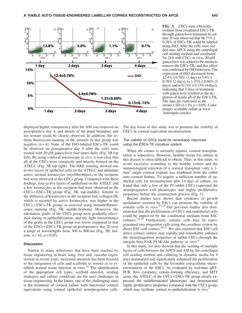
Using flow cytometry analysis, 98.77% – 0.38% of ESCs-TK were labeled by DiO before the reconstruction process.After the cells were cocultured with CECs for 24 h, ap-proximately 12.4% – 0.78% of the cells were DiO positive.After the medium was treated with 20mM ganciclovir for 3days, the expression of DiO markedly decreased from3.4% – 0.78% (2 days) to 1.33% – 0.68% (3 days) and to0.13% – 0. 15% (4 days) (n = 10, p < 0.05) (Fig. 3).
Cocultured ESCs-TK can enhance stem/progenitorcell phenotypes of CECs in ATELC
It has been shown that more basal cells of the ATELCexpress stem/progenitor cell phenotypes in the CECs + ESCsgroup (K3 - , P63 + , ABCG2 + , and integrin-b1 + ) (Fig. 4A).Real-time qRT-PCR analysis demonstrated that the expressionof the corneal specific marker K3 mRNA (1.58 – 0.12) in theCECs + ESCs-TK group was significantly lower compared
with that observed in the CECs group (1.03 – 0.23). In addition,the expression of P63 mRNA and ABCG2 mRNA (1.52 – 0.25)in the CECs + ESCs-TK group was increased from 1.08 – 0.15to 1.69 – 0.26 and from 0.99 – 0.18 to 1.52 – 0.25, respectively(n = 10, p < 0.05) (Fig. 4B). Consistent with the real-time qRT-PCR results, quantitative flow cytometry analyses also showedthat the percentage of Ki67 (15.47% – 2.20%), P63 (14.63% –2.23%), and ABCG2 (13.77% – 1.91%) expression in theATELC in the CECs + ESCs-TK group was significantlyhigher than that found in the CECs group (Ki67 [10.83% –1.53%], P63 [8.50% – 0.96%], ABCG2 [8.13% – 0.85%])(Fig. 4C, n = 10, p < 0.05). Furthermore, the cell-cloningefficiency of the cells in the ATELC of the CECs + ESCs-TK group (9.72% – 3.5%) was also significantly highercompared with that of the CECs group (6.29% – 1.96%)(Fig. 4E, F, n = 10, p < 0.05). MTT proliferation assays alsoshowed that the doubling time of the CECs + ESCs-TKgroup (28.45 – 2.03 h) was significantly shorter than that ofthe CECs group (36.3 – 1.15 h) (Fig. 4G, n = 10, p < 0.05).
Rabbit lamellar keratoplasty
As shown in Figure 5A, slight edema and diffused ex-coriation were observed in the ATELC of the CECs groupafter the AM was removed on postoperative day 4 using slitlamp biomicroscopy and sodium fluorescein staining. Incontrast, the ATELC of the CECs + ESCs-TK group
FIG. 2. Histological evaluation of the morphological and adhesive properties of the auto-tissue-engineered lamellarcornea (ATELC) after 4 days of perfusion culture in vitro. (left column: CECs group, right column: CECs + ESCs-TKgroup). (from top to bottom) Hematoxylin and eosin (H&E) staining shows that two to three layers and four to five layers ofthe corneal epithelium formed between the amniotic membrane and the acellular porcine corneal stroma (APCS) in theCECs group and CECs + ESCs-TK group, respectively. In contrast to the CECs group, the nuclei of the epithelial cells in theCECs + ESCs-TK group were smaller, and the arrangement of the cells was more compact. More microvilli, desmosome(desmocollin-2), and hemidesmosome (integrin-b4) were also observed in the cell–cell and cell–APCS junctions using TEMand immunofluorescence staining, respectively, in the CECs + ESCs-TK group (green). 6 desmosome in the cell–celljunction;[hemidesmosome in the cell–APCS junction. Color images available online at www.liebertpub.com/tec
642 ZHOU ET AL.
displayed higher transparency after the AM was removed onpostoperative day 4, and details of the pupil boundary andiris texture could be clearly observed. In addition, the so-dium fluorescein staining of the animals in this group wasnegative (n = 4). None of the DiO-labeled ESCs-TK couldbe observed on postoperative day 4 after the cells weretreated with 20 mM ganciclovir four times daily (Fig. 5B topleft). By using confocal microscopy in vivo, it was clear thatall of the CECs were compactly and intactly formed on theATELC (Fig. 5B top right). The H&E staining showed oneto two layers of epithelial cells on the ATELC and abundantactive stromal keratocytes (myofibroblasts) in the recipientbed were observed in the CECs group. Compared with thesefindings, four to five layers of epithelium on the ATELC anda few keratocytes in the recipient bed were observed in theCECs + ESCs-TK group (Fig. 5B, top-middle). Similar tothe diffusion of keratocytes in the recipient bed, collagen III,which is secreted by active keratocytes, was higher in theCECs + ESCs-TK group, as assessed using immunofluores-cence staining (Fig. 5B, middle-botttom). Moreover, theedematous grafts of the CECs group were gradually allevi-ated during re-epithelialization, and the light transmittanceof the grafts in the CECs group was lower compared to thatof the CECs + ESCs-TK group on postoperative day 20 overa range of wavelengths from 300 to 800 nm (Fig. 5B, bot-tom, n = 10, p < 0.05).
Discussion
Similar to many milestones that have been reached bytissue engineering in heart, lung, liver and, vascular regen-eration in recent years, increased attention has been focusedto the integration of cells and scaffolds to restore or to es-tablish normal tissue function in vitro.20 The identificationof the appropriate cell types, scaffold material, seedingstrategies and culture conditions are the next challenges intissue engineering. In the future, one of the challenging aimsis the treatment of corneal failure with functional cornealequivalents using corneal epithelial stem/progenitor cells.
The key focus of this study was to promote the viability ofCECs in corneal equivalent reconstruction.
The viability of CECs could be remarkably improvedusing the ESCs-TK coculture system
When the cornea is seriously injured, corneal transplan-tation is imperative. However, healthy tissue for culture inthis disease is often difficult to obtain. Thus, in this study, toavoid excessive wounding to the healthy cornea and theimmunological rejection of a corneal transplant, only a 1-mm2 single corneal explant was trephined from the rabbitauto-corneal limbus. To acquire a sufficient number of ep-ithelial cells for reconstruction after 14 days of culture, wefound that only a few of the P1 rabbit CECs expressed thestem/progenitor cell phenotypes and highly proliferativeproperties before the construction process (Fig. 1).
Recent studies have shown that cytokines or growthmodulators secreted by ESCs can promote the viability ofsomatic cells in vitro.23,24 Our previous studies also dem-onstrated that the proliferation of CECs and endothelial cellscould be improved by the conditional medium from ESCcultures.7,8,25 Furthermore, somatic cells may be repro-grammed into progenitor cells using the coculture system bydirect ESC-cell contact.26,27 We also reported that ESC-celldirect contact culture may rapidly and remarkably enhancethe stem/progenitor properties of rabbit CECs through theintegrin beta-FAK-P13K/Akt pathway in vitro.6
In this study, we also showed that the seeding of multiplelayers of cells between the APCS and AM by the centrifugalcell seeding method and culturing in dynamic media for 4days maintained and significantly enhanced the proliferationof the epithelial cells by the favorable extracellular micro-environment of the ESCs. As evaluated by real-time qRT-PCR, flow cytometry, colony-forming efficiency, and MTTassay, the ATELC of the CECs + ESCs-TK group clearly ex-pressed the undifferentiated phenotypes and demonstratedhighly proliferative properties compared with the CECs group,which may facilitate corneal re-epithelialization in vivo.1
FIG. 3. CECs were efficientlyisolated from cocultured ESCs-TKthrough ganciclovir treatment in cul-ture. It was observed that 98.77% –0.38% of ESCs-TK could be labeledusing DiO. After the cells were see-ded onto APCS using the centrifugalcell seeding method and coculturedfor 24 h with CECs in vitro, 20mMganciclovir was added to the media toremove the ESCs-TK, and this effectwas confirmed by DiO detection. Theexpression of DiO decreased from12.4% – 0.78% (1 day) to 3.4% –0.78% (2 days), to 1.33% – 0.68% (3days), and to 0.13% – 0.15% (4 days),indicating that 3 days of treatmentwith ganciclovir resulted in the de-pletion of nearly all of the ESCs-TK.The data are expressed as themeans – SD (n = 10, p < 0.05). Colorimages available online at www.liebertpub.com/tec
A VIABLE AUTO-TISSUE-ENGINEERED LAMELLAR CORNEA RECONSTRUCTED ON APCS 643

As the immunofluorescence staining images showing,more of the basal cells expressed progenitor/stem pheno-types (K3 - , P63 + , ABCG2 + , and integrin-b1 + ) and pro-liferation marker (Ki67 + ) in the ATELC of the CECs +ESCs-TK group compared with the CECs group (Fig. 4). Bymaintaining the native basement membrane components ofthe cornea, the substrate properties of APCS21 under basalcells may play a critical role in the maintenance of co-cultured epithelial cells in their undifferentiated state,28 whichis required for corneal wound healing.
Multiple layers of the corneal epitheliumcan be rapidly constructed on APCSby centrifugal cell seeding and AM covering
The intact corneal epithelium on the surface can defendagainst external mechanical irritation, protect against in-fection and inflammation, and prevent excessive scar for-mation. In corneal reconstruction engineering, the adhesiveproperties of the epithelium also play a vital role in with-standing the operation of corneal transplantation and the
FIG. 4. The cocultured ESCs-TK promoted the expression of stem/progenitor phenotypes and the proliferative propertiesof epithelial cells in ATELC. (A) (from top to bottom) In contrast to the CECs group, a larger number of low-differentiationphenotypes (K3 - , P63 + , ABCG2 + , integrin-b1 + : green) and high levels of the proliferation marker (Ki67 + ; green) wereobserved in the basal layer of epithelial cells in the CECs + ESCs-TK group, as evaluated by immunofluorescence stainingin vitro. (left column: CECs group, right column: CECs + ESCs-TK group). (B) mRNA expression levels of the stem/progenitor cell markers (ABCG2 + , p63 + ) and corneal specific marker (K3 + ) in the CECs group and the CECs + ESCs-TKgroup, as determined using real-time quantitative reverse transcription polymerase chain reaction with GAPDH as aninternal control. These results are shown as the means – SD (n = 10, p < 0.05) and are expressed as the relative fold increasecompared with the CECs group (normalized to 1). (C) Representative flow cytometry images demonstrating the percentageof cells expressing the proliferation marker (Ki67 + ) and adult stem/progenitor cell-associated markers (p63 + , ABCG2 + )in the CECs group and the CECs + ESCs-TK group. The quantitative data analysis is shown in (D) (n = 10, p < 0.05). (E)Representative cultures stained with Giemsa demonstrating the growth capacity of CECs in both groups on day 6 (colony-forming efficiency, CFE). (F) The CFE of the CECs in the CECs + ESCs-TK group (16.18% – 3.98%) was significantlyhigher compared with that of the CECs group (11.96% – 2.60%, n = 10, p < 0.05). (G) The population doubling time of theCECs in the CECs + ESCs-TK group (28.45 – 2.03 h) was significantly decreased compared with that of the CECs group(36.3 – 1.15 h) (n = 10, p < 0.05). Color images available online at www.liebertpub.com/tec
644 ZHOU ET AL.

FIG. 5. The physiological function, the morphology, light transmittance of ATELC in the auto-rabbit lamellar cornealtransplantation model. (A) (top) In the CECs group, on postoperative day 4, after the removal of the amniotic membrane,slight edema was observed in the ATELC. Punctate sodium fluorescein staining revealed that the barrier function was notcomplete. On postoperative day 7, the transparency of the cornea was improved, and there was a lack of sodium fluoresceinstaining. From postoperative day 12–20, the transparency of the ATELC was increased, and small parts of the cornealopacity could still be observed. (bottom) In the CECs + ESCs-TK group, the ATELC was so transparent that the texture ofthe iris could be observed after the amniotic membrane was removed, and negative staining showed that the barrier functionof the CECs was intact on postoperative day 4. Over 20 days of observation, the sodium fluorescein staining was alwaysnegative, and the transparency of the ATELC was always higher in the CECs + ESCs-TK group compared with the CECsgroup. (B) (top) Using DiO, we found that none of the labeled cells could be observed 4 days after transplantation (left),which revealed that nearly all of the ESCs-TK were eliminated from the ATELC in the CECs + ESCs-TK group. Usingconfocal microscopy in vivo, the epithelial cells on the ATELC were found to be small and homogeneous and were arrangedcompactly (right). (top-middle) In the CECs group (left), as determined using the H&E assay, only one to two layers ofcorneal epithelial cells were observed on the ATELC on postoperative day 4, and the epithelial cells were loose. In addition,a higher number of corneal keratocytes were gathered on the recipient bed. In the CECs + ESCs-TK group (right), four tofive layers of corneal epithelial cells were formed on the ATELC. The cells were small, and the arrangement of the cells wascompact. In addition, few corneal keratocytes were observed on the recipient bed. (middle-bottom) The expression ofcollagen III on the recipient bed was higher in the CECs + ESCs-TK group (right) than in the CECs group (left) 20 days afterkeratoplasty (green). (bottom) Compared with the CECs group, the light transmittance of the ATELC was lower in theCECs + ESCs-TK group over the wavelength range of 300–800 nm on postoperative day 20 (n = 10, p < 0.05).
645
friction of eye blinking in vivo. In this study, during thereconstruction process, all of the cells were cultured be-tween the AM and APCS and fixed onto a chamber of aperfusion culture system. By the protection and limitation ofAM, cell loss may be avoided during construction. Thereby,the density of the cells was increased, which could promotethe proliferative and adhesive properties of cells vice ver-sa.29 Moreover, using the perfusion culture system, the fluidshearing force, supply of sufficient oxygen and nutrition,and the proliferation and adhesion properties could alsobeen improved.30 As shown in the Figure 2, more ultra-structures of desmosomes and hemidesmosomes were formedbetween the cell–cell and cell–matrix junctions in the ATELCof the CECs + ESCs-TK group compared with the CECsgroup. Consistent with these results, more desmocollin-2(desmosome marker) and integrin-b4 (hemidesmosomemarker) were detected using the immunofluorescencestaining in the CECs + ESCs-TK group than the CECs group(Fig. 2). Furthermore, after additional layers and a higherdensity of CECs with higher proliferative and adhesivefunctions were constructed in the ATELC of the CECs +ESCs-TK group in vitro, the corneal transplantation wasenhanced compared with that obtained in the CECs group.
Intact multiple layers of the epithelium could increasethe restoration of transparency and decreasethe formation of scar after corneal transplantation
Covering of the AM could also protect the corneal epi-thelium from transplantation and the formation of celljunctions in the ATELC in vivo. After the AM was removedon postoperative day 4, the corneal barrier functionalproperties were intact, and the corneal transparency wascompletely restored in the ATELC of the CECs + ESCs-TKgroup (Fig. 5A). After 20 days of observation, four to fivelayers of the epithelium were maintained on the ATELC; afew differentiated myofibroblasts and some collagen IIIwere formed on the receiving bed, and the ATELC showedhigh optical transparency and favorable epithelial barrierfunction in the CECs-ESCs + TK group throughout the ob-servational period (Fig. 5B). In contrast, on postoperativeday 4, the barrier function of the ATELC had not completelyformed in the CECs group. Even after 20 days of observation,the ATELC of the CECs group restored its transparency, andthe light transmittance of the cornea was always lower com-pared with that of the CECs-ESCs + TK group. The corneaconsisted of highly organized collagen I and microstructures.If the cornea was deeply wounded, it would be substituted bynewly synthesized type III collagen and lose its spatial orga-nization. However, only a slight alteration in the cornea re-sulted in a marked decrease in visual acuity. Thus, the intactcorneal epithelium of the ATELC in the CECs-ESCs + TKgroup plays an important role in limiting the formation of theECM and decreases the activity of stromal keratocytes(myofibroblasts) in wound healing, both of which are closelycorrelated to the opacity of the corneal stroma.1,2,31
ESCs could be completely removedby ganciclovir treatment
ESCs are undifferentiated resident cells with high pro-liferative capacities and pluripotent potential and may pro-mote the proliferation of cocultured somatic cells by serving
as the stem cell microenvironment; however, this methodstill remains limited by the tumorigenicity or impurity of theinduced differentiation of the cells.22 Thus, in this study, theESCs were pretransfected with the HSV-TK gene (a suicidegene) and were sensitive to ganciclovir. Moreover, after thestem/progenitor phenotypes of CECs were significantly en-hanced using this coculture system and treatment withganciclovir, nearly all of the DiO-labeled ESCs-TK could bedepleted in vitro, with none of the cells detectable 4 daysafter transplantation (Figs. 3 and 5B, top). These resultswere consistent with and supported by our previous reportsthat the ESCs-TK could be completely removed by addingganciclovir in vitro as evaluated by flow cytometry analysisof apoptosis experiments (Annexin V and PI staining), andobservation of undetected Dil track in ESCs-TK after 3 daysof ganciclovir treatment6; as well as in vivo by tracking theESCs-TK with transfected GFP fluorescence.32
During the entire observation period, there was an ab-sence of any tumor formation, which was consistent withour previous studies in cell coculture and leukemia mousetreatment.6,32 These results also showed that, the gradualremoval of ESCs from surrounding cells by treatment withganciclovir in vitro and in vivo, did not destroy the intactbarrier function of the corneal epithelium and maintainedthe corneal transparency consistently, which is also consis-tent with previous studies.33
Although the ESC-TK coculture system has obvious ad-vantages in promoting the proliferation of CECs in therabbit corneal transplantation model, there is still improve-ment in ATELC reconstruction, as assessed using differ-ent parameters, including the percentage or time of theESCs-TK coculture. Furthermore, whether the ATELC isappropriate for other transplantation models, such as corneallimbus transplantation procedures, requires further studies.
Conclusion
In conclusion, this work reconstructed a viable ATELCon APCS using ESCs-TK coculture, the centrifugal seedingmethod, and AM covering. The ATELC could be trans-planted into the rabbit model to rapidly restore cornealtransparency. Moreover, treatment with ganciclovir com-pletely removed the ESCs-TK without forming any tera-toma. This controllable and safe method using the ESCmicroenvironment to construct a more viable ATELCdemonstrates its expansive potential clinical applications.
Acknowledgments
This work was supported by the National High Tech-nology Research and Development Program (863 Program)of China (No.2012AA020507) (W.Z.C.), Natural ScienceFoundation of China (No. 81270971) (W.Z.C.), GuangdongNatural Science Foundation (No. S2012010009113) (W.Z.C.),the Fundamental Research Funds of State Key Laboratoryof Ophthalmology of China (2012PI05) (W.Z.C.). The au-thors thank Professor De-Quan Li from the Baylor Collegeof Medicine for his helpful comments and revision.
Disclosure Statement
No competing financial interests exist.
646 ZHOU ET AL.

References
1. Casaroli-Marano, R.P., Nieto-Nicolau, N., and Martinez-Conesa, E.M. Progenitor cells for ocular surface regenera-tive therapy. Ophthalmic Res 49, 115, 2013.
2. Shah, A., Brugnano, J., Sun, S., Vase, A., and Orwin, E.The development of a tissue-engineered cornea: biomate-rials and culture methods. Pediatr Res 63, 535, 2008.
3. Zhang, X., Sun, H., Li, X., Yuan, X., Zhang, L., and Zhao,S. Utilization of human limbal mesenchymal cells as feederlayers for human limbal stem cells cultured on amnioticmembrane. J Tissue Eng Regen Med 4, 38, 2010.
4. Robertson, D.M., Kalangara, J.P., Baucom, R.B., Petroll,W.M., and Cavanagh, H.D. A reconstituted telomerase-immortalized human corneal epithelium in vivo: a pilotstudy. Curr Eye Res 36, 706, 2011.
5. Liu, J., Song, G., Wang, Z., Huang, B., Gao, Q., Liu, B.,Xu, Y., Liang, X., Ma, P., Gao, N., and Ge, J. Establish-ment of a corneal epithelial cell line spontaneously derivedfrom human limbal cells. Exp Eye Res 84, 599, 2007.
6. Zhou, J., Chen, F., Xiao, J., Li, C., Liu, Y., Ding, Y., Wan,P., Wang, X., Huang, J., and Wang, Z. Enhanced functionalproperties of corneal epithelial cells by coculture withembryonic stem cells via the integrin beta1-FAK-PI3K/Aktpathway. Int J Biochem Cell Biol 43, 1168, 2011.
7. Liu, Z., Wan, P., Duan, H., Zhou, J., Tan, B., Liu, Y., Zhou,Q., Zhou, C., Huang, Z., Tian, B., Li, C., and Wang, Z. ESmicro-environment enhances stemness and inhibits apo-ptosis in human limbal stem cells via the maintenance oftelomerase activity. PLoS One 8, e53576, 2013.
8. Liu, Y., Ding, Y., Ma, P., Wu, Z., Duan, H., Liu, Z., Wan,P., Lu, X., Xiang, P., Ge, J., and Wang, Z. Enhancement oflong-term proliferative capacity of rabbit corneal epithelialcells by embryonic stem cell conditioned medium. TissueEng Part C Methods 16, 793, 2010.
9. Lu, X., Chen, D., Liu, Z., Li, C., Liu, Y., Zhou, J., Wan, P.,Mou, Y.G., and Wang, Z. Enhanced survival in vitroof human corneal endothelial cells using mouse embryonicstem cell conditioned medium. Mol Vis 16, 611, 2010.
10. Ma, D.H., Lai, J.Y., Cheng, H.Y., Tsai, C.C., and Yeh, L.K.Carbodiimide cross-linked amniotic membranes for culti-vation of limbal epithelial cells. Biomaterials 31, 6647,2010.
11. Rama, P., Matuska, S., Paganoni, G., Spinelli, A., DeLuca, M., and Pellegrini, G. Limbal stem-cell therapy andlong-term corneal regeneration. N Engl J Med 363, 147,2010.
12. Bray, L.J., George, K.A., Ainscough, S.L., Hutmacher,D.W., Chirila, T.V., and Harkin, D.G. Human corneal ep-ithelial equivalents constructed on Bombyx mori silk fi-broin membranes. Biomaterials 32, 5086, 2011.
13. Duan, X., McLaughlin, C., Griffith, M., and Sheardown, H.Biofunctionalization of collagen for improved biologicalresponse: scaffolds for corneal tissue engineering. Bioma-terials 28, 78, 2007.
14. Song, J.J., and Ott, H.C. Organ engineering based on decel-lularized matrix scaffolds. Trends Mol Med 17, 424, 2011.
15. Wu, Z., Zhou, Y., Li, N., Huang, M., Duan, H., Ge, J.,Xiang, P., and Wang, Z. The use of phospholipase A(2) toprepare acellular porcine corneal stroma as a tissue engi-neering scaffold. Biomaterials 30, 3513, 2009.
16. Huang, M., Li, N., Wu, Z., Wan, P., Liang, X., Zhang, W.,Wang, X., Li, C., Xiao, J., Zhou, Q., Liu, Z., and Wang, Z.
Using acellular porcine limbal stroma for rabbit limbalstem cell microenvironment reconstruction. Biomaterials32, 7812, 2011.
17. Xiao, J., Duan, H., Liu, Z., Wu, Z., Lan, Y., Zhang, W., Li,C., Chen, F., Zhou, Q., Wang, X., Huang, J., and Wang, Z.Construction of the recellularized corneal stroma usingporous acellular corneal scaffold. Biomaterials 32, 6962,2011.
18. Liu, Z., Zhou, Q., Zhu, J., Xiao, J., Wan, P., Zhou, C.,Huang, Z., Qiang, N., Zhang, W., Wu, Z., Quan, D., andWang, Z. Using genipin-crosslinked acellular porcine cor-neal stroma for cosmetic corneal lens implants. Biomater-ials 33, 7336, 2012.
19. Luo, H., Lu, Y., Wu, T., Zhang, M., Zhang, Y., and Jin, Y.Construction of tissue-engineered cornea composed ofamniotic epithelial cells and acellular porcine cornea fortreating corneal alkali burn. Biomaterials 34, 6748, 2013.
20. Zhang, W., Xiao, J., Li, C., Wan, P., Liu, Y., Wu, Z.,Huang, M., Wang, X., and Wang, Z. Rapidly constructedscaffold-free cornea epithelial sheets for ocular surfacereconstruction. Tissue Eng Part C Methods 17, 569,2011.
21. Wu, Z., Zhou, Q., Duan, H., Wang, X., Xiao, J., Duan, H.,Li, N., Li, C., Wan, P., Liu, Y., Song, Y., Zhou, C., Huang,Z., and Wang, Z. Reconstruction of auto-tissue-engineeredlamellar cornea by dynamic culture for transplantation: arabbit model. PLoS One 9, e93012, 2014.
22. Goldring, C.E., Duffy, P.A., Benvenisty, N., Andrews,P.W., Ben-David, U., Eakins, R., French, N., Hanley, N.A.,Kelly, L., Kitteringham, N.R., Kurth, J., Ladenheim, D.,Laverty, H., McBlane, J., Narayanan, G., Patel, S., Re-inhardt, J., Rossi, A., Sharpe, M., and Park, B.K. Assessingthe safety of stem cell therapeutics. Cell Stem Cell 8, 618,2011.
23. Guo, Y., Graham-Evans, B., and Broxmeyer, H.E. Murineembryonic stem cells secrete cytokines/growth modulatorsthat enhance cell survival/anti-apoptosis and stimulatecolony formation of murine hematopoietic progenitor cells.Stem Cells 24, 850, 2006.
24. Pearton, D.J., Yang, Y., and Dhouailly, D. Transdiffer-entiation of corneal epithelium into epidermis occurs bymeans of a multistep process triggered by dermal devel-opmental signals. Proc Natl Acad Sci U S A 102, 3714,2005.
25. Jiang, X.X., Zhang, Y., Liu, B., Zhang, S.X., Wu, Y., Yu,X.D., and Mao, N. Human mesenchymal stem cells inhibitdifferentiation and function of monocyte-derived dendriticcells. Blood 105, 4120, 2005.
26. Zhang, B., Liu, R., Shi, D., Liu, X., Chen, Y., Dou, X., Zhu,X., Lu, C., Liang, W., Liao, L., Zenke, M., and Zhao, R.C.Mesenchymal stem cells induce mature dendritic cells intoa novel Jagged-2-dependent regulatory dendritic cell pop-ulation. Blood 113, 46, 2009.
27. Orkin, S.H., and Hochedlinger, K. Chromatin connectionsto pluripotency and cellular reprogramming. Cell 145, 835,2011.
28. Jones, R.R., Hamley, I.W., and Connon, C.J. Ex vivo ex-pansion of limbal stem cells is affected by substrate prop-erties. Stem Cell Res 8, 403, 2012.
29. Matter, K., and Balda, M.S. Signalling to and from tightjunctions. Nat Rev Mol Cell Biol 4, 225, 2003.
30. Cattan, V., Bernard, G., Rousseau, A., Bouhout, S.,Chabaud, S., Auger, F.A., and Bolduc, S. Mechanical
A VIABLE AUTO-TISSUE-ENGINEERED LAMELLAR CORNEA RECONSTRUCTED ON APCS 647
stimuli-induced urothelial differentiation in a human tissue-engineered tubular genitourinary graft. Eur Urol 60,1291, 2011.
31. Nakamura, K., Kurosaka, D., Bissen-Miyajima, H., andTsubota, K. Intact corneal epithelium is essential for theprevention of stromal haze after laser assisted in situ ker-atomileusis. Br J Ophthalmol 85, 209, 2001.
32. Zhou, C., Huang, Z., Li, P., Li, W., Liu, Y., Li, C., Liu, Z.,Wang, X., Wan, P., and Wang, Z. Safety and efficacy ofembryonic stem cell microenvironment in a leukemiamouse model. Stem Cells Dev 23, 1741, 2014.
33. Schuldiner, M., Itskovitz-Eldor, J., and Benvenisty, N.Selective ablation of human embryonic stem cells expres-sing a ‘‘suicide’’ gene. Stem Cells 21, 257, 2003.
Address correspondence to:Zhichong Wang, MD, PhD
State Key Laboratory of OphthalmologyZhongshan Ophthalmic Center
Sun Yat-sen University54 Xian Lie Nan Road
Guangzhou 510060China
E-mail: [email protected]
Received: August 9, 2014Accepted: November 20, 2014
Online Publication Date: January 19, 2015
648 ZHOU ET AL.