reconstruction of the heel, middle foot sole and plantar ... · 3 introduction soft tissue...
TRANSCRIPT
Zurich Open Repository andArchiveUniversity of ZurichMain LibraryStrickhofstrasse 39CH-8057 Zurichwww.zora.uzh.ch
Year: 2018
Reconstruction of the heel, middle foot sole and plantar forefoot with themedial plantar artery perforator (MPAP) flap: clinical experience with 28
cases
Scaglioni, Mario F; Rittirsch, Daniel; Giovanoli, Pietro
Abstract: INTRODUCTION: Defects in the weight-bearing region of the foot sole can represent a sub-stantial restriction in quality of life and pose a challenge for reconstructive plastic surgery.The purpose ofthis article is to report our experience with the use of medial plantar artery perforator (MPAP) flap forreconstruction of defects of the foot sole in 3 different regions: heel, middle foot sole and plantar forefoot.PATIENTS AND METHODS: From January 2003 to May 2016, 28 patients (13 males and 15 females)with an average age of 54 years (range 12 to 84 y.o.) received reconstruction with 28 MPAP flaps.26 flapswere harvested as pedicle perforator flaps and 2 as free perforator flaps. All flaps were raised from theipsilateral instep area. The defect locations included the heel (20 cases), middle foot sole (4 cases) andforefoot (4 cases).The causes of reconstruction were tumors in 18 patients (64%), decubitus in 8 patients(29%) and trauma in 2 patients (7%). RESULTS: The flap sizes varied from 2.5 × 2.5 cm to 5.5 × 9.5 cm.All of the flaps survived completely after surgery, apart from one. The donor sites were all covered witha split-thickness skin graft. Follow-up observations were conducted for 4 to 12 months, and all patientshad good functional recovery with satisfactory cosmetic results. CONCLUSION: The MPAP flap can beconsidered an optimal method of foot sole reconstruction not only for covering the weight-bearing areaof the heel but also for the middle and forefoot plantar region.
DOI: https://doi.org/10.1097/PRS.0000000000003975
Posted at the Zurich Open Repository and Archive, University of ZurichZORA URL: https://doi.org/10.5167/uzh-143340Accepted Version
Originally published at:Scaglioni, Mario F; Rittirsch, Daniel; Giovanoli, Pietro (2018). Reconstruction of the heel, middle footsole and plantar forefoot with the medial plantar artery perforator (MPAP) flap: clinical experience with28 cases. Plastic and Reconstructive Surgery, 141(1):200-208.DOI: https://doi.org/10.1097/PRS.0000000000003975
1
Plastic and Reconstructive Surgery Advance Online Article
DOI: 10.1097/PRS.0000000000003975
Reconstruction of the heel, middle foot sole and plantar forefoot with the medial plantar
artery perforator (MPAP) flap: clinical experience with 28 cases.
Mario F. Scaglioni MD1, Daniel Rittirsch MD
1, Pietro Giovanoli MD
1
1Department of Plastic and Hand Surgery, University Hospital Zurich, Zurich, Switzerland
Running Head: Plantaris medialis artery perforator flap for total plantar foot reconstruction
Corresponding Author:
Mario F. Scaglioni, MD
Department of Plastic and Hand Surgery,
University Hospital Zurich, Zurich, Switzerland
Address: Rämistraße 100, 8091, Zurich, Switzerland
Telephone number: 00410442551111
E-mail: [email protected]
Disclosure: The authors have no financial conflicts or commercial associations to disclose.
ACCEPTED
Copyright © American Society of Plastic Surgeons. All rights reserved.
2
ABSTRACT
Introduction:
Defects in the weight-bearing region of the foot sole can represent a substantial restriction in
quality of life and pose a challenge for reconstructive plastic surgery.
The purpose of this article is to report our experience with the use of medial plantar artery
perforator (MPAP) flap for reconstruction of defects of the foot sole in 3 different regions:
heel, middle foot sole and plantar forefoot.
Patients and Methods:
From January 2003 to May 2016, 28 patients (13 males and 15 females) with an average age
of 54 years (range 12 to 84 y.o.) received reconstruction with 28 MPAP flaps.
26 flaps were harvested as pedicle perforator flaps and 2 as free perforator flaps. All flaps
were raised from the ipsilateral instep area. The defect locations included the heel (20 cases),
middle foot sole (4 cases) and forefoot (4 cases).
The causes of reconstruction were tumors in 18 patients (64%), decubitus in 8 patients (29%)
and trauma in 2 patients (7%).
Results:
The flap sizes varied from 2.5 × 2.5 cm to 5.5 × 9.5 cm. All of the flaps survived completely
after surgery, apart from one. The donor sites were all covered with a split-thickness skin
graft. Follow-up observations were conducted for 4 to 12 months, and all patients had good
functional recovery with satisfactory cosmetic results.
Conclusion:
The MPAP flap can be considered an optimal method of foot sole reconstruction not only for
covering the weight-bearing area of the heel but also for the middle and forefoot plantar
region.
Keywords: Plantaris medialis artery perforator flap, plantar foot reconstruction, Instep flap,
free flap
ACCEPTED
Copyright © American Society of Plastic Surgeons. All rights reserved.

3
INTRODUCTION
Soft tissue reconstruction of the foot sole has long been a reconstructive challenge not only to
restore an aesthetic appearance but also to maintain function. In fact, defects of this region of
the body can enormously hamper the patient’s quality of life. Therefore, the choice of
reconstruction of the weight-bearing plantar foot surface is very important. Due to the limited
availability and mobility of the adjacent skin, defects involving the plantar foot area may
require complex reconstruction procedures. One of the most important factors for skin
resurfacing of the weight-bearing area of the foot sole is the provision of an adequate type of
skin with similar properties, of thickness and sensitivity.1
Many techniques, such as skin graft, local flaps, local pedicle flaps and free flaps, have been
already described for the reconstruction of these regions;2–13
however, the reconstructive
options to improve outcomes are still under discussion and there is not a supreme procedure,
which is considered as a gold standard; there are only few flaps which can withstand the
stresses and loads posed each and every day by a person’s body weight.
Ideally, defects involving this area are optimally repaired using thin and pliable flaps. Based
on basic plastic surgery principles to replace “like with like,” with minimal donor site
morbidity and without sacrificing a major vessel, perforator flaps designed from the instep
area of the foot represent a viable option. The term “instep area” refers to the hairless non-
weight-bearing medial plantar region of the foot sole centered over the medial plantar
artery.14,15
The medial plantar artery perforator (MPAP) flap, which is harvested from this
region, is therefore ideal for the reconstruction of soft tissue defects of the foot sole with
minimal donor site morbidity. The MPAP can be defined as a perforator flap based on the
superficial branch of the medial plantar artery (MPA). It can be used as a pedicle or free flap.9
Moreover, it contains a sensitive branch of the medial plantar nerve which supplies the instep
skin area and provides a new sensation in the defect region.5,16
In this article we describe our
ACCEPTED
Copyright © American Society of Plastic Surgeons. All rights reserved.
4
experience using the MPAP flap for the reconstruction of defects of the heel, the middle and
the forefoot sole.
PATIENTS AND METHODS
Between January 2003 and May 2016, 28 patients underwent foot sole reconstructions with
the MPAP flap. There were 13 male patients and 15 female patients. The average age of the
patients was 54 years (range, 12 - 84 years). The causes were various and included soft tissue
defect after cancer resection in 18 cases, trauma in 2 cases and pressure sores in 8 cases. The
defect locations were the heel in 20 patients and the middle plantar foot and forefoot in 4
patients, respectively.
In all cases, the defect dimension and especially the weight-bearing surface precluded skin
grafting and primary closure. The details of patients are presented in Table 1.
Surgical technique
All flaps were designed on the medial part of the sole, the so called “instep area.” To begin, a
surface marking is done in the instep area and also a preoperatively Doppler probe detected
the perforators from the superficial branch of the medial plantar artery system and proved the
patency of the posterior tibial artery (PTA) and dorsalis pedis artery (DPA).
Under tourniquet control without previous exsanguination, the first incision is made parallel
to the abductor hallucis muscle, creating the medial border of the flap. When the flap is
dissected laterally and suprafascially above the flexor hallucis muscle, several perforators of
the medial plantar vessel can be readily identified through the intermuscular septum between
the abductor hallucis muscle and the flexor digitorum muscle (brevis or longus) (Fig. 1).
With the use of retractors, the medial plantar vessel is clearly exposed through the
intermuscular space. The perforator is deeply dissected to the division of the medial plantar
vessel. The lateral border of the MPAP flap is then incised and dissected, taking care to
include the perforators. The perforator arteries are always small in the MPAP flap. The
diameter of the perforated artery is approximately 0.4 to 0.8 mm and the pedicle length
ACCEPTED
Copyright © American Society of Plastic Surgeons. All rights reserved.
5
approximately 4 to 6 cm. There is usually a separate branch arising from the medial plantar
nerve or from the main trunk of the posterior tibial nerve. This branch again divides into 2.
The medial of the 2 branches proceeds to supply the sensation of the medial side of the great
toe and the lateral branch to supply the sensation of the instep area. The lateral branch
supplying the instep area is easily separable from the main branch under loupe magnification.
Smaller branches of the medial plantar nerve around the medial plantar aponeurosis are cut.
Then, the flap is transposed or rotated in propeller fashion into the defect (Fig. 2). The donor
site was covered by a split-thickness skin graft. Postoperatively, patients were maintained on
strict bedrest with the foot elevated. At one week after surgery, patients were allowed to begin
a dangling protocol, and at 2 weeks, patients began light weight-bearing on the operated foot
with the assistance of physical therapy when indicated.
RESULTS
Twenty-eight MPAP flaps were performed. The dimensions of the flap skin paddle ranged
from 2.5 × 2.5 cm to 5.5 × 9.5 cm. The average perforator number included in the flap was 1
(range, 1–2). The mean pedicle length was 5 cm with a range of 4–6 cm.
Regarding the two free MPAP flaps, the recipient vessels were the branch of lateral plantar
artery in 2 patients. The arterial anastomoses were performed in an end-to-end fashion.
All the flaps survived completely, except for one, with a success rate of 96%. One MPAP
distal based flap for forefoot reconstruction was lost due to poor vascular perfusion. No
arterial nor venous thrombosis occurred in the free MPAP flaps. The follow-up time ranged
from 4 to 12 months (mean: 9 months). The flaps healed well and the color matches were
excellent. All donor sites were covered with a split-thickness skin graft, and no early nor late
complications were encountered. None of the patients complained about the donor site scar.
All patients returned to their original jobs and were satisfied with the functional results and
cosmetic appearance. Eventually they could also wear regular footwear. Details are shown in
Table 1.
ACCEPTED
Copyright © American Society of Plastic Surgeons. All rights reserved.

6
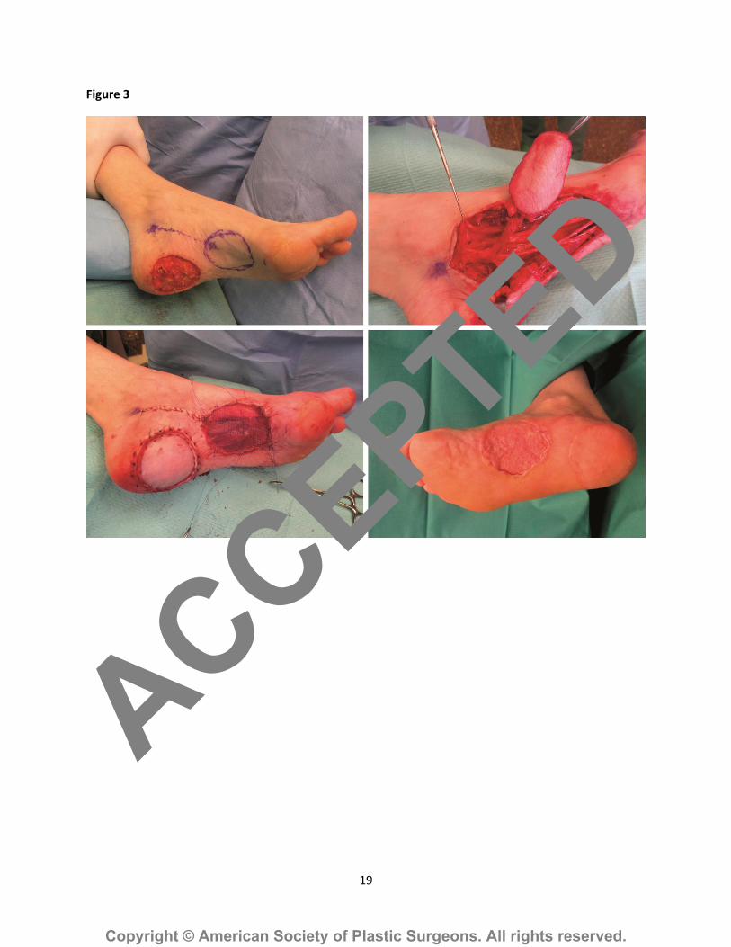
Case 1: Coverage of heel defect
A 55-year-old woman sustained a weight-bearing malignant melanoma on the left heel. After
a wide excision, a tumor free defect measured 5 × 5 cm was noted (Fig. 3, above left). A 5 × 5
cm pedicle MPAP flap was harvested from the ipsilateral instep area based on one perforator.
(Fig.3, above right). Then the flap was transferred to reconstruct the defect (Fig. 3, below
left). The donor site was covered with split-thickness skin graft from the contralateral thigh.
The flap survived completely and the donor site healed uneventfully. The follow-up time was
6 months. The patient had full functional recovery with acceptable cosmetic result (Fig. 3
below right).
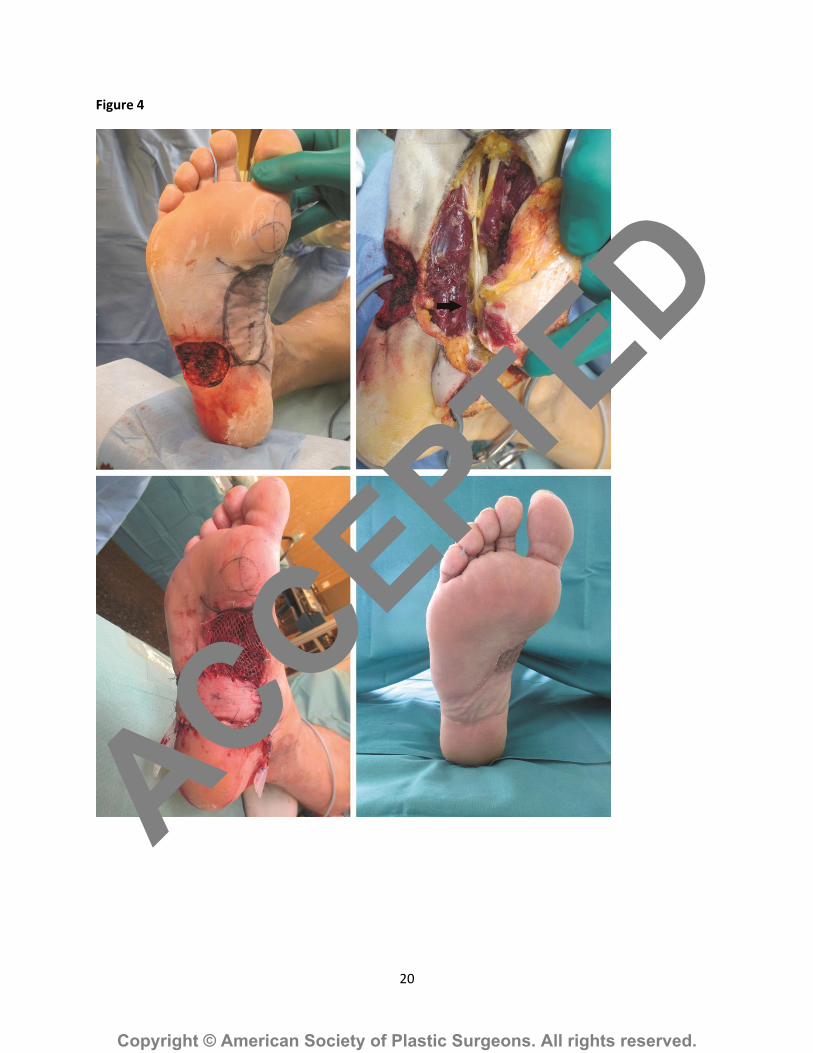
Case 2: Coverage of middle foot sole defect
A 51-year-old man was diagnosed as malignant melanoma on the right middle foot sole. After
wide excision, a tumor free defect measured 6 × 5 cm was noted (Fig. 4, above left). A 6.5 ×
6.5 cm pedicle MPAP flap was harvested from the ipsilateral instep area based on one
perforator. (Fig. 4, above right). Then, the flap was propelled with 90 degree of rotation to
reconstruct the defect (Fig. 4, below left). The donor site was covered with split-thickness
skin graft from the contralateral thigh. At the 8-month follow-up, the flap counter, color and
healing were excellent. The patient had full functional recovery with acceptable cosmetic
result (Fig. 4, below right).
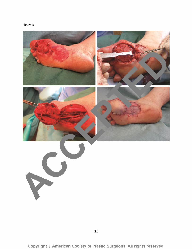
Case 3: Coverage of a forefoot defect
A 53-year-old woman was diagnosed as malignant melanoma on her right forefoot. After
wide excision, a tumor free defect measured 4 × 4.5 cm was noted (Fig. 5, above left). A 5 × 5
cm pedicled MPAP flap was harvested from the ipsilateral instep area based on the distal
perforator of the MPA (Fig. 5, above right). Then the flap was rotated in propeller fashion of
around 180 degrees to reconstruct the forefoot defect (Fig. 5, below). The donor site was
covered with split-thickness skin graft from the contralateral thigh. Postoperatively, the
patient had nothing negative to report regarding ambulation or at the donor site. The follow-
ACCEPTED
Copyright © American Society of Plastic Surgeons. All rights reserved.

7
up time was 6 months. The patient had full functional recovery with acceptable cosmetic
result.
DISCUSSION
Surgical reconstruction of soft tissue defect in the foot sole regions poses one of the greatest
challenges for plastic surgery. The location of the defect enormously hampers the patient’s
quality of life and the peculiar anatomic structure of the plantar foot can make even a small
defect a difficult task. In particular, due to the very robust and thick characteristic of the skin
of this area, it can easily withstand the stresses and loads it is exposed to on a daily basis.
Many soft tissue reconstructions of these areas have been described using traditional local and
free flaps, and the common aim was to use a type of skin with similar properties to the
original skin in order to guaranteeing appropriate coverage and function.1,16
Since the description of the freestyle perforator concept and the perforasome theory pioneered
by Wei and Mardini, the use of local perforator flaps has grown in popularity.17
Hence, local
perforator flaps based on the plantar medial artery have been successfully employed for
plantar foot reconstruction, although the dimension of defect represents the main limitation.
As described by Attinger et al.18
in 2006, the foot sole is perfused by the medial plantar artery
and the lateral plantar artery providing their own corresponding angiosome. The medial
plantar artery perforator (MPAP) flap is based on the perforators arising from the superficial
branch of the medial plantar artery.19
As it has been already reported in the literature, the
perforator nourishing the MPAP flap is constant and can be found between the adductor
allucis muscle and the flexor digitorum brevis muscle in proximity of the navicular
bone.15,16,20
However, the number of suitable perforators and their caliber has not been
described. Moreover, a cutaneous nerve, branch of the medial plantar nerve, can provide
sensation if incorporated within the flap.21
ACCEPTED
Copyright © American Society of Plastic Surgeons. All rights reserved.
8
Due to the non-weight-bearing localization of the instep area, the MPAP flap, harvested from
this region, represents an ideal method of reconstruction of soft tissue defects of the foot sole
and has the great advantage that the donor site morbidity is very low.14–16
The instep flap was first described in 1981 by Harrison and Morgan for soft tissue coverage of
the heel.22
Thereafter, in 1983 Morrison et al.9 developed a free sensory instep flap with
medial plantar artery for heel defects. Since then, it has gained more and more popularity for
plantar foot reconstruction, and it has been employed as free flap for defect coverage of the
hand and fingers as well.23–27
It is important to clarify that the instep flap is based on the deep branch of the MPA and it is
classified as a pedicle fasciocutaneous flap; contrarily, the MPAP flap is based on the
perforators of the superficial branch of the MPA.15,19
Only few reports described the use of the MPAP flap for plantar foot sole reconstruction.
In this article, which covers a period of 13 years, we describe our experience with the use of
the MPAP flap for soft tissue defect reconstruction of the heel, the middle part of the foot
sole, and the forefoot plantar region.
During the flap elevation we could always identify, in the septum between the abductor
hallucis and the flexor digitorum brevis muscle, two perforators: one proximal, few
centimeters after the exit of the MPA from the abductor hallucis muscle, and one more distal.
However, the caliber of the perforators could be variable (around 0.4 to 0.8 mm); in 24 cases
the proximal perforator was bigger than the distal perforator and the flap could be harvested
based on only one perforator. In two cases (two heel reconstructions) 2 perforators were
included in the flap because the distal perforator appeared dominant with a bigger caliber
compared to the proximal one. About the forefoot defect, the distal perforator was the one to
supply the vascularity of the pedicle MPAP flap. Usually a few cm distally to the distal
perforator of the superficial branch of the MPA, perforators, that runs from the superficial
branch of the MPA up to the dorsal foot, can be appreciated; these are the connecting branch
ACCEPTED
Copyright © American Society of Plastic Surgeons. All rights reserved.
9
from dorsal to plantar foot and should be always spared and kept if a distal based MPAP is
planned.
Based on our experience, the selection of the perforators depends on the intraoperative
findings: 1) for heel reconstruction the proximal perforator is the more suitable due its vicinity
to the defect; however, if it has very small size, the second perforator can be included as well.
2) for middle plantar foot reconstruction actually the dissection of the MPA is less important
than heel construction as the MPAP flap needs to be rotated in propeller fashion of around 90
degrees, and it is possible only by dissecting the proximal or distal perforator. The choice of
the perforator depends on the its size. In our series in all four cases the proximal perforator
was selected.
3) for forefoot plantar reconstruction between the first and third metatarsus, our first choice is
the distal perforator of the MPA; however, if it is not to be trusted due to its very small size,
the proximal perforator can be included. We usually tried to dissect only the perforator and to
propeller the flap without divided the superficial branch of the MPA proximally, however
sometimes it was not enough to reach the defect, and consequently we needed to dissect the
superficial branch of the MPA and eventually to divide it proximally to gain more length.
Before dividing it proximally, we always clamped temporarily the vessel proximally and
checked the vascularity of the flap for few minutes. If no signs of congestion appeared, we
proceeded with the division.
In contrast, for forefoot plantar reconstruction between the third and fifth metatarsus, we
proceeded for a free MPAP based on one dominant perforator.
Comparing with the instep flap, the MPAP flap has no fascial component and can be based on
1 or 2 perforators allowing an extra propeller rotation with consequent increase of the pedicle
length if necessary.
About the flap donor site, it interesting to mention that our patient did not experienced early
and late complications of the donor site area such as split-thickness skin graft loss, infection,
ACCEPTED
Copyright © American Society of Plastic Surgeons. All rights reserved.
10
and contracture and pain; it might be due to the preservation of the fascia; however further
investigations are warranted to address this point.
Patient selection is an important aspect to achieve favorable results: the Doppler of the PTA,
DPA and of the perforators of the superficial branch of the MPA were routinely performed in
all our patients before the reconstruction, only if the indicators were positive the patient was
selected for MPAP flap reconstruction. Certainly, patients that have no PTA nor DPA pulse,
nor with diabetes nor a vascular disease should be considered to be at risk and not be a good
candidate for this reconstruction procedure. The patients’ venous condition was investigated
as well and none of our patients had venous incontinence.
The limitation of the MPAP flap is that it is not suitable for the repair of a big defect
involving more than two third of the plantar foot, our biggest flap was 5.5 × 9.5 cm, which
actually represents the whole instep area. However, it is usually big enough for middle and
small defect coverage.
In contrast the MPAP flap has several advantages: 1) optimal flap thickness and quality for
plantar foot reconstruction, 2) good color and texture match, 3) minimal donor site morbidity,
4) lack of functional loss, 5) wide arc of rotation, 6) consistent and reliable vascular anatomy,
7) possibility of a sensible flap including the cutaneous nerve of the medial plantar nerve.
Conclusions
In summary, the MPAP flap can be elevated based on the perforators of the superficial branch
of the MPA. The perforators location is constant and allows a proximal or distally based flap.
The donor site morbidity is minimal. Furthermore, the MPAP flap can be used as a pedicle or
free flap, and in both cases as a sensate flap as well. Our case series shows that the MPAP
flap is an ideal and optimal method for covering small to medium size defects of the heel,
middle foot sole and forefoot plantar region.
ACCEPTED
Copyright © American Society of Plastic Surgeons. All rights reserved.

11
REFERENCES
1. Erdemir A, Sirimamilla PA, Halloran JP, van den Bogert AJ. An Elaborate Data Set
Characterizing the Mechanical Response of the Foot. J Biomech Eng.
2009;131:094502.
2. Bach AD, Leffler M, Kneser U, Kopp J, Horch RE. The versatility of the distally based
peroneus brevis muscle flap in reconstructive surgery of the foot and lower leg. Ann
Plast Surg. 2007;58:397-404.
3. Peek A, Giessler G a. Functional total and subtotal heel reconstruction with free
composite osteofasciocutaneous groin flaps of the deep circumflex iliac vessels. Ann
Plast Surg. 2006;56:628-634.
4. Roblin P, Healy CMJ. Heel Reconstruction with a Medial Plantar V-Y Flap. Plast
Reconstr Surg. 2007;119:927-932.
5. Shanahan RE, Gingrass RP. Medial plantar sensory flap for coverage of heel defects.
Plast Reconstr Surg. 1979;64:295-298.
6. Ulusal BG, Lin Y Te, Yen JT, Ulusal AE, Lin CH. Reconstruction of foot defects with
free lateral arm fasciocutaneous flaps: Analysis of fifty patients. Microsurgery.
2005;25:581-588.
7. Yücel A, Senyuva C, Aydin Y, Cinar C, Güzel Z. Soft-tissue reconstruction of sole and
heel defects with free tissue transfers. Ann Plast Surg. 2000;44:259-268; discussion
268-269.
8. Baker GL, Newton ED, Franklin JD. Fasciocutaneous island flap based on the medial
plantar artery: clinical applications for leg, ankle, and forefoot. Plast Reconstr Surg.
1990;85:47-60.
9. Morrison WA, Crabb DM, O’Brien BM, Jenkins A. The instep of the foot as a
fasciocutaneous island and as a free flap for heel defects. Plast Reconstr Surg.
1983;72:56-65.
ACCEPTED
Copyright © American Society of Plastic Surgeons. All rights reserved.
12
10. Kim ES, Hwang JH, Kim KS, Lee SY. Plantar reconstruction using the medial sural
artery perforator free flap. Ann Plast Surg. 2009;62:679-84.
11. Kuran I, Turgut G, Bas L, Ozkan T, Bayri O, Gulgonen a. Comparison between
sensitive and nonsensitive free flaps in reconstruction of the heel and plantar area. Plast
Reconstr Surg. 2000;105:574-580.
12. Ortak T, Ozdemir R, Ulusoy MG, et al. Reconstruction of heel defects with a
proximally based abductor hallucis muscle flap. J Foot Ankle Surg. 2005;44:265-270.
13. Taylor GA, Gilbert Hopson W. The Cross-Foot Flap. Plast Reconstr Surg.
1975;55:677-681.
14. Siddiqi MA, Hafeez K, Cheema TA, Rashid H ur. The Medial Plantar Artery Flap: A
Series of Cases over 14 Years. J Foot Ankle Surg. 2012;51:790-794.
15. Yang D, Yang JF, Morris SF, Tang M, Nie C. Medial plantar artery perforator flap for
soft-tissue reconstruction of the heel. Ann Plast Surg. 2011;67:294-8.
16. Wan DC, Gabbay J, Levi B, Boyd JB, Granzow JW. Quality of innervation in sensate
medial plantar flaps for heel reconstruction. Plast Reconstr Surg. 2011;127:723-30.
17. Wei F-C, Mardini S. Free-style free flaps. Plast Reconstr Surg. 2004;114:910-6.
18. Attinger CE, Evans KK, Bulan E, Blume P, Cooper P. Angiosomes of the foot and
ankle and clinical implications for limb salvage: reconstruction, incisions, and
revascularization. Plast Reconstr Surg. 2006;117:261S-293S.
19. Tsai F-C, Cheng M-H, Chen H-C, Wei F-C. Microsurgical medialis pedis flaps for
reconstruction of soft-tissue defects in the hand. Ann Plast Surg. 2002;48:41-47.
20. Koshima I, Narushima M, Mihara M, et al. Island medial plantar artery perforator flap
for reconstruction of plantar defects. Ann Plast Surg. 2007;59:558-62.
21. Mourougayan V. Medial plantar artery (instep flap) flap. Ann Plast Surg. 2006;56:160-
3.
ACCEPTED
Copyright © American Society of Plastic Surgeons. All rights reserved.

13
22. Harrison DH, Morgan BDG. The instep island flap to resurface plantar defects. Br J
Plast Surg. 1981;34:315-318.
23. Shanahan RE, Gingrass RP. Medial plantar sensory flap for coverage of heel defects.
Plast Reconstr Surg. 1979 Sep;64(3):295-8.
24. Gu JH, Jeong S-H. Radical resection of a venous malformation in middle finger and
immediate reconstruction using medial plantar artery perforator flap: a case report.
Microsurgery. 2012;32:148-52.
25. Huang SH, Wu SH, Lai CH, et al. Free medial plantar artery perforator flap for finger
pulp reconstruction: Report of a series of 10 cases. Microsurgery. 2010;30:118-124.
26. Lai C-H, Lai C-S, Huang S-H, Lin S-D, Chang K-P. Free medial plantar artery
perforator flaps for the resurfacing of thumb defects. Ann Plast Surg. 2010;65:535-40.
27. Chai Y-M, Wang C-Y, Wen G, Zeng B-F, Cai P-H, Han P. Combined medialis pedis
and medial plantar fasciocutaneous flaps based on the medial plantar pedicle for
reconstruction of complex soft tissue defects in the hand. Microsurgery. 2011;31:45-
50.
ACCEPTED
Copyright © American Society of Plastic Surgeons. All rights reserved.

14
Figure Legend
Figure 1: the perforators of the superficial branch of the medial plantar artery (MPA) can be
identified through the intermuscular septum between the abductor hallucis muscle and the
flexor digitorum muscle. Arrows: proximal and distal perforators of the superficial branch of
the MPA; star left: posterior tibial artery (PTA); dot middle: superficial branch of the MPA;
triangle right: abductor halluces muscle.
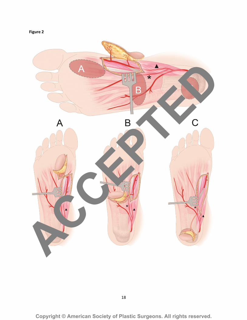
Figure 2: Range of applications of the MPAP flap with two constant perforators of the
superficial branch of the medial plantar artery: Example A shows a defect in the forefoot area.
The flap is rotated 180° and therefore, the blood supply comes retrograde from the dorsalis
pedis artery. Example B shows a defect of the midfoot. The flap needs to be rotated 90° from
the harvest site. Example C shows a defect of the heel. In order to cover the defect, the
superficial branch of the medial plantar artery is tracked proximally until a sufficient length is
needed for flap rotation. Triangle: superficial branch of the MPA, star: deep branch of the
MPA.
Figure 3: Defect measured 5 cm × 5 cm after wide excision of the Melanoma in the left heel
(above left). A 5 cm × 5 cm pedicle MPAP flap was harvested from the ipsilateral instep area
based on one perforator (above right). The MPAP flap was transferred to reconstruct the
defect and the donor site was covered with split-thickness skin graft from the contralateral
thigh (below left). Follow-up at 6 months post-operative (below right).
Figure 4: Defect measured 6 cm × 5 cm after wide excision of the Melanoma in the right
middle foot sole (above left). A 6.5 cm ×5. 5 cm pedicle MPAP flap was harvested from the
ipsilateral instep area based on the proximal perforator (black arrow) (above right). The
MPAP flap was propelled 90 degrees to covert the defect and the donor site was covered with
split-thickness skin graft from the contralateral thigh (below left). Follow-up at 8 months
post-operative (below right).
ACCEPTED
Copyright © American Society of Plastic Surgeons. All rights reserved.

15
Figure 5: Defect measured 4 cm × 4.5 cm after wide excision of the Melanoma in the right
plantar forefoot (above left). Dissection of the distal MPAP perforator (above right). A 5 cm ×
5 cm pedicle MPAP flap was harvested from the ipsilateral instep area based on the distal
perforator (below left). The MPAP flap was propelled 180 degrees to covert the defect and the
donor site was covered with split-thickness skin graft from the contralateral thigh (below
right).
ACCEPTED
Copyright © American Society of Plastic Surgeons. All rights reserved.

Table. 1 Summary of the patients receiving MPAP flap for Heel, middle part and forefoot
reconstruction
No. Age/Sex Etiology Defect
location
Size
Defect
(cm)
Type of
MPAP-flap
No. of
Perforators
MPAP
harvest
ing
time
(min.)
Follow
up Complication
1 55, W Ulcer Heel 5 x 5 proximal
pedicle based
1 P 45 12 none
2 65, M Tumor Heel 5 x 4.5 proximal
pedicle based
1 P 55 8 none
3 31, M Ulcer Heel 7 x 4 proximal
pedicle based
1 P 60 12 none
4 56, M Tumor Heel 2,8 x 3 proximal
pedicle based
1 P 45 6 none
5 83, W Tumor Heel 3.5 x 4 proximal
pedicle based
1 P 50 6 none
6 63, M Tumor Heel 3 x 3 proximal
pedicle based
1 P 40 9 none
7 63, W Tumor Heel 4 x 2 proximal
pedicle based
1 P 45 12 none
8 56, M Ulcer Heel 4 x 5 proximal
pedicle based
1 P 55 12 none
9 37, W Ulcer Heel 5 x 3 proximal
pedicle based
1 P 50 12 none
10 84, M Tumor Heel 5.5 x 9.5 proximal
pedicle based
on
2 P + D 60 9 none
11 80, W Ulcer Heel 3 x 4 proximal
pedicle based
1 P 45 9 none
12 37, M Ulcer Heel 6 x 5 proximal
pedicle based
1 P 60 4 none
13 73, M Tumor Heel 4 x 4 proximal
pedicle based
1 P 65 12 none
14 52, W Ulcer Heel 3 x 4.5 proximal
pedicle based
1 P 50 12 none
15 22, W Trauma Heel 5,5 x 5,5 proximal
pedicle based
1 P 45 6 none
16 70, W Tumor Heel 4 x 3 proximal
pedicle based
1 P 60 12 none
17 48, M Tumor Heel 5 x 4 proximal
pedicle based
1 P 55 8 none
18 12, W Trauma Heel 6 x 7 proximal
pedicle based
2 P + D 60 8 none
19 63, M Ulcer Heel 7 x 5 proximal
pedicle based
1 P 55 12 none
20 44, W Tumor Heel 5 x 5 proximal
pedicle based
1 P 55 4 none
21 19, W Tumor Medial 5 x 5 proximal
pedicle based
1 P 55 4 none
22 68, W Tumor Medial 7 x 5 proximal
pedicle based
1 P 60 12 none
23 47, W Tumor Medial 4 x 4 proximal
pedicle based
1 P 55 6 none
24 51, M Tumor Medial 6 x 5 proximal
pedicle based
1 P 55 9 none
25 53, W Tumor Forefoot 2,5 x 2,5 distal pedicle
based flap
1 D 75 9 none
26
27
28
73, W
60, M
58, M
Tumor
Tumor
Tumor
Forefoot
Forefoot
Forefoot
3 x 4,5
5 x 4
5 x 6
distal pedicle
based flap
free flap
free flap
1 D
1 P
1 P
80
60
55
12
6
8
total lost
none
none
ACCEPTED
Copyright © American Society of Plastic Surgeons. All rights reserved.

17
Figure 1
ACCEPTED
Copyright © American Society of Plastic Surgeons. All rights reserved.
18
Figure 2
ACCEPTED
Copyright © American Society of Plastic Surgeons. All rights reserved.
19
Figure 3
ACCEPTED
Copyright © American Society of Plastic Surgeons. All rights reserved.
20
Figure 4
ACCEPTED
Copyright © American Society of Plastic Surgeons. All rights reserved.
21
Figure 5
ACCEPTED
Copyright © American Society of Plastic Surgeons. All rights reserved.