rectified photocurrent of biophotodiode composed of cytochrome c/chlorophyll a heterostructure
TRANSCRIPT

A
acosib©
K
1
tacouels
p
ER
0d
Available online at www.sciencedirect.com
Colloids and Surfaces A: Physicochem. Eng. Aspects 313–314 (2008) 636–641
Rectified photocurrent of biophotodiode composed ofcytochrome c/chlorophyll a heterostructure
Jeong-Woo Choi a,b,∗, Doo-Bong Lee a, Bumhwan Lee b,Junhong Min c, Byung-Keun Oh a,b
a Department of Chemical and Biomolecular Engineering, Sogang University, #1 Shinsoo-dong,Mapo-gu, Seoul 121-742, Republic of Korea
b Interdisciplinary Program of Integrated Biotechnology, Sogang University, #1 Shinsoo-dong,Mapo-gu, Seoul 121-742, Republic of Korea
c Department of BioNano Technology, Gachon BioNano Research Institute, Kyungwon University,Bokjeong-Dong, Sujung-Gu, Seognam-Si, Kyunggi-Do 461-701, Republic of Korea
Received 19 November 2006; accepted 4 May 2007Available online 12 June 2007
bstract
Photoelectrical rectifying property of biomolecular heterostructure is investigated in molecular scale. Cytochrome c and chlorophyll a were useds an electron acceptor and a sensitizer, respectively, in the molecular layer by mimicking photosynthesis. A self-assembled monolayer of cytochromewas formed on gold surface, and then chlorophyll a was deposited onto the cytochrome c layer by Langmuir–Blodgett method. The formationf cytochrome c/chlorophyll a hetero-layers was confirmed by the surface plasmon resonance spectroscopy, and the surface was observed by the
canning tunneling microscopy. The rectifying property by the scanning-tunneling-spectroscopy based current–voltage characteristics was achievedn the cytochrome c/chlorophyll a hetero-layer. Therefore, proposed hetero-layer can be usefully applied for the development of molecular-scaleioelectronic devices. 2007 Elsevier B.V. All rights reserved.bly; R
tetfsrmecp
eywords: Cytochrome c; Chlorophyll a; Langmuir–Blodgett film; Self-assem
. Introduction
The transfer of an electron from one side of a molecule tohe other or between molecules is one of the most fundamentalnd ubiquitous processes in electronic materials and biologi-al systems [1]. The control and exploitation of this process inrganized molecular systems is a major proposition for molec-lar electronics and bioelectronics [2]. Progress in molecularlectronic devices engineering is still rather modest due to prob-ems associated with the elucidation and effective control of such
tructures and interactions at the nano level.Photoinduced electron transport processes in nature, such ashotoelectric conversion and long-range electron transfer in pho-
∗ Corresponding author at: Department of Chemical and Biomolecularngineering, Sogang University, #1 Shinsoo-dong, Mapo-gu, Seoul 121-742,epublic of Korea. Tel.: +82 2 3273 0331; fax: +82 2 3273 0331.
E-mail address: [email protected] (J.-W. Choi).
tbpae[
b
927-7757/$ – see front matter © 2007 Elsevier B.V. All rights reserved.oi:10.1016/j.colsurfa.2007.05.082
ectifying property; Bioelectronic device
osynthetic organisms, are known to be occurred not only veryfficiently but also unidirectionally guided by molecular func-ional groups [1,3]. The concepts for the development of newunctional electronic devices can be inspired from the biologicalystems such as the electron transfer chain or the photosyntheticeaction center. By mimicking the organization of the functionalolecules in a biological photosynthetic system, the molecular
lectronic devices can be realized artificially. In the initial pro-ess of photosynthesis, a biological electron transfer system,hotoelectric conversion occurs and then long-range electronransfer takes place very efficiently in one direction through theiomolecules [4]. The specific energy and electron transfer takelace on a molecular scale due to the redox potential differences well as the electron transfer property of functional molecules,
specially the electron acceptor, sensitizer and electron donor5].Molecular layer fabricated by the appropriate techniques cane used as model systems of the corresponding photosynthetic

ysicoc
riffontsrhst
tmpsffgtmectb
hcbu(plihsbc
Fc
[ce
caSlbrc
2
2
ca1(pMigabscu
2
w
J.-W. Choi et al. / Colloids and Surfaces A: Ph
eaction center in the biological system. Substantial interestn recent years has focused upon thin film fabrication or theormation of biomaterials mono- and multi-layers on solid sur-aces, by using the Langmuir–Blodgett (LB) film techniquer self-assembly (SA) technique [6,7]. Based on these tech-iques, various artificial molecular devices have been fabricatedo mimic the electron transport function of biological photo-ynthesis [8–11]. And, the photoinduced electron transfer andectifying function of hetero protein layers in molecular-scaleave been previously investigated by the scanning tunnelingpectroscopy (STS) based on current–voltage (I–V) characteris-ics [12–14].
Cytochrome c is one of the most widely studied proteins dueo its stability and solubility in water as well as availability fromany sources. The structure, physiological and physicochemical
roperties and applications of cytochrome c, have been exten-ively studied through the various research fields [15]. The keyeature of cytochrome c, capability of electron transfer, is drivenrom the redox state change and conformational change of hemeroups covalently bound via two thioether linkages formed bywo cysteine side chains and two axial ligands, histidine and
ethionine. Since cytochrome c is a constituent acting as anlectron transfer protein in the bacterial photosynthetic reactionenter, cytochrome c could be used as an electron acceptor inhe development of molecular electronic device by mimickingiological photosynthesis mechanism [9].
Chlorophylls are usually the most abundant pigments andave the most significant roles in light harvesting [16]. Espe-ially, chlorophyll a is the primary photosynthetic pigment iniological photosynthesis. The chlorophyll a molecule is madep of a porphyrin head and phytol tail. The central magnesiumMg) atom of the phophyrin head is electrophilic in behavior andlays an important role in chlorophyll. A phytol tail made up ofong hydrocarbon chain gives chlorophyll a to high insolubilityn water. Thus chlorophyll a forms non-covalent association with
ydrophobic proteins [17]. As the absorption spectrum mea-urement in chlorophyll a solution, the chlorophyll a absorb inoth blue and red region [18]. Also, the absorption spectrum ofhlorophyll a LB film is similar to those of chlorophyll a solutionig. 1. Schematic of an electron transfer diagram of the cytochrome/chlorophyll a hetero-layers.
wm(wcscg(g6rcl
2
mGow
hem. Eng. Aspects 313–314 (2008) 636–641 637
19]. Since chlorophyll a has a very high redox potential [18], itould be chosen as a sensitizer in the development of a molecularlectronic device mimicking biological photosynthesis.
In this study, the photodiode composed of the cytochromeand chlorophyll a was designed to have rectifying property
s shown in Fig. 1 and their hetero layer was fabricated usingA and LB techniques. The rectifying property of this hetero
ayer was then investigated using light illumination STS analysisased on I–V characteristics in molecular scale. It is the firsteport for the biophotodiode composed of cytochrome c andhlorophyll a which shows the rectifying property.
. Experimental
.1. Materials
The cytochrome c (extracted from horse heart, type VI),hlorophyll a (extracted from spinach), 3-murcaptopropanoiccid (3-MPA), 11-murcaptoundecanoic acid (11-MUA) and-ethyl-3-(3-dimethylaminopropyl)carbodiimide hydrochlorideEDAC) were purchased from Sigma–Aldrich Chemical Com-any (St. Louis, USA). All solutions were prepared withillipore (Milli-Q) water followed by distillation. Other chem-
cals used in this study were obtained commercially as reagentrade materials. A p-type silicon wafer was initially coated withCr film (5 nm) and subsequently with a gold film (150 nm)
y using a dc magnetron for ellipsometry measurement. A BK7lide glass (18 mm × 18 mm, Superior, Germany) sequentiallyoated with chromium (5 nm thick) and gold (43 nm thick) wassed as a substrate for surface plasmon resonance analysis.
.2. Units
The cleaning procedure of gold substrate was the sameay as in the cited reference [20]. The prepared gold substrateas immersed into a ethanol solution containing the 10 mMixed carboxyalkanethiols consisting of 3-MPA and 11-MUA
10:1, mol/mol) for 24 h [21]. Then the gold substrate rinsedith absolute ethanol and deionized (DI) water in the ambient
ondition. For chemical binding between the modified goldubstrate and cytochrome c, the carboxyl group in mixedarboxyalkanethiols was activated by submerging the modifiedold substrate into a solution of 10% EDAC in water/ethanol10:1, v/v) for 2 h at room temperature. The EDAC-linkedold surface was then soaked in the cytochrome c solution forh and rinsed with DI water and Tris–HCl buffer solution to
emove the salts remaining on the cytochrome c surface. Thehlorophyll a LB layer was deposited onto the cytochrome cayer at the previously described method [19].
.3. Instrumental analysis
Bi-molecular interaction was monitored by surface plas-
on resonance spectroscopy (MultiskopTM, Optrel GmbH,ermany) using He–Ne laser light source with a wavelengthf 632.8 nm. The p-polarized light beam by the polarizeras used as a reference and the intensity of the reflected
638 J.-W. Choi et al. / Colloids and Surfaces A: Physic
F((
bAagflwpt(ued
2o
SoaaPmugUpm
3
3
Swwcaai
isonvtΔ
soc0
babdSmsogold surface modified with mixed carboxyalkanethiols, the SPRminimum angle was shifted to 43.33 ± 0.018. Finally, The SPRminimum angle shifted from 43.33 ± 0.018 to 43.76 ± 0.044after deposition of chlorophyll a LB layer. From this result, it
ig. 2. Experimental setup for the photoelectrical characteristics measurement.a) 500 W xenon lamp; (b) optical filter (440 nm); (c) mirror; (d) Au STM tip;e) sample; (f) I–V measurement; (g) data acquisition.
eam was measured by photo multiplier tube (PMT) sensor.glass prism (BK 7, n = 1.5168) with 90◦ angle was used asKreschmann ATP coupler [22]. The plane face of the 90◦
lass prism was coupled to cover glass via index matchinguid. The resolution of the angle reading of the goniometeras 0.001◦. All samples were monitored at a constant tem-erature of 20 ◦C. The incidence angle was varied from 38◦o 50◦. In addition, the measurement of the ellipsometric anglesΔ, Ψ ), based on traditional null-ellipsometry, was performedsing the polarizer-compensator-sample-analyzer (PCSA) null-llipsometry system with a photodiode sensor as the opticaletector. The incidence angle of the laser beam was set to 70◦.
.4. Morphological analysis of hetero-layers and analysisf photoelectrical properties
The morphology of the hetero-layers was obtained byTM (XE-100, PSIA, Korea). STM morphology images werebtained by recording the tip height at constant current and at anmbient temperature of 21 ◦C. The tunneling current, bias volt-ge and scan rate were 0.5 nA, 1 V and 1.5–2 Hz, respectively.hotoelectrical response obtained using STS based I–V measure-ent was done by STM (Easy-scan, nanoSurf AG, Switzerland)
nder light illumination. The input light of 440 nm wavelengthenerated by a 500 W xenon lamp (Model-66984, Oriel Co.,SA) was delivered to the hetero-layers while measuring thehotocurrent. Experimental setup for photoelectrical propertyeasurement is shown in Fig. 2.
. Results and discussion
.1. SPR analysys of hetero-layers
For the optimal deposition concentration of cytochrome c,PR spectrums and ellipsometry angle at various concentrationsere obtained. Fig. 3a shows the change of SPR minimum angleith respect to the concentration of cytochrome c. As the con-
entration of cytochrome c was increased, the amount of SPRngle shift was also increased and finally saturated. The self-ssembly property of cytochrome c was similar to the adsorptionsotherm.
Frc
ochem. Eng. Aspects 313–314 (2008) 636–641
In order to confirm this, the ellipsometric angles (Δ,Ψ ) of var-ous concentrations were measured using the null-ellipsometryystem at the incident angle of 70◦. Fig. 3b shows the Δ valuesf each concentration. The Δ value of mixed carboxyalka-ethiol film on gold substrate was 108.67 ± 0.015◦ and the Δ
alues of ellipsometric angles were decreased as the concen-ration of cytochrome c was increased. It is known that the
depends on thickness of the thin film and that the mea-ured Δ is negatively proportional to the thin film thicknessn a solid surface [23]. From these results, the optimal con-entration of cytochrome c for self-assembly was determined to.1 mg/mL.
The results of SPR detection for the bare gold, mixed car-oxyalkanethiols film, cytochrome c SA layer, and chlorophyllLB layers are shown in Fig. 4. Adsorption of mixed car-
oxyalkanethiols, 0.1 mg/mL cytochrome c, and the subsequenteposition of chlorophyll a induced shifts of reflectivity in thePR curves to high angles. As shown in Fig. 4, the SPR mini-um angle of the bare gold was 43.00 ± 0.021 and this was the
hifted to 43.10 ± 0.030 by the adsorption of carboxyalkanethi-ls. When 0.1 mg/mL of cytochrome c was introduced on the
ig. 3. (a) Change of plasmon angle position and (b) change of ellipsomet-ic angle (delta value) with respect to the concentrations of immobilizedytochrome c.

J.-W. Choi et al. / Colloids and Surfaces A: Physicoc
Fc(
iw
3
l
aatcelf2iscsltwc
3
p
ig. 4. Change of change of SPR spectrum with respect to immobilization pro-edure. (a) Bare gold; (b) mixed carboxyalkanethiols film; (c) cytochrome c0.1 mg/mL) SA layer; (d) chlorophyll a LB layer.
s concluded that the cytochrome c/chlorophyll a hetero layersere successfully fabricated.
.2. Surface analysis of hetero-layers with STM
The surface morphologies of bare gold, the cytochrome c SAayer, and chlorophyll a LB layer were investigated using STM
scmm
Fig. 5. Surface morphologies of the (a) bare gold, (b) cytochro
hem. Eng. Aspects 313–314 (2008) 636–641 639
s shown in Fig. 5. Fig. 5a and b shows the surface of bare goldnd the self-assembled layer of cytochrome c molecules, respec-ively. In these results, the size of gold clusters and cytochromeclusters on gold surface was about 50 and 10–20 nm in diam-
ter, respectively. Thus, it is indicated that the self assembledayer of cytochrome c was successfully formed on gold sur-ace. In Fig. 5c, the diameter of the chlorophyll a were about0–30 nm. In this figure, the chlorophyll a LB layer was shownn the bright region. It is indicated that the chlorophyll a formlightly uneven layer on the cytochrome c SA layer. Some ofhlorophyll a molecules slightly aggregated on the layer andome molecules are easily desorbed from the cytochrome c SAayer because the cytochrome c and chlorophyll a cannot formhe covalent bonding [12]. However, the chlorophyll a moleculesas considered to form the molecular layer on the cytochromelayer considering the height of chlorophyll a LB layer.
.3. STS I–V characteristics
The Au STM tip was used as a top electrode and thehotoelectrical characteristics were observed in the current mea-
urement. The electron tunneling in gold substrate/(cytochrome/chlorophyll a hetero-layer)/Au STM tip configuring aetal/insulator/metal (MIM) structure was investigated in theolecular level. Fig. 6 shows the I–V characteristics at differentme c SA layer and (c) chlorophyll a LB layer by STM.
640 J.-W. Choi et al. / Colloids and Surfaces A: Physicochem. Eng. Aspects 313–314 (2008) 636–641
F oleculb
SrwahctnWptac
‘rtaTrTcg
ttreid
4
hocreSbcT
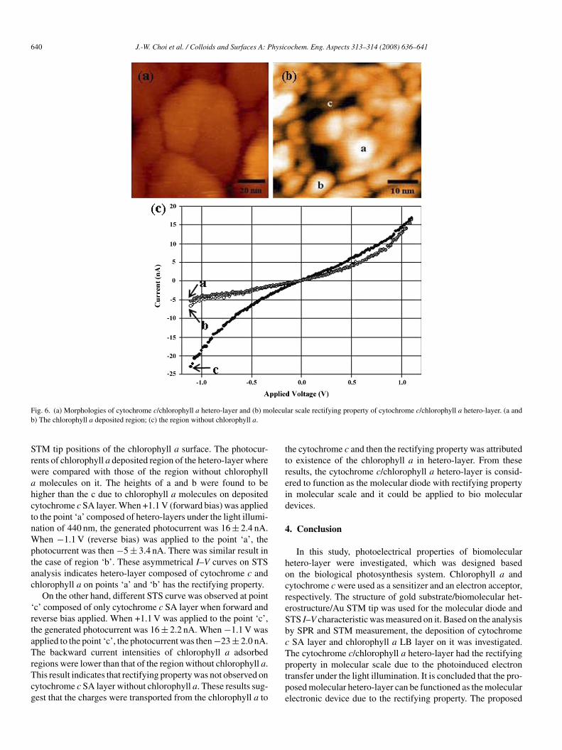
ig. 6. (a) Morphologies of cytochrome c/chlorophyll a hetero-layer and (b) m) The chlorophyll a deposited region; (c) the region without chlorophyll a.
TM tip positions of the chlorophyll a surface. The photocur-ents of chlorophyll a deposited region of the hetero-layer whereere compared with those of the region without chlorophyllmolecules on it. The heights of a and b were found to be
igher than the c due to chlorophyll a molecules on depositedytochrome c SA layer. When +1.1 V (forward bias) was appliedo the point ‘a’ composed of hetero-layers under the light illumi-ation of 440 nm, the generated photocurrent was 16 ± 2.4 nA.hen −1.1 V (reverse bias) was applied to the point ‘a’, the
hotocurrent was then −5 ± 3.4 nA. There was similar result inhe case of region ‘b’. These asymmetrical I–V curves on STSnalysis indicates hetero-layer composed of cytochrome c andhlorophyll a on points ‘a’ and ‘b’ has the rectifying property.
On the other hand, different STS curve was observed at pointc’ composed of only cytochrome c SA layer when forward andeverse bias applied. When +1.1 V was applied to the point ‘c’,he generated photocurrent was 16 ± 2.2 nA. When −1.1 V waspplied to the point ‘c’, the photocurrent was then −23 ± 2.0 nA.he backward current intensities of chlorophyll a adsorbed
egions were lower than that of the region without chlorophyll a.his result indicates that rectifying property was not observed onytochrome c SA layer without chlorophyll a. These results sug-est that the charges were transported from the chlorophyll a to
ptpe
ar scale rectifying property of cytochrome c/chlorophyll a hetero-layer. (a and
he cytochrome c and then the rectifying property was attributedo existence of the chlorophyll a in hetero-layer. From theseesults, the cytochrome c/chlorophyll a hetero-layer is consid-red to function as the molecular diode with rectifying propertyn molecular scale and it could be applied to bio molecularevices.
. Conclusion
In this study, photoelectrical properties of biomolecularetero-layer were investigated, which was designed basedn the biological photosynthesis system. Chlorophyll a andytochrome c were used as a sensitizer and an electron acceptor,espectively. The structure of gold substrate/biomolecular het-rostructure/Au STM tip was used for the molecular diode andTS I–V characteristic was measured on it. Based on the analysisy SPR and STM measurement, the deposition of cytochromeSA layer and chlorophyll a LB layer on it was investigated.he cytochrome c/chlorophyll a hetero-layer had the rectifying
roperty in molecular scale due to the photoinduced electronransfer under the light illumination. It is concluded that the pro-osed molecular hetero-layer can be functioned as the molecularlectronic device due to the rectifying property. The proposed
ysicoc
sbm
A
iMaPSEg
R
[
[
[[
[[
[
[
[
[
[
J.-W. Choi et al. / Colloids and Surfaces A: Ph
ystem which mimics the biological photosynthetic system cane usefully applied as a model system for the development ofolecular scale electronics devices.
cknowledgements
This work was supported by the Korea Science and Engineer-ng Foundation (KOSEF) through the Advanced Environment
onitoring Research Center at Gwangju Institute of Sciencend Technology and by the Nano/Bio Science & Technologyrogram (M10536090001-05N3609-00110) of the Ministry ofcience and Technology (MOST) and by the Korea Science andngineering Foundation (KOSEF) grant funded by the Koreaovernment (MOST) (2006-05374).
eferences
[1] G.J. Kavarnos, Fundamentals of Photoinduced Electron Transfer, VCH,New York, 1993.
[2] H. Kuhn, F.T. Hong, Molecular Electronics—Biosensors and Biocomput-ers, Plenum, New York, 1993.
[3] J. Deisenhofer, O. Epp, K. Miki, R. Huber, H. Michel, Nature 318 (1985)618.
[4] D. Gust, T.A. Moore, Science 244 (1985) 35.[5] M. Fujihira, K. Nichiyama, H. Yamada, Thin Solid Films 132 (1985) 77.[6] Y. Lvov, H. Mohwald, Protein Architecture: Interfacing Molecular Assem-
blies and Immobilization Biotechnology—Langmuir Blodgett Multilayersof Protein, Marcel Dekker Inc., New York, 1999.
[
[[
hem. Eng. Aspects 313–314 (2008) 636–641 641
[7] W. Lee, B.S. Chun, B.-K. Oh, W.H. Lee, J.-W. Choi, Biotechnol. Biopro-cess. Eng. 9 (2004) 241.
[8] M. Fujihira, Photoinduced electron tansfer in monolayer assemblies andits application to artificial photosynthesis and molecular devices, in: A.Ulman (Ed.), Thin Films, Organic Thin Films and Surfaces: Direction forthe Nineties, vol. 20, Academic Press, New York, 1995.
[9] S. Ueyama, FED J. 8 (1997) 33.10] J.-W. Choi, Y.S. Nam, W.H. Lee, D. Kim, M. Fujihira, Appl. Phys. Lett.
79 (2001) 1570.11] J.-W. Choi, Y.S. Nam, M. Fujihira, Biotechnol. Bioprocess. Eng. 9 (2004)
76.12] Y.S. Nam, J.-W. Choi, W.H. Lee, Appl. Phys. Lett. 85 (2004) 6275.13] B. Lee, S. Takeda, K. Nakajima, J. Noh, J. Choi, M. Hara, T. Nagamune,
Biosens. Bioelectron. 19 (2004) 1169.14] J.-W. Choi, M. Fujihira, Appl. Phys. Lett. 84 (2004) 2187.15] S.J. Lippard, J.M. Berg, Principles of Bioinorganic Chemistry, University
Science Books, Mill Vally, 1994.16] H.R. Horton, L.A. Moran, R.S. Ochs, J.D. Rawn, K.G. Scrimgeour, Prin-
ciples of Biochemistry, Prentice-Hall, London, 1996.17] W.G. Hopkins, Introduction to Plant Physiology, second ed., John Wiley
& Sons, New York, 1999.18] A. Agostiano, P. Cosma, M. Trotta, L. Monsu-Scolaro, N. Micali, J. Phys.
Chem. B 106 (2002) 12820.19] Y.S. Nam, J.M. Kim, J.-W. Choi, W.H. Lee, Mater. Sci. Eng. C 24 (2004)
35.20] B.-K. Oh, Y.K. Kim, W. Lee, Y.M. Bae, W.H. Lee, J.-W. Choi, Biosens.
Bioelectron. 18 (2003) 605.21] N. Patel, M.C. Davies, M. Hartshorne, R.J. Heaton, C.J. Roberts, S.J.B.
Tendler, P.M. Williams, Langmuir 13 (1997) 6485.22] E. Kretschmann, Zeitschrift fur Physik 241 (1971) 313.23] A. Tronnin, T. Dubrovsky, C. Nicolini, Langmuir 11 (1995) 385.