recurrent uterine cervical carcinoma: spectrum of … · recurrent uterine cervical carcinoma:...
TRANSCRIPT
198 Korean J Radiol 1(4), December 2000
Recurrent Uterine Cervical Carcinoma:Spectrum of Imaging Findings
Uterine cervical carcinoma is one of the most common malignant tumors occur-ring in females. After primary treatment, patients are usually followed up with CTor MRI and the findings of these modalities may be the first sign of recurrent dis-ease. Because earlier additional treatment by chemotherapy or radiation therapymay improve the prognosis, the early detection of recurrent cervical carcinoma isclinically important. In this article, we review the CT and MR imaging findings ofrecurrent uterine cervical carcinoma, and assign them to one of four groups: a)recurrence at the primary site, involving the intrapelvic organs, b) extension to thepelvic side-wall, c) metastases to pelvic and extrapelvic lymph nodes, or d)metastases to distant organs. A further contribution of CT and MR imaging is thedetection of hydronephrosis due to ureteral obstruction. The cases in each groupare illustrated and discussed, and since an awareness of the spectrum of imagingfindings of recurrent cervical carcinoma is likely to lead to its early detection, radi-ologists should be familiar with the information presented.
espite the decreased incidence of cervical carcinoma associated withwidespread screening using Papanicolaou’s smear test, uterine cervicalcarcinoma is still one of the most common gynecologic malignancies.
Even though patients now survive longer due to radiation therapy and more effectivechemotherapy, it is also one of the most frequent causes of death in women. In spite ofthese advances in the methods of treatment, approximately 30% of women with inva-sive cervical carcinoma die as a result of recurrent or persistent disease (1).
Recurrence is defined as local tumor regrowth or the development of distant metas-tasis discovered six months or more after complete regression of the treated lesion,and uterine cervical carcinoma often recurs within two years of treatment. The inci-dence of recurrence depends primarily on tumor bulk and the stage of the disease atpresentation (1). About one-half of recurrences appear within the pelvis, but distantmetastases may develop in the lung and liver, and in bone and lymph nodes (2).
Conventional radiographic evaluations of these patients include chest radiography,excretory urography, barium enema, and occasionally, lymphangiography. However,improvements in cross-sectional imaging modalities such as CT or MR imaging offerdistinct advantages over conventional radiographic techniques, and nowadays, the fol-low-up of treated cervical carcinoma usually involves the use of these modalities.Because additional chemotherapy or radiation therapy can improve the prognosis, theearly detection of recurrent cervical carcinoma is important. In the interests of earlydetection, radiologists should therefore be familiar with the spectrum of imaging find-ings presented by this condition.
We retrospectively reviewed the CT and MR imaging findings of patients with recur-
Joon-Il Choi, MDSeung Hyup Kim, MDChang Kyu Seong, MDJung Suk Sim, MDHak Jong Lee, MDKyung-Hyun Do, MD
Index terms:Uterine neoplasms, diagnosisUterine neoplasms, CTUterine neoplasms, MR
Korean J Radiol 2000;1:198-207Received January 17, 2000; accepted after revision September 13, 2000.
All authors: Department of Radiology,Seoul National University College ofMedicine; The Institute of RadiationMedicine, SNUMRC; and The ClinicalResearch Institute, Seoul NationalUniversity Hospital, Seoul, Korea.
Address reprint requests to:Seung Hyup Kim, MD, Department ofRadiology, Seoul National UniversityHospital, 28 Yongon-dong, Chongno-gu,Seoul 110-744, Korea.Telephone: (822)760-3259Fax: (822)743-6385e-mail: [email protected]
D

rent cervical carcinoma, classifying them as one of fourtypes according to the site of recurrence: a) recurrence atthe primary site, involving the intrapelvic organs, b) recur-rent mass at the pelvic side-wall, c) metastases to pelvicand extrapelvic lymph nodes and d) metastases to a distantorgan or organs. In this article, we illustrate and discuss thevarious CT and MR imaging findings for each group.
RECURRENCE AT THE PRIMARY SITE
Recurrence at the primary site may involve the cervix,uterus, vagina, parametria or ovary, and is the most com-mon manifestation of recurrent cervical carcinoma (3-5).The incidence of recurrence at the primary site varies withthe stage at presentation, histologic type, adequacy of ther-apy used and host response. Kim et al. (5) reported a high-er incidence of recurrence among patients treated by radia-tion therapy than by surgery. Invasion of the urinary blad-der and rectum, with or without fistula formation, is a se-vere complication which can be demonstrated by CT orMR imaging.
RECURRENCE AS A CENTRAL PELVIC MASSThe evaluation of CT and MR imaging findings of recur-
rence at the primary site requires an understanding of thenormal anatomy of the pelvis after hysterectomy. Axialimages show that the vaginal vault is located posterior tothe bladder and anterior to the rectum at or near the levelof the acetabula. It is roughly rectangular or oblong inshape, and should be symmetric and sharply marginatedfrom surrounding fat. The bladder and loops of the smallintestine occupy the space just superior to this level (1).
The most common CT and MR imaging feature of recur-rent tumor of the primary site is an irregular mass averag-ing 4 7 cm in diameter between the bladder and rectum(3). Central necrosis and irregular enhancement are com-mon findings (Figs. 1, 2), but the lesion may be seen on CTor MR imaging as an area of homogenous soft tissue densi-ty. The vaginal vault may be the first site of recurrence.
INVASION OF THE BLADDER AND RECTUMIf CT reveals loss of the fat plane between the tumor and
the bladder or rectum, invasion of these entities (Figs. 3and 4) has occurred. MR imaging provides superior soft-tis-sue contrast, and its use may therefore more accurately de-tect the occurrence of such invasion. A diagnosis of vesicalor rectal invasion by means of T2-weighted MR imagingcan involve loss of the normal low signal intensity of thebladder or rectal wall continuous to the mass as well as theloss of normal fat planes (6). Kim and Han (6) reportedthat in depicting vesical invasion of the urinary bladder inuterine cervical carcinoma, the sensitivity, specificity, andaccuracy of MR imaging was 83%, 100%, and 99%, re-spectively. The MR imaging findings of vesical invasion in-clude nodularity and irregularity of the bladder wall, mass-es protruding into the bladder lumen, high signal intensityof the anterior aspect of the posterior bladder wall, and ab-normal soft-tissue strands in the uterovesical space, imply-ing desmoplastic reaction (6). A fistula sometimes forms,and urine or fecal material may be present in remainingvagina.
HYDROMETRA DUE TO CERVICAL MASSIn patients who have not undergone hysterectomy, local
Spectrum of Imaging Finding of Recurrent Uterine Cervical Carcinoma
Korean J Radiol 1(4), December 2000 199
Fig. 1. Recurrence at the vagina in a 41-year-old woman who un-derwent hysterectomy and radiation therapy. CT scan reveals anirregular mass (arrows) between the bladder and rectum, and thepresence of a centrally located low-attenuated area indicatesnecrosis and peripheral enhancement. On the left, a double-pig-tail ureteral stent (arrowheads) is visible.
Fig. 2. Recurrence at the uterus following radiation therapy with-out hysterectomy in a 61-year-old-woman. CT scan shows a cen-tral pelvic mass (arrows) replacing the uterus. The central low-at-tenuated area represents necrosis.
tumor recurrence or radiation fibrosis may cause obstruc-tion of the cervical os, leading to hydrometra or pyometra(Fig. 5) (1). The CT appearance is that of a symmetricallyenlarged uterus with a low-attenuation, non-enhancingcentral mass. This central low attenuation and lack of con-trast enhancement help differentiate an obstructed uterusfrom other causes of uterine enlargement (2).
RECURRENT MASS AT PELVIC SIDE-WALL
Recurrent mass at the pelvic side-wall is another commonfinding, and since it may or may not be accompanied by arecurrent mass at the primary site (3, 5) (Fig. 6), is separate-ly discussed. It is characterized either by direct continuationof the mass to the pelvic side-wall by means of a linearstrand of soft tissue, and involving loss of the fat planes, orby confluence of the solid mass and the muscle of the pelvicwall (1) (Fig. 7). Pelvic bone can be destroyed by tumor in-vasion (Fig. 8). A recurrent mass at the pelvic side-wallmost commonly occurs in the parametria adjacent to theuterine corpus and additional lateral extension might occurat the level of the cervix and vagina (1). Kim et al (5) re-ported that the there is no significant difference in the inci-dence of pelvic side-wall extension between patients whoundergo surgery and those treated by radiation therapy.
Choi et al.
200 Korean J Radiol 1(4), December 2000
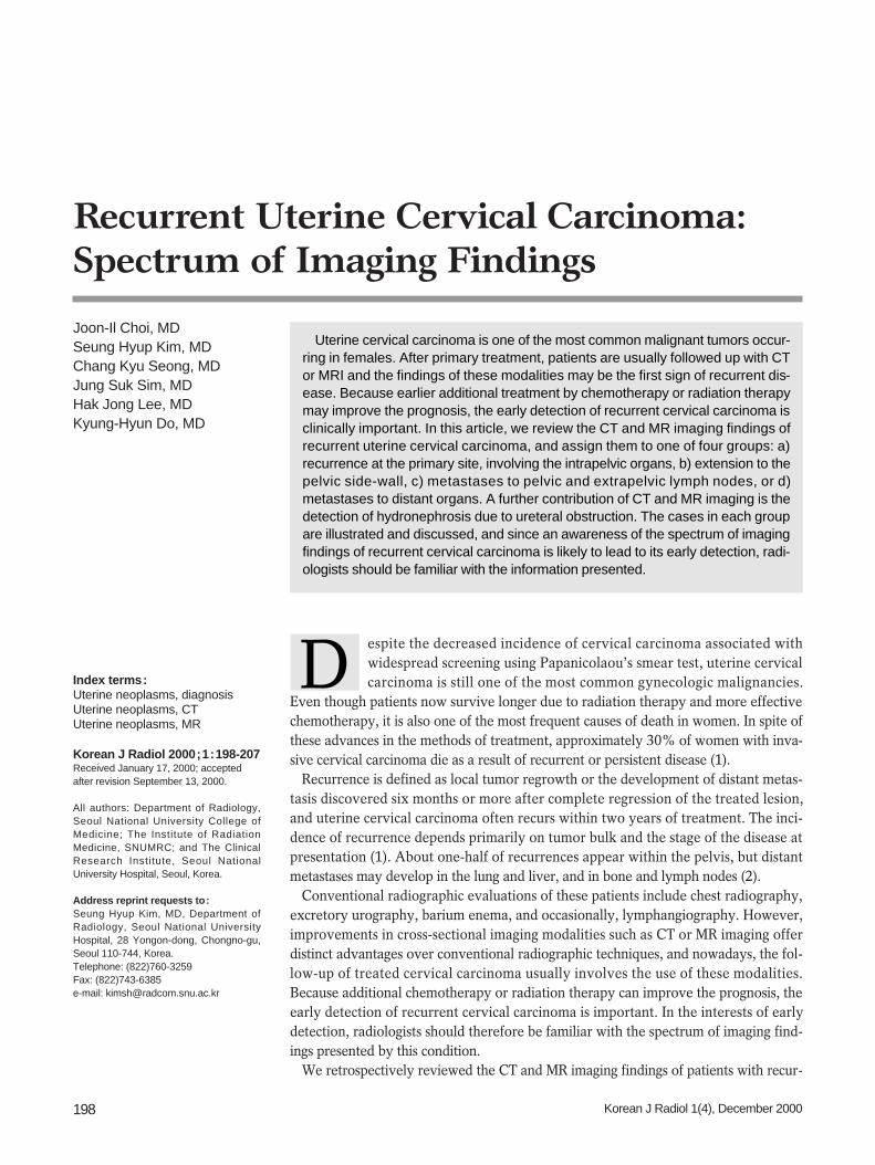
Fig. 3. Recurrence at the primary site with posterior extension to the rectum in an 80-year-old woman who underwent radical hysterecto-my.A. T2-weighted axial MR image shows an irregular shaped, low signal mass between the bladder and rectum. The mass is continuouswith the rectal wall (arrows), and protrudes into the rectal lumen.B. Barium enema shows luminal irregularity and mucosal destruction of the lateral wall of the rectum, indicating the invasion of rectal mu-cosa (arrowheads).
A B
Fig. 4. Recurrence at the primary site, with invasion of the urinarybladder, in a 41-year-old woman. T2-weighted sagittal MR imagereveals a large, necrotic mass in the vagina and uterine cervix. Afistula (arrow) between the necrotic cavity of the mass and theurinary bladder is clearly visible.
LYMPH NODE METASTASIS
The rich cervical lymphatic network drains into the para-metrial, external iliac (including obturator), internal iliac,and lateral sacral lymph nodes, and from there, drainingproceeds into the common iliac and para-aortic nodes. Thelymphatic spread of uterine cervical carcinoma usually fol-lows the pelvic pathway, the common route for the lym-phatic spread of tumors of the pelvic organs, and onewhich can be divided into the anterior, lateral, hypogastric,and presacral sections. In uterine cervical carcinoma, the
lateral route is the most common (7).In uterine cervical carcinoma, lymphatic involvement has
traditionally been separated into primary and secondarynodal groups. The first of these consists of the paracervical,parametrial, internal and external iliac, and obturatornodes, while those comprised by the second include thesacral, common iliac, inguinal and para-aortic (4). Whenthe secondary nodal group is involved, the prognosis wors-ens. In this article, the system used to classify lym-phadenopathy - namely, that this involves either pelvic orextrapelvic lymphnode metastasis - is one suited to the useof cross-sectional imaging techniques.
Spectrum of Imaging Finding of Recurrent Uterine Cervical Carcinoma
Korean J Radiol 1(4), December 2000 201
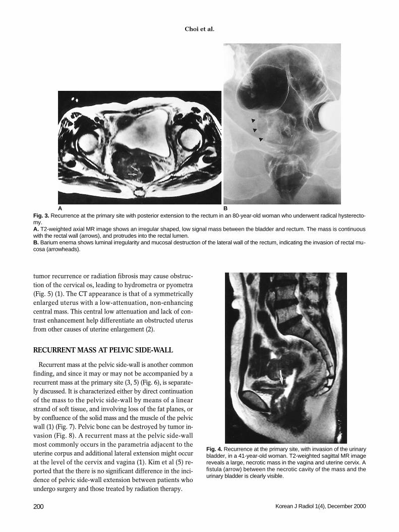
Fig. 5. Hydrometra due to obstruction of the cervical os in a 57-year-old woman who underwent radiation therapy without hysterectomy.A. T2-weighted axial MR image shows a low signal mass (arrows) located in the cervical portion of the uterus and anterior to the rectum. B. In the upper level than in A (also a T2-weighted image), a fluid-filled, dilated uterus is visible. The low signal intensity of fluid in theuterine cavity implies the presence of highly proteineous material.
A B
Fig. 7. Pelvic side-wall extension with destruction of the pelvicbone in a 60-year-old woman. CT image shows a necrotic mass(arrows) with bulging contour in the posterior pelvic wall, andsacral destruction has occurred.
Fig. 6. Pelvic side-wall extension with invasion of gluteal musclein a 28-year-old woman. CT scan demonstrates the presence ofa necrotic mass (arrows) in the posterolateral aspect of the uri-nary bladder. The mass extends to the left posterior pelvic side-wall and invades the gluteus muscle.
PELVIC LYMPH NODE METASTASISThe incidence of pelvic lymph node metastases in uterine
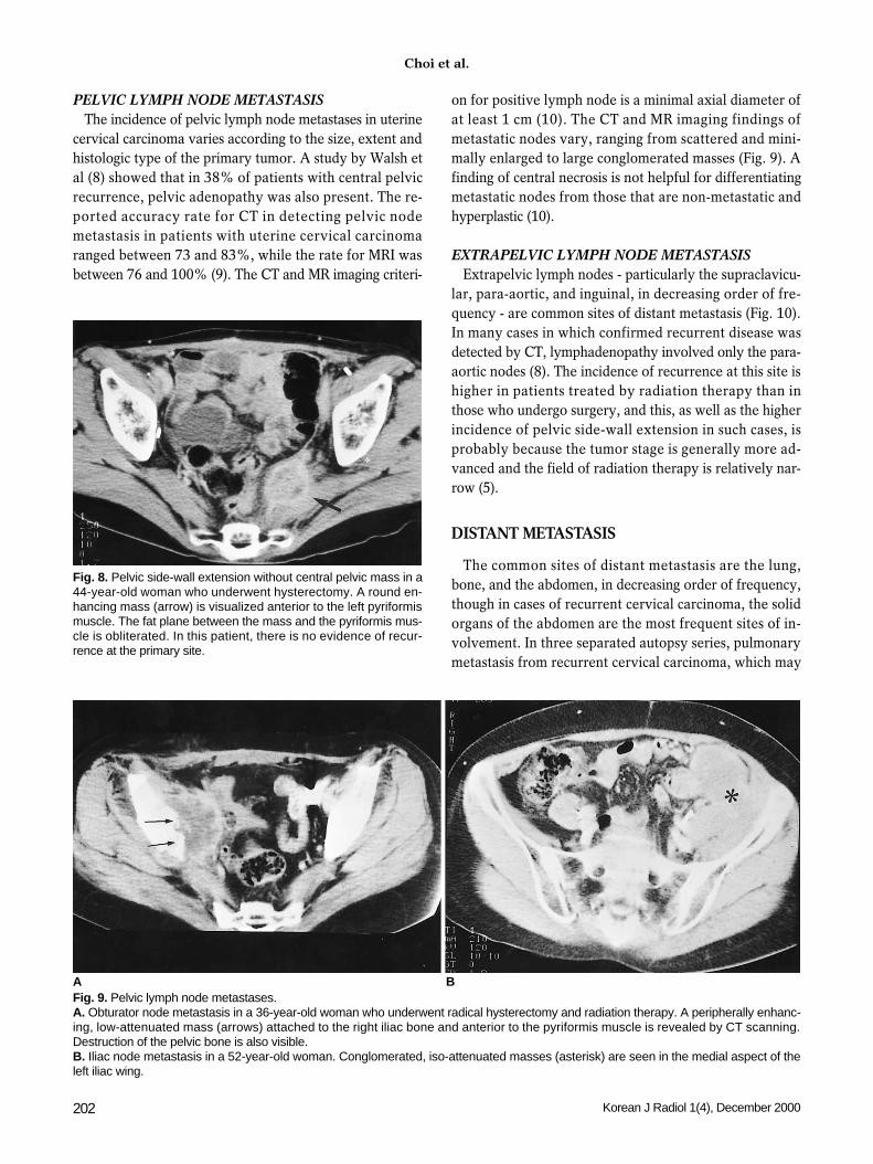
cervical carcinoma varies according to the size, extent andhistologic type of the primary tumor. A study by Walsh etal (8) showed that in 38% of patients with central pelvicrecurrence, pelvic adenopathy was also present. The re-ported accuracy rate for CT in detecting pelvic nodemetastasis in patients with uterine cervical carcinomaranged between 73 and 83%, while the rate for MRI wasbetween 76 and 100% (9). The CT and MR imaging criteri-
on for positive lymph node is a minimal axial diameter ofat least 1 cm (10). The CT and MR imaging findings ofmetastatic nodes vary, ranging from scattered and mini-mally enlarged to large conglomerated masses (Fig. 9). Afinding of central necrosis is not helpful for differentiatingmetastatic nodes from those that are non-metastatic andhyperplastic (10).
EXTRAPELVIC LYMPH NODE METASTASISExtrapelvic lymph nodes - particularly the supraclavicu-
lar, para-aortic, and inguinal, in decreasing order of fre-quency - are common sites of distant metastasis (Fig. 10).In many cases in which confirmed recurrent disease wasdetected by CT, lymphadenopathy involved only the para-aortic nodes (8). The incidence of recurrence at this site ishigher in patients treated by radiation therapy than inthose who undergo surgery, and this, as well as the higherincidence of pelvic side-wall extension in such cases, isprobably because the tumor stage is generally more ad-vanced and the field of radiation therapy is relatively nar-row (5).
DISTANT METASTASIS
The common sites of distant metastasis are the lung,bone, and the abdomen, in decreasing order of frequency,though in cases of recurrent cervical carcinoma, the solidorgans of the abdomen are the most frequent sites of in-volvement. In three separated autopsy series, pulmonarymetastasis from recurrent cervical carcinoma, which may
Choi et al.
202 Korean J Radiol 1(4), December 2000
Fig. 9. Pelvic lymph node metastases.A. Obturator node metastasis in a 36-year-old woman who underwent radical hysterectomy and radiation therapy. A peripherally enhanc-ing, low-attenuated mass (arrows) attached to the right iliac bone and anterior to the pyriformis muscle is revealed by CT scanning.Destruction of the pelvic bone is also visible.B. Iliac node metastasis in a 52-year-old woman. Conglomerated, iso-attenuated masses (asterisk) are seen in the medial aspect of theleft iliac wing.
A B
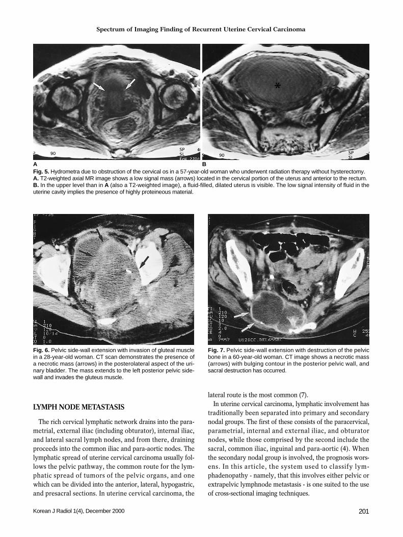
Fig. 8. Pelvic side-wall extension without central pelvic mass in a44-year-old woman who underwent hysterectomy. A round en-hancing mass (arrow) is visualized anterior to the left pyriformismuscle. The fat plane between the mass and the pyriformis mus-cle is obliterated. In this patient, there is no evidence of recur-rence at the primary site.

Spectrum of Imaging Finding of Recurrent Uterine Cervical Carcinoma
Korean J Radiol 1(4), December 2000 203
A B
Fig. 10. Extrapelvic node metastases.A. Para-aortic node metastasis in a 45-year-old woman. CT scanshows an enlarged para-aortic lymph node (asterisk), which in thispatient is the only site involved by recurrent disease.B. Retroperitoneal lymph node metastasis in a 40-year-old woman.Extensive lymphadenopathy in the para-aortic region and aroundthe celiac trunk is detected by CT. This lesion extends into thesplenic hilum (arrows).C. Inguinal node metastasis. CT scan indicates the presence of anenlarged right inguinal lymph node (arrow). Also note the presenceof swelling and soft tissue edema in the right lower extremity (ar-rowheads) due to lymphatic and venous obstruction by metastaticlymphadenopathy.
C
Fig. 11. Pulmonary metastases.A. Hematolymphangitic metastasis in a 47-year-old woman. Lung window setting of the lower part of the lung demonstrates multiple nod-ules and interlobular septal thickening.B. Pulmonary metastasis manifested as mediastinal lymphadenopathy in a 45-year-old woman who two years earlier underwent hys-terectomy. CT scan shows large mediastinal (para-aortic) lymph nodes (asterisk). Percutaneous needle biopsy confirmed that adenocar-cinoma had metastasized from the uterine cervix.
A B

exist for a significant period of time before becomingsymptomatic and may be either solitary or multiple, oc-curred in 33 35% of cases (4). Typical patterns of lung in-volvement are pulmonary nodules (71%), mediastinal andhilar lymphadenopathy (32%), and pleural metastasis(27%). Rare findings include endobronchial obstruction(5%), and lymphangitic carcinomatosis (3%) (Fig. 11).
The spine, especially the lumbar spine, is the most com-mon site of osseous metastasis. Involvement of the verte-bral bodies, the mechanism of which is direct extension ofthe tumor subsequent to para-aortic lymphadenopathy, ismost frequent, followed by that of the pelvis, ribs, and ex-tremities (2).
Choi et al.
204 Korean J Radiol 1(4), December 2000
Fig. 12. Metastasis to the peritoneal cavity in a 48-year-oldwoman. Abdominal CT scan indicates the presence of a mass(arrow) with extensive central necrosis and lobulated contour inthe right upper quadrant.
Fig.15. Splenic metastasis in a 53-year-old woman who under-went hysterectomy. Enhanced CT scan reveals the presence inthe spleen of an ill-defined low-attenuated mass (arrow).
Fig. 13. Multiple hepatic metastases in a 44-year-old woman.Contrast-enhanced T1-weighted axial MR image shows multiple,target-shaped lesions with peripheral enhancement in the liver.
Fig. 14. Adrenal metastasis in a 61-year-old female. CT scan in-dicates the presence in the right adrenal gland area of an oval-shaped low-attenuated, 4 cm-sized mass (arrow). Needle biopsyconfirmed the occurrence of metastatic carcinoma.
Fig.16. Subcutaneous metastasis to a surgical scar in a 55-year-old woman who underwent radical hysterectomy and radiationtherapy. CT scan shows a round heterogenous mass (arrow) insubcutaneous abdominal tissue in the region of the scar.

Abdominal metastasis occurred in the peritoneal cavity(Fig. 12) and solid organs such as the liver (Fig. 13) andadrenal gland (Fig. 14). Peritoneal metastasis, includingperitoneal carcinomatosis, is more common than metastasisto the liver. The CT and MR imaging findings of metastasesto the peritoneal cavity are ascites, implants scalloping theliver contour, peritoneal thickening with nodularity, andsoft tissue mass(4). The CT and MR imaging findings of he-patic and adrenal involvement in recurrent cervical carci-noma are non-specific and indistinguishable from involve-ment by other primary malignancies. The involvement ofanother solid abdominal solid organ such as the spleen(Fig. 15), kidney, pancreas or gastrointestinal tract is rare,and in almost all cases, widespread metastasis involvingother organs also occurs.
In the detection of abdominal recurrence, the most im-portant problem related to the use of CT and MR imagingis the fact that because the known pattern of spread of thedisease is through the iliac and aortic lymph nodes, most
such scanning routinely covers the region from the iliaccrests to the symphisis pubis. So that other recurrent le-sions are not missed, scanning of a cervical neoplasmshould, however, include the upper abdomen.
The common primary sites in patients with skin or subcu-taneous metastasis are the breast, colon, lung, and ovary;skin metastasis from uterine cervical carcinoma is extreme-ly rare. The most common sites of skin lesions are the ab-dominal wall and vulva, followed by the anterior chestwall, and in most patients there are multiple lesions.Recurrence at the site of a surgical scar is rare, but is a pos-sible complication of hysterectomy (Fig. 16). It is classifiedas skin and subcutaneous involvement, and occurs mainlyin patients who have not undergone combined radiationtherapy or whose radiotherapeutic field did not include thesurgical scar.
Spectrum of Imaging Finding of Recurrent Uterine Cervical Carcinoma
Korean J Radiol 1(4), December 2000 205
A B
Fig.17. Hydronephrosis due to ureteral obstruction caused by re-currence at the primary site in a 41-year-old woman who under-went radical hysterectomy and radiation therapy. A. CT scan demonstrates hydronephrosis of the left kidney andthat the patient had undergone percutaneous nephrostomy of theright kidney. A dilated left ureter (arrow) is depicted.B. In the lower level than in A, dilated bilateral ureters (arrows) canbe traced.C. Anterior to the rectum, an irregular shaped mass (arrowheads)encased the dilated left ureter. Pelvic side-wall extension is also vi-sualized.
C

URETERAL OBSTRUCTION
Another contribution of CT and MR imaging in recurrentuterine cervical carcinoma is the detection of ureteral ob-struction, and determination of the etiology and level (3).Such obstruction is due to the encasement or extrinsic com-pression of a recurrent pelvic mass (Fig. 17) or metastaticlymph nodes (Figs. 18, 19), or tumor infiltration of the blad-der wall, which results in obstruction at the ureteral orifice.The site of ureteral obstruction is usually revealed by scan-
ning of the retroperitoneal or pelvic area (3).
CONCLUSION
The recurrence of uterine cervical carcinoma can involvevarious organs, with various patterns of imaging findings.CT and MR imaging are the primary imaging modalitiesused during the follow-up of treated cervical carcinoma.Since the early detection of recurrent cancer makes for abetter prognosis, radiologists should be familiar with the
Choi et al.
206 Korean J Radiol 1(4), December 2000
Fig. 18. Hydronephrosis due to ureteral obstruction by retroperitoneal lymph node metastasis in a 61-year-old woman. A. Enhanced CT scan shows bilateral hydronephrosis and decreased perfusion of the left kidney.B. In the lower level than in A, a dilated right ureter (arrow) is visualized, and the presence of retroperitoneal lymphadenopathy is also vi-su alized (arrowheads).
A B
Fig. 19. Hydronephrosis due to ureteral obstruction by periureteral lymph node in a 49-year-old woman.A. CT scan shows hydronephrosis of the left kidney, with decreased renal perfusion.B. In the lower level than in A, a periureteral lymph node (arrow) obstructing the left ureter is depicted. The ureter can not be traced in thislevel.
A B
Spectrum of Imaging Finding of Recurrent Uterine Cervical Carcinoma
Korean J Radiol 1(4), December 2000 207
spectrum of CT and MR imaging findings of recurrent uter-ine cervical carcinoma. Recurrence which is extrapelvic orinvolves the pelvic side-wall is more common in patientswho undergo radiation therapy only, a fact probably dueto the more advanced stage of the disease in such patientsand the limited field within which radiation therapy is pos-sible. More careful examination is, therefore, necessary forthe patients treated by radiation therapy only. In the eval-uation of pelvic malignancies, including uterine cervicalcarcinoma, careful examination of the upper abdomen isrequired in addition to routine pelvic scanning.
References1. Walsh JW. Computed tomography of the pelvis. Churchill New
York: Livingstone 1985:195-1982. Carlson V, Delclos L, Fletcher GH. Distant metastases in squa-
mous-cell carcinoma of the uterine cervix. Radiology 1967;88:961-966
3. Walsh JW, Amendola MA, Hall DJ, Tisnado J, Goplerud DR.Recurrent carcinoma of the pelvis: CT diagnosis. AJR 1981;136:117-122
4. Fulcher AS, O’Sullivan SG, Segreti EM, Kavanagh BD.Recurrent cervical carcinoma: Typical and atypical manifesta-tions. RadioGraphics 1999;19:s103-s116
5. Kim JE, Park HA, Kim KH, Lim D, Chin SY. Patterns of recur-rent cervical carcinoma on CT. J Korean Radiol Soc 1988;24:1130-1134
6. Kim SH, Han MC. Invasion of the urinary bladder by uterinecervical carcinoma: evaluation with MR imaging. AJR 1997;168:393-397
7. Park JM, Charnsangavej C, Yoshimitsu K, Herron DH, RobinsonTJ, Wallace S. Pathways of nodal metastasis from pelvic tumors:CT demonstration. RadioGraphics 1994;14:1309-1321
8. Walsh JW, Amendola MA, Konerding KF, Tisnado J, Hazra TA.Computed tomographic detection of pelvc and inguinal lymph-node metastases from primary and recurrent malignant pelvicdisease. Radiology 1980;137:157-166
9. Kim SH, Choi BI, Lee HP, Kang SB, Choi YM, Han MC, KimCW. Uterine cervical carcinoma: Comparison of CT and MRfindings. Radiology 1990;175:45-51
10. Kim SH, Kim SC, Choi BI, Han MC. Uterine cervical carcinoma:Evaluation of pelvic lymph node metastasis with MR imaging.Radiology 1994;190:807-811