redox regulation of apurinic/apyrimidinic endonuclease 1 activity in long-evans cinnamon rats during...
TRANSCRIPT
Redox regulation of apurinic/apyrimidinic endonuclease 1 activityin Long-Evans Cinnamon rats during spontaneous hepatitis
Soumendra Krishna Karmahapatra •
Tapas Saha • Sanjay Adhikari • Jordan Woodrick •
Rabindra Roy
Received: 3 June 2013 / Accepted: 15 November 2013 / Published online: 15 December 2013
� Springer Science+Business Media New York 2013
Abstract The Long-Evans Cinnamon (LEC) rat is an
animal model for Wilson’s disease. This animal is geneti-
cally predisposed to copper accumulation in the liver,
increased oxidative stress, accumulation of DNA damage,
and the spontaneous development of hepatocellular carci-
noma. Thus, this animal model is useful for studying the
relationship of endogenous DNA damage to spontaneous
carcinogenesis. In this study, we have investigated the
apurinic/apyrimidinic endonuclease 1 (APE1)-mediated
excision repair of endogenous DNA damage, apurinic/
apyrimidinic (AP)-sites, which is highly mutagenic and
implicated in human cancer. We found that the activity was
reduced in the liver extracts from the acute hepatitis period
of LEC rats as compared with extracts from the age-mat-
ched Long-Evans Agouti rats. The acute hepatitis period
had also a heightened oxidative stress condition as assessed
by an increase in oxidized glutathione level and loss of
enzyme activity of glyceraldehyde 3-phosphate dehydro-
genase, a key redox-sensitive protein in cells. Interestingly,
the activity reduction was not due to changes in protein
expression but apparently by reversible protein oxidation
as the addition of reducing agents to extracts of the liver
from acute hepatitis period reactivated APE1 activity and
thus, confirmed the oxidation-mediated loss of APE1
activity under increased oxidative stress. These findings
show for the first time in an animal model that the repair
mechanism of AP-sites is impaired by increased oxidative
stress in acute hepatitis via redox regulation which con-
tributed to the increased accumulation of mutagenic AP-
sites in liver DNA.
Keywords APE1 � DNA damage � DNA repair �HCC
Abbreviation
LEC Long-Evans Cinnamon
LEA Long-Evans Agouti
APE1 AP-endonuclease 1
ROS Reactive oxygen species
WD Wilson’s disease
GAPDH Glyceraldehyde 3-phosphate dehydrogenase
Introduction
Considerable evidence has linked chronic inflammation
and oxidative damage with increased risk of cancer [1, 2].
Reactive oxygen species (ROS) are generated in different
subcellular locations as a function of oxygen-mediated
biochemical reactions [3]. The ROS levels are also ele-
vated by transition metals, such as iron or copper, and by
exogenous agents, such as ionizing radiation or ozone [4].
Certain diseases that are characterized by oxyradical
overload, such as Wilson’s disease (WD), have been
associated with chronic inflammation and a higher risk of
hepatocellular carcinoma (HCC) [5]. The Long-Evans
Cinnamon (LEC) rat is a model for WD and is character-
ized by a mutation in the Atp7b gene. As in WD, this
mutation leads to defective copper excretion and copper
accumulation in the liver, thereby leading to increased
ROS production. This may result in oxidative damage in
S. K. Karmahapatra � T. Saha � S. Adhikari � J. Woodrick �R. Roy (&)
Department of Oncology, Lombardi Comprehensive Cancer
Center, Georgetown University Medical School, Georgetown
University Medical Center, LL level, S-122 3800 Reservoir
Road, NW, Washington, DC 20057, USA
e-mail: [email protected]
123
Mol Cell Biochem (2014) 388:185–193
DOI 10.1007/s11010-013-1909-y
susceptible regions such as the liver, brain, and kidney. The
LEC rat has been shown to be very useful for studying the
mechanisms of inflammation-mediated DNA damage and
spontaneous carcinogenesis. Recent investigations revealed
considerable increases in DNA single-strand breaks and
8-oxoguanine levels in the brain, liver, and kidney of
affected animals [6–10]. In addition, other findings have
suggested the involvement of lipid peroxidation in Cu-
mediated toxicity in the LEC liver [11]. To eliminate the
deleterious effects of oxidized bases, organisms have
developed efficient repair mechanisms. Oxidized base
lesions are removed by enzymes of the base excision repair
(BER) pathway. Endonuclease III (NTH1) and 8-oxogua-
nine DNA glycosylase (OGG1) play significant roles in the
removal of oxidized bases such as oxidized pyrimidines
and 8-oxoguanine [12–15]. Previously, we found that
expression and activity of both NTH1 and OGG1 were
significantly reduced during the course of liver tumori-
genesis in LEC rats [10].
Mammalian apurinic/apyrimidinic (AP)-endonuclease
(APE1), a major enzyme in BER pathway, initiates the
repair of abasic sites (AP-sites) which are directly formed
by free radicals during cellular metabolism, inflammatory
diseases, carcinogenesis, aging, anti-tumoricidal agents,
and by excision of damaged bases by DNA glycosylases
via the BER pathway. APE1 catalyzes the hydrolytic
cleavage of the phosphodiester bond immediately 50 to the
AP-site [16]. Being a multifunctional protein, APE1 has
recently been found to be involved in RNA metabolism
[17] and acts as a redox factor to activate various tran-
scription factors and is involved in gene regulation [18,
19]. Moreover, APE1 or acetylated APE1 interacts with
various transcription factors to control transcription of
several genes in various biological pathways (19). APE1
itself is a redox-sensitive protein, and its activity is redox
regulated by glyceraldehyde 3-phosphate dehydrogenase
(GAPDH) [20–24]. The nuclear redox function of GAPDH
is involved in transcriptional regulatory function of various
transcription factors as well [25].
To understand the biology of APE1 and its role in car-
cinogenesis, transgenic mice with conditional APE1 gene
knock-out have been developed [26–28]. It has not been
possible yet to show the direct relationship of APE1 defi-
ciency and AP-site accumulation with tumorigenesis as the
APE1 null mouse is embryonically lethal and in condi-
tional knock-out mice tumorigenesis studies are compli-
cated by high levels of apoptosis [26]. This complication
was demonstrated in both APE1-null mouse and APE1
knockdown in cultured human cells, which underwent cell
death due to AP-site accumulation and deficiency in tran-
scriptional regulation in part [26, 29].
In this study, we have investigated the excision repair
capacity of AP-sites, a potentially protective cellular
mechanism. We found that APE1 activity was reduced in
the liver extracts from the acute hepatitis period of LEC
rats as compared with extracts from age-matched Long-
Evans Agouti (LEA) rats carrying wild type Atp7b gene.
The activity reduction was not due to expressional changes,
but by reversible protein oxidation. These findings suggest
that the repair of endogenous AP-sites is marred by
heightened oxidative stress in acute hepatitis through redox
regulation which contributed to the increased accumulation
of mutagenic AP-sites in liver DNA.
Materials and methods
Animals and liver tissues
LEC and LEA animals were maintained under standard
conditions as described earlier following IACUC approved
protocol [10]. Liver tissues were collected from LEC and
LEA rats of various age groups (8–40 weeks), as described
previously [10].
DNA isolation
Genomic DNA from the liver tissues was extracted by a
guanidine/detergent-based DNA isolation method using
DNeasy Blood and Tissue kit (Qiagen). The DNA was then
measured by Nano-Drop ND1000 spectrophotometer
(Thermo scientific, DE, USA) and stored in aliquots at
-80 �C for AP-site measurements.
Protein isolation
Liver nuclear extracts were prepared under usual reducing
conditions from 20 mg of liver tissues of LEC and LEA
rats using NE-PER nuclear extraction kit (Thermo scien-
tific, DE, USA) following manufacturer’s protocol. How-
ever, the nuclear extracts in the non-reducing condition
were prepared based on a published procedure, except the
reducing agent, dithiothreitol (DTT) was omitted in the
buffers [30].
Western blot analysis
The liver nuclear extracts (20 lg) from LEC and LEA rats
of various ages were electrophoresed on 12 % sodium
dodecyl sulfate–polyacrylamide gel and transferred onto a
nitrocellulose membrane. Western blotting was carried out
with the affinity-purified anti-APE1 (1:1,000; Novus Bio-
logicals, CO, USA), anti-GAPDH (1:1,000), and b-actin
(1:1,000; Sigma-Aldrich, MO, USA) monoclonal antibod-
ies and a horseradish peroxidase-linked anti-mouse sec-
ondary antibody (1:1,000; Amersham Pharmacia Biotech,
186 Mol Cell Biochem (2014) 388:185–193
123
NJ, USA) using the enhanced chemiluminescence tech-
nique (Santa Cruz Biotechnology, CA, USA) according to
the manufacturer’s protocol. Image capture and quantifi-
cation of the protein bands were carried out using the
Chemi Genius 2 Bio-Imaging System.
AP-site detection
AP-sites were measured in DNA from liver tissues of LEC
and LEA rats of different ages using aldehyde reactive
probe (ARP) assay-based DNA damage quantification kit
(Dojindo Molecular Technologies, Inc., MD, USA) fol-
lowing manufacturers protocol and as described previously
[31].
Substrate preparation
A 50-mer double-stranded oligonucleotide containing a
tetrahydrofuran (THF) at the 27th position was [32P]
labeled at the 50-end as described previously [32] and was
used as substrate for APE1 activity assay.
AP-endonuclease 1 activity assay
APE1 activity assay was performed following a published
method [32]. Briefly, liver nuclear extracts (10 ng) from
various ages of LEC and LEA rats were incubated with
5032P-labeled THF-containing duplex oligonucleotide sub-
strate (1.5 nM) in the presence or absence of 1.0 mM DTT
in an assay buffer containing 50 mM Tris HCl (pH 7.5),
0.1 mg/ml bovine serum albumin (BSA), 2 mM MgCl2,
50 mM KCl for 7 min at 37 �C in 20 ll of reaction vol-
ume. The reactions were then terminated followed by
separation of the substrate and the product in the reaction
mixture as described previously [32].
Effect of DTT and a-Lipoic acid on AP-endonuclease 1
activity assay
Prior to performing the assay, liver tissue extracts from
16 weeks of LEC and LEA rats were incubated with
varying concentrations of DTT (0–2 mM) and a-lipoic acid
(0–1 lM) for 1 h on ice and the extracts (10 ng) were used
for APE1 activity assay as described previously [32].
GAPDH activity assay
GAPDH activity from liver tissue nuclear extract was
measured by using a 96-well plate format colorimetric
assay kit (KD alert GAPDH assay kit, Ambion Inc., TX,
USA). Briefly, liver nuclear extracts (5 lg) from various
ages of LEC and LEA rats or the diluted GAPDH pure
enzyme were used with the GAPDH substrate, the
appropriate reaction buffer (provided in the kit) and fol-
lowed manufacturer’s instructions for measurement.
Assay of glutathione
Total [reduced (GSH) and oxidized glutathione (GSSG)]
and GSSG from the liver tissue extracts of different ages of
LEC and LEA rat were measured using a 96-well plate
glutathione assay kit (Cayman Chemical Co, Ann Arbor,
MI, USA). Briefly, 50 ll of liver tissue extracts and the
standards (provided in the kit) were used for the mea-
surement of total glutathione and GSSG levels following
manufacturer’s instructions.
Statistical analysis
Statistical analyses were performed on data collected from
three to six independent experiments using three animals
per group. Student’s t test (one-tailed) was used to evaluate
the statistical difference between LEC and LEA rats. The
differences were considered statistically significant when
P \ 0.05.
Results
Increased accumulation of AP-sites in LEC rat liver
DNA during acute hepatitis
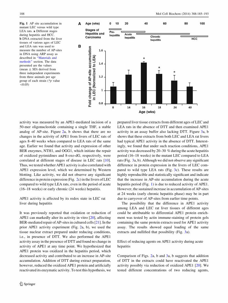
Comparison of AP-site accumulation in genomic liver DNA
from age-matched mutant LEC and wild type LEA rats
showed a statistically significant increase (up to 30–40 %) in
AP-site levels, particularly during the period of acute hepa-
titis (16–18 weeks) and early chronic hepatitis (24 weeks;
Fig. 1b). However, both the pre-hepatitis (Fig. 1a, 8 weeks)
and the chronic hepatitis periods (Fig. 1a, 40 weeks) dis-
played no significant changes in AP-site levels in LEC
compared to LEA rat livers (Fig. 1b). To our knowledge, this
is the first time that an increase in AP-sites, which can be
generated by oxidative stress [26], has been observed during
hepatitis and inflammation prior to preneoplastic foci for-
mation and HCC.
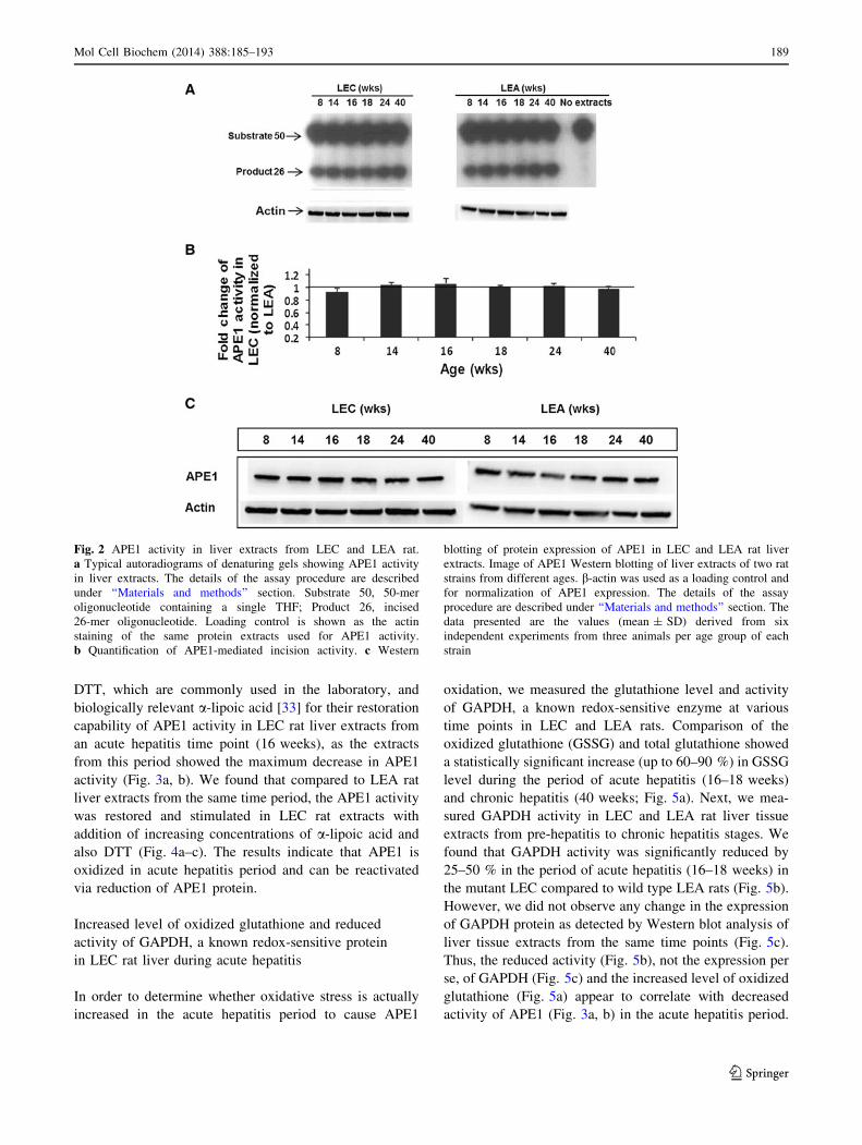
APE1 activity is unchanged in LEC rat liver
during hepatitis
Increase in AP-site generation can be due to reduced repair
activity of APE1-initiated BER pathway; so, we tested this
idea during different disease stages of LEC rats. It is well
known that APE1 is an essential enzyme to initiate the repair
of AP-sites by the BER pathway [16]. We examined APE1
activity in liver tissue extracts from different ages of wild
type LEA and copper overloaded mutant LEC rats. The
Mol Cell Biochem (2014) 388:185–193 187
123
activity was measured by an APE1-mediated incision of a
50-mer oligonucleotide containing a single THF, a stable
analog of AP-site. Figure 2a, b shows that there are no
changes in the activity of APE1 from livers of LEC rats of
ages 8–40 weeks when compared to LEA rats of the same
age. Earlier we found that activity and expression of other
BER enzymes, NTH1, and OGG1, which initiate the repair
of oxidized pyrimidines and 8-oxo-dG, respectively, were
correlated at different stages of disease in LEC rats [10].
Thus, we tested whether APE1 activity is also correlated with
APE1 expression level, which we determined by Western
blotting. Like activity, we did not observe any significant
difference in protein expression (Fig. 2c) in the livers of LEC
compared to wild type LEA rats, even in the period of acute
(16–18 weeks) or early chronic (24 weeks) hepatitis.
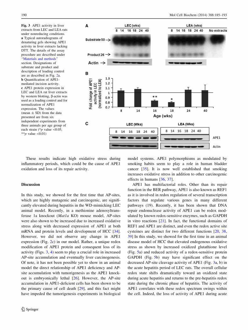
APE1 activity is affected by its redox state in LEC rat
liver during hepatitis
It was previously reported that oxidation or reduction of
APE1 can markedly alter its activity in vitro [20], affecting
BER-mediated repair of AP-sites in cultured cells [21]. In the
prior APE1 activity experiment (Fig. 2a, b), we used the
tissue nuclear extract prepared under reducing conditions,
i.e., in presence of DTT. We also performed the APE1
activity assay in the presence of DTT and found no change in
activity of APE1 at any time point. We hypothesized that
APE1 protein was oxidized in the hepatitis period, which
decreased activity and contributed to an increase in AP-site
accumulation. Addition of DTT during extract preparation,
however, reduced the oxidized APE1 protein and artificially
reactivated its enzymatic activity. To test this hypothesis, we
prepared liver tissue extracts from different ages of LEC and
LEA rats in the absence of DTT and then examined APE1
activity in an assay buffer also lacking DTT. Figure 3a, b
shows that these extracts from both LEC and LEA rat livers
had typical APE1 activity in the absence of DTT. Interest-
ingly, we found that under such reaction conditions, APE1
activity was decreased by 20–30 % during the acute hepatitis
period (16–18 weeks) in the mutant LEC compared to LEA
rats (Fig. 3a, b). Although we did not observe any significant
difference in protein expression in the livers of LEC com-
pared to wild type LEA rats (Fig. 3c). These results are
highly reproducible and statistically significant and indicate
that the increase in AP-site accumulation during the acute
hepatitis period (Fig. 1) is due to reduced activity of APE1.
However, the sustained increase in accumulation of AP-sites
at 24 weeks (early chronic hepatitis phase) may be in part
due to carryover of AP-sites from earlier time points.
The possibility that the difference in APE1 activity
among LEA and LEC rat liver tissues of different ages
could be attributable to differential APE1 protein enrich-
ment was tested by actin immune-staining of protein gels
containing the same protein extracts used for APE1 activity
assay. The results showed equal loading of the same
extracts and nullified that possibility (Fig. 3a).
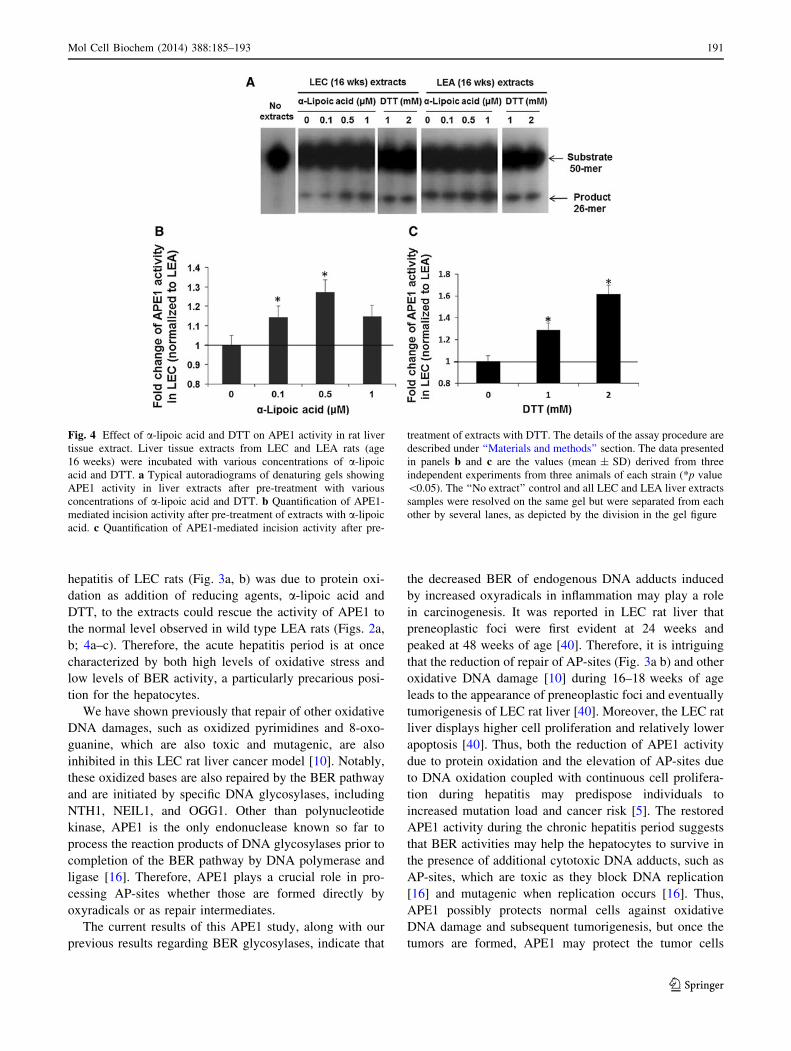
Effect of reducing agents on APE1 activity during acute
hepatitis
Comparison of Figs. 2a, b and 3a, b suggests that addition
of DTT in the extracts could have reactivated the APE1
activity possibly via reduction of oxidized APE1 [20]. We
tested different concentrations of two reducing agents,
Fig. 1 AP site accumulation in
mutant LEC versus wild type
LEA rats. a Different stages
during hepatitis and HCC.
b DNA extracted from the liver
tissues of various ages of LEC
and LEA rats was used to
measure the number of AP-sites
in DNA using ARP assay as
described in ‘‘Materials and
methods’’ section. The data
presented are the values
(mean ± SD) derived from
three independent experiments
from three animals per age
group of each strain (*p value
\0.05)
188 Mol Cell Biochem (2014) 388:185–193
123
DTT, which are commonly used in the laboratory, and
biologically relevant a-lipoic acid [33] for their restoration
capability of APE1 activity in LEC rat liver extracts from
an acute hepatitis time point (16 weeks), as the extracts
from this period showed the maximum decrease in APE1
activity (Fig. 3a, b). We found that compared to LEA rat
liver extracts from the same time period, the APE1 activity
was restored and stimulated in LEC rat extracts with
addition of increasing concentrations of a-lipoic acid and
also DTT (Fig. 4a–c). The results indicate that APE1 is
oxidized in acute hepatitis period and can be reactivated
via reduction of APE1 protein.
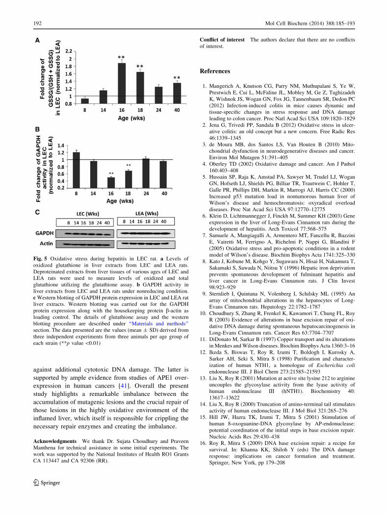
Increased level of oxidized glutathione and reduced
activity of GAPDH, a known redox-sensitive protein
in LEC rat liver during acute hepatitis
In order to determine whether oxidative stress is actually
increased in the acute hepatitis period to cause APE1
oxidation, we measured the glutathione level and activity
of GAPDH, a known redox-sensitive enzyme at various
time points in LEC and LEA rats. Comparison of the
oxidized glutathione (GSSG) and total glutathione showed
a statistically significant increase (up to 60–90 %) in GSSG
level during the period of acute hepatitis (16–18 weeks)
and chronic hepatitis (40 weeks; Fig. 5a). Next, we mea-
sured GAPDH activity in LEC and LEA rat liver tissue
extracts from pre-hepatitis to chronic hepatitis stages. We
found that GAPDH activity was significantly reduced by
25–50 % in the period of acute hepatitis (16–18 weeks) in
the mutant LEC compared to wild type LEA rats (Fig. 5b).
However, we did not observe any change in the expression
of GAPDH protein as detected by Western blot analysis of
liver tissue extracts from the same time points (Fig. 5c).
Thus, the reduced activity (Fig. 5b), not the expression per
se, of GAPDH (Fig. 5c) and the increased level of oxidized
glutathione (Fig. 5a) appear to correlate with decreased
activity of APE1 (Fig. 3a, b) in the acute hepatitis period.
Fig. 2 APE1 activity in liver extracts from LEC and LEA rat.
a Typical autoradiograms of denaturing gels showing APE1 activity
in liver extracts. The details of the assay procedure are described
under ‘‘Materials and methods’’ section. Substrate 50, 50-mer
oligonucleotide containing a single THF; Product 26, incised
26-mer oligonucleotide. Loading control is shown as the actin
staining of the same protein extracts used for APE1 activity.
b Quantification of APE1-mediated incision activity. c Western
blotting of protein expression of APE1 in LEC and LEA rat liver
extracts. Image of APE1 Western blotting of liver extracts of two rat
strains from different ages. b-actin was used as a loading control and
for normalization of APE1 expression. The details of the assay
procedure are described under ‘‘Materials and methods’’ section. The
data presented are the values (mean ± SD) derived from six
independent experiments from three animals per age group of each
strain
Mol Cell Biochem (2014) 388:185–193 189
123
These results indicate high oxidative stress during
inflammatory periods, which could be the cause of APE1
oxidation and loss of its repair activity.
Discussion
In this study, we showed for the first time that AP-sites,
which are highly mutagenic and carcinogenic, are signifi-
cantly elevated during hepatitis in the WD-mimicking LEC
animal model. Recently, in a methionine adenosyltrans-
ferase 1a knockout (Mat1a KO) mouse model, AP-sites
were also shown to be increased due to increased oxidative
stress along with decreased expression of APE1 at both
mRNA and protein levels and development of HCC [34].
However, we did not observe any change in APE1
expression (Fig. 2c) in our model. Rather, a unique redox
modification of APE1 protein and consequent loss of its
activity (Figs. 3, 4) seem to play a crucial role in increased
AP-site accumulation and eventually liver carcinogenesis.
Of note, it has not been possible yet to show in an animal
model the direct relationship of APE1 deficiency and AP-
site accumulation with tumorigenesis as the APE1 knock-
out is embryonically lethal [26]. However, the AP-site
accumulation in APE1-deficient cells has been shown to be
the primary cause of cell death [29], and this fact might
have impeded the tumorigenesis experiments in biological
model systems. APE1 polymorphisms as modulated by
smoking habits seem to play a role in human bladder
cancer [35]. It is now well established that smoking
increases oxidative stress in addition to other carcinogenic
effects in humans [36, 37].
APE1 has multifactorial roles. Other than its repair
function in the BER pathway, APE1 is also known as REF1
as it is involved in redox regulation of several transcription
factors that regulate various genes in many different
pathways (19). Recently, it has been shown that DNA
repair endonuclease activity of APE1 can be redox mod-
ulated by known redox-sensitive enzymes, such as GAPDH
in vitro reactions [21]. In fact, the functional domains of
REF1 and APE1 are distinct, and even the redox active site
cysteines are distinct for two different functions [20, 38,
39] In this study, we showed for the first time in an animal
disease model of HCC that elevated endogenous oxidative
stress as shown by increased oxidized glutathione level
(Fig. 5a) and reduced activity of a redox-sensitive protein
GAPDH (Fig. 5b) may have significant effect on the
decreased AP-site cleavage activity of APE1 (Fig. 3a, b) in
the acute hepatitis period of LEC rats. The overall cellular
redox state shifts dramatically toward an oxidized state
during acute hepatitis and returns to the pre-hepatitis redox
state during the chronic phase of hepatitis. The activity of
APE1 correlates with these redox spectrum swings within
the cell. Indeed, the loss of activity of APE1 during acute
Fig. 3 APE1 activity in liver
extracts from LEC and LEA rats
under nonreducing conditions.
a Typical autoradiograms of
denaturing gels showing APE1
activity in liver extracts lacking
DTT. The details of the assay
procedure are described under
‘‘Materials and methods’’
section. Designations of
substrate and product and
description of loading control
are as described in Fig. 2a.
b Quantification of APE1-
mediated incision activity.
c APE1 protein expression in
LEC and LEA rat liver extracts
by western blotting, b-actin was
used as a loading control and for
normalization of APE1
expression. The values
(mean ± SD) from the data
presented are from six
independent experiments from
three animals per age group of
each strain (*p value \0.05,
**p value \0.01)
190 Mol Cell Biochem (2014) 388:185–193
123
hepatitis of LEC rats (Fig. 3a, b) was due to protein oxi-
dation as addition of reducing agents, a-lipoic acid and
DTT, to the extracts could rescue the activity of APE1 to
the normal level observed in wild type LEA rats (Figs. 2a,
b; 4a–c). Therefore, the acute hepatitis period is at once
characterized by both high levels of oxidative stress and
low levels of BER activity, a particularly precarious posi-
tion for the hepatocytes.
We have shown previously that repair of other oxidative
DNA damages, such as oxidized pyrimidines and 8-oxo-
guanine, which are also toxic and mutagenic, are also
inhibited in this LEC rat liver cancer model [10]. Notably,
these oxidized bases are also repaired by the BER pathway
and are initiated by specific DNA glycosylases, including
NTH1, NEIL1, and OGG1. Other than polynucleotide
kinase, APE1 is the only endonuclease known so far to
process the reaction products of DNA glycosylases prior to
completion of the BER pathway by DNA polymerase and
ligase [16]. Therefore, APE1 plays a crucial role in pro-
cessing AP-sites whether those are formed directly by
oxyradicals or as repair intermediates.
The current results of this APE1 study, along with our
previous results regarding BER glycosylases, indicate that
the decreased BER of endogenous DNA adducts induced
by increased oxyradicals in inflammation may play a role
in carcinogenesis. It was reported in LEC rat liver that
preneoplastic foci were first evident at 24 weeks and
peaked at 48 weeks of age [40]. Therefore, it is intriguing
that the reduction of repair of AP-sites (Fig. 3a b) and other
oxidative DNA damage [10] during 16–18 weeks of age
leads to the appearance of preneoplastic foci and eventually
tumorigenesis of LEC rat liver [40]. Moreover, the LEC rat
liver displays higher cell proliferation and relatively lower
apoptosis [40]. Thus, both the reduction of APE1 activity
due to protein oxidation and the elevation of AP-sites due
to DNA oxidation coupled with continuous cell prolifera-
tion during hepatitis may predispose individuals to
increased mutation load and cancer risk [5]. The restored
APE1 activity during the chronic hepatitis period suggests
that BER activities may help the hepatocytes to survive in
the presence of additional cytotoxic DNA adducts, such as
AP-sites, which are toxic as they block DNA replication
[16] and mutagenic when replication occurs [16]. Thus,
APE1 possibly protects normal cells against oxidative
DNA damage and subsequent tumorigenesis, but once the
tumors are formed, APE1 may protect the tumor cells
Fig. 4 Effect of a-lipoic acid and DTT on APE1 activity in rat liver
tissue extract. Liver tissue extracts from LEC and LEA rats (age
16 weeks) were incubated with various concentrations of a-lipoic
acid and DTT. a Typical autoradiograms of denaturing gels showing
APE1 activity in liver extracts after pre-treatment with various
concentrations of a-lipoic acid and DTT. b Quantification of APE1-
mediated incision activity after pre-treatment of extracts with a-lipoic
acid. c Quantification of APE1-mediated incision activity after pre-
treatment of extracts with DTT. The details of the assay procedure are
described under ‘‘Materials and methods’’ section. The data presented
in panels b and c are the values (mean ± SD) derived from three
independent experiments from three animals of each strain (*p value
\0.05). The ‘‘No extract’’ control and all LEC and LEA liver extracts
samples were resolved on the same gel but were separated from each
other by several lanes, as depicted by the division in the gel figure
Mol Cell Biochem (2014) 388:185–193 191
123
against additional cytotoxic DNA damage. The latter is
supported by ample evidence from studies of APE1 over-
expression in human cancers [41]. Overall the present
study highlights a remarkable imbalance between the
accumulation of mutagenic lesions and the crucial repair of
those lesions in the highly oxidative environment of the
inflamed liver, which itself is responsible for crippling the
necessary repair enzymes and creating the imbalance.
Acknowledgments We thank Dr. Sujata Choudhury and Praveen
Manthena for technical assistance in some initial experiments. The
work was supported by the National Institutes of Health RO1 Grants
CA 113447 and CA 92306 (RR).
Conflict of interest The authors declare that there are no conflicts
of interest.
References
1. Mangerich A, Knutson CG, Parry NM, Muthupalani S, Ye W,
Prestwich E, Cui L, McFaline JL, Mobley M, Ge Z, Taghizadeh
K, Wishnok JS, Wogan GN, Fox JG, Tannenbaum SR, Dedon PC
(2012) Infection-induced colitis in mice causes dynamic and
tissue-specific changes in stress response and DNA damage
leading to colon cancer. Proc Natl Acad Sci USA 109:1820–1829
2. Jena G, Trivedi PP, Sandala B (2012) Oxidative stress in ulcer-
ative colitis: an old concept but a new concern. Free Radic Res
46:1339–1345
3. de Moura MB, dos Santos LS, Van Houten B (2010) Mito-
chondrial dysfunction in neurodegenerative diseases and cancer.
Environ Mol Mutagen 51:391–405
4. Oberley TD (2002) Oxidative damage and cancer. Am J Pathol
160:403–408
5. Hussain SP, Raja K, Amstad PA, Sawyer M, Trudel LJ, Wogan
GN, Hofseth LJ, Shields PG, Billiar TR, Trautwein C, Hohler T,
Galle PR, Phillips DH, Markin R, Marrogi AJ, Harris CC (2000)
Increased p53 mutation load in nontumorous human liver of
Wilson’s disease and hemochromatosis: oxyradical overload
diseases. Proc Nat Acad Sci USA 97:12770–12775
6. Klein D, Lichtmannegger J, Finckh M, Summer KH (2003) Gene
expression in the liver of Long-Evans Cinnamon rats during the
development of hepatitis. Arch Toxicol 77:568–575
7. Samuele A, Mangiagalli A, Armentero MT, Fancellu R, Bazzini
E, Vairetti M, Ferrigno A, Richelmi P, Nappi G, Blandini F
(2005) Oxidative stress and pro-apoptotic conditions in a rodent
model of Wilson’s disease. Biochim Biophys Acta 1741:325–330
8. Kato J, Kobune M, Kohgo Y, Sugawara N, Hisai H, Nakamura T,
Sakamaki S, Sawada N, Niitsu Y (1996) Hepatic iron deprivation
prevents spontaneous development of fulminant hepatitis and
liver cancer in Long-Evans Cinnamon rats. J Clin Invest
98:923–929
9. Sternlieb I, Quintana N, Volenberg I, Schilsky ML (1995) An
array of mitochondrial alterations in the hepatocytes of Long-
Evans Cinnamon rats. Hepatology 22:1782–1787
10. Choudhury S, Zhang R, Frenkel K, Kawamori T, Chung FL, Roy
R (2003) Evidence of alterations in base excision repair of oxi-
dative DNA damage during spontaneous hepatocarcinogenesis in
Long-Evans Cinnamon rats. Cancer Res 63:7704–7707
11. DiDonato M, Sarkar B (1997) Copper transport and its alterations
in Menkes and Wilson diseases. Biochim Biophys Acta 1360:3–16
12. Ikeda S, Biswas T, Roy R, Izumi T, Boldogh I, Kurosky A,
Sarker AH, Seki S, Mitra S (1998) Purification and character-
ization of human NTH1, a homologue of Escherichia coli
endonuclease III. J Biol Chem 273:21585–21593
13. Liu X, Roy R (2001) Mutation at active site lysine 212 to arginine
uncouples the glycosylase activity from the lyase activity of
human endonuclease III (hNTH1). Biochemistry 40:
13617–13622
14. Liu X, Roy R (2000) Truncation of amino-terminal tail stimulates
activity of human endonuclease III. J Mol Biol 321:265–276
15. Hill JW, Hazra TK, Izumi T, Mitra S (2001) Stimulation of
human 8-oxoguanine-DNA glycosylase by AP-endonuclease:
potential coordination of the initial steps in base excision repair.
Nucleic Acids Res 29:430–438
16. Roy R, Mitra S (2009) DNA base excision repair: a recipe for
survival. In: Khanna KK, Shiloh Y (eds) The DNA damage
response: implications on cancer formation and treatment.
Springer, New York, pp 179–208
Fig. 5 Oxidative stress during hepatitis in LEC rat. a Levels of
oxidized glutathione in liver extracts from LEC and LEA rats.
Deproteinated extracts from liver tissues of various ages of LEC and
LEA rats were used to measure levels of oxidized and total
glutathione utilizing the glutathione assay. b GAPDH activity in
liver extracts from LEC and LEA rats under nonreducing condition.
c Western blotting of GAPDH protein expression in LEC and LEA rat
liver extracts. Western blotting was carried out for the GAPDH
protein expression along with the housekeeping protein b-actin as
loading control. The details of glutathione assay and the western
blotting procedure are described under ‘‘Materials and methods’’
section. The data presented are the values (mean ± SD) derived from
three independent experiments from three animals per age group of
each strain (**p value \0.01)
192 Mol Cell Biochem (2014) 388:185–193
123
17. Tell G, Wilson DM, Lee CH (2010) Intrusion of a DNA repair
protein in the RNome world: is this the beginning of a new era?
Mol Cell Biol 30:366–371
18. Chattopadhyay R, Das S, Maiti AK, Boldogh I, Xie J, Hazra TK,
Kohno K, Mitra S, Bhakat KK (2008) Regulatory role of human
AP-Endonuclease (APE1/REF-1) in YB-1-mediated activation of
the multidrug resistance gene MDR1. Mol Cell Biol
28:7066–7080
19. Bhakat KK, Mantha AK, Mitra S (2009) Transcriptional regula-
tory functions of mammalian AP-endonuclease (APE11/Ref-1),
an essential multifunctional protein. Antioxid Redox Signal
11:621–638
20. Kelley MR, Parsons SH (2001) Redox regulation of the DNA
repair function of the human AP endonuclease Ape1/ref-1. An-
tioxid Redox Signal 3:671–683
21. Azam S, Jouvet N, Jilani A, Vongsamphanh R, Yang X, Yang S,
Ramotar D (2008) Human glyceraldehyde-3-phosphate dehy-
drogenase plays a direct role in reactivating oxidized forms of the
DNA repair enzyme APE1. J Biol Chem 283:30632–30641
22. Sirover MA (1999) New insights into an old protein: the func-
tional diversity of mammalian glyceraldehyde-3-phosphate
dehydrogenase. Biochim Biophys Acta 1432:159–184
23. Meyer-Siegler K, Mauro DJ, Seal G, Wurzer J, deRiel JK, Sirover
MA (1991) A human nuclear uracil DNA glycosylase is the
37-kDa subunit of glyceraldehyde-3-phosphate dehydrogenase.
Proc Natl Acad Sci USA 88:8460–8464
24. Dastoor Z, Dreyer JL (2001) Potential role of nuclear transloca-
tion of glyceraldehyde-3-phosphate dehydrogenase in apoptosis
and oxidative stress. J Cell Sci 114:1643–1653
25. Sen N, Hara MR, Kornberg MD, Cascio MB, Bae BI, Shahani N,
Thomas B, Dawson TM, Dawson VL, Snyder SH, Sawa A (2008)
Nitric oxide-induced nuclear GAPDH activates p300/CBP and
mediates apoptosis. Nat Cell Biol 10:866–873
26. Izumi T, Brown DB, Naidu CV, Bhakat KK, Macinnes MA, Saito
H, Chen DJ, Mitra S (2005) Two essential but distinct functions
of the mammalian abasic endonuclease. Proc Natl Acad Sci USA
102:5739–5743
27. Xanthoudakis S, Smeyne RJ, Wallace JD, Curran T (1996) The
redox/DNA repair protein, Ref-1, is essential for early embryonic
development in mice. Proc Natl Acad Sci USA 93:8919–8923
28. Meira LB, Devaraj S, Kisby GE, Burns DK, Daniel RL, Hammer
RE, Grundy S, Jialal I, Friedberg EC (2001) Heterozygosity for
the mouse Apex gene results in phenotypes associated with oxi-
dative stress. Cancer Res 61:5552–5557
29. Fung H, Demple B (2005) A vital role for Ape1/Ref1 protein in
repairing spontaneous DNA damage in human cells. Mol Cell
17:463–470
30. Schreiber E, Matthias P, Muller MM, Schaffner W (1989) Rapid
detection of octamer binding proteins with ‘mini-extracts’, pre-
pared from a small number of cells. Nucleic Acids Res 17:6419
31. Nakamura J, Walker VE, Upton PB, Chiang SY, Kow YW,
Swenberg JA (1998) Highly sensitive apurinic/apyrimidinic site
assay can detect spontaneous and chemically induced depurina-
tion under physiological conditions. Cancer Res 58:222–225
32. Adhikari S, Manthena PV, Kota KK, Karmahapatra SK, Roy G,
Saxena R, Uren A, Roy R (2012) A comparative study of
recombinant mouse and human apurinic/apyrimidinic endonu-
clease. Mol Cell Biochem 362:195–201
33. Packer L, Witt EH, Tritschler HJ (1995) Alpha-lipoic acid as a
biological antioxidant. Free Radic Biol Med 19:227–250
34. Tomasi ML, Iglesias-Ara A, Yang H, Ramani K, Feo F, Pascale
MR, Martınez-Chantar ML, Mato JM, Lu SC (2009) S-adeno-
sylmethionine regulates apurinic/apyrimidinic endonuclease 1
stability: implication in hepatocarcinogenesis. Gastroenterology
136:1025–1036
35. Terry PD, Umbach DM, Taylor JA (2006) APE1 genotype and
risk of bladder cancer: evidence for effect modification by
smoking. Int J Cancer 118:3170–3173
36. Pryor WA, Stone K (1993) Oxidants in cigarette smoke. Radicals,
hydrogen peroxide, peroxynitrate, and peroxynitrite. Ann N Y
Acad Sci 686:12–27
37. Abdi S, Ali A (1999) Role of ROS modified human DNA in the
pathogenesis and etiology of cancer. Cancer Lett 142:1–9
38. Walker LJ, Robson CN, Black E, Gillespie D, Hickson ID (1993)
Identification of residues in the human DNA repair enzyme
HAP1 (Ref-1) that are essential for redox regulation of Jun DNA
binding. Mol Cell Biol 13:5370–5376
39. Georgiadis MM, Luo M, Gaur RK, Delaplane S, Li X, Kelley MR
(2008) Evolution of the redox function in mammalian apurinic/
apyrimidinic endonuclease. Mutat Res 643:54–63
40. Jia G, Tohyama C, Sone H (2002) DNA damage triggers
imbalance of proliferation and apoptosis during development of
preneoplastic foci in the liver of Long-Evans Cinnamon rats. Int J
Oncol 21:755–761
41. Jiang Y, Zhou S, Sandusky GE, Kelley MR, Fishel ML (2010)
Reduced expression of DNA repair and redox signaling protein
APE1/Ref-1 impairs human pancreatic cancer cell survival, pro-
liferation, and cell cycle progression. Cancer Invest 28:885–895
Mol Cell Biochem (2014) 388:185–193 193
123