reduced field-of-view mri with two-dimensional spatially-selective rf excitation and unfold
TRANSCRIPT

Reduced Field-of-View MRI With Two-DimensionalSpatially-Selective RF Excitation and UNFOLD
Lei Zhao, Bruno Madore, and Lawrence P. Panych*
When the region of interest (ROI) is smaller than the object, onecan increase MRI speed by reducing the imaging field of view(FOV). However, when such an approach is used, features outsidethe reduced FOV will alias into the reduced-FOV image along thephase-encoding direction. Reduced-FOV methods are designedto correct this aliasing problem. In the present study, we proposea combination of two different approaches to reduce the acquiredFOV: 1) two-dimensional (2D) spatially-selective RF excitation, and2) the unaliasing by Fourier-encoding the overlaps using the tem-poral dimension (UNFOLD) technique. While 2D spatially-selectiveRF excitation can restrict the spins excited within a reduced FOV,the UNFOLD technique can help to eliminate any residual aliasedsignals and thus relaxes the requirement for a long RF excitationpulse. This hybrid method was implemented for MR-based tem-perature mapping, and resulted in artifact-free images with a four-fold improvement in temporal resolution. Magn Reson Med 53:1118–1125, 2005. © 2005 Wiley-Liss, Inc.
Key words: reduced FOV; spatially-selective 2D RF excitation;UNFOLD; dynamic MRI; fast imaging
Dynamic MRI has become increasingly feasible over thelast several years because of technological advances, espe-cially the increased data acquisition speed allowed byhigher-performance hardware systems. However, in anumber of dynamic applications the currently achievabletemporal resolution may still be unsatisfactory. Severalapproaches have been developed to further increase imag-ing speed. Multiple-echo data acquisition strategies, suchas echo-planar imaging (EPI) (1), rapid acquisition withrelaxation enhancement (RARE) (2), and gradient andspin-echo (GRASE) (3), increase imaging speed by acquir-ing multiple k-space lines after each excitation. Comple-mentary approaches to enhance imaging speed includemethods whereby images are reconstructed using only areduced set of the k-space data. Examples of such methodsinclude partial-Fourier imaging (4,5), keyhole techniques(6,7), MR fluoroscopy (8,9), feature-recognizing MRI (10),multiple-region MRI (11), reduced-encoding MRI withgeneralized-series reconstruction (RIGR) (12,13), and re-duced field-of-view (FOV) methods (such as that describedby Hu and Parrish (14)), unaliasing by Fourier-encodingthe overlaps using the temporal dimension (UNFOLD)(15,16), and 2D spatially-selective RF excitation (17,18).The performance of such methods depends on how well
the assumptions upon which they are based are justified ina particular application. The present work focuses on com-bining two of these approaches, UNFOLD and 2D spa-tially-selective RF excitation, which are believed to behighly compatible.
In dynamic imaging applications, such as cardiovascu-lar MRI, interventional MRI, or, in some cases, functionalMRI (fMRI), only a small portion of the FOV may be ofclinical interest. In cardiac imaging, for example, the en-tire chest is typically within the FOV even though only theheart is of special interest. In MR temperature mapping tomonitor procedures such as focused ultrasound ablation oftissue, only the small portion of the FOV in which heat isconcentrated may be of special interest. In fMRI for pre-surgical planning, the region around a tumor may be theonly part of the FOV that has real clinical value. With areduced FOV, fewer k-space lines are needed to achievethe same spatial resolution, leading to faster imaging.However, simply reducing the imaging FOV is normallyunacceptable because it typically results in aliasing arti-facts (also called “wraparound” artifacts). A straightfor-ward way to avoid such artifacts is to avoid exciting anysignal outside the desired ROI, as can be done using 2Dspatially-selective RF excitation pulses (17,18). Suchpulses perform a spatial selection along the slice directionin addition to selecting a limited region in the phase-encode direction.
Along the phase-encoding direction, a selection profilewith sharp edges is needed to completely avoid any alias-ing while the full extent of the reduced FOV is used forimaging. Usually, such sharp profiles require RF pulsesthat are very long in duration. For example, Reiseberg et al.(19) reported the application of 2D RF excitation in EPIusing a 26-ms pulse to achieve a reduced FOV in thephase-encode direction. Even with this relatively long RFpulse, edges for the 40-mm FOV extended an additional10 mm. If one used a shorter, more practical RF pulse for2D excitation, the corresponding excitation profile wouldbecome even wider. Such a compromised profile wouldresult in a severe aliasing artifact from the excited materiallocated outside the boundaries of the reduced FOV.
In a preliminary study, Zhao and Panych (20) combineda dynamic rFOV method similar to that reported by Huand Parrish (14) with a short-duration 2D spatially-selec-tive RF excitation. Typically, neither method alone(whether the 2D RF excitation or the rFOV method) wassufficient to satisfactorily eliminate the aliasing artifacts.The rFOV method eliminated aliasing from static signalcomponents outside the reduced FOV, but not dynamicsignal components. The RF excitation method signifi-cantly suppressed signal outside the reduced FOV, but leftaliased signals near the boundaries. Although the twomethods combined performed significantly better than ei-
Department of Radiology, Harvard Medical School, Brigham and Women’sHospital, Boston, Massachusetts, USA.Grant sponsor: NIH; Grant numbers: P01-CA67165; R29-CA7314; R01-NS37922; R01-HL073319; Grant sponsor: Whitaker Foundation.*Correspondence to: Lawrence P. Panych, Ph.D., Department of Radiology,Brigham and Women’s Hospital, Harvard Medical School, 75 Francis Street,Boston, MA 02115. E-mail: [email protected] 5 October 2004; revised 9 December 2004; accepted 14 December2004.DOI 10.1002/mrm.20458Published online in Wiley InterScience (www.interscience.wiley.com).
Magnetic Resonance in Medicine 53:1118–1125 (2005)
© 2005 Wiley-Liss, Inc. 1118

ther did individually, this hybrid approach failed in casesin which dynamic material was found outside and near thereduced FOV boundaries. Furthermore, the reference im-age required by this approach sometimes proved difficultto obtain in practice.
On the other hand, the UNFOLD method (15,16) doesnot require a reference image, it can handle dynamic vari-ations in the aliasing artifacts to be suppressed, and it isoptimal in terms of SNR. In the current work, we used 2Dspatially-selective RF excitations to reduce the excitedFOV and speed up the acquisition process, and used UN-FOLD to suppress any leftover aliasing artifact. We foundthe combination of 2D RF excitation and UNFOLD to be avery reliable hybrid fast imaging approach. The applica-tion of a 2D excitation pulse can reduce the FOV manyfold, and the addition of UNFOLD serves to relax therequirement for a sharp RF excitation profile, thus allow-ing for an RF pulse of more practical duration. As reportedhere, this hybrid method was successfully implemented inboth dynamic phantom and in vivo imaging. Artifact-freeimages with a fourfold improvement in temporal resolu-tion were obtained that are suitable for MRI-based temper-ature mapping studies.
THEORY
2D RF Pulse Design
We used the small-tip-angle model developed by Pauly etal. (17) in this study for the design of our 2D selective RFpulse. An on-resonance RF field with envelope B1(t) isapplied in conjunction with a magnetic field gradient G(t)to generate an excited profile Mxy(r). With W(k(t)) � B1(t)/
|�G(t)| and k(t) � –� �t
T G(s)ds, where T is the time at
which the RF excitation pulse ends, it can be shown that(17):
Mxy�r� � i�M0 � W�k�S�k�exp�ir � k�dk. [1]
where S(k) represents the sampling function in excitationk-space, and W(k) represents the spatial frequency weight-ing function, which is the Fourier transform of the desiredspatial profile, w(r). The domain of integration extendsover the central region in excitation k-space, where thenonzero (i.e., sampled) locations of S(k) are found.
To reduce pulse duration, it is advantageous to choose aW(k) that is as compact as possible in excitation k-space.Of course, the need for a compact W(k) conflicts with theneed for a compact profile w(r), i.e., one that has sharptransitions between excited and non-excited regions. Be-cause of the conflicting need for compactness in the recip-rocal k and r spaces, a 2D Gaussian shape for the targetedspatial profile, w(r), appears to be a good compromise.
In the reduced-FOV application, the slice thicknessalong z will be much smaller (�1 cm) than the desiredexcited region along the phase-encoded direction y(��1 cm). Thus, the sampling function S(k) must cover alarger extent along kz than along ky. For this reason anecho-planar trajectory, as shown in Fig. 1, would be ap-propriate for S(k). Figure 2 shows the spatially-selectiveRF pulse and the gradient waveforms that can be used togenerate a Gaussian-shaped 2D profile. The slice (Gz) andphase (Gy) gradients produce the echo-planar k-space tra-jectory S(k), while the RF pulse (of magnitude |B1(t)| andphase �(t) � angle(B1(t))) produces the spatial frequencyweighting W(k).
The sampling function S(k) must be designed to not onlygive an RF pulse of practical duration (�5 ms), but to alsoensure that the periodicity of the excitation in the y direc-tion is greater than the expected object size in that direc-tion. The number of ky lines in the trajectory (i.e., thesampling density in the ky direction) affects both the pulseduration and the excitation periodicity along y. This num-ber must be small enough to keep the pulse duration short,and at the same time be large enough to ensure that thedistance between the excited lobes is greater than theobject size.
FIG. 1. Weighted echo-planar excitation k-spacetrajectory of a 2D RF pulse that can be used forselective excitation of a slice in the z-direction(�1 cm) and a region in y (��1 cm).
FIG. 2. RF and gradient diagram of a 2D RFpulse with excitation k-space trajectory shown inFig. 1. At the end of the RF excitation, rephrasinggradient pulses are used to rewind the samplingtrajectory to the center of k-space.
2D RF Excitation and UNFOLD Reduced-FOV MRI 1119
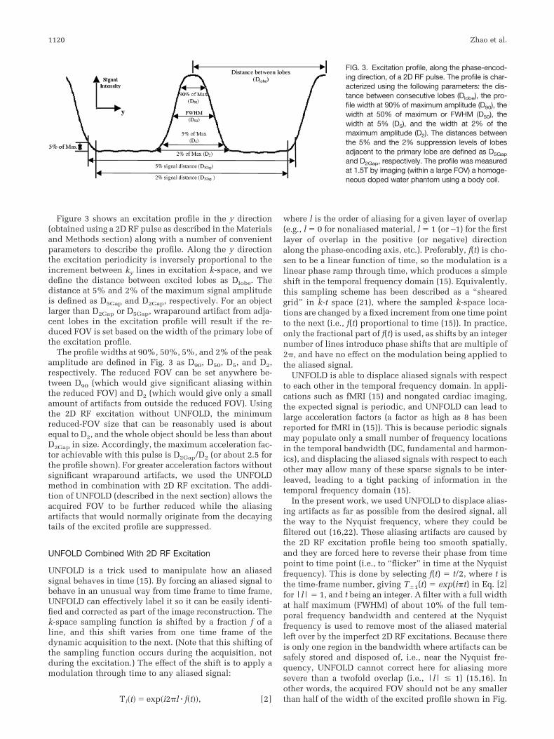
Figure 3 shows an excitation profile in the y direction(obtained using a 2D RF pulse as described in the Materialsand Methods section) along with a number of convenientparameters to describe the profile. Along the y directionthe excitation periodicity is inversely proportional to theincrement between ky lines in excitation k-space, and wedefine the distance between excited lobes as Dlobe. Thedistance at 5% and 2% of the maximum signal amplitudeis defined as D5Gap and D2Gap, respectively. For an objectlarger than D2Gap or D5Gap, wraparound artifact from adja-cent lobes in the excitation profile will result if the re-duced FOV is set based on the width of the primary lobe ofthe excitation profile.
The profile widths at 90%, 50%, 5%, and 2% of the peakamplitude are defined in Fig. 3 as D90, D50, D5, and D2,respectively. The reduced FOV can be set anywhere be-tween D90 (which would give significant aliasing withinthe reduced FOV) and D2 (which would give only a smallamount of artifacts from outside the reduced FOV). Usingthe 2D RF excitation without UNFOLD, the minimumreduced-FOV size that can be reasonably used is aboutequal to D2, and the whole object should be less than aboutD2Gap in size. Accordingly, the maximum acceleration fac-tor achievable with this pulse is D2Gap/D2 (or about 2.5 forthe profile shown). For greater acceleration factors withoutsignificant wraparound artifacts, we used the UNFOLDmethod in combination with 2D RF excitation. The addi-tion of UNFOLD (described in the next section) allows theacquired FOV to be further reduced while the aliasingartifacts that would normally originate from the decayingtails of the excited profile are suppressed.
UNFOLD Combined With 2D RF Excitation
UNFOLD is a trick used to manipulate how an aliasedsignal behaves in time (15). By forcing an aliased signal tobehave in an unusual way from time frame to time frame,UNFOLD can effectively label it so it can be easily identi-fied and corrected as part of the image reconstruction. Thek-space sampling function is shifted by a fraction f of aline, and this shift varies from one time frame of thedynamic acquisition to the next. (Note that this shifting ofthe sampling function occurs during the acquisition, notduring the excitation.) The effect of the shift is to apply amodulation through time to any aliased signal:
Tl�t� � exp�i2�l � f�t��, [2]
where l is the order of aliasing for a given layer of overlap(e.g., l � 0 for nonaliased material, l � 1 (or –1) for the firstlayer of overlap in the positive (or negative) directionalong the phase-encoding axis, etc.). Preferably, f(t) is cho-sen to be a linear function of time, so the modulation is alinear phase ramp through time, which produces a simpleshift in the temporal frequency domain (15). Equivalently,this sampling scheme has been described as a “shearedgrid” in k-t space (21), where the sampled k-space loca-tions are changed by a fixed increment from one time pointto the next (i.e., f(t) proportional to time (15)). In practice,only the fractional part of f(t) is used, as shifts by an integernumber of lines introduce phase shifts that are multiple of2�, and have no effect on the modulation being applied tothe aliased signal.
UNFOLD is able to displace aliased signals with respectto each other in the temporal frequency domain. In appli-cations such as fMRI (15) and nongated cardiac imaging,the expected signal is periodic, and UNFOLD can lead tolarge acceleration factors (a factor as high as 8 has beenreported for fMRI in (15)). This is because periodic signalsmay populate only a small number of frequency locationsin the temporal bandwidth (DC, fundamental and harmon-ics), and displacing the aliased signals with respect to eachother may allow many of these sparse signals to be inter-leaved, leading to a tight packing of information in thetemporal frequency domain (15).
In the present work, we used UNFOLD to displace alias-ing artifacts as far as possible from the desired signal, allthe way to the Nyquist frequency, where they could befiltered out (16,22). These aliasing artifacts are caused bythe 2D RF excitation profile being too smooth spatially,and they are forced here to reverse their phase from timepoint to time point (i.e., to “flicker” in time at the Nyquistfrequency). This is done by selecting f(t) � t/2, where t isthe time-frame number, giving T1(t) � exp(i�t) in Eq. [2]for |l| � 1, and t being an integer. A filter with a full widthat half maximum (FWHM) of about 10% of the full tem-poral frequency bandwidth and centered at the Nyquistfrequency is used to remove most of the aliased materialleft over by the imperfect 2D RF excitations. Because thereis only one region in the bandwidth where artifacts can besafely stored and disposed of, i.e., near the Nyquist fre-quency, UNFOLD cannot correct here for aliasing moresevere than a twofold overlap (i.e., |l| � 1) (15,16). Inother words, the acquired FOV should not be any smallerthan half of the width of the excited profile shown in Fig.
FIG. 3. Excitation profile, along the phase-encod-ing direction, of a 2D RF pulse. The profile is char-acterized using the following parameters: the dis-tance between consecutive lobes (Dlobe), the pro-file width at 90% of maximum amplitude (D90), thewidth at 50% of maximum or FWHM (D50), thewidth at 5% (D5), and the width at 2% of themaximum amplitude (D2). The distances betweenthe 5% and the 2% suppression levels of lobesadjacent to the primary lobe are defined as D5Gap
and D2Gap, respectively. The profile was measuredat 1.5T by imaging (within a large FOV) a homoge-neous doped water phantom using a body coil.
1120 Zhao et al.
3. For example, to avoid having aliased material of inten-sity greater than 2% of its fully-excited magnitude, onewould select an acquired FOV no smaller than D2/2 (seeFig. 3). Clearly, a larger (or smaller) acquired FOV wouldlead to a more benign (or more severe) level of residualartifacts after the application of UNFOLD.
MATERIALS AND METHODS
All experiments were performed using a modified versionof a gradient-echo sequence, implemented on a 1.5 T GESigna scanner (General Electric Medical Systems, Milwau-kee, WI) with echo-speed gradient set (40 mT/m maximumand a slew rate of 150 T/m/s). As described below, exper-iments were performed in a static water phantom, in adynamic water phantom, in a piece of meat while heat wasapplied, as well as in vivo without applying heat.
Figure 2 shows the RF and the gradient waveforms de-signed to generate a Gaussian-shaped 2D profile. The slice(Gz) and phase (Gy) gradients were chosen to produce anecho-planar k-space trajectory (Fig. 1), while the RF pro-duces the spatial frequency weighting. The waveform wasfurther adjusted to make maximum use of the availableslew rate of the system in order to minimize the RF pulseduration. We also minimized the pulse duration by apply-ing a Gaussian weighting to the width of the kz coverage asa function of ky (Fig. 1). This ky modulation of the kz
coverage resulted in a reduction of the RF pulse durationby more than 50%.
The RF pulse design features nine segments along the ky
direction, with gradient pulses applied at the end of theexcitation to rephase the magnetization. The RF and gra-dient waveforms were designed for a system with a max-imum gradient strength of 4 mT/m and a maximum slewrate of 150 T/m/s. The RF pulse waveform consisted of1054 points with 4 s dwell time, for a total duration of4.2 ms.
A large homogeneous doped water phantom was imagedusing the body coil, and a large FOV was set so that the fullRF excitation profile (including its periodic lobes) couldbe visualized. The excitation profile shown in Fig. 3 wasobtained with the 2D selective excitation pulse describedabove.
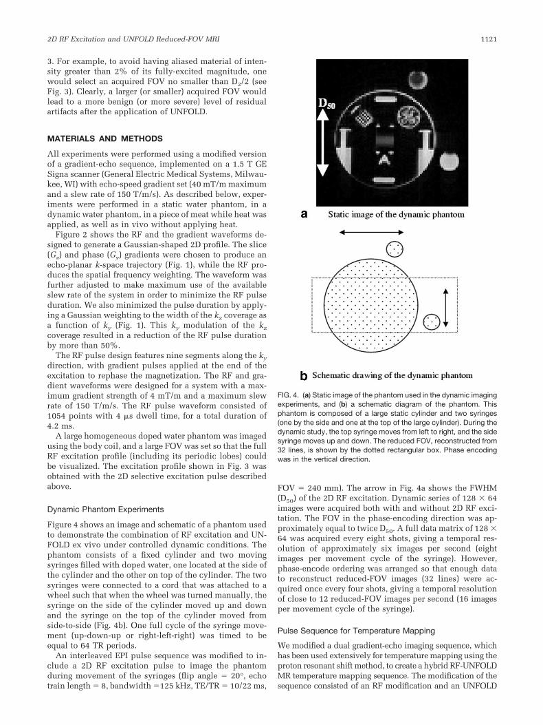
Dynamic Phantom Experiments
Figure 4 shows an image and schematic of a phantom usedto demonstrate the combination of RF excitation and UN-FOLD ex vivo under controlled dynamic conditions. Thephantom consists of a fixed cylinder and two movingsyringes filled with doped water, one located at the side ofthe cylinder and the other on top of the cylinder. The twosyringes were connected to a cord that was attached to awheel such that when the wheel was turned manually, thesyringe on the side of the cylinder moved up and downand the syringe on the top of the cylinder moved fromside-to-side (Fig. 4b). One full cycle of the syringe move-ment (up-down-up or right-left-right) was timed to beequal to 64 TR periods.
An interleaved EPI pulse sequence was modified to in-clude a 2D RF excitation pulse to image the phantomduring movement of the syringes (flip angle � 20°, echotrain length � 8, bandwidth �125 kHz, TE/TR � 10/22 ms,
FOV � 240 mm). The arrow in Fig. 4a shows the FWHM(D50) of the 2D RF excitation. Dynamic series of 128 � 64images were acquired both with and without 2D RF exci-tation. The FOV in the phase-encoding direction was ap-proximately equal to twice D50. A full data matrix of 128 �64 was acquired every eight shots, giving a temporal res-olution of approximately six images per second (eightimages per movement cycle of the syringe). However,phase-encode ordering was arranged so that enough datato reconstruct reduced-FOV images (32 lines) were ac-quired once every four shots, giving a temporal resolutionof close to 12 reduced-FOV images per second (16 imagesper movement cycle of the syringe).
Pulse Sequence for Temperature Mapping
We modified a dual gradient-echo imaging sequence, whichhas been used extensively for temperature mapping using theproton resonant shift method, to create a hybrid RF-UNFOLDMR temperature mapping sequence. The modification of thesequence consisted of an RF modification and an UNFOLD
FIG. 4. (a) Static image of the phantom used in the dynamic imagingexperiments, and (b) a schematic diagram of the phantom. Thisphantom is composed of a large static cylinder and two syringes(one by the side and one at the top of the large cylinder). During thedynamic study, the top syringe moves from left to right, and the sidesyringe moves up and down. The reduced FOV, reconstructed from32 lines, is shown by the dotted rectangular box. Phase encodingwas in the vertical direction.
2D RF Excitation and UNFOLD Reduced-FOV MRI 1121
modification. The RF modification involved replacing theslice-selective RF pulse with a 2D RF pulse as describedabove. The UNFOLD modification involved shifting the ac-quisition k-space trajectory by �k/2 on every second acqui-sition in the dynamic imaging series, where �k is the incre-ment between acquisition k-space lines.
Abdominal Imaging Experiments
We tested the modified imaging sequence described in theprevious section in vivo by acquiring several dynamicseries of abdominal images from a human subject. Heatingwas not performed in the in vivo imaging experiments.The goal of the first set of experiments was simply to testthe feasibility of using the new method in the presence ofphysiological motion to obtain images free of both motionand wraparound artifact. The abdominal imaging serieswere acquired 1) at full FOV without 2D RF excitation orUNFOLD, 2) at half FOV with 2D RF excitation but with-out UNFOLD, and 3) at quarter FOV with both 2D RF andwith UNFOLD. The full FOV in both the phase- and fre-quency-encoding directions was 25 cm. The image acqui-sition matrix for full-FOV imaging was 192 in the fre-quency-encoding direction, and 128, 64, and 32 in thephase-encoding direction for full-, half-, and quarter-FOVimaging, respectively. Therefore, with a TR of 50 ms, thetemporal resolution for full-, half-, and quarter-FOV imag-ing was 6.4 s, 3.2 s, and 1.6 s. The duration of the 2D RFpulse was 4.2 ms. The acquisition bandwidth was 16 khz,and the TEs of the two gradient echoes were 5.6 ms and20 ms. Images were acquired using an eight-channel flex-ible phased-array abdominal RF coil. The subject breathedfreely during the acquisitions. Informed consent was ob-tained before each subject study under an approved IRB.
Temperature Mapping Experiments
In a separate set of experiments, the hybrid RF-UNFOLDsequence was applied for temperature mapping during heat-ing of a phantom. In these experiments, a gel pack (Rapid AidReusable Cold & Hot Compress, Oakville, Canada) washeated in a microwave and then placed on top of a large pieceof meat, which was then moved to the MRI scanner forimaging. Dynamic sets of images were acquired during threeseparate periods: 1) a period immediately after placement inthe scanner, 2) a period beginning 5 min after the end of the
first imaging period, and 3) a period beginning 15 min afterthe end of the second period. Each imaging period was ap-proximately 100 s in duration. The imaging parameters werethe same as those used in the abdominal imaging experi-ments with a quarter FOV (i.e., acquisition matrix � 192 �32, TR � 50 ms), and thus the temporal resolution for thedynamic imaging was 1.6 s per scan.
The complex data were processed to produce maps ofthe change in phase over each dynamic series. Phasewraps in the data were removed voxel by voxel along thetime dimension using the “unwrap” function in Matlab.The slope of phase change over the heating period wasthen computed from the unwrapped data by least-squarefitting. Change in the water resonant frequency is temper-ature-dependent with a change of 0.01 PPM (.64 Hz at1.5T) per degree Celsius of temperature change. Thus,using this conversion factor and the TEs of the sequence,we obtained maps of temperature change directly from thephase difference maps.
RESULTS
Dynamic Phantom Experiments
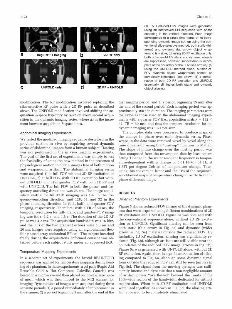
Figure 5 shows reduced-FOV images of the dynamic phan-tom that were acquired using different combinations of 2DRF excitation and UNFOLD. Figure 5a was obtained withthe conventional sequence alone, without 2D RF excita-tion or UNFOLD. Significant aliasing can be seen fromboth static (thin arrow in Fig. 5a) and dynamic (widerarrow in Fig. 5a) material outside the reduced FOV. Byincluding 2D RF excitation, aliasing was significantly re-duced (Fig. 5b), although artifacts are still visible near theboundaries of the reduced FOV image (arrows in Fig. 5b).Figure 5c was generated with UNFOLD alone, without 2DRF excitation. Again, there is significant reduction of alias-ing compared to Fig. 5a, although some dynamic signalfrom outside the reduced FOV can still be seen (arrows inFig. 5c). The signal from the moving syringes was suffi-ciently intense and dynamic that a non-negligible amountof artifact power “overflowed” beyond the limits of the10%-wide region of the bandwidth dedicated for artifactsuppression. When both 2D RF excitation and UNFOLDwere used together, as shown in Fig. 5d, the aliasing arti-fact appeared to be completely eliminated.
FIG. 5. Reduced-FOV images were generatedusing an interleaved EPI sequence with phaseencoding in the vertical direction. Each imagecorresponds to a single time frame of its corre-sponding dynamic image set: (a) using the con-ventional slice-selective method, both static (thinarrow) and dynamic (fat arrow) object wrap-around is visible; (b) using 2D RF excitation only,both outside-of-FOV static and dynamic objectsare suppressed, however, suppression is incom-plete at the boundary of the FOV (see arrows); (c)using the UNFOLD method alone, outside-of-FOV dynamic object wraparound cannot becompletely eliminated (see arrow); (d) a combi-nation of both 2D RF excitation and UNFOLDessentially eliminates both static and dynamicobject aliasing.
1122 Zhao et al.

Figure 6 presents a more extreme example than Fig. 5, toshow more clearly the potential advantages of combiningthe two approaches. In Fig. 6, the reduced FOV is furtherreduced by a factor of 2 (matrix � 128 � 16). The imagesin Fig. 6 were obtained using the same method as Fig. 5(including the same 2D RF excitation) except that onlyevery second k-space line was used for reconstruction. Asexpected, Fig. 6a (without UNFOLD and RF excitation),Fig. 6b (with RF excitation alone), and Fig. 6c (with UN-FOLD alone) all show significant increases in the amountof artifacts as compared to their counterparts in Fig. 5.Note that the result with UNFOLD only (Fig. 6c) also hasaliasing due to static signal contributions (thin arrow).This is because, as described in the Theory section, theUNFOLD method used here is not able to correct for anoverlap factor of more than 2. Using both 2D RF excitationand UNFOLD gives a result (Fig. 6d) in which aliasingartifacts are essentially eliminated. (Note that the artifactalong the vertical direction of the moving syringe, which isindicated with an arrow in Fig. 6d, is due to unresolvedmotion and is not an aliasing artifact.)
Abdominal Imaging Experiments
Figure 7 shows results of the abdominal imaging experi-ments for full-FOV imaging, half-FOV imaging with 2D RFexcitation, and quarter-FOV imaging with RF-UNFOLD. Inthe time that one full-FOV image is acquired, two half-FOV or four quarter-FOV images can be acquired. To em-phasize this difference in temporal resolution, variablenumbers of time frames are shown for the three cases suchthat the total imaging time for each of the acquisitions inFig. 7a–c is constant.
Figure 7a, which we refer to as the “full-FOV” image,was obtained with an FOV of 25 cm, the minimum FOVnecessary to avoid serious wraparound artifact. (In fact,there is some wraparound of signal at the top of the imagefrom a heating pad that was placed under the back of thesubject.) Reference boxes showing the limits of the half-
FOV and quarter-FOV are drawn on the full-FOV image inFig. 7a. There is significant artifact in this full-FOV imagedue to physiological motion, which suggests that at thistemporal resolution, temperature mapping likely would becompromised.
The half-FOV images (Fig. 7b) acquired with 2D RFexcitation do not show any obvious artifact. Note that theRF excitation has effectively suppressed the bright fat sig-nal from the thoracic cage such that there is no evidentwraparound. The temporal resolution of the half-FOV ac-quisition appears to be sufficient to avoid any seriousmotion-related artifact; however, there is a significant evo-lution between successive time frames that is especiallyevident when the image series is played in a movie loop.
The quarter-FOV images (Fig. 7c) were acquired withRF-UNFOLD at four times the temporal resolution of thefull-FOV acquisitions and twice the temporal resolution ofthe half-FOV acquisitions. The combination of UNFOLDwith 2D RF excitation has effectively eliminated any wrap-around in these images. There are no motion-related arti-facts in the images; furthermore, the dynamic series ofimages shows a relatively smooth transition from frame toframe. There are subtle frame-to-frame differences in theimages that are most likely due to the effects of respiration.
Temperature Mapping Experiments
Figure 8 shows the results from the phantom heating ex-periments using the RF-UNFOLD acquisition. Results fromthe three heating periods are presented as temperaturechange contours, with separate contours A–K in steps of0.4° (centigrade) per minute. The contours are overlaid ona gray-level image of the meat phantom. Figure 8a is fromthe first heating period and, as expected, shows the great-est rate of temperature change, up to 4°/min in the hottestregion (bounded by the “K” contour). This region coin-cides with the location at which the heated gel pack wasplaced. Figure 8b and c show results from the second andthird heating periods, respectively. These results also
FIG. 6. Reduced-FOV images were generatedusing the same method as in Fig. 5 except thatonly every second k-space line was used, result-ing in a further reduction (by 2) in the FOV. Eachimage corresponds to a single time frame of itscorresponding dynamic image set: (a) using theconventional slice-selective method, (b) using 2DRF excitation only, (c) using the UNFOLD methodonly, and (d) using a combination of both 2D RFexcitation and UNFOLD.
FIG. 7. Results from abdominal imaging experi-ments using a sequence appropriate for re-duced-FOV temperature mapping. a: Full-FOVimage from series of five acquired at a temporalresolution of 6.4 s per image. b: Half-FOV imagesfrom series of 10 acquired with 2D RF excitationat a temporal resolution of 3.2 s per image. c:Quarter-FOV images from series of 20 acquiredwith RF-UNFOLD at a temporal resolution of1.6 s per image. Phase encoding is in the verticaldirection.
2D RF Excitation and UNFOLD Reduced-FOV MRI 1123

show the greatest rate of heating at the location at whichthe gel pack was placed. As expected, the rate of heating ismuch lower in the second and third periods than in theinitial period. The lowest rate of temperature change is inthe third heating period (Fig. 8c), rising only to 0.4°/min inthe region of maximum temperature rise (bounded by the“A” contour in the figure).
DISCUSSION
When a 2D RF excitation pulse is designed for reduced-FOV imaging, there is a tradeoff between spatial selectivityand pulse duration. Usually a small width and sharp edgeexcitation profile are desired, but they would require adense sampling of a large region of excitation k-space.Such a sampling trajectory usually requires very long RFexcitations. In practice, long-duration RF excitations areoften not acceptable because they prolong the TE, resultingin loss in signal from the T2 or T2* decay. Another draw-back of long RF excitations relates to off-resonance effectsduring the excitation (17). Off-resonance adds spectralselectivity and introduces phase shifts that cannot be eas-ily handled, degrading spatial selectivity. In general, off-resonance effects introduce losses in image resolution, andimperfect spatial phase coherence.
If the duration of the 2D RF is long, it is also lesseffective for increasing temporal resolution in dynamicapplications. For example, consider a multishot EPI se-quence whose sequence length with standard slice-selectRF excitation is equal to TR. The replacement of the stan-dard excitation pulse with a 2D RF excitation adds � to thesequence length. If we assume that N phase encodes areacquired for a full-FOV image, and the echo-train length isM, then the minimum time required to acquire one slicewould normally be (N/M)TR. If 2D RF excitation is usedand the FOV is reduced by a factor of R, then the time toacquire N/R phase encodes is (N/M)(TR �)/R. The sliceimaging time for full-FOV vs. reduced-FOV imaging isR(TR)/(TR �). When � is insignificant with respect toTR, the acceleration factor due to reduced FOV imaging isequal to the FOV reduction factor, R. In the case in which� would be equal to TR, however, the acceleration factorbecomes only R/2. Even in this case, if R is 4 or 8, therewould still be a significant acceleration, although the SNRper unit time might suffer. Clearly, � should be kept asshort as possible to derive the maximum benefit from
reduced-FOV imaging. The theoretical maximum acceler-ation factor, which preserves the SNR per unit time, isreduced by a factor of TR/(TR �).
For the reasons discussed above, the principal goal ofour RF excitation design was to keep the 2D excitation RFpulse as short as possible. The 2D RF pulse designed forthis study had a total pulse length of only 4.2 ms. Thereduction in RF pulse duration was due in part to the useof a non-rectangular (Gaussian weighted) k-space trajec-tory. If a full k-space echo planar trajectory were usedinstead, the pulse duration would roughly double. How-ever, the major factors that keep the pulse short are 1)minimizing the range of excitation k-space covered in theky direction, and 2) minimizing the number of ky lines inthe sampling trajectory. Therefore, much of the reductionin RF pulse length was at the expense of sharpness in theexcitation profile and a reduction in the distance betweenside lobes in the excitation profile.
In reduced-FOV imaging with 2D RF excitation, aliasingartifacts can be caused by the vanishing tails of the excitationprofile (due to the smoothness of the excited profile) or by theside excitation lobes (due to periodicity of the excited pro-file). To prevent side lobe aliasing, the object size must besmaller than D5Gap or D2Gap (Fig. 3). To avoid aliasing fromthe vanishing tails of the profile, the minimum reduced-FOVsize must be no less than D5 or D2. A sharper profile (i.e.,smaller D2 and D5) or a larger distance between lobes (i.e.,large D2Gap and D5Gap) would come at the cost of a longer RFpulse. The addition of UNFOLD allows the acquired FOV tobe reduced from about D5 down to D5/2.
The spatial excitation profile in our abdominal imagingand temperature mapping experiments had a width at 5%of the maximum (D5) of 12.5 cm. If we assume that allspins outside the 5% range are completely suppressed bythis 2D RF excitation, then the minimum reduced FOVwithout any major aliasing artifact using the hybrid 2D RFand UNFOLD is D5/2, or 6.25 cm. In our abdominal imag-ing experiments, D5Gap was set around 37 cm, setting amaximum allowable size for the object. Thus, the maxi-mum acceleration factor using this Gaussian-shaped RFexcitation was approximately 6, i.e., D5Gap/(D5/2) � 37/6.25. The maximum value of 6 is greater than the fourfoldacceleration we actually obtained in our studies, becausethe “full FOV” was set at 25 cm according to the actualobject size, which was smaller than the maximum allow-able object size of 37 cm.
FIG. 8. Temperature mapping results are shown for the heating of a meat phantom. RF-UNFOLD method was used for all acquisitions witha temporal resolution of 1.6 s per image acquisition. a: Temperature change contours for the first heating period are overlaid on an imageof the phantom. b: Temperature change contours for the second heating period, which began 5 min after the end of the first period. c:Temperature change contours for the third heating period, which began 15 min after the end of the second heating period.
1124 Zhao et al.
As discussed above, the maximum acceleration factor ofD5Gap/(D5/2) is based on an assumption that the object sizeis equal to D5Gap. It is also based on the assumption thatthe excited ROI is at the center of the object. If it is desired(as in the case of our abdominal imaging experiments) toexcite a region off-center (see Fig. 7a), the profile shown inFig. 3 must be shifted to the right or left with respect to theobject in the FOV. The maximum allowable object size isthen reduced from D5Gap by the amount that the excitedregion is shifted with respect to the center of the object. Inour experiments, the offset was 5.5 cm from the center ofthe object. Thus, in this case the maximum allowableobject size for a D5Gap of 37 cm is only 31.5 cm (i.e.,37–5.5). The maximum acceleration factor, assuming anobject of size 31.5 cm, is now only 5 (i.e., 31.5/6.25).
In the present implementation of our 2D RF pulse, weuse an echo-planar excitation k-space trajectory. Onemight expect spiral trajectories to be a better choice be-cause spirals are generally efficient and spiral-in trajecto-ries leave the magnetization refocused at the end of thepulse. In our application, however, a spiral trajectory wasnot favored because of the large asymmetry of k-space inthe two dimensions (i.e., the span in kz is much greaterthan in ky). The sampling density in excitation k-spacedepends on the number of spirals, which must be a smallnumber to ensure an RF pulse of short duration. In the ky
direction the sampling density would be similar to thedensity for an echo-planar trajectory. In the kz direction,however, the density would be much lower and the periodof the excitation in the z direction would be too short.
As a potential alternative to 2D RF excitation with UN-FOLD for reduced-FOV imaging, one could select a rect-angular box using standard slice selection along one di-mension and a refocusing pulse along a second orthogonaldirection. This approach has been used in the past ininterventional MRI for MR-guided biopsy (23) and forcryotherapy monitored by MRI (24). The approach is in-herently spin-echo based, however, and is somewhat lim-ited for dynamic applications because the same volumecannot be excited rapidly without saturating the spins.The RF-UNFOLD method presented here can be imple-mented with either spin-echo or gradient-echo sequences.
In conclusion, the combination of 2D RF excitation andUNFOLD can allow significant reductions in the acquiredFOV (and thereby increase temporal resolution) while keep-ing the RF pulse to a practical duration. In the proposedhybrid method, 2D spatially-selective RF excitation can beseen as a means of accelerating the acquisition by reducingthe FOV, while UNFOLD can be seen as a way to relax therequirement for a sharp excitation profile and allow RFpulses to be shorter. With the combination of 2D excitationand the UNFOLD technique, it becomes possible to obtainessentially artifact-free, high-speed dynamic and even real-time images for clinical applications. To even further reducethe image FOV, it may be possible to form combinations withother methods, such as parallel imaging, that are compatiblewith RF excitation and UNFOLD. We are investigating opti-mal ways of combining parallel imaging with 2D RF excita-tion and UNFOLD, and our initial results show that, as couldbe expected, accelerations greater than those obtained by 2DRF excitation and UNFOLD, or by parallel imaging alone, arepossible (25).
ACKNOWLEDGMENTS
The authors thank Dr. Christopher Hardy of GE GlobalResearch for providing pulse sequence support. This workwas supported in part by bioengineering research grantsfrom the Whitaker Foundation to B.M. and L.P.P.
REFERENCES
1. Mansfield P. Multi-planar image formation using NMR spin echoes. JPhys 1977;C10:L55–L58.
2. Hennig J, Nauerth A, Friedburg H. RARE imaging: fast imaging methodfor clinical MRI. Magn Reson Med 1986;3:823–833.
3. Oshio K, Friedburg H. GRASE (gradient and spin-echo) imaging: anovel fast MRI technique. Magn Reson Med 1991;10:344–349.
4. Margosian P, Schmitt F, Purdy D. Faster MR imaging: imaging with halfthe data. Health Care Instrum 1986;1:195–197.
5. Feinberg DA, Hale JD, Watts JC, Kaufman L, Mark A. Halving MRimaging time by conjugation: demonstration at 3.5 kg. Radiology 1986;161:527–531.
6. van Vaals JJ, Tuithof HH, Dixon WT. Increased time resolution indynamic imaging. In: Proceedings of the 11th Annual Meeting ofSMRM, Berlin, Germany, 1992.
7. Brummer ME, et al. Composite k-space windows (keyhole technique) toimprove temporal resolution in a dynamic series of images followingcontrast administration. In: Proceedings of the 11th Annual Meeting ofSMRM, Berlin, Germany, 1992.
8. Riederer SJ, et al. MR fluoroscopy: technical feasibility. Magn ResonMed 1988;8:1–15.
9. Wright RC, et al. Real-time MR fluoroscopic data acquisition and imagereconstruction. Magn Reson Med 1989;12:407–415.
10. Cao Y, Levin DN. Feature-recognizing MRI. Magn Reson Med 1993;30:305–317.
11. Nagle SK, Levin DN. Multiple region MRI. Magn Reson Med 1999;41:774–786.
12. Webb AG, et al. Applications of reduced-encoding MR imaging withgeneralized-series reconstruction (RIGR). J Magn Reson Imaging 1993;3:925–928.
13. Chandra S, et al. Applications of reduced-encoding MR imaging withgeneralized-series reconstruction (RIGR). J Magn Reson Imaging 1996;6:783–797.
14. Hu X, Parrish T. Reduction of field of view for dynamic imaging. MagnReson Med 1994;31:691–694.
15. Madore B, Glover GH, Pelc NJ. Unaliasing by Fourier-encoding theoverlaps using the temporal dimension (UNFOLD), applied to cardiacimaging and fMRI. Magn Reson Med 1999;42:813–828.
16. Madore B. Using UNFOLD to remove artifacts in parallel imaging andin partial-Fourier imaging. Magn Reson Med 2002;48:493–501.
17. Pauly J, Nishimura D, Macovski A. A k-space analysis of small-tip-angle excitation. J Magn Reson 1989;81:43–56.
18. Hardy CJ, Cline HE. Spatial localization in two dimensions using NMRdesigner pulses. J Magn Reson 1989;82:647–654.
19. Reiseberg S, Frahm J, Finsterbusch J. Two dimensional spatially-selec-tive RF excitation pulses in echo-planar imaging. Magn Reson Med2002;47:1186–1193.
20. Zhao L, Panych L. Reduced field-of-view dynamic imaging using thecombination of a two-dimensional spatial excitation pulse and a rFOVreconstruction. In: Proceedings of the 87th Scientific Assembly andAnnual Meeting of the Radiologic Society of North America, Chicago,IL, 2001.
21. Tsao J. On the UNFOLD method. Magn Reson Med 2002;47:202–207.22. Kellman P, Epstein FH, McVeigh ER. Adaptive sensitivity encoding
incorporating temporal filtering (TSENSE). Magn Reson Med 2001;45:846–852.
23. Buecker A, et al. MR-guided biopsy using a T2-weighted single-shotzoom imaging sequence (Local Look technique). J Magn Reson Imaging1998;8:955–959.
24. Tacke J, et al. MR-guided percutaneous cryotherapy of the liver: in vivoevaluation with histologic correlation in an animal model. J MagnReson Imaging 2001;13:50–56.
25. Madore B, Zhao L, Panych LP. Combining 2D RF, UNFOLD and parallelimaging. In: Proceedings of the 11th Annual Meeting of ISMRM, To-ronto, Canada, 2003.
2D RF Excitation and UNFOLD Reduced-FOV MRI 1125