reduces zika virus transmission in south texas
TRANSCRIPT
viruses
Article
High Rate of Non-Human Feeding by Aedes aegyptiReduces Zika Virus Transmission in South Texas
Mark F. Olson 1, Martial L. Ndeffo-Mbah 2, Jose G. Juarez 1 , Selene Garcia-Luna 1,Estelle Martin 1, Monica K. Borucki 3, Matthias Frank 3, José Guillermo Estrada-Franco 4,Mario A. Rodríguez-Pérez 4 , Nadia A. Fernández-Santos 4, Gloria de Jesús Molina-Gamboa 5,Santos Daniel Carmona Aguirre 5, Bernardita de Lourdes Reyes-Berrones 5,Luis Javier Cortés-De la cruz 5, Alejandro García-Barrientos 5, Raúl E. Huidobro-Guevara 5,Regina M. Brussolo-Ceballos 5, Josue Ramirez 6, Aaron Salazar 7, Luis F. Chaves 8 ,Ismael E. Badillo-Vargas 1 and Gabriel L. Hamer 1,*
1 Department of Entomology, Texas A&M University, College Station, TX 77843, USA;[email protected] (M.F.O.); [email protected] (J.G.J.); [email protected] (S.G.-L.);[email protected] (E.M.); [email protected] (I.E.B.-V.)
2 Veterinary Integrative Biosciences, College of Veterinary Medicine, Texas A&M University, College Station,TX 77843, USA; [email protected]
3 Biosciences and Biotechnology Division, Chemistry, Materials and Life Sciences Directorate, LawrenceLivermore National Laboratory, Livermore, CA 94550, USA; [email protected] (M.K.B.);[email protected] (M.F.)
4 Instituto Politécnico Nacional, Centro de Biotecnología Genómica, Cd. Reynosa 88710, Tamaulipas, Mexico;[email protected] (J.G.E.-F.); [email protected] (M.A.R.-P.);[email protected] (N.A.F.-S.)
5 Secretary of Health of the State of Tamaulipas, Epidemiology Directorate, Cd. Victoria 87000, Tamaulipas,Mexico; [email protected] (G.d.J.M.-G.); [email protected] (S.D.C.A.);[email protected] (B.d.L.R.-B.); [email protected] (L.J.C.-D.l.c.);[email protected] (A.G.-B.); [email protected] (R.E.H.-G.);[email protected] (R.M.B.-C.)
6 Health Department, City of Harlingen, TX 78550, USA; [email protected] Hidalgo County Health & Human Services, Edinburg, TX 78539, USA; [email protected] Instituto Costarricense de Investigación y Enseñanza en Nutrición y Salud (INCIENSA), Apartado Postal,
Tres Ríos, Cartago 4-2250, Costa Rica; [email protected]* Correspondence: [email protected]; Tel.: +1-979-862-4067
Received: 23 March 2020; Accepted: 14 April 2020; Published: 17 April 2020�����������������
Abstract: Mosquito-borne viruses are emerging or re-emerging globally, afflicting millions of peoplearound the world. Aedes aegypti, the yellow fever mosquito, is the principal vector of dengue, Zika,and chikungunya viruses, and has well-established populations across tropical and subtropical urbanareas of the Americas, including the southern United States. While intense arboviral epidemicshave occurred in Mexico and further south in the Americas, local transmission in the United Stateshas been minimal. Here, we study Ae. aegypti and Culex quinquefasciatus host feeding patterns andvertebrate host communities in residential environments of South Texas to identify host-utilizationrelative to availability. Only 31% of Ae. aegypti blood meals were derived from humans, while 50%were from dogs and 19% from other wild and domestic animals. In Cx. quinquefasciatus, 67% of bloodmeals were derived from chicken, 22% came from dogs, 9% from various wild avian species, and2% from other mammals including one human, one cat, and one pig. We developed a model for thereproductive number, R0, for Zika virus (ZIKV) in South Texas relative to northern Mexico usinghuman disease data from Tamaulipas, Mexico. We show that ZIKV R0 in South Texas communitiescould be greater than one if the risk of human exposure to Ae. aegypti bites in these communities is atleast 60% that of Northern Mexico communities. The high utilization of non-human vertebrates and
Viruses 2020, 12, 453; doi:10.3390/v12040453 www.mdpi.com/journal/viruses

Viruses 2020, 12, 453 2 of 20
low risk of human exposure in South Texas diminishes the outbreak potential for human-amplifiedurban arboviruses transmitted by Ae. aegypti.
Keywords: Zika virus; Aedes aegypti; Culex quinquefasciatus; host selection; reproductive number
1. Introduction
Mosquito-borne viruses driven principally by Aedes aegypti, have emerged and re-emerged globally,resulting in a large burden of human disease [1]. Globalization and other anthropogenic factors haveallowed this mosquito to thrive in diverse landscapes and facilitate urban transmission cycles ofdengue virus (DENV), chikungunya virus (CHIKV), Zika virus (ZIKV), and others [2]. In the Americas,all four serotypes of DENV have re-emerged causing consistent epidemics from South America toMexico and the Caribbean [3]. The Asian lineage of CHIKV first arrived in the Caribbean in 2014 andspread throughout the Americas in just a few years [4], resulting in 338,963 confirmed human cases [5].ZIKV invaded Brazil in 2013 [6] and rapidly swept through the Americas in a similar fashion, resultingin an estimated 8.5 million cases in Brazil alone [7].
In the continental United States, Ae. aegypti is found throughout the southern states, andrecent enhanced surveys of Stegomyia mosquitoes have documented the presence of this species in26 states [8]. Despite this wide distribution of the primary vector and the Asian tiger mosquito(Ae. albopictus), a secondary vector for these viruses in many locations, the only regions experiencingautochthonous transmission of DENV, CHIKV, and ZIKV by mosquito exposure, are South Floridaand South Texas [9,10]. While the Mexico cities along the U.S.—Mexico border have experiencedconsistent epidemics of Ae. aegypti-driven viruses, markedly fewer human cases have occurred in thecommunities of the Lower Rio Grande Valley (LRGV) on the Texas side of the border. For example, thestate of Tamaulipas, Mexico, recorded an estimated 11,760 probable cases of DENV and 2677 cases ofDengue Hemorrhagic Fever between 2009 and 2019 [11]. In contrast, in the LRGV, local mosquito-borneDENV epidemics occurred only in 2005 and 2013 [9], and the outbreaks were associated with relativelysmall numbers of human cases. For example, in 2005 the LRGV documented three symptomatic casesand six asymptomatic cases of DEN with no travel history [12], compared to 7062 reported DEN casesin Tamaulipas the same year [13]. In contrast, these viruses are recorded with only isolated cases oflocal transmission in South Texas, including a single CHIKV case in Brownsville, TX in 2015 (TexasDepartment of State Health Services, 2016) and 11 cases of locally acquired ZIKV in the LRGV between2016–2017 [14].
Aedes aegypti-driven viruses continue to have intense epidemics in the Americas, resulting in highrates of viremic humans entering the U.S. [15], but minimal local transmission has occurred [16]. Thisdiscrepancy in the magnitude of virus transmission along geo-political boundaries of the U.S.—Mexicoborder has attracted research attention to identify the mechanisms responsible for these patterns. Thisis especially perplexing given that Ae. aegypti in U.S. border communities has comparable relativeabundances in residential neighborhoods to areas with a much higher burden of human diseaseacross the border in Mexico [14,17]. Prior studies have identified several factors contributing tothis discrepancy, which have identified social-ecological factors, such as window screens and airconditioning, that reduce the risk of exposure to the viruses [13,18]. However, despite evidence thathousing quality is associated with virus transmission [18], there remains limited knowledge of howthis influences the ability of Ae. aegypti to feed on humans and how proportional human feeding mightdrive virus transmission potential on both sides of the border.
This study quantifies Ae. aegypti host feeding patterns and vertebrate host availability in residentialenvironments in South Texas, to compare the observed frequency of blood meals relative to the expectedfrequency in the study location. We rely on empirical data on Ae. aegypti abundance, human populationdensity, and epidemiological data of epidemics in South Texas and in Tamaulipas, Mexico, to evaluate
Viruses 2020, 12, 453 3 of 20
the risk of human-amplified urban arbovirus transmission. We present evidence contrary to the mostcommonly reported observation that Ae. aegypti feeds mostly on humans by showing a high utilizationof dogs and other non-human hosts in South Texas, and that this contributes to a lower risk of humanexposure to ZIKV, which reduces epidemic potential.
2. Materials and Methods
2.1. Study Site and Mosquito Collection
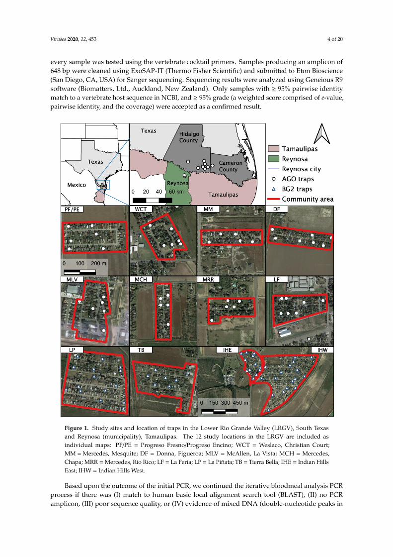
Blood-engorged mosquitoes were collected from several neighborhoods in the Lower Rio GrandeValley (LRGV) on the U.S. side of the U.S.–Mexico border (see Figure 1) from September, 2016 throughDecember, 2018. The climate of Weslaco, Texas, which was used as a representation of the general climatefor the LRGV, includes an average annual high and low temperature of Weslaco (28.7 and 17.4 ◦C) andReynosa, Mexico (29.2 and 17.3 ◦C; climate-data.org). Average annual precipitation (in mm) is 609for Weslaco and 532 for Reynosa. We sampled mosquitoes from eight lower-income (15,000–29,999USD annual household income) neighborhoods (Mercedes, Mesquite (MM); Donna, Figueroa (DF);Mercedes, Chapa (MCH); Progreso Fresno/Progreso Encino (PF/PE); Indian Hills East (IHE); IndianHills West (IHW); La Piñata (LP); Tierra Bella (TB)) and four middle-income (30,000–40,000 USD annualhousehold income) neighborhoods (La Feria, La Bonita (LF); Mercedes, Rio Rico (MRR); McAllen, LaVista (MLV); Weslaco, Christian Court (WCT)) as described by Martin et al. [14]. We used BiogentsSentinel 2 traps (BGS2; Biogents, Germany) with BG lures at IHE, IHW, LP, TB, and DF neighborhoods,placing one trap outside homes once per week for a 24-h trapping period. We also used AutocidalGravid Ovitraps (AGO) at PF/PE, DF, LF, MCH, MLV, MM, and MCH. One AGO was placed inside thehome and an additional trap was placed outside the home, serviced weekly. More details about AGOpreparation and deployment are described in Martin et al. [14]. The selection of homes was largelydictated by obtaining permission from the homeowners to place traps in and around the property. Uponcollection, mosquitoes were identified morphologically based upon illustrations and dichotomous keysfound in The Illustrated Key to Common Mosquitoes of Louisiana [19]. While processing mosquitoes toidentify species and sex, all bloodfed mosquitoes were placed individually into nuclease-free, 1.5 mLmicro-centrifuge tubes, labeled with species, sex, date and house identification number and stored at−20 or −80 ◦C until further processing.
2.2. Blood Meal Analysis
Mosquito samples were identified under microscope, photographed, and given a Sella score (stagesof blood digestion and ovary development) based upon observation of the engorged abdomen [20].The Sella score was used to identify the engorged mosquitoes that had the highest likelihood of yieldinga DNA sequence. To minimize exogenous DNA on the mosquito exoskeleton, each whole mosquitowas washed in 10% bleach followed by two rinses with nuclease-free water [21–24]. On a clean,chilled microscope slide, the abdomen was carefully separated from the rest of the mosquito body,and the abdominal contents were expressed into a new, labeled, DNA-free 1.5 mL microcentrifugetube. A homogenizing bead and 200 µL of lysis solution were added to the tube with blood andshaken for 1 min at 30 Hz in the Qiagen Tissue Lyser (Qiagen, Germantown, MD, USA). DNA wasextracted using the Thermo Scientific™ Kingfisher™ Flex Purification System, along with the MagMAXCore Nucleic Acid Purification Kit (Thermo Fisher Scientific, Waltham, MA, USA) following themanufacturer’s instructions.
We adopted previously-published protocols to conduct a PCR-Sanger sequencing blood mealanalysis [25–27]. Three primer pairs were used in a tiered approach: (I) A vertebrate cocktail targeting a648 base pair region of the cytochrome c oxidase 1 (COI) gene, (II) blood meal (BM) primers targeting a358 base pair region of the cytochrome b gene, and (III) ‘Herp’ primers that target a 228 base pair regionof the cytochrome b gene (Table S1) [27]. This three-tiered approach has the benefits of cost efficiency,maximizing the number of identified blood meals, and increasing reliability of results [25–27]. First,
Viruses 2020, 12, 453 4 of 20
every sample was tested using the vertebrate cocktail primers. Samples producing an amplicon of648 bp were cleaned using ExoSAP-IT (Thermo Fisher Scientific) and submitted to Eton Bioscience(San Diego, CA, USA) for Sanger sequencing. Sequencing results were analyzed using Geneious R9software (Biomatters, Ltd., Auckland, New Zealand). Only samples with ≥ 95% pairwise identitymatch to a vertebrate host sequence in NCBI, and ≥ 95% grade (a weighted score comprised of e-value,pairwise identity, and the coverage) were accepted as a confirmed result.
Viruses 2020, 12, x FOR PEER REVIEW 4 of 20
Figure 1. Study sites and location of traps in the Lower Rio Grande Valley (LRGV), South Texas and Reynosa (municipality), Tamaulipas. The 12 study locations in the LRGV are included as individual maps: PF/PE = Progreso Fresno/Progreso Encino; WCT = Weslaco, Christian Court; MM = Mercedes, Mesquite; DF = Donna, Figueroa; MLV = McAllen, La Vista; MCH = Mercedes, Chapa; MRR = Mercedes, Rio Rico; LF = La Feria; LP = La Piñata; TB = Tierra Bella; IHE = Indian Hills East; IHW = Indian Hills West.
2.2. Blood Meal Analysis
Mosquito samples were identified under microscope, photographed, and given a Sella score (stages of blood digestion and ovary development) based upon observation of the engorged abdomen [20]. The Sella score was used to identify the engorged mosquitoes that had the highest likelihood of yielding a DNA sequence. To minimize exogenous DNA on the mosquito exoskeleton, each whole mosquito was washed in 10% bleach followed by two rinses with nuclease-free water [21–24]. On a clean, chilled microscope slide, the abdomen was carefully separated from the rest of the mosquito body, and the abdominal contents were expressed into a new, labeled, DNA-free 1.5 mL microcentrifuge tube. A homogenizing bead and 200 µL of lysis solution were added to the tube with blood and shaken for 1 min at 30 Hz in the Qiagen Tissue Lyser (Qiagen, Germantown, MD, USA).
Figure 1. Study sites and location of traps in the Lower Rio Grande Valley (LRGV), South Texasand Reynosa (municipality), Tamaulipas. The 12 study locations in the LRGV are included asindividual maps: PF/PE = Progreso Fresno/Progreso Encino; WCT = Weslaco, Christian Court;MM = Mercedes, Mesquite; DF = Donna, Figueroa; MLV = McAllen, La Vista; MCH = Mercedes,Chapa; MRR = Mercedes, Rio Rico; LF = La Feria; LP = La Piñata; TB = Tierra Bella; IHE = Indian HillsEast; IHW = Indian Hills West.
Based upon the outcome of the initial PCR, we continued the iterative bloodmeal analysis PCRprocess if there was (I) match to human basic local alignment search tool (BLAST), (II) no PCRamplicon, (III) poor sequence quality, or (IV) evidence of mixed DNA (double-nucleotide peaks in
Viruses 2020, 12, 453 5 of 20
chromatograph) [27]. If we obtained any of these four outcomes, a second PCR utilizing the BM1:BM2primers was conducted [25,27]. Finally, using the same criteria, the analysis was either concluded orsubjected to a third primer pair and PCR thermal profile, the ‘Herp’ primers [25,27].
We followed the protocol of Medeiros et al. [27] for the vertebrate cocktail reaction, but modifiedthe thermal cycling conditions as follows: after denaturation we ran nine cycles of 94 ◦C for 30 s, agradient from 45 to 54 ◦C for 40 s, and 72 ◦C for 1 min. The remaining thermal cycling conditionswere identical to the Medeiros protocol. For the BM and ‘Herp’ reactions, we followed the protocolof Hamer et al. [25] with the following modification: we lowered the annealing temperature for the‘Herp’ reaction from 50 to 47 ◦C. The vertebrate cocktail, BM1:BM2, and ‘herp’ PCR reactions used thefollowing reagents and quantities per reaction: 8.59 µL Nuclease-free H2O, 12.5 µL FailSafe™ PCR2X Premix E (Lucigen, Middleton, WI, USA), 0.83 µL forward primer, 0.83 µL reverse primer, 0.25 µLFailSafe™ PCR Enzyme Mix (Lucigen), and 2 µL DNA template.
The protocol was tested using lab-raised, Aedes aegypti and Culex quinquefasciatus mosquitoes fedon defibrinated sheep blood (HemoStat Laboratories, Dixon, CA, USA). Adult female mosquitoeswere offered artificial blood meals using a Hemotek membrane feeder (Hemotek Ltd., Blackburn, UK).Each specimen was observed under a dissecting light microscope to confirm species, give a Sella score,and capture a digital photograph. We also extracted DNA directly from the blood of several controlsincluding iguana (Iguana iguana), white-tailed deer (Odocoileus virginianus), tiger (Panthera tigris),sandhill crane (Grus canadensis), and sheep (Ovis aries). These vertebrate species were selected becausethey are unlikely to be found in mosquito blood meals from this region and would thus minimize therisk of downstream amplicon contamination.
2.3. Molecular Verification of Mosquito Species
While most mosquitoes captured via BGS2 traps could be taxonomically classified frommorphological features, those collected from the glue boards of the AGO traps are often damaged andmore difficult to identify morphologically. Therefore, molecular identification of mosquito species wasconfirmed using a modified version of the protocol designed by Folmer et al. [28]. Briefly, a primerpair that amplifies a 710-bp fragment of the cytochrome c oxidase subunit I gene (LCO 1490 and HCO2198; Table S2) was used with the following reagents and quantities per reaction: 8 µL Nuclease-freeH2O, 12.5 µL FailSafe™ PCR 2X Premix E (Lucigen), 1 µL forward primer (LCO 1490), 1 µL reverseprimer (HCO 2198), 0.5 µL FailSafe™ PCR Enzyme Mix (Lucigen), and 2 µL DNA template. The PCRthermal cycling profile included initial denaturation for three minutes at 95 ◦C followed by 35 cycles of95 ◦C for 1 min, 45 ◦C for 1.5 min, and 72 ◦C for 2 min, followed by a final extension at 72 ◦C for 5 min.Amplified PCR products were purified using Exo-SAP-IT™ (ThermoFisher Scientific) and sent to EtonBioscience (San Diego) for Sanger sequencing.
2.4. Quantitative Synthesis of Published Literature
We compiled all the published data on Ae. aegypti host feeding patterns from around theworld. To systematically review the literature, we searched PubMed, Web of Science, and GoogleScholar for published literature using keywords “Aedes aegypti host feeding”, and a second search of“Aedes aegypti blood meal analysis”. These queries in Web of Science yielded 50 and seven results,respectively, in PubMed yielded 13 and four results, respectively, and in Google Scholar yielded 1970and 450 results, respectively. We also tracked references from key review papers and other primaryliterature. Inclusion criteria included blood meal results from wild-caught mosquitoes. Studies usinglaboratory-raised mosquitoes, as well as studies which indicated samples were likely from the formAe. aegypti formosus [29–31], were excluded. The form formosus was excluded to allow a focus on theurban form of Ae. Aegypti, which is globally distributed.
Viruses 2020, 12, 453 6 of 20
2.5. Mosquito Relative Abundance
Female Ae. aegypti relative abundance was estimated in the LRGV and the city of Reynosa,Tamaulipas, Mexico, using AGO traps that were deployed concurrently on both sides of the border in2017 (Figure 1). Eighty AGO traps were deployed outside residential homes in Reynosa and checkedweekly between May 7 and Aug 12 (trapping data were unavailable for two weeks in this period dueto adverse weather or trap failure) [32]. In the LRGV, 30 AGO traps were deployed outside residentialhomes during the same weeks as a subset of the data presented in a previous study [14].
2.6. Vertebrate Surveys
In order to estimate mosquito host selection, a questionnaire related to vertebrate availabilitywas developed and conducted in the four primary communities where blood-fed mosquitoes werecollected. Project personnel visited all the homes containing BG Sentinel 2 traps in these communities:14 (out of 307 total homes present) in Indian Hills East, 10 (96) in Indian Hills West, 13 (160) in LaPiñata, and seven (49) in Tierra Bella. An adult from each home was asked for the number of personsliving at each residence (further categorized by age group < 5; 5–17; 18–65; > 65), the number of dogs,cats, pet birds, chickens, pigs, horses, and other animals. Of the dogs and cats, the number of themroaming outside of the property was also noted. From previous observations, we suspected that alarge number of stray dogs and cats live in some of these neighborhoods, therefore a final questionregarding the number of strays that the adult resident is aware of was also asked. Results from thesurveys were tabulated and relative abundance calculated with 95% confidence intervals for selectedvertebrates, using the Wilson/Brown method (Table S3) [33].
Populations of human, dog, cat, chicken, pig, and opossum were estimated by extrapolating thevertebrates documented from survey homes to create estimates of the number of each vertebrate perunit area in the entire community. To achieve this, the average number of vertebrates in the surveyedhomes was multiplied by the total number of homes in the defined community to arrive at an estimateddensity of each vertebrate per unit area. This number was divided by the total estimated number of allpotential hosts to obtain relative abundance. Population estimates of wild birds and wild mammals(rodents, meso-predators, etc.) were not obtained.
2.7. Human Density Estimation
We used remote sensing satellite imagery (Google Earth, California, USA) to map the communitieswithin the LRGV using QGIS 3.4 (QGIS Development team 2019). We estimated household densitiesusing the 2010 US census blocks shape file and extracted the information regarding number of houses,number people/house and area of the community. We also quantified household density in NuevoAmanecer as a representative community in the city of Reynosa with prior DENV transmission activity(Rodríguez-Pérez Mario A, A. M. A., Russell Tanya L, Olguin-Rodriguez Omar, Laredo-TiscareñoStephanie V, Garza-Hernandez Javier A, Reyes-Villanueva Filiberto. Host-seeking Aedes aegyptilinked to dengue seropositive households at northeastern Mexico. Journal of Vector Borne Diseases(in press)). We used satellite imagery (Google Earth, California, USA) to quantify homes manuallyand census block information to identify the boundaries of the neighborhood. Nuevo Amanecer waschosen because of its known dengue endemicity.
2.8. Host Selection Indices
We estimated the Forage Ratio (FR), the frequency at which a mosquito selects a vertebrate hostover other available vertebrate hosts, by dividing the observed frequency of bloodmeals divided bythe expected frequency of bloodmeals of a given species [34]
FR = s/a, (1)
Viruses 2020, 12, 453 7 of 20
where s = the percent of female mosquitoes containing blood of a particular host, and a = percent of thetotal available host population represented by that particular host [35]. A forage ratio of 1.0 indicatesmosquitoes are feeding on hosts in equal proportion to availability, whereas values >1.0 indicateover-utilization and values <1.0 indicate under-utilization. We used the Wilson/Brown statisticalmethod to calculate 95% confidence intervals [33].
We also estimated the human blood index (HBI), which measures the frequency at which femalemosquitoes feed on human hosts and is the number of human blood meals divided by the number ofengorged females [36].
2.9. Tamaulipas Human Disease Data
The General Directorate of Epidemiology, Secretariat of Health, México aggregates probable andconfirmed empirical cases of DEN, CHIK, and ZIK in the state of Tamaulipas (Figure S1). Patientswith history of travel outside of Tamaulipas in the month prior to onset of symptoms were notincluded in the modeling of R0. Physicians of symptomatic patients use a case definition of DEN:fever with ≥2 signs or symptoms such as retro-orbital or ocular pain, rash, headache, arthralgia,myalgia, leukopenia or hemorrhagic manifestations; CHIK: severe arthralgia, intense asymmetric,debilitating joint pain, swelling associated with tenosynovitis; and, ZIK: pruritic maculopapular rash,for differential clinical diagnosis between the three viruses and are required to report cases to theSecretary of Health of Tamaulipas. Clinical serum samples receiving laboratory confirmation weresent by sanitary jurisdictions of the state to the Molecular Biology Laboratory of the Tamaulipas StatePublic Health Laboratory. They were stored at −20 ◦C until further processing.
Nucleic acid extraction was performed using a MagNA Pure LC total nucleic acid isolation kit ina MagNA Pure LC 2.0 Instrument (Roche Applied Science, Germany). The extracted viral RNA wasstored at −70 ◦C. We used RT-PCR to detect the presence of arboviruses using protocols previouslydescribed [37]. We used the SuperScript III Platinum® One-Step qRT-PCR System enzyme (Invitrogen,Carlsbad, CA, USA). A 7500 Fast Real-Time Thermocycler from Applied Biosystems (Foster City, CA,USA) was used, and reportable positive values were below a Ct value of 38.
For ZIKV, the primary patients that received laboratory confirmation using RT-PCR were pregnantfemales. For the modeling in this study, we used the empirical data of probable cases of ZIKV in themunicipality of Reynosa in 2017 with no recent travel history (Figure S2).
2.10. Mathematical Modeling
The Ross MacDonald formulation of the basic reproductive number for mosquito-bornediseases [38] is defined as the average number of secondary human cases generated by an index casein an otherwise susceptible population
R0 =m(a f )2bce −µEIP
µ r, (2)
where m: the density of female mosquitoes to human, a: female mosquito biting rate, f : the proportionof mosquito feeding on human, b: mosquito-to-human transmission probability, c: human-to-mosquitotransmission probability, EIP: extrinsic incubation period, 1/r: human average infectious period, µ:adult mosquito mortality rate [38]. The density of Ae. aegypti to humans is a function of mosquitodensity and human exposure to mosquitoes [38–40] m = D × E where D is mosquito density andE is the human risk of exposure to Ae. aegypti. The risk of exposure to mosquitoes is a functionof socioeconomic variables, such as the availability of air conditioning, which can drastically limitmosquito–human contacts and virus transmission, even when mosquitoes are abundant [18]. Studieshave shown that population mobility may also play a role in individuals exposure risk to Ae. aegypti [41].Furthermore, fine-scale variation in population susceptibility, immunity, or social structures mayalso be factors contributing to vector-borne disease transmission heterogeneity amongst neighboring
Viruses 2020, 12, 453 8 of 20
communities [42]. Though these parameters may be hard to measure empirically, they can play apivotal role in the risk of mosquito borne disease outbreaks.
Given the geographical proximity and similarities between the city of Reynosa and neighborhoodsin the LRGV of South Texas, any difference in the risk of outbreak (R0), for a newly introduced Ae.Aegypti-borne disease such as Zika, between the two communities would be due to the density ofmosquito to human (m) or the proportion of mosquito feeding on humans (f ). Therefore, R0 in the LRGV
and Reynosa can be written as RLRGV0 =
mLRGV(a fLRGV)2bce −µEIP
µ r and RRey0 =
mRey(a fRey)2bce−µEIP
µ r , respectively.
We have RRey0 = mRey
(fRey
)2 (a)2bce−µEIP
µ r , which is rearranged as (a)2bce −µEIP
µ r = RRey0
1mRey( fRey)
2 This
implies that
RLRGV0 = mLRGV( fLRGV)
2 (a)2bce −µEIP
µ r= mLRGV( fLRGV)
2RRey0
1
mRey(
fRey)2
RLRGV0 = RRey
0mLRGVmRey
(fLRGV
fRey
)2
, (3)
where RLRGV0 and RRey
0 are the basic reproductive numbers in the LRGV and Reynosa, respectively.mLRGV and mRey are the density of female Ae. aegypti to human in the LRGV and Reynosa, respectively;and fLRGV and fRey are the proportion of Ae. aegypti feeding on humans in the LRGV and Reynosa,respectively. We estimated the ratio mLRGV
mRey=
DLRGVELRGVDReyERey
. Female Ae. aegypti relative abundance wasestimated using AGO data collected in 2017 during the same weeks in the city of Reynosa (5.16 femaleAe. aegypti per AGO per week) and in the LRGV (4.16 female Ae. aegypti per AGO per week) [14]. SomLRGVmRey
= 0.8 ELRGVERey
. As the proportion of feeding on human in Reynosa is not currently available, weconsidered a range of values informed by available data from the Americas (Table 1): LRGV, PuertoRico, and Florida.
R0 in Reynosa municipality was estimated using case data for the 2017 Zika epidemic and theEstimateR function from the EpiEstim R library [43,44] to estimate the time-dependent reproductivenumber, R(t), based on the method introduced by Cori et al. [43]. We derive R0 using the fact that R0 isequal to R(t) at the start of the outbreak, such as Zika, for which we do not have pre-existing immunityin the population [44]. This approach would not be applicable to endemic diseases such as dengue.The instantaneous reproduction number R(t) was computed over 4-week sliding windows using themethod introduced by Cori et al. [43]. This approach uses a Bayesian inference method to propagateuncertainty of data and generation time into R0 estimate. Following Ferguson et al. [44], we assumethat the ZIKV generation time is gamma-distributed with a mean of 20.0 days and a standard deviation(s.d.) of 7.4 days. The incidence data themselves may contain many potential sources of uncertaintysuch as misdiagnosis, variable time-dependent case detection rate, and asymptomatic cases, which arenot explicitly taken into account into our analysis.
Viruses 2020, 12, 453 9 of 20
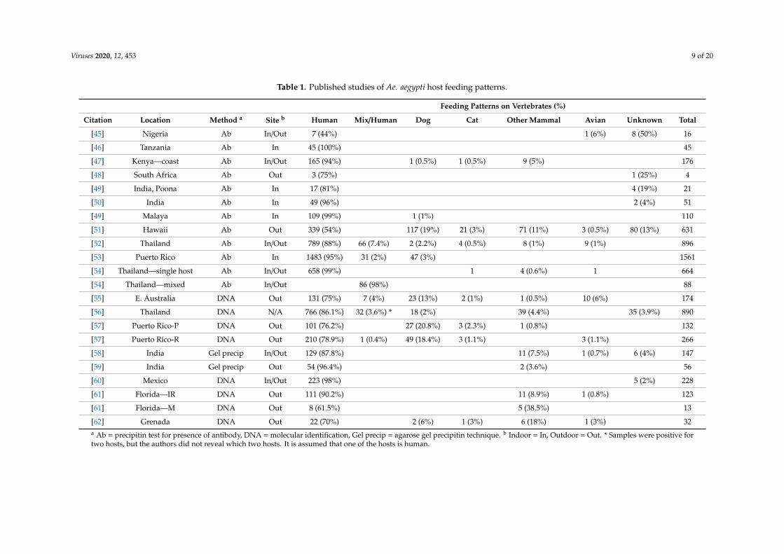
Table 1. Published studies of Ae. aegypti host feeding patterns.
Feeding Patterns on Vertebrates (%)
Citation Location Method a Site b Human Mix/Human Dog Cat Other Mammal Avian Unknown Total
[45] Nigeria Ab In/Out 7 (44%) 1 (6%) 8 (50%) 16
[46] Tanzania Ab In 45 (100%) 45
[47] Kenya—coast Ab In/Out 165 (94%) 1 (0.5%) 1 (0.5%) 9 (5%) 176
[48] South Africa Ab Out 3 (75%) 1 (25%) 4
[49] India, Poona Ab In 17 (81%) 4 (19%) 21
[50] India Ab In 49 (96%) 2 (4%) 51
[49] Malaya Ab In 109 (99%) 1 (1%) 110
[51] Hawaii Ab Out 339 (54%) 117 (19%) 21 (3%) 71 (11%) 3 (0.5%) 80 (13%) 631
[52] Thailand Ab In/Out 789 (88%) 66 (7.4%) 2 (2.2%) 4 (0.5%) 8 (1%) 9 (1%) 896
[53] Puerto Rico Ab In 1483 (95%) 31 (2%) 47 (3%) 1561
[54] Thailand—single host Ab In/Out 658 (99%) 1 4 (0.6%) 1 664
[54] Thailand—mixed Ab In/Out 86 (98%) 88
[55] E. Australia DNA Out 131 (75%) 7 (4%) 23 (13%) 2 (1%) 1 (0.5%) 10 (6%) 174
[56] Thailand DNA N/A 766 (86.1%) 32 (3.6%) * 18 (2%) 39 (4.4%) 35 (3.9%) 890
[57] Puerto Rico-P DNA Out 101 (76.2%) 27 (20.8%) 3 (2.3%) 1 (0.8%) 132
[57] Puerto Rico-R DNA Out 210 (78.9%) 1 (0.4%) 49 (18.4%) 3 (1.1%) 3 (1.1%) 266
[58] India Gel precip In/Out 129 (87.8%) 11 (7.5%) 1 (0.7%) 6 (4%) 147
[59] India Gel precip Out 54 (96.4%) 2 (3.6%) 56
[60] Mexico DNA In/Out 223 (98%) 5 (2%) 228
[61] Florida—IR DNA Out 111 (90.2%) 11 (8.9%) 1 (0.8%) 123
[61] Florida—M DNA Out 8 (61.5%) 5 (38.5%) 13
[62] Grenada DNA Out 22 (70%) 2 (6%) 1 (3%) 6 (18%) 1 (3%) 32a Ab = precipitin test for presence of antibody, DNA = molecular identification, Gel precip = agarose gel precipitin technique. b Indoor = In, Outdoor = Out. * Samples were positive fortwo hosts, but the authors did not reveal which two hosts. It is assumed that one of the hosts is human.
Viruses 2020, 12, 453 10 of 20
3. Results
3.1. Blood Meal Analysis
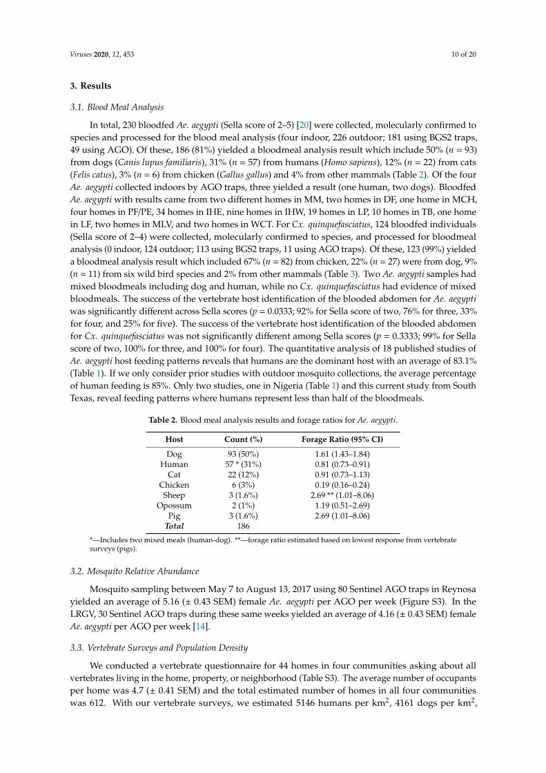
In total, 230 bloodfed Ae. aegypti (Sella score of 2–5) [20] were collected, molecularly confirmed tospecies and processed for the blood meal analysis (four indoor, 226 outdoor; 181 using BGS2 traps,49 using AGO). Of these, 186 (81%) yielded a bloodmeal analysis result which include 50% (n = 93)from dogs (Canis lupus familiaris), 31% (n = 57) from humans (Homo sapiens), 12% (n = 22) from cats(Felis catus), 3% (n = 6) from chicken (Gallus gallus) and 4% from other mammals (Table 2). Of the fourAe. aegypti collected indoors by AGO traps, three yielded a result (one human, two dogs). BloodfedAe. aegypti with results came from two different homes in MM, two homes in DF, one home in MCH,four homes in PF/PE, 34 homes in IHE, nine homes in IHW, 19 homes in LP, 10 homes in TB, one homein LF, two homes in MLV, and two homes in WCT. For Cx. quinquefasciatus, 124 bloodfed individuals(Sella score of 2–4) were collected, molecularly confirmed to species, and processed for bloodmealanalysis (0 indoor, 124 outdoor; 113 using BGS2 traps, 11 using AGO traps). Of these, 123 (99%) yieldeda bloodmeal analysis result which included 67% (n = 82) from chicken, 22% (n = 27) were from dog, 9%(n = 11) from six wild bird species and 2% from other mammals (Table 3). Two Ae. aegypti samples hadmixed bloodmeals including dog and human, while no Cx. quinquefasciatus had evidence of mixedbloodmeals. The success of the vertebrate host identification of the blooded abdomen for Ae. aegyptiwas significantly different across Sella scores (p = 0.0333; 92% for Sella score of two, 76% for three, 33%for four, and 25% for five). The success of the vertebrate host identification of the blooded abdomenfor Cx. quinquefasciatus was not significantly different among Sella scores (p = 0.3333; 99% for Sellascore of two, 100% for three, and 100% for four). The quantitative analysis of 18 published studies ofAe. aegypti host feeding patterns reveals that humans are the dominant host with an average of 83.1%(Table 1). If we only consider prior studies with outdoor mosquito collections, the average percentageof human feeding is 85%. Only two studies, one in Nigeria (Table 1) and this current study from SouthTexas, reveal feeding patterns where humans represent less than half of the bloodmeals.
Table 2. Blood meal analysis results and forage ratios for Ae. aegypti.
Host Count (%) Forage Ratio (95% CI)
Dog 93 (50%) 1.61 (1.43–1.84)Human 57 * (31%) 0.81 (0.73–0.91)
Cat 22 (12%) 0.91 (0.73–1.13)Chicken 6 (3%) 0.19 (0.16–0.24)Sheep 3 (1.6%) 2.69 ** (1.01–8.06)
Opossum 2 (1%) 1.19 (0.51–2.69)Pig 3 (1.6%) 2.69 (1.01–8.06)
Total 186
*—Includes two mixed meals (human-dog). **—forage ratio estimated based on lowest response from vertebratesurveys (pigs).
3.2. Mosquito Relative Abundance
Mosquito sampling between May 7 to August 13, 2017 using 80 Sentinel AGO traps in Reynosayielded an average of 5.16 (± 0.43 SEM) female Ae. aegypti per AGO per week (Figure S3). In theLRGV, 30 Sentinel AGO traps during these same weeks yielded an average of 4.16 (± 0.43 SEM) femaleAe. aegypti per AGO per week [14].
3.3. Vertebrate Surveys and Population Density
We conducted a vertebrate questionnaire for 44 homes in four communities asking about allvertebrates living in the home, property, or neighborhood (Table S3). The average number of occupantsper home was 4.7 (± 0.41 SEM) and the total estimated number of homes in all four communitieswas 612. With our vertebrate surveys, we estimated 5146 humans per km2, 4161 dogs per km2,

Viruses 2020, 12, 453 11 of 20
1751 cats per km2, 2299 chickens per km2, and 75 pigs per km2 (Table S4). Independent from thehousehold questionnaires, our analysis of US Census data using QGIS for the combined communitiesin the current study where blood-fed individuals were collected and in the eight communities withAGO surveillance [14], we estimate that on average the human density was 3,597 per km2 (Table S5).In Nuevo Amanecer, Reynosa, we identified 885 homes in the neighborhood minus the soccer fieldopen space. The area with homes is 0.27 km2 and using an average occupancy of 4.2 persons per home(based on unpublished data from co-author M. A. Rodríguez-Pérez), the estimated human density forthis area is 13,767 per km2. The human density in Reynosa is between 2.7- and 3.8-fold higher thancomparable low-income communities in the LRGV.
Table 3. Blood meal analysis results and forage ratios for Cx. quinquefasciatus.
Host Count (%) Forage Ratio (95% CI)
Chicken 82 (67%) 3.92 (3.33–4.87)Dog 27 (22%) 0.71 (0.63–0.81)
House sparrow 6 (5%) -Western kingbird 1 (0.8%) -
Human 1 (0.8%) 0.02 (0.02–0.02)Cat 1 (0.8%) 0.06 (0.05–0.08)Pig 1 (0.8%) 1.36 (0.51–4.07)
Plain chachalaca 1 (0.8%) -Curvebilled
thrasher 1 (0.8%) -
Northernmockingbird 1 (0.8%) -
Rock dove 1 (0.8%) -Total 123
3.4. Host Selection
Forage ratios for Ae. aegypti and Cx. quinquefasciatus were calculated with host availabilityestimated from our vertebrate surveys in the neighborhoods where we collected engorged mosquitoes.The Ae. aegypti forage ratio (observed frequency of bloodmeals from a given host divided by theexpected frequency) for dogs (1.61) was nearly twice the forage ratio for humans (0.81; Table 2). Incontrast, the highest forage ratio for Cx. quinquefasciatus was on chicken (3.92; Table 3). The humanblood index (total number of human blood meals divided by the total number of engorged femaleswith a confirmed result) for Ae. aegypti and Cx. quinquefasciatus was 30.7% and 0.8% respectively.
3.5. Mathematical Modeling
Using the Ross MacDonald equation for the basic reproductive number, R0, and based on the 2017cases of ZIKV in Reynosa (Figure S2) and data on Ae. aegypti collected in the LRGV and Reynosa, weestimated ZIKV RLRGV
0 in the LRGV. We started by estimating RRey0 in Reynosa using case data from
the 2017 Zika outbreak in Reynosa, where R0 was 2.2 (95% Confidence Interval: 1.1—3.8) (Figure S2).A total of 330 cases of Zika were observed in Reynosa in 2017; only a subset were tested by PCR,of which 81 were confirmed positive for ZIKV RNA. Seven Zika cases were not included, given ahistory of travel in the prior month. Because of the geographical proximity between Reynosa and theLRGV, we assumed that all parameters, except for mosquito abundance and human biting rates, in theRoss MacDonald R0 equation were equal across the US-Mexico border. We obtained the followingexpression for R0 in the LRGV
RLRGV0 = RRey
0 0.8ELRGVERey
(0.31fRey
)2
(4)
where 0.8 is the ratio between mosquito abundance in LRGV and Reynosa estimated with AGO traps,and 0.31 is the proportion of Ae. aegypti feeding on humans in LRGV. fRey is the proportion of Ae. aegypti
Viruses 2020, 12, 453 12 of 20
feeding on human in Reynosa, and ERey (ELRGV) is the risk of human exposure to Ae. aegypti bites in
Reynosa (LRGV). Then, with the RRey0 estimate and using different combinations for the unknown
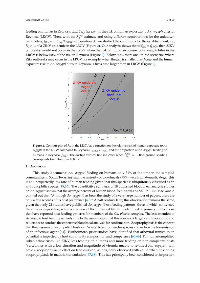
parameters, fRey and ERey/ELRGV, of Equation (4) we studied the conditions for the establishment, i.e.,R0 > 1, of a ZIKV epidemic in the LRGV (Figure 2). Our analysis shows that if fRey = fLRGV then ZIKVoutbreaks would not occur in the LRGV when the risk of human exposure to Ae. aegypti bites in theLRGV is below 60% of the risk in Reynosa (Figure 2). Below 60%, there are limited scenarios whereZika outbreaks may occur in the LRGV: for example, when the fRey is smaller than fLRGV and the humanexposure risk to Ae. aegypti bites in Reynosa is fives time larger than in LRGV (Figure 2).
Viruses 2020, 12, x FOR PEER REVIEW 12 of 20
where 0.8 is the ratio between mosquito abundance in LRGV and Reynosa estimated with AGO traps, and 0.31 is the proportion of Ae. aegypti feeding on humans in LRGV. 𝑓 is the proportion of Ae. aegypti feeding on human in Reynosa, and 𝐸 (𝐸 ) is the risk of human exposure to Ae. aegypti bites in Reynosa (LRGV). Then, with the 𝑅 estimate and using different combinations for the unknown parameters, fRey and ERey/ELRGV, of Equation (4) we studied the conditions for the establishment, i.e., R0 > 1, of a ZIKV epidemic in the LRGV (Figure 2). Our analysis shows that if fRey = fLRGV then ZIKV outbreaks would not occur in the LRGV when the risk of human exposure to Ae. aegypti bites in the LRGV is below 60% of the risk in Reynosa (Figure 2). Below 60%, there are limited scenarios where Zika outbreaks may occur in the LRGV: for example, when the fRey is smaller than fLRGV and the human exposure risk to Ae. aegypti bites in Reynosa is fives time larger than in LRGV (Figure 2).
Figure 2. Contour plot of 𝑅 in the LRGV as a function on the relative risk of human exposure to Ae. aegypti in the LRGV compared to Reynosa (𝐸 /𝐸 ), and the proportion of Ae. aegypti feeding on humans in Reynosa (fRey). The dashed vertical line indicates when = 1. Background shading
corresponds to contour predictions.
4. Discussion
This study documents Ae. aegypti feeding on humans only 31% of the time in the sampled communities in South Texas; instead, the majority of bloodmeals (50%) were from domestic dogs. This is an unexpectedly low rate of human feeding given that this species is ubiquitously classified as an anthropophilic species [18,63]. The quantitative synthesis of 18 published blood meal analysis studies on Ae. aegypti shows that the average percent of human blood-feeding was 83.8%. In 1967, MacDonald pointed out that “Although Ae. aegypti has been the study of a very large number of papers, there are only a few records of its host preference [49].” A half century later, this observation remains the same, given that only 21 studies have published Ae. aegypti host feeding patterns, three of which concerned the subspecies formosus, while our review of the published literature identified 86 primary publications that have reported host feeding patterns for members of the Cx. pipiens complex. The less attention to Ae. aegypti host feeding is likely due to the assumption that this species is largely anthropophilic and reluctance to conduct the expensive bloodmeal analysis for confirmation. Zooprophylaxis is the concept that the presence of incompetent hosts can ‘waste’ bites from vector species and reduce the transmission of an infectious agent [64]. Furthermore, prior studies have identified that arboviral transmission potential is impacted by host community composition and competence [65,66]. For human-amplified urban arboviruses like ZIKV, less feeding on humans and more feeding on non-competent hosts (vertebrates with a low duration and magnitude of viremia unable to re-infect Ae. aegypti), will have a zooprophylactic effect on
Figure 2. Contour plot of R0 in the LRGV as a function on the relative risk of human exposure to Ae.aegypti in the LRGV compared to Reynosa (ELRGV/ERey), and the proportion of Ae. aegypti feeding on
humans in Reynosa (fRey). The dashed vertical line indicates when fLRGVfRey
= 1. Background shadingcorresponds to contour predictions.
4. Discussion
This study documents Ae. aegypti feeding on humans only 31% of the time in the sampledcommunities in South Texas; instead, the majority of bloodmeals (50%) were from domestic dogs. Thisis an unexpectedly low rate of human feeding given that this species is ubiquitously classified as ananthropophilic species [18,63]. The quantitative synthesis of 18 published blood meal analysis studieson Ae. aegypti shows that the average percent of human blood-feeding was 83.8%. In 1967, MacDonaldpointed out that “Although Ae. aegypti has been the study of a very large number of papers, there areonly a few records of its host preference [49].” A half century later, this observation remains the same,given that only 21 studies have published Ae. aegypti host feeding patterns, three of which concernedthe subspecies formosus, while our review of the published literature identified 86 primary publicationsthat have reported host feeding patterns for members of the Cx. pipiens complex. The less attention toAe. aegypti host feeding is likely due to the assumption that this species is largely anthropophilic andreluctance to conduct the expensive bloodmeal analysis for confirmation. Zooprophylaxis is the conceptthat the presence of incompetent hosts can ‘waste’ bites from vector species and reduce the transmissionof an infectious agent [64]. Furthermore, prior studies have identified that arboviral transmissionpotential is impacted by host community composition and competence [65,66]. For human-amplifiedurban arboviruses like ZIKV, less feeding on humans and more feeding on non-competent hosts(vertebrates with a low duration and magnitude of viremia unable to re-infect Ae. aegypti), willhave a zooprophylactic effect on transmission, as originally observed with cattle when describingzooprophylaxis in malaria transmission [67,68]. This has principally been considered an important
Viruses 2020, 12, 453 13 of 20
phenomenon in human malaria transmission with Anopheles spp. wasting bites on cattle, somethingthat protects humans from malaria infectious bites [69–71]. However, Hess and Hayes [64] determinedthat potential for zooprophylaxis exists in Cx. tarsalis, Cx. pipiens, Cx. quinquefasciatus and Ae. albopictus.Moreover, in East Africa the observation of non-human host feeding by Ae. aegypti led to the conclusionthat these were likely to be poor vectors of yellow fever virus [29].
Non-human feeding by Ae. aegypti may have important consequences for arboviral pathogentransmission. For example, the receptivity of certain regions of the world to Ae. Aegypti-drivenarboviruses might vary not simply due to the abundance of Ae. aegypti, but due to the proclivity andavailability of Ae. aegypti to feed on humans relative to other hosts. In that sense, threshold indices,such as R0, that indicate when the transmission of viruses will persist, can guide management activitiesand even inform urban planning and home modification to further reduce the probability of Ae. aegyptifeeding on humans.
Collection technique and location can influence the apparent host feeding patterns. While only 2%of the collections in this study were from inside homes, all of the specimens were collected within theresidential yard. While our previous study documented Ae. aegypti inside homes of the LRGV [14], wedid not target indoor collections with aspirators, given the unique socio-demographics and politicalclimate of the LRGV, which makes indoor access challenging. Prior studies on Ae. aegypti host feedingpatterns are limited, with only ten studies (56%) with indoor collections, seven (39%) with collectionsfrom residential yards, and two studies (11%) with collections in non-residential locations (Table 1).Of the published studies reporting Ae. aegypti blood meal results, most (69%) have been conducted inregions of the world where dengue is endemic (Table 1). This identifies a research gap, with a fewstudies such as this one reporting Ae. aegypti host feeding patterns in areas where the environmentmay be suitable for arboviral transmission, but risk appears to be diminished by limited access tohumans, and, possibly easier access to non-human vertebrates. We also did not process blood-engorgedmosquitoes collected in Reynosa in the same laboratory as the current study, which is a priority forfuture research.
The low rate of Ae. aegypti anthropophily in the current study could be explained by severalreasons. The most parsimonious explanation is the higher availability of non-human hosts, limitedopportunity for human biting, and lower human density [18,63]. In our Texas study sites, humansmake up 41% of all domestic hosts in the four study sites combined. Our analysis does not accountfor wild birds and wild mammals which, if included, would further reduce the relative abundanceof humans compared to non-human animals. Although many human-amplified urban arbovirusesoccur in densely populated settings, a study in Thailand found the largest dengue epidemics occurredin low to moderate population densities, where water storage and the production of mosquitoes isan additional factor driving transmission [72]. Another factor influencing the ability of Ae. aegyptito feed on humans is the integrity of the home and frequency of indoor feeding. Our recent studyin nearby neighborhoods shows that the outdoor Ae. aegypti relative abundance is about eight timesthat of the indoor population [14]. Prior studies in the Texas–Mexico border region have shown thatthe presence of air conditioning units and larger lot size are associated with a lower probability ofhomeowners being exposed to DENV [13,18]. A final hypothesis explaining the low rate of Ae. aegyptifeeding on humans is that there is a genetic basis. A genetic basis for host selection has been welldocumented in Culex spp. [73] and Anopheles spp. [74]. In 1967, Macdonald [49] reviewed the progresson understanding the ecology of multiple forms of Ae. Aegypti, including Ae. aegypti formosus foundin east Africa which tended to be more exophilic and frequently fed on non-human hosts. Morecontemporary population genetics studies have confirmed that Ae. aegypti formosus is the ancestral formof the domesticated Ae. aegypti aegypti, which lives in tight association with human landscapes and ismore anthropophilic [75]. The host preference of the ancestral and domesticated forms of Ae. aegypti ineast Africa is considered to have a genetic basis [76,77]. Although the domesticated form of Ae. aegyptihas spread around the world, Macdonald [49] postulated that, outside Africa, the plasticity of the
Viruses 2020, 12, 453 14 of 20
species means the potential for non-human feeding and exophily exists, and the current study supportsthe ability of Ae. aegypti to adapt to an environment with lower availability of human hosts [63,78].
With Ae. aegypti feeding on non-human hosts about 70% of the time, this study highlights thepotential role of Ae. aeygyti in contributing to enzootic transmission among animals or even bridgetransmission of zoonotic agents to humans. Of the non-human bloodmeals, 50% were from domesticdogs. A recent study testing dogs from animal shelters in the LRGV (Edinburg, TX, USA) identified20.9% of the dogs to be infected with dog heartworm, Dirofilaria immitis [79]. Several studies suggestAe. aegypti as an efficient vector of D. immitis in dogs [80,81], and a study in Florida found Ae. aegyptiinfected with D. immitis [82]. Given that Ae. aegypti is the dominant mammalophilic mosquito speciesin low- and middle-income LRGV residential communities [14], these observations suggest that Ae.aegypti may play a role in D. immitis transmission, which warrants further research. Prior studies havealso documented the potential spill-over of human-amplified urban arboviruses into wild or domesticanimals [83,84], suggesting that Ae. aegypti could play the role of a bridge vector in this context.
The blood meal analysis results for Cx. quinquefasciatus yielded 75.6% of the bloodmeals frombirds, with chickens being the dominant species (Table 3). These results are consistent with priorstudies which show that Culex are principally ornithophilic [85]. Both the bloodfed Culex and Aedeswere processed with the exact same protocol, and the contrasting results provide more confidence in theaccuracy of the identified blood meals. The high host chicken use is consistent with Cx. quinquefasciatushost feeding patterns in tropical and subtropical regions [86]. The inclusion of Passerines as hosts byCx. quinquefasciatus would suggest their potential role as an amplification vector for West Nile virus(WNV) in South Texas, while the observation of human feeding would suggest the potential to bridgeWNV to humans. In some regions of the world, Cx. quinquefasciatus can be highly anthropophilic [87]and it was surprising to not find more human-derived bloodmeals. A good example of a study thatanalyzed both forage ratio (FR) and the human blood index (HBI) for Cx. quinquefasciatus mosquitoeswas conducted by Garcia-Rejon et al. in Yucatan State, Mexico [35]. They found an HBI of 6.7,but the FR for humans was < 1 when compared to that of other vertebrate hosts, indicating thatCx. quinquefasciatus mosquitoes in this area under-utilized humans as hosts. In fact, species of thePasseriformes and Galliformes orders were the only hosts that had a FR >1 [35]. This ornithophilicpattern of Cx. quinquefasciatus mosquitoes was also demonstrated in College Station, Texas (95.5%blood meals on birds) [34]; and Harris County, Texas (39.1% on birds) [88].
5. Conclusions
In conclusion, we identify a potential mechanism explaining how ZIKV resulted in large epidemicsin Reynosa, Tamaulipas, Mexico but did not result in widespread transmission in the LRGV of SouthTexas. The population of Ae. aegypti in South Texas fed on humans only 31% of the time, which is likelydue to the abundance of non-human hosts in the residential neighborhood, the low human density,and social practices of minimizing risk of exposure to Ae. aegypti [18]. The high rate of non-humanblood meals of Ae. aegypti occurring in the LRGV is likely reducing the risk of human-amplified urbanarboviruses such as DENV, ZIKV, and CHIKV. However, the high number of blood meals from dogsand cats is concerning for zoonotic agents such as dog heartworm transmission and the potential forbridge transmission to human populations [89]. The population of Cx. quinquefasciatus in the LRGVwas ornithophilic, which likely contributes to the local transmission of WNV observed in the region.This study revealed high non-human host utilization in Ae. aegypti mosquitoes, which warrants furtherresearch to determine factors driving the variation in mosquito–human contact.
Supplementary Materials: The following are available online at http://www.mdpi.com/1999-4915/12/4/453/s1,Figure S1: Probable and confirmed human cases of DENV, CHIKV and ZIKV from 2015 to 2019 in Tamaulipas,México, Figure S2: Weekly human Zika cases in Reynosa in 2017, and estimated effective reproductive number,Figure S3: Weekly Autocidal Gravid Ovitrap (AGO) counts for Ae. aegypti in the Lower Rio Grande Valley(LRGV) and Reynosa between 13 May and 12 August, 2017, Table S1: Vertebrate-specific primers used in thisstudy, Table S2: Universal invertebrate primers used in this study, Table S3: Vertebrate densities resulting fromcommunity surveys, Table S4: Estimated vertebrate population densities based upon community surveys, Table S5:
Viruses 2020, 12, 453 15 of 20
Estimated number of homes, population sizes and area in the regions of the LRGV receiving mosquito samplingin the current study and Martin et al., 2019.
Author Contributions: This study was conceived by G.L.H., J.G.E.-F., and M.A.R.-P. Data was procured byM.F.O., M.L.N.-M., J.R., A.S., J.G.E.-F., M.A.R.-P., N.A.F.-S., G.d.J.M.-G., S.D.C.A., B.d.L.R.-B., L.J.C.-D.l.c., A.G.-B.,R.E.H.-G., and R.M.B.-C. Data and results were formally analyzed by M.F.O., J.G.J., and L.F.C. including additionalmathematical modeling by M.L.N.-M. The methodology and protocol were refined by G.L.H., E.M., and M.L.N.-M.Study materials, laboratory equipment, instrumentation and reagents were provided by G.L.H., I.E.B.-V., M.A.R.-P.,M.K.B., M.F. Mosquito samples were collected under the supervision of G.L.H., I.E.B.-V., J.G.J. and S.G.-L.Additionally, project-related activities were supervised by G.L.H., J.G.E.-F., M.A.R.-P., G.d.J.M.-G., and S.D.C.A.The first draft of this manuscript was written by M.F.O. and G.L.H. All authors approved and contributed tosubsequent drafts of the manuscript and agree with the results presented. All authors have read and agreed to thepublished version of the manuscript.
Funding: This work was performed, in part, under the auspices of the U.S. Department of Energy by LawrenceLivermore National Laboratory under Contract DE-AC52-07NA27344 to G.L.H., M.F., M.K.B. Additional supportcame from NIH R21AI128953 (G.L.H. and I.E.B.-V.), K01AI128005 (G.L.H.), Texas A&M AgriLife Insect Vector SeedGrant (G.L.H. and I.E.B.-V.), CONACyT-MEXBOL (No. 295569), IPN (SIP 20181120 and 20195706) to M.A.R.-P.This publication was supported by Cooperative Agreement Number U01CK000512 (G.L.H. and I.E.B.-V.) andcontract 200-2017-93141 (G.L.H. and I.E.B.-V.), funded by the Centers for Disease Control and Prevention. Itscontents are solely the responsibility of the authors and do not necessarily represent the official views of theCenters for Disease Control and Prevention or the Department of Health and Human Services.
Acknowledgments: We are grateful to the residents of our study locations in the Rio Grande Valley, TX thatgranted us permission to trap mosquitoes on their property. We thank Ester Carbajal, Edwin Valdez, and CourtneyAvila for assistance in the field and Helena Hopson, Victoria Kamilar, and Wendy Tang for assistance in the lab.We appreciate the helpful review and comments from Sarah Hamer, Roberto Barrera, Ryan Hemme, and NgaVuong on an earlier version of this manuscript. Three anonymous reviewers provided constructive reviews whichimproved the manuscript. The graphical abstract was created with BioRender.com.
Conflicts of Interest: The authors declare no conflict of interest. The funders had no role in the design of thestudy; in the collection, analyses, or interpretation of data; in the writing of the manuscript, or in the decision topublish the results.
References
1. Huang, Y.-J.S.; Higgs, S.; Vanlandingham, D.L. Emergence and re-emergence of mosquito-borne arboviruses.Curr. Opin. Virol. 2019, 34, 104–109. [CrossRef]
2. Gould, E.; Pettersson, J.; Higgs, S.; Charrel, R.; de Lamballerie, X. Emerging arboviruses: Why today?One Health 2017, 4, 1–13. [CrossRef]
3. Espinal, M.A.; Andrus, J.K.; Jauregui, B.; Waterman, S.H.; Morens, D.M.; Santos, J.I.; Horstick, O.; Francis, L.A.;Olson, D. Emerging and reemerging Aedes-transmitted arbovirus infections in the region of the Americas:Implications for health policy. Am. J. Public Health 2019, 109, 387–392. [CrossRef]
4. Macpherson, C.; Noël, T.; Fields, P.; Jungkind, D.; Yearwood, K.; Simmons, M.; Widjaja, S.; Mitchell, G.;Noel, D.; Bidaisee, S. Clinical and serological insights from the Asian lineage chikungunya outbreak inGrenada, 2014: An observational study. Am. J. Trop. Med. Hyg. 2016, 95, 890–893. [CrossRef]
5. PAHO. Chikungunya. Available online: https://www.paho.org/hq/index.php?option=com_topics&view=
article&id=343&Itemid=40931&lang=en (accessed on 12 July 2019).6. Faria, N.R.; da Silva Azevedo, R.d.S.; Kraemer, M.U.; Souza, R.; Cunha, M.S.; Hill, S.C.; Thézé, J.; Bonsall, M.B.;
Bowden, T.A.; Rissanen, I. Zika virus in the Americas: Early epidemiological and genetic findings. Science2016, 352, 345–349. [CrossRef]
7. Brady, O.J.; Osgood-Zimmerman, A.; Kassebaum, N.J.; Ray, S.E.; de Araújo, V.E.; da Nóbrega, A.A.;Frutuoso, L.C.; Lecca, R.C.; Stevens, A.; de Oliveira, B.Z. The association between Zika virus infection andmicrocephaly in Brazil 2015–2017: An observational analysis of over 4 million births. PLoS Med. 2019, 16,e1002755. [CrossRef]
8. Hahn, M.B.; Eisen, R.J.; Eisen, L.; Boegler, K.A.; Moore, C.G.; McAllister, J.; Savage, H.M.; Mutebi, J.-P.Reported distribution of Aedes (stegomyia) aegypti and Aedes (stegomyia) albopictus in the United States,1995–2016 (diptera: Culicidae). J. Med. Entomol. 2016, 53, 1169–1175. [CrossRef]
9. Thomas, D.L.; Santiago, G.A.; Abeyta, R.; Hinojosa, S.; Torres-Velasquez, B.; Adam, J.K.; Evert, N.; Caraballo, E.;Hunsperger, E.; Muñoz-Jordán, J.L. Reemergence of dengue in southern Texas, 2013. Emerg. Infect. Dis. 2016,22, 1002. [CrossRef]
Viruses 2020, 12, 453 16 of 20
10. Rey, J.R. Dengue in Florida (USA). Insects 2014, 5, 991–1000. [CrossRef]11. Secretaría de Salud, M. Dirección General de Epidemiología. 2019. Available online: https://www.gob.mx/
salud/acciones-y-programas/historico-boletin-epidemiologico (accessed on 21 February 2020).12. Centers for Disease Control and Prevention. Dengue hemorrhagic fever—US-Mexico border, 2005. MMWR.
Morbidity and Mortality Weekly Report. 2007. Available online: https://www.ncbi.nlm.nih.gov/pubmed/
17687243 (accessed on 6 July 2019).13. Ramos, M.M.; Mohammed, H.; Zielinski-Gutierrez, E.; Hayden, M.H.; Lopez, J.L.R.; Fournier, M.; Trujillo, A.R.;
Burton, R.; Brunkard, J.M.; Anaya-Lopez, L. Epidemic dengue and dengue hemorrhagic fever at theTexas–Mexico border: Results of a household-based seroepidemiologic survey, December 2005. Am. J. Trop.Med. Hyg. 2008, 78, 364–369. [CrossRef]
14. Martin, E.; Medeiros, M.C.; Carbajal, E.; Valdez, E.; Juarez, J.G.; Gracia-Luna, S.; Salazar, A.; Qualls, W.A.;Hinojosa, S.; Borucki, M.K. Surveillance of Aedes aegypti indoors and outdoors using Autocidal GravidOvitraps in South Texas during local transmission of Zika virus, 2016 to 2018. Acta Trop. 2019, 192, 129–137.[CrossRef]
15. Grubaugh, N.D.; Ladner, J.T.; Kraemer, M.U.; Dudas, G.; Tan, A.L.; Gangavarapu, K.; Wiley, M.R.; White, S.;Thézé, J.; Magnani, D.M. Genomic epidemiology reveals multiple introductions of Zika virus into the UnitedStates. Nature 2017, 546, 401. [CrossRef]
16. Adams, L.E.; Martin, S.W.; Lindsey, N.P.; Lehman, J.A.; Rivera, A.; Kolsin, J.; Landry, K.; Staples, J.E.;Sharp, T.M.; Paz-Bailey, G. Epidemiology of dengue, chikungunya, and Zika virus disease in the US statesand territories, 2017. Am. J. Trop. Med. Hyg. 2019, 101, 884–890. [CrossRef]
17. Walker, K.R.; Williamson, D.; Carrière, Y.; Reyes-Castro, P.A.; Haenchen, S.; Hayden, M.H.; Jeffrey Gutierrez, E.;Ernst, K.C. Socioeconomic and human behavioral factors associated with Aedes aegypti (Diptera: Culicidae)immature habitat in Tucson, AZ. J. Med. Entomol. 2018, 55, 955–963. [CrossRef]
18. Reiter, P.; Lathrop, S.; Bunning, M.; Biggerstaff, B.; Singer, D.; Tiwari, T.; Baber, L.; Amador, M.; Thirion, J.;Hayes, J. Texas lifestyle limits transmission of dengue virus. Emerg. Infect. Dis. 2003, 9, 86. [CrossRef]
19. Fox, M. Illustrated key to common mosquitoes of Louisiana. In Mosquito Control Training Manual; LouisianaMosquito Control Association: Baton Rouge, LA, USA, 2007; pp. 86–150.
20. Sella, M. The antimalaria campaign at Fiumicino (Rome), with epidemiological and biological notes. Int. J.Public Health 1920, 1, 316–346.
21. Graham, C.B.; Black Iv, W.C.; Boegler, K.A.; Montenieri, J.A.; Holmes, J.L.; Gage, K.L.; Eisen, R.J. Combiningreal-time polymerase chain reaction using SYBR Green I detection and sequencing to identify vertebratebloodmeals in fleas. J. Med. Entomol. 2012, 49, 1442–1452. [CrossRef]
22. Hamer, S.A.; Weghorst, A.C.; Auckland, L.D.; Roark, E.B.; Strey, O.F.; Teel, P.D.; Hamer, G.L. Comparison ofDNA and carbon and nitrogen stable isotope-based techniques for identification of prior vertebrate hosts ofticks. J. Med. Entomol. 2015, 52, 1043–1049. [CrossRef]
23. Greenstone, M.H.; Weber, D.C.; Coudron, T.A.; Payton, M.E.; Hu, J.S. Removing external DNA contaminationfrom arthropod predators destined for molecular gut-content analysis. Mol. Ecol. Resour. 2012, 12, 464–469.[CrossRef]
24. Hughes, G.L.; Dodson, B.L.; Johnson, R.M.; Murdock, C.C.; Tsujimoto, H.; Suzuki, Y.; Patt, A.A.; Cui, L.;Nossa, C.W.; Barry, R.M.; et al. Native microbiome impedes vertical transmission of Wolbachia in Anophelesmosquitoes. Proc. Natl. Acad. Sci. USA 2014, 111, 12498–12503. [CrossRef]
25. Hamer, G.L.; Kitron, U.D.; Goldberg, T.L.; Brawn, J.D.; Loss, S.R.; Ruiz, M.O.; Hayes, D.B.; Walker, E.D. Hostselection by Culex pipiens mosquitoes and West Nile virus amplification. Am. J. Trop. Med. Hyg. 2009, 80,268–278. [CrossRef]
26. Hernández-Triana, L.M.; Brugman, V.A.; Prosser, S.W.J.; Weland, C.; Nikolova, N.; Thorne, L.; De Marco, M.F.;Fooks, A.R.; Johnson, N. Molecular approaches for blood meal analysis and species identification ofmosquitoes (Insecta: Diptera: Culicidae) in rural locations in southern England, United Kingdom. Zootaxa2017, 4250, 67–76. [CrossRef]
27. Medeiros, M.C.; Ricklefs, R.E.; Brawn, J.D.; Hamer, G.L. Plasmodium prevalence across avian host species ispositively associated with exposure to mosquito vectors. Parasitology 2015, 142, 1612–1620. [CrossRef]

Viruses 2020, 12, 453 17 of 20
28. Folmer, O.; Black, M.; Hoeh, W.; Lutz, R.; Vrijenhoek, R. DNA primers for amplification of mitochondrialcytochrome c oxidase subunit I from diverse metazoan invertebrates. Mol. Mar. Biol. Biotechnol. 1994, 3,294–299.
29. McClelland, G.; Weitz, B. Serological identification of the natural hosts of Aedes aegypti (L.) and some othermosquitoes (Diptera, Culicidae) caught resting in vegetation in Kenya and Uganda. Ann. Trop. Med. Parasitol.1963, 57, 214–224. [CrossRef]
30. Teesdale, C. Studies on the bionomics of Aedes aegypti (L.) in its natural habitats in a coastal region of Kenya.Bull. Entomol. Res. 1955, 46, 711–742. [CrossRef]
31. Chepkorir, E.; Venter, M.; Lutomiah, J.; Mulwa, F.; Arum, S.; Tchouassi, D.; Sang, R. The occurrence, diversityand blood feeding patterns of potential vectors of dengue and yellow fever in Kacheliba, West Pokot County,Kenya. Acta Trop. 2018, 186, 50–57. [CrossRef]
32. Feria-Arroyo, T.P.; Aguilar, C.; Oraby, T. A tale of two cities: Aedes Mosquito surveillance across theTexas-Mexico Border. Subtrop. Agric. Environ. 2020, 71, 12.
33. Brown, L.D.; Cai, T.T.; DasGupta, A. Interval estimation for a binomial proportion. Stat. Sci. 2001, 16,101–117.
34. Komar, N.; Panella, N.A.; Golnar, A.J.; Hamer, G.L. Forage ratio analysis of the southern house mosquito inCollege Station, Texas. Vector Borne Zoonotic Dis. 2018, 18, 485–490. [CrossRef]
35. Garcia-Rejon, J.E.; Blitvich, B.J.; Farfan-Ale, J.A.; Lorono-Pino, M.A.; Chi Chim, W.A.; Flores-Flores, L.F.;Rosado-Paredes, E.; Baak-Baak, C.; Perez-Mutul, J.; Suarez-Solis, V.; et al. Host-feeding preference of themosquito, Culex quinquefasciatus, in Yucatan State, Mexico. J. Insect. Sci. 2010, 10, 32. [CrossRef]
36. Garrett-Jones, C. The human blood index of malaria vectors in relation to epidemiological assessment.Bull. World Health Organ. 1964, 30, 241–261.
37. Lanciotti, R.S.; Kosoy, O.L.; Laven, J.J.; Velez, J.O.; Lambert, A.J.; Johnson, A.J.; Stanfield, S.M.; Duffy, M.R.Genetic and serologic properties of Zika virus associated with an epidemic, Yap State, Micronesia, 2007.Emerg. Infect. Dis. 2008, 14, 1232. [CrossRef]
38. Perkins, T.A.; Siraj, A.S.; Ruktanonchai, C.W.; Kraemer, M.U.; Tatem, A.J. Model-based projections of Zikavirus infections in childbearing women in the Americas. Nat. Microbiol. 2016, 1, 16126. [CrossRef]
39. Ndeffo-Mbah, M.L.; Pandey, A. Global risk and elimination of yellow fever epidemics. J. Inf. Dis. 2019.[CrossRef]
40. Zhang, Q.; Sun, K.; Chinazzi, M.; y Piontti, A.P.; Dean, N.E.; Rojas, D.P.; Merler, S.; Mistry, D.; Poletti, P.;Rossi, L. Spread of Zika virus in the Americas. Proc. Natl. Acad. Sci. USA 2017, 114, E4334–E4343. [CrossRef]
41. Reiner, R.C., Jr.; Stoddard, S.T.; Scott, T.W. Socially structured human movement shapes dengue transmissiondespite the diffusive effect of mosquito dispersal. Epidemics 2014, 6, 30–36. [CrossRef]
42. Padmanabha, H.; Correa, F.; Rubio, C.; Baeza, A.; Osorio, S.; Mendez, J.; Jones, J.H.; Diuk-Wasser, M.A.Human social behavior and demography drive patterns of fine-scale Dengue transmission in endemic areasof Colombia. PLoS ONE 2015, 10, e0144451. [CrossRef]
43. Cori, A.; Ferguson, N.M.; Fraser, C.; Cauchemez, S. A new framework and software to estimate time-varyingreproduction numbers during epidemics. Am. J. Epidemiol. 2013, 178, 1505–1512. [CrossRef]
44. Ferguson, N.M.; Cucunubá, Z.M.; Dorigatti, I.; Nedjati-Gilani, G.L.; Donnelly, C.A.; Basáñez, M.-G.;Nouvellet, P.; Lessler, J. Countering the Zika epidemic in Latin America. Science 2016, 353, 353–354.[CrossRef]
45. Davis, G.E.; Philip, C.B. The Identification of the Blood-meal in West African Mosquitoes by means of thePrecipitin Test. A preliminary Report. Am. J. Hyg. 1931, 14, 130–141. [CrossRef]
46. Lumsden, W. An epidemic of virus disease in Southern Province, Tanganyika territory, in 1952–1953 II.General description and epidemiology. Trans. R. Soc. Trop. Med. Hyg. 1955, 49, 33–57. [CrossRef]
47. Heisch, R.; Nelson, G.; Furlong, M. Studies in filariasis in East Africa. 1. Filariasis on the Island of Pate,Kenya. Trans. R. Soc. Trop. Med. Hyg. 1959, 53, 41–53. [CrossRef]
48. Paterson, H.; Bronsden, P.; Levitt, J.; Worth, C. Some Culicine Mosquitoes (Diptera, Culicidae) at Ndumu,Republic of South. Africa. A Study of Their Host Preferences and Host Range. Med. Proc. 1964, 10, 188–192.
49. Macdonald, W. Host feeding preferences. Bull. World Health Organ. 1967, 36, 597.

Viruses 2020, 12, 453 18 of 20
50. Krishnamurthy, B.; Kalra, N.; Joshi, G.; Singh, N. Reconnaissance Survey of Aedes Mosquitoes in Delhi.Bull. Indian Soc. Malar. Commun. Dis. 1965, 2, 56–67.
51. Tempelis, C.; Hayes, R.; Hess, A.; Reeves, W. Blood-feeding habits of four species of mosquito found inHawaii. Am. J. Trop. Med. Hyg. 1970, 19, 335–341. [CrossRef]
52. Scott, T.W.; Chow, E.; Strickman, D.; Kittayapong, P.; Wirtz, R.A.; Lorenz, L.H.; Edman, J.D. Blood-feedingpatterns of Aedes aegypti (Diptera: Culicidae) collected in a rural Thai village. J. Med. Entomol. 1993, 30,922–927. [CrossRef]
53. Scott, T.W.; Amerasinghe, P.H.; Morrison, A.C.; Lorenz, L.H.; Clark, G.G.; Strickman, D.; Kittayapong, P.;Edman, J.D. Longitudinal studies of Aedes aegypti (Diptera: Culicidae) in Thailand and Puerto Rico: Bloodfeeding frequency. J. Med. Entomol. 2000, 37, 89–101. [CrossRef]
54. Ponlawat, A.; Harrington, L.C. Blood feeding patterns of Aedes aegypti and Aedes albopictus in Thailand. J.Med. Entomol. 2005, 42, 844–849. [CrossRef]
55. Jansen, C.C.; Webb, C.E.; Graham, G.C.; Craig, S.B.; Zborowski, P.; Ritchie, S.A.; Russell, R.C.; Van denHurk, A.F. Blood sources of mosquitoes collected from urban and peri-urban environments in easternAustralia with species-specific molecular analysis of avian blood meals. Am. J. Trop. Med. Hyg. 2009, 81,849–857. [CrossRef]
56. Siriyasatien, P.; Pengsakul, T.; Kittichai, V.; Phumee, A.; Kaewsaitiam, S.; Thavara, U.; Tawatsin, A.;Asavadachanukorn, P.; Mulla, M.S. Identification of blood meal of field caught Aedes aegypti (L.) by multiplexPCR. Southeast Asian J. Trop. Med. Public Health 2010, 41, 43.
57. Barrera, R.; Bingham, A.M.; Hassan, H.K.; Amador, M.; Mackay, A.J.; Unnasch, T.R. Vertebrate hosts of Aedesaegypti and Aedes mediovittatus (Diptera: Culicidae) in rural Puerto Rico. J. Med. Entomol. 2012, 49, 917–921.[CrossRef]
58. Sivan, A.; Shriram, A.; Sunish, I.; Vidhya, P. Host-feeding pattern of Aedes aegypti and Aedes albopictus(Diptera: Culicidae) in heterogeneous landscapes of South Andaman, Andaman and Nicobar Islands, India.Parasitol. Res. 2015, 114, 3539–3546. [CrossRef]
59. Wilson, J.J.; Sevarkodiyone, S. Host preference of blood feeding mosquitoes in rural areas of southern TamilNadu, India. Acad. J. Entomol. 2015, 8, 80–83.
60. Baak-Baak, C.M.; Cigarroa-Toledo, N.; Cruz-Escalona, G.A.; Machain-Williams, C.; Rubi-Castellanos, R.;Torres-Chable, O.M.; Torres-Zapata, R.; Garcia-Rejon, J.E. Human blood as the only source of Aedes aegypti inchurches from Merida, Yucatan, Mexico. J. Vector Borne Dis. 2018, 55, 58.
61. Stenn, T.; Peck, K.J.; Rocha Pereira, G.; Burkett-Cadena, N.D. Vertebrate Hosts of Aedes aegypti, Aedes albopictus,and Culex quinquefasciatus (Diptera: Culicidae) as Potential Vectors of Zika Virus in Florida. J. Med. Entomol.2018, 56, 10–17. [CrossRef]
62. Fitzpatrick, D.M.; Hattaway, L.M.; Hsueh, A.N.; Ramos-Niño, M.E.; Cheetham, S.M. PCR-Based BloodmealAnalysis of Aedes aegypti and Culex quinquefasciatus (Diptera: Culicidae) in St. George Parish, Grenada.J. Med. Entomol. 2019, 56, 1170–1175. [CrossRef]
63. Chaves, L.F.; Harrington, L.C.; Keogh, C.L.; Nguyen, A.M.; Kitron, U.D. Blood feeding patterns of mosquitoes:Random or structured? Front. Zool. 2010, 7, 3. [CrossRef]
64. Hess, A.; Hayes, R.O. Relative potentials of domestic animals for zooprophylaxis against mosquito vectorsof encephalitis. Am. J. Trop. Med. Hyg. 1970, 19, 327–334. [CrossRef]
65. Hamer, G.L.; Chaves, L.F.; Anderson, T.K.; Kitron, U.D.; Brawn, J.D.; Ruiz, M.O.; Loss, S.R.; Walker, E.D.;Goldberg, T.L. Fine-scale variation in vector host use and force of infection drive localized patterns of WestNile virus transmission. PLoS ONE 2011, 6, e23767. [CrossRef]
66. Koolhof, I.; Carver, S. Epidemic host community contribution to mosquito-borne disease transmission: RossRiver virus. Epidemiol. Infect. 2017, 145, 656–666. [CrossRef]
67. Hackett, L.; Missiroli, A. The Natural Disappearance of Malaria in Certain Regions of Europe. Am. J. Hyg.1931, 13, 57–78. [CrossRef]
68. Fantini, B. Anophelism without malaria: An ecological and epidemiological puzzle. Parassitologia 1994, 36,83–106.
69. Russell, P.F.; West, L.S.; Manwell, R.D.; Macdonald, G. Practical malariology. In Practical Malariology; OxfordUniversity Press: London, UK, 1963.
Viruses 2020, 12, 453 19 of 20
70. Mahande, A.; Mosha, F.; Mahande, J.; Kweka, E. Feeding and resting behaviour of malaria vector,Anopheles arabiensis with reference to zooprophylaxis. Malar. J. 2007, 6, 100. [CrossRef]
71. Iwashita, H.; Dida, G.O.; Sonye, G.O.; Sunahara, T.; Futami, K.; Njenga, S.M.; Chaves, L.F.; Minakawa, N.Push by a net, pull by a cow: Can zooprophylaxis enhance the impact of insecticide treated bed nets onmalaria control? Parasit. Vectors 2014, 7, 52. [CrossRef]
72. Schmidt, W.-P.; Suzuki, M.; Thiem, V.D.; White, R.G.; Tsuzuki, A.; Yoshida, L.-M.; Yanai, H.; Haque, U.;Tho, L.H.; Anh, D.D. Population density, water supply, and the risk of dengue fever in Vietnam: Cohortstudy and spatial analysis. PLoS Med. 2011, 8, e1001082. [CrossRef]
73. Fritz, M.; Walker, E.; Miller, J.; Severson, D.; Dworkin, I. Divergent host preferences of above-and below-groundCulex pipiens mosquitoes and their hybrid offspring. Med. Vet. Entomol. 2015, 29, 115–123. [CrossRef]
74. Main, B.J.; Lee, Y.; Ferguson, H.M.; Kreppel, K.S.; Kihonda, A.; Govella, N.J.; Collier, T.C.; Cornel, A.J.;Eskin, E.; Kang, E.Y. The genetic basis of host preference and resting behavior in the major African malariavector, Anopheles arabiensis. PLoS Genet. 2016, 12, e1006303. [CrossRef]
75. Kotsakiozi, P.; Evans, B.R.; Gloria-Soria, A.; Kamgang, B.; Mayanja, M.; Lutwama, J.; Le Goff, G.; Ayala, D.;Paupy, C.; Badolo, A. Population structure of a vector of human diseases: Aedes aegypti in its ancestral range,Africa. Ecol. Evolut. 2018, 8, 7835–7848. [CrossRef]
76. Crawford, J.E.; Alves, J.M.; Palmer, W.J.; Day, J.P.; Sylla, M.; Ramasamy, R.; Surendran, S.N.; Black, W.C.;Pain, A.; Jiggins, F.M. Population genomics reveals that an anthropophilic population of Aedes aegyptimosquitoes in West Africa recently gave rise to American and Asian populations of this major disease vector.BMC Biol. 2017, 15, 16. [CrossRef]
77. McBride, C.S.; Baier, F.; Omondi, A.B.; Spitzer, S.A.; Lutomiah, J.; Sang, R.; Ignell, R.; Vosshall, L.B. Evolutionof mosquito preference for humans linked to an odorant receptor. Nature 2014, 515, 222–227. [CrossRef]
78. Paris, V.; Cottingham, E.; Ross, P.A.; Axford, J.K.; Hoffmann, A.A. Effects of alternative blood sources onWolbachia infected Aedes aegypti females within and across generations. Insects 2018, 9, 140. [CrossRef]
79. Hodo, C.L.; Rodriguez, J.Y.; Curtis-Robles, R.; Zecca, I.B.; Snowden, K.F.; Cummings, K.J.; Hamer, S.A.Repeated cross-sectional study of Trypanosoma cruzi in shelter dogs in Texas, in the context of Dirofilaria immitisand tick-borne pathogen prevalence. J. Vet. Intern. Med. 2019, 33, 158–166. [CrossRef]
80. Tiawsirisup, S.; Nithiuthai, S. Vector competence of Aedes aegypti (L.) and Culex quinquefasciatus (Say) forDirofilaria immitis (Leidy). Southeast Asian J. Trop. Med. Public Health 2006, 37, 110.
81. Serrão, M.L.; Labarthe, N.; Lourenço-de-Oliveira, R. Vectorial competence of Aedes aegypti (Linnaeus 1762)Rio de Janeiro strain, to Dirofilaria immitis (Leidy 1856). Mem. Inst. Oswaldo Cruz 2001, 96, 593–598. [CrossRef]
82. Ledesma, N.A.; Kaufman, P.E.; Xue, R.-D.; Leyen, C.; Macapagal, M.J.; Winokur, O.C.; Harrington, L.C.Entomological and sociobehavioral components of heartworm (Dirofilaria immitis) infection in two Floridacommunities with a high or low prevalence of dogs with heartworm infection. J. Am. Vet. Med. Assoc. 2019,254, 93–103. [CrossRef]
83. Thoisy, B.D.; Lacoste, V.; Germain, A.; Muñoz-Jordán, J.; Colón, C.; Mauffrey, J.F.; Delaval, M.; Catzeflis, F.;Kazanji, M.; Matheus, S.; et al. Dengue infection in neotropical forest mammals. Vector Borne Zoonotic Dis.2009, 9, 157–170. [CrossRef]
84. Pauvolid-Corrêa, A.; Gonçalves Dias, H.; Marina Siqueira Maia, L.; Porfírio, G.; Oliveira Morgado, T.;Sabino-Santos, G.; Helena Santa Rita, P.; Teixeira Gomes Barreto, W.; Carvalho de Macedo, G.; MarinhoTorres, J. Zika Virus Surveillance at the Human–Animal Interface in West-Central Brazil, 2017–2018. Viruses2019, 11, 1164. [CrossRef]
85. Farajollahi, A.; Fonseca, D.M.; Kramer, L.D.; Kilpatrick, A.M. “Bird biting” mosquitoes and human disease:A review of the role of Culex pipiens complex mosquitoes in epidemiology. Inf. Genet. Evolut. 2011, 11,1577–1585. [CrossRef]
86. Kent, R.J.; Reiche, A.S.G.; Morales-Betoulle, M.E.; Komar, N. Comparison of engorged Culex quinquefasciatuscollection and blood-feeding pattern among four mosquito collection methods in Puerto Barrios, Guatemala,2007. J. Am. Mosq. Control. Assoc. 2010, 26, 332–337. [CrossRef]
87. Janssen, N.; Fernandez-Salas, I.; Diaz Gonzalez, E.E.; Gaytan-Burns, A.; Medina-de la Garza, C.E.;Sanchez-Casas, R.M.; Borstler, J.; Cadar, D.; Schmidt-Chanasit, J.; Jost, H. Mammalophilic feeding behaviourof Culex quinquefasciatus mosquitoes collected in the cities of Chetumal and Cancun, Yucatan Peninsula,Mexico. Trop. Med. Int. Health 2015, 20, 1488–1491. [CrossRef]
Viruses 2020, 12, 453 20 of 20
88. Molaei, G.; Andreadis, T.G.; Armstrong, P.M.; Bueno, R., Jr.; Dennett, J.A.; Real, S.V.; Sargent, C.; Bala, A.;Randle, Y.; Guzman, H.; et al. Host feeding pattern of Culex quinquefasciatus (Diptera: Culicidae) and its rolein transmission of West Nile virus in Harris County, Texas. Am. J. Trop. Med. Hyg. 2007, 77, 73–81. [CrossRef]
89. McCall, J.W.; Genchi, C.; Kramer, L.H.; Guerrero, J.; Venco, L. Heartworm disease in animals and humans.Adv. Parasitol. 2008, 66, 193–285.
© 2020 by the authors. Licensee MDPI, Basel, Switzerland. This article is an open accessarticle distributed under the terms and conditions of the Creative Commons Attribution(CC BY) license (http://creativecommons.org/licenses/by/4.0/).