reduction and fusion in grade iv l5-s1 spondylolisthesis. case
TRANSCRIPT

546 M. Catana and M. Gorgan Reduction and fusion in spondylolisthesis
Reduction and fusion in grade IV L5-S1 spondylolisthesis. Case presentation
M. Catana1, M. Gorgan2
1PhD Student “Grigore T. Popa” UMPh, Iasi 2I-st Neurosurgical Clinic, 4-th Neurosurgical Department, “Bagdasar Arseni” Clinical University Hospital, Bucharest
Abstract Grade IV spondylolisthesis in Meyerding
classification is a special pathology given the particular anatomy, biomechanics, clinical presentation or surgical options.
The clinical presentation may include a vertebral instability syndrome with various degrees of presentation but also radicular syndromes and caudaequina syndrome.
Surgical treatment is a difficult attempt, and the available techniques are subject to controversy. The objectives of surgery are decompression of the neural elements, lumbar spine alignment, lordosys correction, with a normal disc space, and calibration of neural foramina. The gold standard is represented by reduction and fusion, but as an alternative option is “in situ” fixation, if the first attempt failed.
Keywords: grade IV spondylolisthesis, vertebral instability, reduction, fusion
Background With bipedal locomotion, the human
skeleton, including the spine, suffered important changes. The centre of gravity moved forward, anterior to the lombosacral junction, and the physiological lumbar lordosis appeared, the intervertebral discs especially L5 were oriented forwards and downwards. In the same time the L5 neural
arch had a reactive development and articular facets were oriented in a coronal plane being in this way adapted to their function to prevent anterior sliding.
An anatomical defect at the level of “pars interarticulars”, called spondylolysis make this anterior movement possible, and the vertebral body slides anteriorly on the subjacent vertebra – spondylolisthesis.
In 1782, Herbiniaux, a belgian obstetrician first described a vertebral sliding. He was concerned with the pelvic outlet narrowing, as a consequence of an deformity at the lobo-sacral junction (12). The term spondylolisthesis was introduced by Kilian in 1854 and comes from Greeks pondylos (vertebra) andolisthesis (sliding) (6, 21, 22).
Case presentation A 16 years old male was admitted in our
department in October 2010 with lumbalgia, walking deficit and bladder symptoms. The clinical exam showed a severe lumbar instability (less than 30 min. tolerance to standing upright), lumbar blockage with paravertebral muscle spasm. A caudaequina syndrome was diagnosed with bilateral sciatalgia, L5-S1 bilateral paresthesia, Frankel D paraparesis, predominantly in bilateral L5 myotomes

Romanian Neurosurgery (2011) XVIII 4: 546 - 550 547
(ASIA 3), and bladder disturbances including dysuria and polakiuria.
The diagnosis of a L5-S1 isthmic grade IV spondylolisthesis was confirmed on lumbar radiographs.
The surgical treatment, consisted in dural sac and bilateral L5 intraforaminal roots decompression, followed by complete L5 discectomy and L5-S1 reduction and fusion with a PEEK cage, completed with bilateral L4-L5-S1 posterior fusion with transpedicular screws.
The patient remained immobilised in bed for 3 days. Lumbago and sciatica remitted immediately and the motor deficits and the bladder dysfunction remitted 4 to 6 weeks postoperative. Lumbar blockage persisted for two more months.
At 6 months follow up the patient was walking and running without difficulty, he had a good lumbar mobility and no lumbago or sciatica, also no motor deficits or bladder dysfunction. A control hyperflexion-hyperextension radiographs showed a good alignment of L4-L5 and S1 vertebral bodies, a normal lumbar lordosis, and normal discal space height with a tendency towards a vertebral block. Surgical technique
Given the evolution of the symptomatology the only available treatment was surgery, with a defined purpose of treating the lumbalgia and alleviate the neurological suffering.
The goals of the surgery were nerve roots decompression with reduction and fusion of spondylolisthesis, regaining the lumbar lordosis, and the discal space and neural foramina height (Figures 1 and 2). We also thought of a second option of decompression and “in situ” fusion.
Figure 1 Preoperative X-Ray
Figure2 Postoperative X-Ray (3 months)
A 12-15 cm midspinal lumbar incision
was performed, and the muscles were slipped to give a good view of the transverses. A bilateral L5 spondylolysis was observed along with L5 and S1 spina bifida
548 M. Catana and M. Gorgan Reduction and fusion in spondylolisthesis
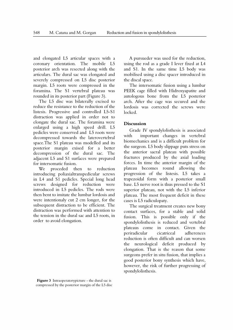
and elongated L5 articular spaces with a coronary orientation. The mobile L5 posterior arch was resected along with the articulars. The dural sac was elongated and severely compressed on L5 disc posterior margin. L5 roots were compressed in the foramina. The S1 vertebral plateau was rounded in its posterior part (Figure 3).
The L5 disc was bilaterally excised to reduce the resistance to the reduction of the listesis. Progressive and controlled L5-S1 distraction was applied in order not to elongate the dural sac. The foramina were enlarged using a high speed drill. L5 pedicles were conserved and L5 roots were decompressed towards the laterovertebral space.The S1 plateau was modelled and its posterior margin exised for a better decompression of the dural sac. The adjacent L5 and S1 surfaces were prepared for intersomatic fusion.
We proceded then to reduction introducing poliaxialtranspedicular screws in L4 and S1 pedicles. Special long head screws designed for reduction were introduced in L5 pedicles. The rods were then bent to imitate the lumbar lordosis and were intentionaly cut 2 cm longer, for the subsequent distraction to be efficient. The distraction was performed with attention to the tension in the dural sac and L5 roots, in order to avoid elongation.
Figure 3 Intraoperatorypicture - the dural sac is
compressed by the posterior margin of the L5 disc
A pursueder was used for the reduction, using the rod as a grade I lever fixed at L4 and S1. In the same time L5 body was mobilised using a disc spacer introduced in the discal space.
The intersomatic fusion using a lumbar PEEK cage filled with Hidroxyapatite and autologous bone from the L5 posterior arch. After the cage was secured and the lordosis was corrected the screws were locked.
Discussion Grade IV spondylolisthesis is associated
with important changes in vertebral biomechanics and is a difficult problem for the surgeon. L5 body slippage puts stress on the anterior sacral plateau with possible fractures produced by the axial loading forces. In time the anterior margin of the plateau becomes round allowing the progression of the listesis. L5 takes a trapezoidal form with a posterior small base. L5 nerve root is thus pressed to the S1 superior plateau, not with the L5 inferior plateau. The most frequent deficit in these cases is L5 radiculopaty.
The surgical treatment creates new bony contact surfaces, for a stable and solid fusion. This is possible only if the spondylolisthesis is reduced and vertebral plateaus come in contact. Given the periradicular cicatriceal adherences reduction is often difficult and can worsen the neurological deficit produced by elongation. That is the reason that some surgeons prefer in situ fusion, that implies a good posterior bony synthesis which have, however, the risk of further progressing of spondylolisthesis.

Romanian Neurosurgery (2011) XVIII 4: 546 - 550 549
Conclusion Grade IV spondylolisthesis remains a
challenging surgical problem and the best treatment is reduction and solid fusion, which can be accomplished if the anatomical and biomechanical changes associated to this condition are well understood, and the surgical fusion techniques are safely performed.
References 1. Baker DR, McHolick W. Spondylolischisis and spondylolisthesis in children. J Bone Joint Surg Am 38:933-934, 1956; 2. Boxall D, Bradford DS, Winter RB. Management of severe spondylolisthesis in children and adolescents. J Bone Joint Surg Am 61:479-495, 1979; 3. Crock HV, Normal and pathological anatomy of thelumbar spinal nerve rootcanals, J of Bone and Joint Surgery, 63B, No 4:487-490, 1981; 4. Dick WT Schnebel B: Severe spondylolisthesis. Reduction and internal fixatio. Clin Orthop 232:70-79, 1988; 5. Fairbank JC, Pynsent PB, The Oswestry Disability Index, Spine 25(22):2940-52, 1976; 6. Fredrickson BE, Baker D, McHolick WJ. The natural history of spondylolysis and spondylolisthesis. J Bone Joint Surg 66A:699-707, 1984; 7. Frobin W, Brinckmann P, Kramer M, Hartwig E. Height of lumbar discs measured from radiographs compared with degeneration and height classified from MR images. Eur Radiol. 11:263–269, 2001; 8. Gill GG, Manneng FG, While HL: Surgical treatment of spondylolisthesis without spine fusion. J Bone Joint Surg Am 37:493-520, 1955; 9. Gould D, et al.: Visual Analogue Scale (VAS), Journal of Clinical Nursing, 10, 697-706, 2001; 10. Harvell JC, Hanley EN: Spondylolysis and spondylolisthesis, Disorders of the Pediatric Spine: 561-674, 1995; 11. Hersinger RN, Lang JR, MacEwen GD: Surgical management of spondylolisthesis in children and adolescents. Spine I:207-216, 1976; 12. Herbiniaux G. Traite sur Divers Accouchemens Laborieux et sur les Polypes de la matrice. Brussels: JL De Boubers, 1782; 13. Hutton WC, Cynon BM. Spondylolysis. The role of the posterior elements in resisting the intervertebral compressive force. Acta OrthopScand 49:604-609, 1978; 14. Jackson DW, Wiltse LL, Cirincione RJ. Spondylolysis in thefemalegymnast. Clin Orthop 117:68-73, 1976;
15. Junghanns H. Spondylolisthes enohne Spalt im Zwischengelenkstuck, Arch of Orthop 29:118-127,1930; 16. Lambl D. Zehn Thesenuber Spondylolisthesis. Zentralbl Gynak 9:356-357, 1885; 17. Lehmann TR, Spratt KF, Tozzi JE, et al. Long-term follow-up of lower lumbar fusion patients. Spine (Phila Pa 1976). 12:97–104, 1987; 18. Leon L, Wiltse LL, Winter RB: Terminology and measurement of spondylolisthesis, J of Bone and Joint Surg, 65A, 6:768-772; 19. Matsunaga S, Sakou T, Morizono Y, et al.: Natural history of degenerative spondylolisthesis: pathogenesisand natural course of slippage. Spine 16: 1204-1210, 1990; 20.Mayerding HW. Spondylolisthesis. Surg Gynecol Obstet 54:371-377, 1932; 21.MacAfee PC, Weiland DJ, Carlow JJ: Survivor ship analysis of pedicle spinal instrumentation. Spine 16: S422-S427, 1991; 22.McPhee B: Spondylolisthesis and spondylolysis. In Youmans JR (ed): Neurological Surgery. Philadelphia, WB Saunders, pp 2749-2784, 1990; 23. Neugebauer Fl. The classic: a new contribution to the historyandetiology of spondylolisthesis. Clin Orthop 117:4-22, 1976; 24. Ohmori K, Suzuki K, Ishida Y: Translamino-pedicular screw fixation with bone grafting for symptomatic isthmic lumbar spondylolysis. Neurosurgery 30:379-384, 1992; 25. Osterman K, Lindholm TS, Laurent LE: Late results of removal of thelooses posterior element (Gill’s operation) in thetreatment of lytic lumbar spondylolisthesis. Clin Osthop 117:121-128, 1976; 26. Peek RD, Wiltse LL, Reynolds JB et al: In situ arthrodesis without decompression for Grade-III or IV isthmic spondylolisthesis in adultswhohave severe sciatica. J Bone Joint Surgery Am 71:62-68, 1989; 27. Pizzutillo PD, Hummer CD III: Nonoperative treatment for painful adolescent spondylolisis or spondylolisthesis. J Pediatr Orthop 6:311-316, 1986; 28. Pizzutillo PD, Mirenda W, MacEwen GD: Postero lateral fusion for spondylolisthesis in adolescence. J Pediatr Orthop 6:311-316, 1986; 29. Rowe GG, Roche MB: The etiology of separate neural arch, J Bone Joint Surg Am 35: 102-110, 1953 30. Saraste H: The etiology of spondylolysis. A retrospective radiographicstudy. Acta OrthopScand 56:253-255, 1985; 31. Semon RL, Spengler D. Significance of lumbarspondylolysis in collegefootballplayers. Spine 6:172-174, 1981; 32. Simper LB: Spondylolysis in Eskimoskeletons. Acta OrthopScand 57:78-80, 1986; 33. Rosenberg NJ. Degenerative spondylolisthesis:

550 M. Catana and M. Gorgan Reduction and fusion in spondylolisthesis
surgicaltreatment. Clin Orthop 117:112-120, 1976; 34. Rosenberg NJ, Bargar WL, Friedman B. The incidence of spondylolysis and spodylolisthesis in non ambulatory patients. Spine 6:35-38, 1981; 35. Vital JM, Lavignolle B, Grenier N, et al.: Anatomy of the lumbar radicular canal, Anatomia Clinica, 5:141, 1983; 36. Wenger DR, Lee CS. Spondylolisthesis in children andadolescents. Semin Spine Surg 5:308-319; 1993; 37. Wiltse LL, Newman PH, Mac Nab I. Classification of spondylolysis and spondylolisthesis. Clin Orthop
117:23-29, 1976; 38. Wiltse LL, Rothman SLG. Spondylolisthesis: Clasification, diagnosisand natural history. Semin Spine Surg I:78-94, 1989; 39.Wiltse LL, Widell EH Jr, Jackson DW: Fatiguefracture: the basic lesion in isthmic spondylolisthesis. J Bone Joint Surg 61B:301-305, 1979; 40.Wynne-Davies R, Scott JH: Inheritance and spondylolisthesis: a radiographic family survey. J Bone Joint Surg Br 61:301-305, 1979.