regeneration-associated wnt signaling is activated in...
TRANSCRIPT

Volume 14 Number 12 December 2012 pp. 1236–1248 1236
AbbreviatimacrophagAddress allMilano, It1This worRegionale2This artic3F. CorlazReceived 7
CopyrightDOI 10.1
www.neoplasia.com
Regeneration-associatedWNT Signaling Is Activatedin Long-term ReconstitutingAC133bright Acute MyeloidLeukemia Cells1,2
ons: AML, acute myeloid leukemia; LSC, leukemic stem cell; LT-HSCs, longe; BM MNCs, bone marrow mononuclear cells; LIC, leukemia-initiating cellcorrespondence to: Alessandro Beghini, PhD, Department of Medical Biotechnoaly. E-mail: [email protected] was supported in part by PUR 2008 (to A.B.), Progetto Integrato OncoloSangue-Regione Lombardia 2006 (DDG 7917). The authors declare that thele refers to supplementary materials, which are designated by Table W1 andzoli and L. Del Giacco contributed equally to this work.September 2012; Revised 15 October 2012; Accepted 18 October 2012
© 2012 Neoplasia Press, Inc. All rights reserved 1522-8002/12/$25.00593/neo.121480
Alessandro Beghini*, Francesca Corlazzoli*,3,Luca Del Giacco†,3, Matteo Re‡,Francesca Lazzaroni*, Matteo Brioschi§,Giorgio Valentini‡, Fulvia Ferrazzi¶,#,Anna Ghilardi†, Marco Righi*,**, Mauro Turrini††,Marco Mignardi‡‡,§§, Clara Cesana††,Vincenzo Bronte¶¶, Mats Nilsson‡‡,§§,Enrica Morra†† and Roberto Cairoli††,##
*Department of Medical Biotechnology and TranslationalMedicine, Università degli Studi di Milano, Milano, Italy;†Department of Biosciences, Università degli Studi diMilano, Milano, Italy; ‡Department of Computer Science,Università degli Studi di Milano, Milano, Italy; §Dipartimentodi Scienze Oncologiche e Chirurgiche, Università degliStudi di Padova, Padova, Italy; ¶Dipartimento di Informaticae Sistemistica, Università degli Studi di Pavia, Pavia, Italy;#Gene Center, Ludwig-Maximilians-Universität München,Munich, Germany; **Consiglio Nazionale delle Ricerche,Institute of Neuroscience, Milano, Italy; ††Department ofOncology, Niguarda Hospital, Milano, Italy; ‡‡Departmentof Immunology, Genetics and Pathology, RudbeckLaboratory, Uppsala Universitet, Uppsala, Sweden;§§Science for Life Laboratory, Department of Biochemistryand Biophysics, Stockholm University, Solna, Sweden;¶¶Istituto Oncologico Veneto, Padova, Italy; ##Divisionedi Medicina Interna, Sezione di Ematologia Clinica,Ospedale Valduce, Como, Italy
AbstractAcute myeloid leukemia (AML) is a genetically heterogeneous clonal disorder characterized by two molecularly dis-tinct self-renewing leukemic stem cell (LSC) populations most closely related to normal progenitors and organizedas a hierarchy. A requirement for WNT/β-catenin signaling in the pathogenesis of AML has recently been suggestedby a mouse model. However, its relationship to a specific molecular function promoting retention of self-renewingleukemia-initiating cells (LICs) in human remains elusive. To identify transcriptional programs involved in the main-tenance of a self-renewing state in LICs, we performed the expression profiling in normal (n= 10) and leukemic (n =33) human long-term reconstituting AC133+ cells, which represent an expanded cell population in most AMLpatients. This study reveals the ligand-dependentWNTpathway activation inAC133bright AMLcells and shows a diffuse
-term hematopoietic stem cells; CFU, colony forming units; CFU-GM, granulocyte/
logy and Translational Medicine, Università degli Studi di Milano, via Viotti 3/5, 20133
gia 2006 (RO 4/2007), Associazione Malattie del Sangue Onlus (AMS), and Pianoy have no competing financial interests.W2 and Figure W1 and are available online at www.neoplasia.com.

Neoplasia Vol. 14, No. 12, 2012 Regenerative WNT Signaling Activated in AC133+ Beghini et al. 1237
expression and release ofWNT10B, a hematopoietic stemcell regenerative-associatedmolecule. The establishmentofa primary AC133+ AML cell culture (A46) demonstrated that leukemia cells synthesize and secrete WNT ligands, in-creasing the levels of dephosphorylated β-catenin in vivo. We tested the LSC functional activity in AC133+ cells andfound significant levels of engraftment upon transplantation of A46 cells into irradiated Rag2−/−γc−/− mice. Owingto the link between hematopoietic regeneration and developmental signaling, we transplanted A46 cells into develop-ing zebrafish. This system revealed the formation of ectopic structures by activating dorsal organizer markers that actdownstream of the WNT pathway. In conclusion, our findings suggest that AC133bright LSCs are promoted by mis-appropriating homeostatic WNT programs that control hematopoietic regeneration.
Neoplasia (2012) 14, 1236–1248
IntroductionDifferent genetic causes result in variable clinical courses of acutemyeloid leukemia (AML) and different responses to standard chemo-therapy including stem cell transplant. Despite the genetic differencesamong individual patients, most AML clones display certain commonfeatures. Ample evidence exists in mouse models that AML developsthrough the stepwise acquisition of collaborating genetic and epigeneticchanges in self-renewing LICs, which exhibit a committed myeloidimmunophenotype and give rise to nonleukemogenic progeny in amyeloid-restricted hierarchy [1–3]. An important issue to understandthe early events in the origin of AML is the observation that long-termhematopoietic stem cell (LT-HSC) expansion precedes the generationof committed myeloid LICs [4].Although well-orchestrated cell intrinsic programs and environmental
cues represent the main contributory factors for normal LT-HSC ex-pansion, it is still unclear if transcriptional programs responsible for theexpansion of premalignant LT-HSC populations and leukemia initiationshare common embryonic or post-embryonic functions, such as stemcell renewal, tissue repair, and regeneration [5,6]. Even though recentstudies have addressed the role of Hedgehog signaling for maintenanceof cancer stem cells in myeloid leukemia [7], its requirement in AMLremains controversial [8].Recently, the notion that LICs are restricted only to the CD34+CD38−
population has been challenged [9,10] and it has been suggested thatmore cell surface markers could be appropriately used to enrich theleukemia-initiating cell (LIC)–containing fraction. One of such markersis the AC133 antigen (a glycosylation-dependent epitope of CD133)that defines a desirable population of stem and progenitor cells con-taining in turn all the CD34brightCD38− progenitors, as well as theCD34brightCD38+ cells committed to the granulocytic/monocytic line-age [11]. In addition, AC133 represents a well-documented marker oftumor-initiating cells in a number of human cancers [12]. In this study,fluorescence-activated cell sorter (FACS) analysis demonstrates thatAC133+ cell population is dramatically expanded in 25 AML cases ana-lyzed. We carried out genome-wide transcriptional analysis of AC133+
cells isolated from newly diagnosed non-promyelocytic AML patients(n = 33) and healthy donors (n = 10). Results obtained from a multistepanalysis of the generated data defined the involvement of the ligand-dependentWNT receptor signaling pathway as the self-renewal associatedsignature in the AC133-enriched fraction in human AML. Furthermore,the results presented here suggested thatWNT10B and otherWNTgenesexpressed during the regenerative process of the hematopoietic system[13,14] are aberrantly upregulated in AC133bright AML cells. To obtain
a localized detection of each single transcript, we first applied an in situdetection of individual mRNA molecules [15] on bone marrow (BM)sections from AML patients. By the establishment of a primary cultureof AC133+ AML cells (termed A46 hereafter), we confirmed that secretedWNTs activated a β-catenin/human T-cell factor (TCF) transcription–based reporter construct. Moreover, we intend to clarify the relationshipbetween the abnormal WNT activation in AC133+ population and theleukemic stem cell (LSC) activity.UsingRag2−/−γc−/− as immunodeficientxenotransplantmodel [16], AC133+ A46 cells were injected intravenouslyinto sublethally irradiated mice.
To achieve a complete view of how AC133+ A46 cells modulatedthe microenvironment and given that hematopoietic regenerationconverge to developmental signaling, we used zebrafish embryonicmodel as an in vivo biosensor.
Our results confirmed previously reported data [17] and raise newimportant implications for the involvement of the ligand-dependentcanonical WNT pathway in AML. These suggestive findings are sup-ported by the pivotal function of WNT in promoting self-renewal[18,19], its emerging role in myeloid leukemogenesis [20,21], andthe effects of its constitutive activation through a stabilized form ofβ-catenin, by inducing quiescent stem cells to enter the cell cycle andarresting their differentiation [22,23].
Materials and Methods
Collection of Patient Samples and Normal Hematopoietic CellsBM MNCs were collected from 33 newly diagnosed, unselected
non-promyelocytic AML patients, according to Niguarda Hospital’sEthical Board–approved protocols (116_04/2010). According to therevised Medical Research Council risk group stratification, based oncytogenetic and molecular markers/mutations [24], samples included14 adverse, 13 intermediate, and 6 favorable risk patients. Humanadult BM cells obtained from 10 consenting healthy donors wereprocessed as previously described [25].
Cell Sorting and Flow CytometryWe carried out AC133+ cell separation based onMACSMicroBeads
and cytofluorometric determinations, as previously described [25].
Microarray Expression AnalysisTotal RNA for expression profiling was extracted using RNAqueous-
4PCR kit (Ambion, Austin, TX) from AC133-selected cells. Expressionprofiling was performed on Affymetrix HGU133plus2.0 GeneChip

1238 Regenerative WNT Signaling Activated in AC133+ Beghini et al. Neoplasia Vol. 14, No. 12, 2012
arrays according to the manufacturer’s procedures. The bioinformaticsanalysis performed in this study was realized using the R language for sta-tistical computing (http://www.r-project.org/) and the annotation librar-ies provided by the Bioconductor project (http://www.bioconductor.org/). Microarray data have been deposited in ArrayExpress (http://www.ebi.ac.uk/arrayexpress/), with accession number E-MTAB-220.We performed a genome-wide analysis to select genes differentially ex-pressed between AML AC133+ patients and AC133+ healthy donors(Welch t test, 0.05 significance level). The resulting set of differentiallyexpressed genes has been analyzed for functional enrichment with re-spect to the terms of the Biological Process (BP) branch of the GeneOntology (GO) and the pathways of the KEGG database. We reliedon three different methods for functional enrichment analysis: GOStats(version 2.12.0 of the Bioconductor package, http://www.bioconductor.org/packages/release/bioc/html/GOstats.html) [26], Database for Anno-tation, Visualization and Integrated Discovery (DAVID; http://david.abcc.ncifcrf.gov/home.jsp) [27], and the iterative procedure of dysregu-lated pathway analysis proposed by Majeti et al. [17]. The first two testsare based on the hypergeometric distribution, whereas the last one isbased on a non-parametric test and on an iterative procedure. Over-represented GO terms and KEGG pathways have been selected at0.05 significance level.
WNT/β-catenin Responsive Luciferase AssayHEK293T cells grown in 24-well plates at a density of 1.7 × 10⁵ cells
per well were transfected with M50 Super 8x TOPFlash (plasmid12456; Addgene, Cambridge, MA) and pRL-TK (Renilla luciferase;Promega, Madison, WI) using jetPEI (Polyplus, New York, NY). Cellswere treated for 12 hours with A46 conditioned medium (CM) orHEK293T cells transfectedwithBA-WNT10B (plasmid 1831; Addgene)CM as positive control. WNT10B expression in HEK293T transfectedwith BA-WNT10B was evaluated by SYBR Green–based real-time re-verse transcription–polymerase chain reaction (RT-PCR) usingWNT10B FW-5′-GCTGTAACCATGACATGGAC-3′ and RW-5′-CTGCCTGATGTGCCATGAC-3′ specific primers. Luciferase activitymeasurement was performed with the Dual-Luciferase Reporter AssaySystem (Promega).
ImmunoblotProtein expression was assessed by immunoblot analysis using stan-
dard procedures, applying anti–active β-catenin (ABC) monoclonalmouse (anti-ABC clone 8E7; Millipore, Billerica, MA), anti–β-cateninmonoclonal rabbit (E247; Abcam, Cambridge, United Kingdom), an-ti–WNT10B polyclonal rabbit (H-70; Santa Cruz Biotechnology, Inc,Santa Cruz, CA), anti–WNT10B monoclonal mouse (5A7; Abcam),anti–glyceraldehyde-3-phosphate dehydrogenase (GAPDH) polyclonalrabbit (ab97626; Abcam), and anti–Pygopus 2 polyclonal rabbit (H-216;Santa Cruz Biotechnology, Inc) antibodies. Secondary antibodiesused were anti–mouse HRP, anti–goat HRP, and anti–rabbit HRP(Thermo Fisher Scientific, Waltham, MA).
In Situ mRNA DetectionIn situ detection of individual mRNA molecules was performed as
described [15]. One micromolar of locked nucleic acid–modified cDNAprimer (WNT10B, 5′-C+A+G+G+C+CGGACAGCGTCAAGC-ACACG-3′; β-actin, 5′-C+TG+AC+CC+AT+GCCCACCATCA-CGCCC-3′; Exiqon, Vedbaek, Denmark) was added to the reversetranscriptase reaction. Ligation was then carried out with 0.1 μM ofthe WNT10B padlock probe or β-actin padlock probe (WNT10B,
5′-[Phos]ACCGTGCCTGTCGGACCCTCCTCTATGATTA-CTGACCTAAGTCGGAAGTACTACTCTCTTCTTC-TTTTAGTGAAGCCCAGGCAACCCA-3′; β-actin, 5′-[Phos]GCCGGCTTCGCGGGCGACGATTCCTCTATGATTACTGA-CCTATGCGTCTATTTAGTGGAGCCTCTTCTTTA-CGGCGCCGGCATGTGCAAG-3′; Sigma-Aldrich, St Louis, MO).Rolling circle products (RCPs) were visualized using 100 nM of detec-tion probe (WNT10B, 5′-Cy5-AGTCGGAAGTACTACTCTCT-3′and β-actin, 5′-Cy3-TGCGTCTATTTAGTGGAGCC-3′; Sigma-Aldrich). Nuclei were counterstained with 100 ng/ml Hoechst 33258(Sigma-Aldrich). Images of BM tissue slides were acquired using anAxioplan II epifluorescence microscope (Zeiss, Munchen, Germany)equipped with a charge-coupled device (CCD) camera (HRM, Zeiss)and a computer-controlled filter wheel with excitation and emission filtersfor visualization of 4′-6-diamidino-2-phenylindole (DAPI), Cy3, and Cy5.A ×20 objective (Plan-Apocromat, Zeiss) was used for capturing the images.Images were collected using the Axiovision software (release 4.3, Zeiss).Images were collected as z-stacks to ensure that all RCPs were imaged, witha maximum intensity project created in Axiovision. For quantification, thenumbers of RCPs and cell nuclei in images were counted digitally usingCellProfiler software (www.cellprofiler.org) on three ×20 microscopeimages. The total number of RCPs was divided by the number of nucleifor each image. The average for each sample was then calculated from theresult of the three images and is reported as RCPs per cell.
ImmunostainingDirect and indirect immunostaining were performed following stan-
dard procedures. BM biopsies of AML patients, previously embedded inparaffin blocks, were cut in 5-μm-thick sections and mounted on slides.Slides were incubated with primary antibody mouse anti-CD133.1(AC133) (1:100;Miltenyi Biotec GmBH, BergischGladbach, Germany),directly labeled with 488-nm dye (Sigma), and with primary antibodiessuch as mouse anti-ABC (1:100; Millipore), rabbit anti-WNT10B(H70) (1:100; Santa Cruz Biotechnology, Inc), rabbit anti–Pygopus2 (H-216) (1:100; Santa Cruz Biotechnology, Inc), goat anti-SMYD3(F-19) (1:100; Santa Cruz Biotechnology, Inc). Samples were incubatedwith the secondary antibodies donkey anti-mouse Alexa Fluor 488(1:500; Life Technologies, Carlsband, CA), donkey anti-rabbit AlexaFluor 568 (1:500; Life Technologies), and donkey anti-goat Alexa Fluor568 (1:500; Life technologies). Nuclei were counterstained with 100 ng/mlDAPI (Sigma-Aldrich). Cells were analyzed using the upright micro-scope (Leica, DM 4000B).
Immunofluorescence Image AnalysisImage analysis was performed bymeans of a custom automatic routine
and the ImageJ program (1.43s; National Institutes of Health, Bethesda,MD). Maps of ABC+/WNT10B+ cells were obtained using an auto-matic threshold based on moments algorithm newly implemented inthe ImageJ program and mathematical morphology plugin developedby D. Prodanov (http://rsbweb.nih.gov/ij/plugins/gray-morphology.html). DAPI staining was used to identify nuclei. Finally, images ofnuclei for positive cells were obtained by Boolean AND operations be-tween DAPI staining and cell maps. The resulting images were used todetermine the percentage of ABC+/WNT10B+ cells both manually orthrough the Analyze Particles function of ImageJ.
Cell CultureSelected AC133+ cells from BM at AML diagnosis were cultured
for 16 weeks. The culture was performed using synthetic medium

Neoplasia Vol. 14, No. 12, 2012 Regenerative WNT Signaling Activated in AC133+ Beghini et al. 1239
StemSpam H3000 (StemCell Technologies, Vancouver, Canada) inthe absence of serum and cytokines. StemSpan H3000 and HPGMmedia, conditioned by the cell culture, were collected after 12 weeks,refined with 0.2-μm filter and stored at −20°C. HEK293T cellswere grown in Dulbecco’s modified Eagle’s medium (high glucosewith sodium pyruvate and L-glutamine; Euroclone), supplementedwith 10% FBS (Euroclone) and penicillin-streptomycin solution(100×) (Euroclone, Milano, Italy).
Mice and Xenogeneic TransplantationRag2−/−γc−/− BALB/c mice were bred and maintained under specific
pathogen-free conditions in the mouse facility of Istituto OncologicoVeneto, and experiments were performed according to state guidelinesand approved by the local ethics committee. Rag2−/−γc−/− mice at6 weeks were sublethally irradiated with 5 Gy and transplanted via thetail vein with 1 × 10⁶ of human AC133+ AML cells (A46).
Evaluation of Hematopoietic Chimerism by Flow CytometryThree weeks after transplantation, recipient mice were sacrificed and
BM cells were harvested by flushing femurs and tibias. BM engraftmentwas evaluated using human antibodies CD34, CD38, AC133, andCD45 [BD Biosciences (Bedford, MA) and Miltenyi Biotec GmBH].Multicolor flow cytometric analyses were performed using FACSCaliburflow cytometer (BD Biosciences) and analyzed by FlowJo software (TreeStar, Ashland, OR). Cell engraftment was determined by expression ofthe human panleukocyte marker CD45.
Zebrafish Models and Transplantation ProceduresEmbryos were handled according to relevant guidelines. Fish of the
AB strain were maintained at 28°C on a 14-hour light/10-hour darkcycle and collected by natural spawning. Transplantation of humanA46 cells into zebrafish embryos was performed as previously reported[28]. Briefly, fluorescently labeled A46 cells were resuspended in 1×phosphate-buffered saline and injected into zebrafish blastulae (be-tween 100 and 200 cells per injection) at 3 hours post-fertilization(hpf ). Injected live embryos were observed under a fluorescent micro-scope at 30% of epiboly to ensure the presence of labeled A46 cells.Embryos were collected at the desired developmental stages, immediatelyfixed, and processed for whole-mount in situ hybridization accordingto Thisse et al. [29], using gsc, ntl, and pax2a DIG-labeled riboprobes.
Figure 1. Human AC133+ cells are strongly expanded in AML. Repres(A) a healthy donor and (B) a patient with AML (AML No. 42 in Table W1ages on total cellularity are shown for gated normal and AML populatioof healthy donors (n=10) and AML patients (n=25). The IQR for eachto calculate the P value (α = 0.001).
Results
AC133+ Cells Are Highly Expanded in AMLAC133 antigen is restricted to a rare cell population with long-term
reconstituting activity, ranging from 20% to 60% of all CD34+ cells,and resulting barely detectable in CD34−Lin− cells [11–30]. We havepreviously shown that AC133+ LT-HSCs are also highly enriched incolony forming units and have a stronger granulocyte/macrophagedifferentiation potential relative to unselected BM MNCs. However,their burst-forming units–erythroid forming potential is lower [25]. Todetermine the range of expansion of AC133+ cell fraction in AML, flowcytometric quantification of CD133.1 (AC133) expression either insingle staining or in combination with the pan-hematopoietic markerCD45 was performed in BM on 25 primary non-promyelocytic AMLsamples and 10 age-matched healthy volunteer adult donors. The re-sulting CD133.1+ cell fraction results expanded among AML patientsby a median of 31.5% [interquartile range (IQR), 16.5%–53.4%] withrespect to normal donors (median, 0.54%; IQR, 0.17%–1.14%; P <.0001; Figure 1, A–C). To directly compare the gene expression profilesof purified populations highly enriched in LSCs or HSCs, positive selec-tion of CD133.1+ cells was performed on all the 33 non-promyelocyticAML patients (25 de novo, 7 secondary to myelodysplasia, and 1 second-ary therapy-related; Table W1) as well as on the 10 healthy donorsrecruited to this study, respectively. MNC selection in samples fromAML patients led to an average of 236-fold enrichment of AC133-positive cells (IQR, 142.72–419.44, data not shown).
Identification of Dysregulated Pathways in AC133+ AMLCell Fraction
To identify the dysregulated pathways in AC133+ AML cell frac-tion versus normal long-term reconstituting AC133+ HSC cells, weperformed a genome-wide functional enrichment analysis on gene ex-pression microarray data of 33 AML patients and 10 healthy donors.The identification of overrepresented pathways in AC133+ AML cellswas realized through three computational tools: GOStats [26], DAVID[27], and dysregulated pathway analysis according to Majeti et al. [17].Employing the functional terms from GO BPs and the pathway infor-mation from KEGG databases, we found 212 functionally enrichedGOBP terms with GOStats (P < .01), 284 GOBP terms with DAVID(P < .05), and 616 GO BP terms with the non-parametric test ofMajeti et al. (P < .05). Moreover, GOStats selected 16 KEGG pathways
entative dot plots of the immunophenotype analysis from the BM of). The CD45/CD133.1 co-staining was gated on BMMNCs; percent-ns. (C) Flow cytometry analysis of the CD133.1 antigen in BMMNCssample group is indicated in the dot. Mann-WhitneyU test was used

Figure 2. Schematic visualization of dysregulated networks in KEGG database and representation of WNT gene expression profiles.(A) Visualization of WNT signaling pathway in the KEGG database. Highlighted genes are differentially expressed and red-starred path-ways are overrepresented in AC133+ AML cells. (B) Heat map of the differentially expressed WNT-associated genes. The color bar underthe patient sample dendrogram identifies AML samples (red) and healthy controls (blue). The names, accession numbers, as well asP values for the differentially overexpressed and underexpressed genes are shown in the right panels.
1240 Regenerative WNT Signaling Activated in AC133+ Beghini et al. Neoplasia Vol. 14, No. 12, 2012

Neoplasia Vol. 14, No. 12, 2012 Regenerative WNT Signaling Activated in AC133+ Beghini et al. 1241
(P < .05), DAVID selected 24 KEGG pathways (P < .05), and thedysregulated pathway analysis selected 3 KEGG pathways (P < .05).As shown in Table W2, DAVID ranks the WNT signaling pathway(P = .022) among the first 10 dysregulated pathways in AC133+
AML cells. The visualization of the WNT molecular interaction inKEGG database is presented in Figure 2A.It is worth noting that different statistical methods agree in identify-
ing theWNT pathway as a significant dysregulated pathway in AC133+
AML cells. Moreover, the WNT signaling pathway in KEGG is se-lected as overrepresented in AC133+ AML stem cells by both GOStatsand DAVID. Taken together, the functional enrichment methodsselect the term “WNT receptor signaling pathway” (GO:0016055)as the most specific self-renewal associated dysregulated pathway.
WNT Gene Expression Profiles of Normal and LeukemiaLong-term Reconstituting AC133+ CellsTo obtain insights into theWNTpathway in AC133+ leukemia cells,
we focused our analysis on the probe sets annotated to “WNT receptorsignaling pathway” GO class or to any of its children GO terms. Theresulting probe set list was complemented with the probes mapping togenes in a manually curated list of WNT target genes and with probesmapping to other well-known WNT genes, not included in the previ-ous two lists. The total list of analyzed WNT genes includes 480 probesets, mapping to 193 different genes (Table_A in http://homes.dsi.unimi.it/~re/TRdataset/). To assess differential expression of WNTgenes, we employed Welch t tests (two-tailed) on the 480 probe sets,thus identifying 103 differentially expressed genes (P < .05; Figure 2B).
Figure 3. Altered WNT signaling in AC133+ AML cells. (A) Detectionindividual WNT10B transcripts on BM slides from AML patients. Greβ-actin transcripts in consecutive sections. Cell nuclei are shown in blu(B) The quantification of RCPs was done on three ×20 images of BMof each image were counted digitally using CellProfiler software. The aa ratio between β-actin and WNT RCPs. (C) Immunoblot analysis of ABCWNT10B, and Pygopus 2 protein expression in AC133+ cell fractionsGAPDH, loading control. HD, healthy donor; *therapy-related secondary
Genes shown to be highly AML-specific include the WNT ligandsWNT2B, WNT6, WNT10A, and WNT10B [14], the WNT/β-cateninsignaling agonists including SMYD3 [31], DKK2 [32], SOX4 [33],PROP-1 [34], and PYGO2 [35,36], antagonists including WIF-1[37], KLHL12 [38], LRP6 [39], KREMEN1 [40], E2F1 [41], DACT1[42], and HBP1 [43], and the deregulated WNT targets includingSTAT3, MYCN, ABCC4, DLX3, MARK4, RUNX2, CD24, andCD44 [44]. Collectively, these data are consistent with ligand-dependentactivation of the regeneration-associated WNT pathway [14].
We also investigated whether the expression of the WNT genesvaried between the risk groups in AML (favorable, intermediate, andadverse) revised by Smith et al. [24]. Clustering results of AML casesrestricted to WNT genes show that the risk groups are not clearlydistinguishable in separate clusters (Figure 2B). Analysis of variancebetween groups confirms these results: only 3 (ABCC4, HBP1, andNDP) of 193 WNT-related genes are differentially expressed acrossthe three categories (P < .05). Thus, our data do not support a signif-icant distinction between risk groups in AML patients. This state-ment should be considered with caution, because the cardinalityof the subgroups is relatively low (6 favorable, 13 intermediate, and14 adverse samples).
Hyperactive WNT Signaling Resulted Activated inDiffuse AC133bright AML Cells Expressing theHematopoietic Regenerative Molecule WNT10B
Most of what is known about hematopoietic regeneration point toWNT signaling pathway and specifically toWNT10B [13,45]; therefore,
with padlock probe and target-primed rolling circle amplification ofen RCPs represent WNT10B transcripts, and red RCPs represente. Images were acquired with ×20 magnification. Scale bar, 10 μm.biopsies of AML patients. For quantification, the numbers of RCPsverage of RCPs for each sample was calculated and it is reported as, β-catenin (as detected with the N-terminal pan–β-catenin antibody),from 18 patient samples and 1 healthy donor introduced as control.AML.

Figure 4. β-Catenin activation in the subpopulation of AC133bright
AML cells expressing WNT10B. (A) Representative immunostainingmicrographs show green fluorescence of cells expressing AC133 ina BM section of AML No. 9 (TableW1). Cell nuclei are shown in blue.Scale bar represents 10 μm. (B) Co-staining of BM from AML No. 9adjacent serial section for expression of ABC (green) and WNT10B(red). Cell nuclei are shown in blue (DAPI). (C) False color maps ofABC/WNT10B double positive cells (blue) were obtained using anautomatic threshold based on the moments algorithm implementedin the ImageJ program. DAPI staining was used to identify nuclei.Images obtained crossing ABC masks with WNT10B signals wereused to count the percentage of ABC/WNT10B double positive cellsover the total number of cells. The macro was validated againsta trained experimenter over a sample of 830 total cells from eightdifferent images. Differences in results were restricted to less than0.01%. (D) Morphologic detail of cells showing intense specificstaining for ABC (top panels) and WNT10B (bottom panels). Allimages were acquired with a ×40 objective. Scale bars represent10 μm. Representative images of at least three serial slides fromfive randomly selected patients.
1242 Regenerative WNT Signaling Activated in AC133+ Beghini et al. Neoplasia Vol. 14, No. 12, 2012
we investigated how expression of WNT10B is related to AMLphenotype. To fully understand mRNA distribution of WNT10Bwithin BM sections, we focused on the application of single moleculedetection methods. In situ mRNA detection by target-primed rollingcircle amplification analysis offers high sensitivity and localized detec-tion within single cells and tissues [15]. Following this approach, wedetected WNT10B-related transcript in BM sections obtained at diag-nosis from two randomly selected patients. β-Actin was included as areference transcript in consecutive sections. Visualization by using high-performance fluorescence microscopy showed a diffuse localizationpattern in the tissues (Figure 3A). Signal distribution and RCP quantifi-cation showed a β-actin/WNT10B ratio close to 1, suggesting a con-stitutive activation of WNT10B transcription in the BM (Figure 3B).In addition, we analyzed transcriptional activation of canonical WNTsfocusing on genes that have been shown to be potent regulators of stemcell functions. N-terminally dephosphorylated β-catenin (ABC) wasincreasingly accumulated as determined by immunoblot analysis (Fig-ure 3C). Remarkably, we confirmed a dramatic increase in WNT10Bexpression in all patient samples, except for one, reanalyzed by immuno-blot (Figure 3C). Interestingly, the only AML patient negative for theWNT10B expression (AML No. 40 in Table W1) was affected bytherapy-related AML. According to the biologic relevance of PYGO2in promoting the responsiveness ofWNT signaling [35,36], diffuse over-expression was detected by immunoblot (Figure 3C). To better elucidatethe impact of the broad WNT10B overexpression on the leukemicmicroenvironment, we examined its expression in histologic preparationsof BM from five randomly selected AML patients at diagnosis. Doubleimmunostaining for WNT10B and ABC, followed by ImageJ analysis,confirmed that WNT10B was expressed by a high proportion of leuke-mic cells.We next examined by immunostaining a number of BMbiopsysections from five randomly selected cases. In all the analyzed samples,the AC133 immunostaining revealed islands of highly positive cells(AC133bright) in an estimated proportion of 8% of cells, amid AC133dim
or negative tumor blasts (Figure 4A). AC133bright cells correlated withaccumulation of ABC (Figure 4, B and C). WNT10B antibody stainingwas also detectable in interstitial spaces, suggesting its secretion and re-lease in the BMmicroenvironment (Figure 4D, top). The slides revealedthatWNT10B is diffusely expressed (Figure 4,A andD) but that only theAC133bright small cells (8–10 μm diameter of the nuclei), with a clonalappearance and increased N/C ratio, shared the WNT signaling activa-tion signature represented by accumulation of ABC [46], likely inducedthrough an autocrine/paracrine mechanism (Figure 4, B and D, top).
AC133+ Leukemic Cells Express and Secrete WNT10Bin a Primary Cell Culture
The notion that primary cell cultures closely mimic the in vivo stateand generate more physiologically relevant data led us to establish aprimary AC133+ cell culture (A46). A46 cells, with diploid karyotype,were selected from a 66-year-old male at diagnosis of AML-M2 (AMLNo. 46 in Table W1). Immunophenotype of MNCs before selectionrevealed a dominant CD133.1+CD34+CD38−CD45+CD117+ blastpopulation (59%; Figure 5A), representing an optimal source to establishan LIC-enriched primary culture. Recently published data implicate theexistence of an immunophenotypic hierarchy in AML, with a minorityof CD38−CD45+ cells giving rise to CD38+CD45+ “GMP-like” cellsat the apex of an LIC hierarchy [10]. The latter finding seems to suggestthat CD38−CD45+ cells start to gain CD38 expression after AC133 se-lection procedures as observed in sorted cells (Figure 5A). Comparativeanalysis of spotted AC133-selected A46 leukemic and normal cells by

Figure 5. AC133+ A46 cells express and release WNT10B. Dot plots of the immunophenotype analysis from AML No. 46 BM MNCs atdiagnosis and after selection. (A) Patterns of CD38/CD133.1 (top left), CD117/CD45 (top right), and CD34/CD38 (bottom left) co-stainingweregated on BM AML cells before selection. Representative CD34 and CD38 expression on Ficoll-selected MNCs (bottom center) and AC133-sorted cells before culture (bottom right) is shown. Percentages on total cellularity are shown for gated AML populations. (B) Immuno-staining assessment for WNT10B in AC133+ populations from A46 (top panels) or healthy donor (bottom panels). Representative imagesof at least five serial slides. Blue, nuclei; red, WNT10B; merge, WNT10B/DAPI. All images were acquired with a ×40 objective. Scale barsrepresent 10 μm. (C and D) TOPFlash reporter assay showing luciferase expression driven by eight TCF/lymphoid-enhancing factor (LEF)binding sites. (C) Positive control was obtained by CM of pBA-WNT10B–transfected H293T cells. Expression of WNT10B was evaluatedin pBA-WNT10B–transfected H293T by real-time PCR (left). TOPFlash reporter assay shows luciferase expression induced in Super8x TOPFlash–H293T cells by pBA–WNT10B H293T CM (right). (D) TOPFlash reporter assay showing dose-dependent luciferase expressioninduced in Super 8x TOPFlash–H293T cells by A46 CM. Significance was evaluated by the unpaired Student’s t test: *P < .05; **P < .001.Data represent the mean ± SD of triplicate reactions and are representative of three independent experiments.
Neoplasia Vol. 14, No. 12, 2012 Regenerative WNT Signaling Activated in AC133+ Beghini et al. 1243

1244 Regenerative WNT Signaling Activated in AC133+ Beghini et al. Neoplasia Vol. 14, No. 12, 2012
immunostaining revealed WNT10B expression in all A46 AC133+ cells(Figure 5B, top), whereas normal BM-derived AC133+ cells resultednegative (Figure 5B, bottom). To further investigate whether the endoge-nous WNT production had any paracrine effect, we used A46 CMto evaluate β-catenin–mediated transcriptional activation. To thisaim, HEK293T cells (H293T), transfected with Super 8x TOPFlashβ-catenin/TCF transcription–based reporter construct, were exposedeither to pBA–WNT10B H293T CM as control (Figure 5C) or A46CM. This construct could be efficiently expressed in a dose-dependentmanner when transiently exposed to A46 CM for 12 hours (Figure 5D).
AC133 Is Expressed on Functional A46 AML LSCTo complement the in vitro observations, we tested whether AC133
was expressed on functional A46 AML LSC. We transplanted A46cells into sublethally irradiated (5 Gy) 6-week-old Rag2−/−γc−/− mice viathe tail vein. This highly immunodeficient mouse strain lacks mature B,
Figure 6. AC133+ A46 cell transplantation in Rag2−/−γc−/− mice. (A) Oinjected into sublethally irradiated Rag2−/−γc−/− mice through the tailanalyzed by flow cytometry. (B) Expression profiles of three recipient mmice, and within hCD45+, the expression patterns of hCD34 and hCD1according to the expression of hCD34 and hCD133.1 and then analyz
T, and natural killer (NK) cells, supporting efficient engraftment ofhuman AML [16]. Transplanted mice were killed at 3 to 8 weeks aftertransplantation and analyzed for engraftment of human leukemia cellsin BM (Figure 6A). The results of a typical experiment from threetransplanted mice (M1–M3) are shown in Figure 6, with AC133+
A46 cells showing engraftment of human CD45 (hCD45) cells. Weconfirmed that engrafted hCD45+ cells were human myeloid leukemiablasts by measuring CD34/CD38/CD133 expression (Figure 6B).
Transplantation of A46 Induces Ectopic AxialStructure Formation in Zebrafish Embryo by WNTSignaling Activation
To bring our functional analyses full circle, we explored the physio-logical relevance of tumor-derived WNT signals by using the develop-ing zebrafish as a biosensor. We hypothesized that WNT-secretingA46 cells transplanted into developing zebrafish embryos might act as
verview of the experimental design: 1 × 10⁶ AC133+ A46 cells werevein. Three weeks after transplantation, BM cells were collected andice (M1–M3) are shown. hCD45+ population was identified in BMs of33.1 (AC133) were analyzed. Cells were separated in subpopulationsed for hCD38 and hCD133.1 expressions.

Figure 7. A46AML cells induce ectopic gene expression and second-ary body axis formation upon transplantation in zebrafish embryos.(A) Fluorescence microscopy of a live zebrafish embryo at 30% ofepiboly (lateral view) transplanted at 3 hpf at the animal pole regionwith A46 cells previously blue-stained with Hoechst 33342. (B–D)Dorsal side view of 70% epiboly-stage embryos hybridized witha gsc-specific probe. Embryos have been injected with (B) normalAC133+ cells as control or (C and D) A46 AML cells. The arrow-heads indicate the gsc endogenous signal, while arrows specifythe position of zebrafish cells expressing ectopic gsc. (E) Bright-fieldmicroscopy of a 24-hpf zebrafish embryo injected with A46 AMLcells (lateral view). The arrowhead and the dotted line indicate thesecondary trunk/tail induced by A46 cells. (F, G) The embryo in Ehas been hybridized with a probe specific for the notochord and tailbud marker ntl. (F) The probe labels the notochord (n) in the endoge-nous trunk and the chordoneural hinge (cnh) in both tails (G, highermagnification). (H) Tail of a 24-hpf embryo hybridized with ntl-specific probe. The endogenous (n) and ectopic (*n) ntl signals runparallel along the axis of the embryo, indicating the presence ofadditional axial structures. (I) Dorsal view of a 24-hpf embryo that de-veloped an ectopic head on the side of the endogenous one, as indi-cated by the expression of the brain marker gene pax2a. The dottedlines indicate the main (M) and secondary (S) axes. The optic stalk(OS) in close vicinity to the eye (e), the midbrain-hindbrain boundary(MHB), and the otic vesicles (OV) of the embryo are stained with thepax2a riboprobe, as well as several areas of the ectopic head (theasterisks indicate the twoclearly recognizable additional otic vesicles).The image is composed of different pictures corresponding to severalfocal planes, since the embryo is not flat, and a single focal planecannot comprise all the labeled structures belonging to the mainand secondary axes. Scale bars represent 125 μm (A–D), 150 μm(E), 40 μm (F), 15 μm (G and I), or 25 μm (H).
Neoplasia Vol. 14, No. 12, 2012 Regenerative WNT Signaling Activated in AC133+ Beghini et al. 1245
ectopic sources of maternal WNT ligands. Cell-grafted embryos,injected at or before the mid-blastula transition stage (∼3 hpf) withHoechst 33342 fluorescently labeled A46 cells (Figure 7A), showedthe expression of the organizer-specific gene goosecoid (gsc). Expressionof gsc is typically initiated by the nuclear translocation of maternalβ-catenin triggered by the activation of the WNT signaling. Whilenormal AC133+ cells did not alter the normal expression of the gene(Figure 7B), approximately 30% of the embryos grafted with A46 cells(n = 208) displayed both the expansion of the gsc endogenous domainand the activation of the gene in ectopic positions (Figure 7, C and D).
Consistent with our hypothesis, the A46 cells retain a dorsal organizer–inducing activity possibly correlated with their strong WNT signalingactivation. In addition, grafted embryos developed secondary axial struc-tures, ranging from additional tail tissues (Figure 7, E–H) to an almostfully formed ectopic head (Figure 7I ). Control embryos grafted withnormal BM-derived AC133+ cells, devoid of any WNT signaling ac-tivity, did not display alterations of the normal phenotype (data notshown). The identity of the additional tail structures was confirmedby in situ hybridization staining for the notochord and tail bud markerntl [47], whereas the emergence of ectopic head structures was high-lighted by pax2a labeling optic stalk, midbrain–hindbrain boundary,and otic vesicles. These results imply that A46 cells might determinethe establishment of an additional source of signal, with a Nieuwkoopcenter–like activity, able to induce an extra dorsal organizer, similarlyto the endogenous situation at mid-blastula transition, when maternalWNT/β-catenin signaling initiates the formation of the dorsal orga-nizer [48]. In zebrafish, secondary axis can be also induced by Nodal,a highly evolutionarily conserved morphogen belonging to the trans-forming growth factor–β superfamily and able to activate gsc expression[49]. Therefore, we investigated the possible involvement of the Nodalsignaling pathway in the dorsal organizer induction mediated by A46cells. The complete lack of Nodal expression observed after microarrayanalyses was confirmed by qualitative RT-PCR in A46 and samplesfrom six different AML patients (Figure W1), allowing to exclude anyNodal contribution from A46 cells to the formation of the secondaryaxis and induction of gsc expression.
DiscussionThe deceivingly homogeneous, undifferentiated morphology of theAML blasts is now known to mask a heterogeneous collection of cellsthat recapitulate the hierarchy of precursor cells that characterize thenormal process of blood-cell differentiation. The concept that LICproperties occur in a self-renewing non-HSC progenitor cell popula-tion, preceded by the expansion of a preleukemic LT-HSC, has beenrecently reinforced [4,10]. However, the molecular functions respon-sible for the preleukemic LT-HSC expansion and the acquisition ofself-renewal ability in AML remained poorly defined.
The WNT/β-catenin pathway has been show to play a critical rolein the regulation of cell proliferation, differentiation, and apoptosis ofdifferent malignant entities. It is highly regulated in AML [50] andalso involved in the self-renewal process of HSCs. WNT/β-cateninpathway requirement for LIC development in AML has emergedin mouse model [21]. Recent studies revealed aberrant WNT signal-ing in AML cells that is independent from the occurrence of AML-associated fusion proteins or mutations in tyrosine kinase receptors[reviewed in 20].
The results presented here using gene expression microarrays andpathway analysis provide direct evidence that the WNT/β-catenin sig-naling is diffusely activated in the AC133+ AML population, with a spe-cific transcriptional signature involving overexpression of the WNTpathway agonists and down-modulation of the major antagonists.
Although the long-term reconstituting human HSC marker AC133has been detected in a majority of CD34+ AML [51], no extensive dataconcerning the role of AC133 in AML were available.
Analysis of freshly fractionated cells from AML patients showed thatactive WNT signaling was predominant in the population highly en-riched for the AC133 marker. Notably, WNT2B, WNT6, WNT10A,and WNT10B, known to promote hematopoietic tissue regeneration[13,14], are the WNT mediators specifically upregulated in the AC133+

1246 Regenerative WNT Signaling Activated in AC133+ Beghini et al. Neoplasia Vol. 14, No. 12, 2012
AML cells. In addition, there is evidence that in the hematopoieticsystemWNT10B is specifically and significantly upregulated followingan injury and that WNT10B acts to enhance the growth of HSCs [13].It is also worth noting that greater fold expansion of murine HSCprogenitors was obtained after WNT10B CM exposure [52]. Consis-tent with the latter observations, we showed a dramatic increase ofWNT10B expression and protein release within the microenviron-ment in the large majority of samples from AML patients recruitedto this study, with the exception of the unique therapy-related AMLpatient. In accordance with previous reports [52], we have not detectedWNT10B gene expression in normal AC133+ hematopoietic cells.Moreover, our data also point to the strong expression of the WNTtarget gene CD44 and the loss of CD24, according to the expressionof CD44+CD24−AC133+ phenotype that defines a putative cancerstem cell population also in breast tumors [53]. Interestingly, Dicket al. have shown that CD44 is a key regulator of AML LSC func-tion and that targeting it eradicates LSCs [54]. In light of the higherhomeostatic range of WNT/β-catenin signaling occurring upon an acuteinjury [55], our study demonstrated the involvement of the regeneration-associated WNT signaling in AC133bright AML cell fraction. The term“regeneration” has been used to define the physiological phenomena ofreconstitution from damage due to injury or disease. Hematopoieticregenerative-associated WNT ligand (WNT10B) is expressed at mRNAand protein levels on both leukemic blasts and stromal-like cells, indi-cating a possible autocrine/paracrine involvement of WNT in the BMmicroenvironment. Conversely, activation of WNT signaling markedby expression of the dephosphorylated β-catenin was restricted to thesmaller population of AC133bright leukemic cells. The reasons for thesedifferences are unclear, but it is possible that β-catenin activation byWNTs requires the expression of specific Fzd receptors, conferring a“responsive” phenotype, only restricted to a rare population of cells. Inthe HSC biology, a fundamental question is how self-renewal is con-trolled; several lines of evidence point to Notch for a WNT-mediatedmaintenance of undifferentiated HSCs [56]. Recent studies, examiningthe molecular targets of WNT10B, have highlighted the nuclear factorκB and Notch pathways as downstream targets of WNT10B [57]. It istempting to speculate that WNT10B and other regeneration-associatedWNTs (i.e., WNT2B, WNT6, and WNT10A) integrate with othersignals, such as Notch, to drive oncogenic renewal. Because activationof WNT signaling can increase the ability of HSCs to reconstitute thehematopoietic system of lethally irradiated mice, we transplanted theAC133+ A46 cells into irradiated Rag2−/−γc−/− mice. Different papershave been recently published in which comparisons in the engraftmentability of different immunodeficient murine models were made. Thevariability of the reconstitution seemed to be mainly linked to the degreeof immunodepression of the murine host. Rag2−/−γc−/− represents apowerful model to verify the biologic and malignant characteristics ofhuman tumor cells. It is characterized by immunodeficiency of T andB cells caused by Rag2 knockout, coupled with the NK deficit mediatedby the absence of the γc interleukin receptor chain [16]. The results of ourexperiments indicate that AC133 is expressed on AML LSC in the A46primary cells, suggesting that regeneration-associated WNT expressionsignature is enriched in primary human AML LSC–containing fraction.
Regeneration requires the rapid expansions of HSCs; this processis often mediated by reactivation of developmental signal transductionpathways, such as BMP and WNT [45,58]. In the early embryo, theWNT factors induce the nuclear translocation of β-catenin, which trig-gers the formation of the dorsal organizer through the activation ofzygotic dorsal-specific genes [59]. SuchWNT-induced cascade of events
results in the establishment of the embryonic dorsoventral axis [47]. Totest the hypothesis that regeneration-associated WNT signaling is in-volved in AML LSC expansion, we used a zebrafish embryonic modelas a tool by examining the ability of A46 primary leukemia cells tomodulate embryonic microenvironment. Therefore, we used zebrafishembryos to show that A46 cells prompt secondary axis development,inducing the formation of a dorsal organizer–like structure, possiblythrough the secretion of different WNT ligands. The mechanisms pro-moting organizer formation are known to involve cooperation betweenNodal andWNT signaling. However, implanted cell lines with differentorigin induce the dorsal organizer independently from the Nodal sig-naling [60]. Here, we demonstrated that the A46-dependent alterationof zebrafish development and activation of the organizer-specific gene gscare not reliant on Nodal activity.
In summary, the findings we report here implicate, for the first time,that regeneration-associated ligand-dependent WNT signaling exceedsthe homeostatic range in the majority of human AML cases and affectsresponsive AC133bright cells whose renewal is promoted byWNT path-way activity in vivo. These results not only support that AC133 is ex-pressed on functional leukemia stem cells, but because developmentalsignal transduction pathways are often reactivated during regeneration,we also show that AC133+ AML cells induce the formation of a dorsalorganizer–like structure in zebrafish embryos.
Finally, these studies suggest that the regenerative WNT signalingis a stem cell–associated function altered, as a common feature, inAC133bright AML leukemia stem cell fraction. Future studies will berequired to demonstrate a pathogenic association of AC133bright LSCregeneration response with AML, in terms of tumorigenicity, clinico-pathologic features, and patient outcomes.
AcknowledgmentsWe thank the patients with AML and their families. We also thankE. Cattaneo, G. Simonutti, R. Bacchetta, and N. Santo for supportwith microscopy facilities, U. Landegren and O. Soderberg for support-ing our activities at Rudbeck Laboratory, B. Scarpati and E. Calzavarafor assistance with flow cytometry, R. Brusamolino and L. Pezzetti forassistance with patients’ samples, L. Prosperi for support with zebrafishprocedures, and S. Pozzi for helpful discussion.
References[1] Cozzio A, Passegué E, Ayton PM, Karsunky H, Cleary ML, and Weissman IL
(2003). Similar MLL-associated leukemias arising from self-renewing stem cellsand short-lived myeloid progenitors. Genes Dev 17, 3029–3035.
[2] Somervaille TC, Matheny CJ, Spencer GJ, Iwasaki M, Rinn JL, Witten DM,Chang HY, Shurtleff SA, Downing JR, and Cleary ML (2006). Hierarchicalmaintenance of MLL myeloid leukemia stem cells employs a transcriptionalprogram shared with embryonic rather than adult stem cells. Cell Stem Cell 4,129–140.
[3] Kirstetter P, Schuster MB, Bereshchenko O, Moore S, Dvinge H, Kurz E,Theilgaard-Mönch K, Månsson R, Pedersen TA, Pabst T, et al. (2008). Modelingof C/EBPα mutant acute myeloid leukemia reveals a common expression signa-ture of committed myeloid leukemia-initiating cells. Cancer Cell 13, 299–310.
[4] Bereshchenko O, Mancini E, Moore S, Bilbao D, Månsson R, Luc S, Grover A,Jacobsen SE, Bryder D, and Nerlov C (2009). Hematopoietic stem cell expansionprecedes the generation of committed myeloid leukemia-initiating cells in C/EBPαmutant AML. Cancer Cell 16, 390–400.
[5] Beachy PA, Karhadkar SS, and Berman DM (2004). Tissue repair and stem cellrenewal in carcinogenesis. Nature 432, 324–331.
[6] Goessling W, North TE, Loewer S, Lord AM, Lee S, Stoick-Cooper CL,Weidinger G, Puder M, Daley GQ, Moon RT, et al. (2009). Genetic interactionof PGE2 and Wnt signaling regulates developmental specification of stem cellsand regeneration. Cell 136, 1136–1147.

Neoplasia Vol. 14, No. 12, 2012 Regenerative WNT Signaling Activated in AC133+ Beghini et al. 1247
[7] Zhao C, Chen A, Jamieson CH, Fereshteh M, Abrahamsson A, Blum J, KwonHY, Kim J, Chute JP, Rizzieri D, et al. (2009). Hedgehog signalling is essentialfor maintenance of cancer stem cells in myeloid leukaemia. Nature 458, 776–779.
[8] Hofmann I, Stover EH, Cullen DE, Mao J, Morgan KJ, Lee BH, Kharas MG,Miller PG, Cornejo MG, Okabe R, et al. (2009). Hedgehog signaling isdispensable for adult murine hematopoietic stem cell function and hema-topoiesis. Cell Stem Cell 4, 559–567.
[9] Taussig DC, Miraki-Moud F, Anjos-Afonso F, Pearce DJ, Allen K, Ridler C,Lillington D, Oakervee H, Cavenagh J, Agrawal SG, et al. (2008). Anti-CD38antibody-mediated clearance of human repopulating cells masks the heterogeneityof leukemia-initiating cells. Blood 112, 568–575.
[10] Goardon N, Marchi E, Atzberger A, Quek L, Schuh A, Soneji S, Woll P, Mead A,Alford KA, Rout R, et al. (2011). Coexistence of LMPP-like and GMP-likeleukemia stem cells in acute myeloid leukemia. Cancer Cell 19, 138–152.
[11] Yin AH, Miraglia S, Zanjani ED, Almeida-Porada G, Ogawa M, Leary AG,Olweus J, Kearney J, and Buck DW (1997). AC133, a novel marker for humanhematopoietic stem and progenitor cells. Blood 90, 5002–5012.
[12] Mizrak D, Brittan M, and Alison MR (2007). CD133: molecule of the moment.J Pathol 214, 3–9.
[13] Congdon KL, Voermans C, Ferguson EC, DiMascio LN, Uqoezwa M, Zhao C,and Reya T (2008). Activation of Wnt signaling in hematopoietic regeneration.Stem Cells 26, 1202–1210.
[14] Katoh M and Katoh M (2006). Cross-talk of WNT and FGF signaling path-ways at GSK3β to regulate β-catenin and SNAIL signaling cascades. Cancer BiolTher 5, 1059–1064.
[15] Larsson C, Grundberg I, Söderberg O, and Nilsson M (2010). In situ detectionand genotyping of individual mRNA molecules. Nat Methods 7, 395–397.
[16] Shinkai Y, Rathbun G, Lam KP, Oltz EM, Stewart V, Mendelsohn M,Charron J, Datta M, Young F, Stall AM, et al. (1992). RAG-2-deficient micelack mature lymphocytes owing to inability to initiate V(D)J rearrangement. Cell68, 855–867.
[17] Majeti R, Becker MW, Tian Q, Lee TL, Yan X, Liu R, Chiang JH, Hood L,Clarke MF, and Weissman IL (2009). Dysregulated gene expression networksin human acute myelogenous leukemia stem cells. Proc Natl Acad Sci USA 106,3396–3401.
[18] Reya T, Duncan AW, Ailles L, Domen J, Scherer DC, Willert K, Hintz L,Nusse R, and Weissman IL (2003). A role for Wnt signalling in self-renewalof haematopoietic stem cells. Nature 423, 409–414.
[19] Staal FJT and Luis TC (2010). Wnt signaling in hematopoiesis: crucial factors forself-renewal, proliferation, and cell fate decisions. J Cell Biochem 109, 844–849.
[20] Mikesch JH, Steffen B, Berdel WE, Serve H, and Müller-Tidow C (2007). Theemerging role of Wnt signaling in the pathogenesis of acute myeloid leukemia.Leukemia 21, 1638–1647.
[21] Wang Y, Krivtsov AV, Sinha AU, North TE, Goessling W, Feng Z, Zon LI,and Armstrong SA (2010). The Wnt/β-catenin pathway is required for thedevelopment of leukemia stem cells in AML. Science 327, 1650–1653.
[22] Scheller M, Huelsken J, Rosenbauer F, Taketo MM, Birchmeier W, TenenDG, and Leutz A (2006). Hematopoietic stem cell and multilineage defectsgenerated by constitutive β-catenin activation. Nat Immunol 7, 1037–1047.
[23] Kirstetter P, Anderson K, Porse BT, Jacobsen SE, and Nerlov C (2006). Acti-vation of the canonical Wnt pathway leads to loss of hematopoietic stem cell re-population and multilineage differentiation block. Nat Immunol 7, 1048–1056.
[24] Smith ML, Hills RK, and Grimwade D (2011). Independent prognostic vari-ables in acute myeloid leukaemia. Blood Rev 25, 39–51.
[25] Brioschi M, Fischer J, Cairoli R, Rossetti S, Pezzetti L, Nichelatti M, Turrini M,Corlazzoli F, Scarpati B, Morra E, et al. (2010). Down-regulation of micro-RNAs 222/221 in acute myelogenous leukemia with deranged core-bindingfactor subunits. Neoplasia 12, 866–876.
[26] Falcon S and Gentleman R (2007). Using GOstats to test gene lists for GOterm association. Bioinformatics 23(2), 257–258.
[27] Huang DW, Sherman BT, and Lempicki RA (2009). Systematic and integrativeanalysis of large gene lists using DAVID bioinformatics resources. Nat Protoc 4(1), 55–57.
[28] Hatta K and Takahashi Y (1996). Secondary axis induction by heterospecificorganizers in zebrafish. Dev Dyn 205, 183–195.
[29] Thisse C, Thisse B, Schilling TF, and Postlethwait JH (1993). Structure of thezebrafish snail1 gene and its expression in wild-type, spadetail and no tail mutantembryos. Development 119, 1203–1215.
[30] Hess DA, Wirthlin L, Craft TP, Herrbrich PE, Hohm SA, Lahey R, Eades WC,Creer MH, and Nolta JA (2006). Selection based on CD133 and high aldehyde
dehydrogenase activity isolates long-term reconstituting human hematopoieticstem cells. Blood 107, 2162–2169.
[31] Hamamoto R, Silva FP, Tsuge M, Nishidate T, Katagiri T, Nakamura Y, andFurukawa Y (2006). Enhanced SMYD3 expression is essential for the growth ofbreast cancer cells. Cancer Sci 97, 113–118.
[32] Mao B and Niehrs C (2003). Kremen2 modulates Dickkopf2 activity duringWnt/LRP6 signaling. Gene 302, 179–183.
[33] Sinner D, Kordich JJ, Spence JR, Opoka R, Rankin S, Lin SC, Jonatan D, ZornAM, and Wells JM (2007). Sox17 and Sox4 differentially regulate β-catenin/T-cell factor activity and proliferation of colon carcinoma cells. Moll Cell Biol27, 7802–7815.
[34] Olson LE, Tollkuhn J, Scafoglio C, Krones A, Zhang J, Ohgi KA, Wu W,Taketo MM, Kemler R, Grosschedl R, et al. (2006). Homeodomain-mediatedβ-catenin-dependent switching events dictate cell-lineage determination. Cell 125,593–605.
[35] Mieszczanek J, de la Roche M, and Bienz M (2008). A role of Pygopus as ananti-repressor in facilitating Wnt-dependent transcription. Proc Natl Acad SciUSA 105, 19324–19329.
[36] Gu B, Sun P, Yuan Y, Moraes RC, Li A, Teng A, Agrawal A, Rhéaume C,Bilanchone V, Veltmaat JM, et al. (2009). Pygo2 expands mammary pro-genitor cells by facilitating histone H3 K4 methylation. J Cell Biol 185,811–826.
[37] Gehrke I, Gandhirajan RK, and Kreuzer KA (2009). Targeting the WNT/β-catenin/TCF/LEF1 axis in solid and haematological cancers: multiplicity oftherapeutic options. Eur J Cancer 45, 2759–2767.
[38] Angers S, Thorpe CJ, Biechele TL, Goldenberg SJ, Zheng N, MacCoss MJ, andMoon RT (2006). The KLHL12–Cullin-3 ubiquitin ligase negatively regulatesthe Wnt–β-catenin pathway by targeting Dishvelled for degradation. Nat CellBiol 8, 348–357.
[39] Li Y, LuW, King TD, Liu CC, Bijur GN, and Bu G (2010). Dkk1 stabilizes Wntco-receptor LRP6: implication for Wnt ligand-induced LRP6 down-regulation.PloS One 5, e11014.
[40] Wang K, Zhang Y, Li X, Chen L, Wang H, Wu J, Zheng J, and Wu D(2008). Characterization of the Kremen-binding site on Dkk1 and elucidationof the role of Kremen in Dkk-mediated Wnt antagonism. J Biol Chem 22,23371–23375.
[41] Morris EJ, Ji JY, Yang F, Di Stefano L, Herr A, Moon NS, Kwon EJ, HaigisKM, Näär AM, and Dyson NJ (2008). E2F1 represses β-catenin transcriptionand is antagonized by both pRB and CDK8. Nature 25, 552–556.
[42] Gao X,Wen J, Zhang L, Li X, Ning Y, Meng A, and Chen YG (2008). Dapper1 isa nucleocytoplasmic shuttling protein that negatively modulates Wnt signaling inthe nucleus. J Biol Chem 283, 35679–35688.
[43] Sampson EM, Haque ZK, Ku MC, Tevosian SG, Albanese C, Pestell RG,Paulson KE, and Yee AS (2001). Negative regulation of the Wnt–β-catenin path-way by the transcriptional repressor HBP1. EMBO J 20, 4500–4511.
[44] Quéré R, Andradottir S, Brun AC, Zubarev RA, Karlsson G, Olsson K,Magnusson M, Cammenga J, and Karlsson S (2011). High levels of the adhesionmolecule CD44 on leukemic cells generate acute myeloid leukemia relapse afterwithdrawal of the initial transforming event. Leukemia 25, 515–526.
[45] Bowman TV, Tropouki E, and Zon LI (2012). Linking hematopoietic regenera-tion to developmental signaling pathways: a story of BMP and Wnt. Cell Cycle 11,424–425.
[46] Staal FJ, Noort Mv M, Strous GJ, and Clevers HC (2002). Wnt signals aretransmitted through N-terminally dephosphorylated β-catenin. EMBO Rep 31,63–68.
[47] Lin X, Rinaldo L, Fazly AF, and Xu X (2007). Depletion of Med10 enhancesWnt and suppresses Nodal signaling during zebrafish embryogenesis. Dev Biol303, 536–548.
[48] Langdon YG and Mullins MC (2011). Maternal and zygotic control of zebrafishdorsoventral axial patterning. Annu Rev Genet 45, 357–377.
[49] Schier AF (2003). Nodal signaling in vertebrate development. Annu Rev Cell DevBiol 19, 589–621.
[50] Siapati EK, Papadaki M, Kozaou Z, Rouka E, Michali E, Savvidou I, Gogos D,Kyriakou D, Anagnostopoulos NI, and Vassilopoulos G (2010). Proliferationand bone marrow engraftment of AML blasts is dependent on β-catenin signaling.Br J Haematol 152, 164–174.
[51] Fauth F, Weidmann E, Martin H, Schneider B, Sonnhoff S, and Hoelzer D(2001). AC133 expression on acute myeloid leukemia blasts: correlation to FABand to CD34 expression and possible implications for peripheral blood progenitorcell purging in AML. Leuk Res 25(3), 191–196.

1248 Regenerative WNT Signaling Activated in AC133+ Beghini et al. Neoplasia Vol. 14, No. 12, 2012
[52] Austin TW, Solar GP, Ziegler FC, Liem L, and Matthews W (1997). A role forthe Wnt gene family in hematopoiesis: expansion of multilineage progenitorcells. Blood 89, 3624–3635.
[53] Wright MH, Calcagno AM, Salcido CD, Carlson MD, Ambudkar SV, andVarticovski L (2008). Brca1 breast tumors contain distinct CD44+/CD24− andCD133+ cells with cancer stem cell characteristics. Breast Cancer Res 10, 1–16.
[54] Jin L, Hope KJ, Zhai Q, Smadja-Joffe F, and Dick JE (2006). Targeting of CD44eradicates human acute myeloid leukemic stem cells. Nat Med 12, 1167–1174.
[55] Angers S and Moon RT (2009). Proximal events in Wnt signal transduction.Nat Rev Mol Cell Biol 10, 468–477.
[56] Duncan AW, Rattis FM, DiMascio LN, Congdon KL, Pazianos G, Zhao C, YoonK, Cook JM, Willert K, Gaiano N, et al. (2005). Integration of Notch and Wntsignaling in hematopoietic stem cell maintenance. Nat Immunol 6(3), 314–322.
[57] Mödder UI, Oursler MJ, Khosla S, and Monroe DG (2011). Wnt10b activatesthe Wnt, Notch and NFκB pathways in U2OS osteosarcoma cells. J Cell Biochem112(5), 1392–1402.
[58] Trompouki E, Bowman TV, Lawton LN, Fan ZP, Wu DC, DiBiase A, MartinCS, Cech JN, Sessa AK, Leblanc JL, et al. (2011). Lineage regulators directBMP and WNT pathways to cell-specific programs during differentiation andregeneration. Cell 147, 577–589.
[59] Lu FI, Thisse C, and Thisse B (2011). Identification and mechanism ofregulation of the zebrafish dorsal determinant. Proc Natl Acad Sci USA 108,15876–15880.
[60] Hashiguchi M, Shinya M, Tokumoto M, and Sakai N (2008). Nodal/Bozozok-independent induction of the dorsal organizer by zebrafish cell lines. Dev Biol321, 387–396.
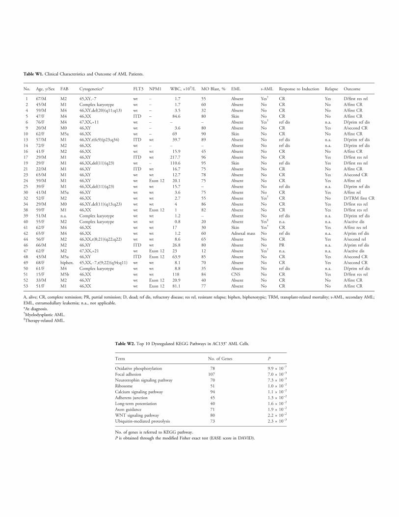
Table W1. Clinical Characteristics and Outcome of AML Patients.
No.
Age, y/Sex FAB Cytogenetics* FLT3 NPM1 WBC, ×109/L MO Blast, % EML s-AML Response to Induction Relapse Outcome1
67/M M2 45,XY,−7 wt – 1.7 55 Absent Yes† CR Yes D/first res rel 2 45/M M1 Complex karyotype wt – 1.7 60 Absent No CR No A/first CR 4 59/M M4 46,XY,del(20)(q11;q13) wt – 3.5 32 Absent No CR No A/first CR 5 47/F M4 46,XX ITD – 84.6 80 Skin No CR No A/first CR 6 76/F M4 47,XX,+11 wt – – – Absent Yes† ref dis n.a. D/prim ref dis 9 20/M M0 46,XY wt – 3.6 80 Absent No CR Yes A/second CR 10 62/F M5a 46,XX wt – 69 90 Skin No CR No A/first CR 13 57/M M1 46,XY,t(6;9)(p23;q34) ITD wt 39.7 89 Absent No ref dis n.a. D/prim ref dis 14 72/F M2 46,XX wt – – – Absent No ref dis n.a. D/prim ref dis 16 41/F M2 46,XX wt wt 15.9 45 Absent No CR No A/first CR 17 29/M M1 46,XY ITD wt 217.7 96 Absent No CR Yes D/first res rel 19 29/F M1 46,XX,del(11)(q23) wt – 110.6 95 Skin No ref dis Yes D/first res rel 21 22/M M1 46,XY ITD wt 16.7 75 Absent No CR No A/first CR 23 65/M M1 46,XY wt wt 12.7 78 Absent No CR Yes A/second CR 24 59/M M1 46,XY wt Exon 12 20.1 75 Absent No CR Yes A/first rel 25 39/F M1 46,XX,del(11)(q23) wt wt 15.7 – Absent No ref dis n.a. D/prim ref dis 30 41/M M5a 46,XY wt wt 3.6 75 Absent No CR Yes A/first rel 32 52/F M2 46,XX wt wt 2.7 55 Absent Yes† CR No D/TRM first CR 34 29/M M0 46,XY,del(11)(q13;q23) wt wt 4 86 Absent No CR Yes D/first res rel 38 59/F M1 46,XX wt Exon 12 1 82 Absent No CR Yes D/first res rel 39 51/M n.a. Complex karyotype wt wt 1.2 – Absent No ref dis n.a. D/prim ref dis 40 55/F M2 Complex karyotype wt wt 0.8 20 Absent Yes‡ n.a. n.a. A/active dis 41 62/F M4 46,XX wt wt 17 30 Skin Yes† CR Yes A/first res rel 42 65/F M4 46,XX wt wt 1.2 60 Adnexal mass No ref dis n.a. A/prim ref dis 44 56/F M2 46,XX,t(8;21)(q22;q22) wt wt 8.6 65 Absent No CR Yes A/second rel 46 66/M M2 46,XY ITD wt 26.8 80 Absent No PR n.a. A/prim ref dis 47 62/F M2 47,XX,+21 wt Exon 12 23 12 Absent Yes† n.a. n.a. A/active dis 48 43/M M5a 46,XY ITD Exon 12 63.9 85 Absent No CR Yes A/second CR 49 68/F biphen. 45,XX,−7,t(9;22)(q34;q11) wt wt 8.1 70 Absent No CR Yes A/second CR 50 61/F M4 Complex karyotype wt wt 8.8 35 Absent No ref dis n.a. D/prim ref dis 51 15/F M5b 46,XX wt wt 118 84 CNS No CR Yes D/first res rel 52 33/M M2 46,XY wt Exon 12 20.9 40 Absent No CR No A/first CR 53 51/F M1 46,XX wt Exon 12 81.1 77 Absent No CR No A/first CRA, alive; CR, complete remission; PR, partial remission; D, dead; ref dis, refractory disease; res rel, resistant relapse; biphen, biphenotypic; TRM, transplant-related mortality; s-AML, secondary AML;EML, extramedullary leukemia; n.a., not applicable.*At diagnosis.†Myelodysplastic AML.‡Therapy-related AML.
Table W2. Top 10 Dysregulated KEGG Pathways in AC133+ AML Cells.
Term
No. of Genes POxidative phosphorylation
78 9.9 × 10⁻⁷Focal adhesion
107 7.0 × 10⁻³Neurotrophin signaling pathway
70 7.3 × 10⁻³Ribosome
51 1.0 × 10⁻²Calcium signaling pathway
94 1.1 × 10⁻²Adherens junction
45 1.3 × 10⁻²Long-term potentiation
40 1.6 × 10⁻²Axon guidance
71 1.9 × 10⁻²WNT signaling pathway
80 2.2 × 10⁻²Ubiquitin-mediated proteolysis
73 2.3 × 10⁻³No. of genes is referred to KEGG pathway.P is obtained through the modified Fisher exact test (EASE score in DAVID).

Figure W1. Nodal expression analysis on BM samples from AML patients. RT-PCR analysis has been performed to investigate the level ofNodal expression in AML samples from six different patients (lanes 1 to 6). The analysis has also been performed on BM specimen sampledfrom a healthy donor (lane HD), and monitored for quality with a positive control (lane HS401, human embryonic stem cell line HS401) and ano cDNA negative control (lane neg). A 100-bp molecular weight DNA ladder (Genespin) has been loaded onto the gel for amplicon sizing(lane L). The analysis pointed out the complete lack ofNodal expression in all the six samples tested, aswell as, unsurprisingly, in the healthydonor. Arrowhead indicates the size of the Nodal-specific PCR product (392 bp) obtained with the following pair of primers: HuNODAL_ff2:5′AGGGCGAGTGTCCTAATCCT and HuNODAL_rr: 5′CAGACTCCACTGAGCCCTTC. To further increase the sensitivity of the assay, we havesubjected the PCRs to a second round of amplification using a semi-nested approach with the same reverse primer and with a nestedforward primer (HuNODAL_ff1: 5′GAGGAGTTTCATCCGACCAA). The semi-nested PCR confirmed the total absence of Nodal expressionin all the six samples analyzed, with the expected 366-bp product detected exclusively in the positive control (not shown, figure availableupon request). To further increase the strength of the data, we have performed the same analysis on a second set of seven AML BM speci-mens, resulting in the exact same outcome (not shown, figure available upon request). GAPDHwas used as internal control to test the goodquality of all the cDNAs used in the experiments (not shown, figure available upon request).