regenerative medicine – a new industry ~ remedi ~ a grand ... · remedi, regenerative medicine...
TRANSCRIPT

European Cells and Materials Vol. 16. Suppl. 3, 2008 (page 1) ISSN 1473-2262
Regenerative Medicine – A New Industry ~ remedi ~
A Grand Challenge David J. Williams1, Richard Archer2, Paul Hourd1, John Crowe3, Brian Meenan4, Richard Lilford5,
Paul Martin3, Kevin Shakesheff3, Nick Rhodes6, Finbarr Livesey7
1Loughborough University, 2Two BC Cambridge, 3Nottingham University, 4Ulster University, 5Birmingham University, 6Liverpool University, 7Cambridge University
INTRODUCTION: Realisation of regenerative medicine therapies using human cells and tissues as a 21st century industry requires consistent manufacturing and appropriate business and cost structures. The goal of the remedi grand challenge project is to realise delivery at an acceptable price and to work to make a key contribution to the growth of regenerative medicine as an industry and unlock its potential to contribute to the UK economy. A key aim of the project is demonstrating how established bio-science can be transformed into profitable commercial practice and generate affordable therapies. Remedi is a £7m programme, the core runs from Sep 2005 to Feb 2010. More details at www.remedigc.org.
MANAGEMENT AND ORGANISATION: Remedi is carefully managed. It is chaired by Richard Archer; PI, David J. Williams; and Project Manager, Paul Hourd report to a Steering Group of project partners. Operational management is via a board of Workpackage managers reporting against deliverables, with parallel informal Researchers Meetings. December 2007 saw a major review.
There are five workpackages in the project. WP1 understands the market and WP2 the policy environment; WP3 focusses on product processing of scaffolds, cells, and tissues and WP4 on characterisation and control of the product; and WP5 aims to facilitate industry and SME growth.
SOME KEY PUBLICATIONS: Cosh, E., Girling, A., Lilford, R., McAteer, H., Young, T., (2007) "Investing in new medical technologies: A decision framework". Journal of Commercial Biotechnology, 2007, Vol. 13, No. 4, pp. 263-271
Kulkarni, R. P, Livesey, F., Dodin, L. D., (2007) “Will regulation determine the science agenda? A look at hESCs”, Regenerative Medicine, 2(5), 839-844.
Ginty, P. J., Barry, J. J. A, White, L. J., Howdle, S. M., Shakesheff, K. M., (2007) “Controlling protein release from scaffolds using polymer blends and composites”, European Journal of Pharmaceutics and Biopharmaceutics, in print.
Thomas, R. J., Chandra, A., Liu, Y., Hourd, P. C., Conway, P. P., Williams, D. J., (2007) “Manufacture of a human mesenchymal stem cell population using an automated cell culture platform”, Cytotechnology, 55(1), 31-39.
Sebastine, I.M. and Williams, D.J., ''The Role of Mechanical Stimulation in Engineering of Extracellular Matrix (ECM)'', Proceedings of the 28th IEEE EMBS Annual International Conference, New York City, USA, September 2006, 3648-3651, ISBN 1-4244-0033-3
Mather, M. L., Morgan, S. P., White, L. J., Tai, H., Kockenberger, W., Howdle, S. M., Shakesheff K. M., Crowe, J. A., (2008), “Image-based characterisation of foamed polymeric tissue scaffolds”, Biomedical Materials, 3, pp.11.
PLANS FOR THE FUTURE
ACKNOWLEDGEMENTS: EPSRC; Partners including The Automation Partnership, Regentec, Critical Pharmaceuticals, Intercytex, Association of British Healthcare Industries, NHS Futures, National Physical Laboratory, National Institute of Biological Standards and Control, East Midlands Development Agency, East Midlands NHS Innovations Hub, and Medlink East Midlands; and remedi researchers: George Burke, Amit Chandra, Judith Curran, Richard Elliot, Patrick Ginty, Lloyd Hamilton, Sally Jackson, Jasmin Kee, Yang Liu, Melissa Mather, Helen McAteer, Richard McCann, Ed Morris, Nikolay Nikolaev, Aleksandar Nikolic, Elizabeth Pearson, Abdel Rabi, Yvonne Reinwald, Emma Rowley, Immanuel Sebastine, Pawanbir Singh, Rob Thomas, and Lisa White.
European Cells and Materials Vol. 16. Suppl. 3, 2008 (page 2) ISSN 1473-2262
Mesenchymal Stem Cells: Characterization, Therapeutic Evaluation and Manufacturing
Frank Barry, Mary Murphy, Cindy Coleman, Tim O’Brien, Margaret Rae & Karen Duffy
REMEDI, Regenerative Medicine Institute, National University of Ireland, Galway
Stem cell therapy can be defined as the treatment of disease by the mobilization or transplantation of autologous or allogeneic stem cells into a host. Mesenchymal stem cells (MSCs) represent especially attractive candidates for therapeutic use because of their ease of isolation, expansion in culture and clear multipotent nature. While the therapeutic testing of these cells has progressed well, there are still many questions to be addressed concerning their mechanism of action, the role of endogenous populations of stem cells in the adult and the function of various stem cell niches. In addition, there are several aspects to the implanted cell–host interaction that need to be addressed as we attempt to understand the mechanisms underlying these therapies. There is no consensus regarding a method for the isolation of MSCs from any human tissue. There is some consensus on what are the defining characteristics of MSCs, based on a recent position paper from the International Society for Cellular Therapy1. However, this is based on a somewhat slender list of biological attributes and, in the words of the authors, represents research criteria that “should not be confused with release specifications for clinical studies”. They further indicated that the criteria are based on “the best currently available data” and “future research will probably mandate a revision”. The lack of agreed clinical release specifications is a serious impediment to progress in assessing the therapeutic potential of MSCs in humans. There is a risk that current clinical studies will be rendered useless by the lack of consensus on the definition of MSC and the lack of standardised isolation protocols2. The ISCT position paper relates to adherence to plastic, specific surface antigen expression and multipotent differentiation potential as the criteria for defining MSCs. There is not one criterion in this list that is specific to MSCs and not shared with many other cells. Indeed it is probably the case that other cells types share ALL of the MSC-selected criteria outlined in this position paper. Certainly this merely reflects the inadequacy of our understanding of the phenotype, genotype and
plasticity of MSCs. There is a conundrum in stem cell technology that is highlighted in the application of MSCs more than any other stem cell:
• the absence of a rigorous and foolproof set of criteria for a standard definition of an MSC means that universal standards of preparation are essential
• the absence of universal standards of preparation means that release criteria must be solid and cell-specific.
There has been relatively little progress in the development of new culture technologies for MSC manufacture and most efforts, including those in small-scale GMP facilities, rely on laboratory-scale methods with little improvement over earlier efforts. There is a strong possibility that progress in delivering therapy to patients will be hampered by a poor ability to produce stem cells. In the event that therapeutic applications are successful, it is likely that there will be a significant gap between demand and supply. Current approaches, which often rely on serum-containing media and relatively low volume methods, will not be effective and methods for testing MSC preparations rely on panels of surface antigens which may be non-specific3, 4. REFERENCES: 1 Dominici et al (2006) Cytotherapy. 8, 315-317. 2 Barry FP, Murphy JM (2004) Int J Biochem Cell Biol. 36, 568-584. 3 Barry FP, Boynton RE, Haynesworth S, Murphy JM, Zaia J. (1999) Biochem Biophys Res Commun. 265, 134-139.

European Cells and Materials Vol. 16. Suppl. 3, 2008 (page 3) ISSN 1473-2262
Please Tick your preferred Presentation Option
X
Oral Presentation Poster Presentation with Oral PresentationPoster Presentation
Development and manufacture of a human living dermal equivalent (ICX-SKN) M. Flasza-Baron, P. Kemp, D. Shering, D. Marshall, Z. Denham & P.A. Johnson 1
1 Intercytex Ltd, Manchester, UK INTRODUCTION: ICX-SKN has been designed as a sterile allogeneic dermal substitute comprising a matrix composed largely of collagen and allogeneic neonatal foreskin derived human dermal fibroblast cells (HDFs). ICX-SKN is first formed from a provisional matrix of fibrin into which HDFs are seeded. Through a process of maturation, HDFs are induced to lay down collagen and other extra-cellular matrix materials and, as the construct matures, the original fibrin is replaced or augmented by collagen that becomes cross-linked. This gives tensile strength and flexibility to the construct, but more importantly is designed to resist the enzymatic activity of the wound environment. ICX-SKN is intended to perform similarly to a skin graft and to have similar persistence and function in wound-healing.
In addition to its functional properties ICX-SKN has been designed to provide several key characteristics identified by experts in the field as critical to success of a dermal or skin substitute. These include efficacy, convenient size and shape, ease of use, off-the-shelf availability, and permanent healing effect. In here we describe the development of a discontinuous manufacturing process [1] that resulted in the production of a human living dermal substitute suitable for use in a first-in-man clinical trial [2]. METHODS: ICX-SKN was manufactured in accordance with EU standards of Good Manufacture Practice (GMP) in licensed GMP manufacturing facility at Intercytex Ltd., UK according to protocol described in [1]. ICX-SKN was analyzed for its composition using histology and DSC methods; its performance by using strength test, resistance to collagenase and in vivo open wound assay. Cell attachment and viability was analyzed by metabolic activity Alamar Blue assay and phalloidin staining [1].
RESULTS: The novel process of manufacturing ICX-SKN resulted in robust in its handling properties matrix which was composed mainly of human collagen I. ICX-SKN can be repopulated with HDFs and human keratinocytes (HKs) which can attach and differentiate on the matrix. The graft replacements applied to excision wounds in mice [1] and human volunteers [2] healed and were rapidly re-epithelialized, and persisted for over 28 days post grafting.
Fig. 1: Process flow of ICX-SKN production. Stages of production are marked by numbers and points of exit/entry to GMP are indicated by dashed lines. Casting of the constructs within GMP facility occurs at stage 1: Reagents are chilled, mixed sequentially and cast immediately upon the addition of thrombin as described [1]. Maturation occurs at stage 2, where constructs are fed 3 times per week with maturation for 7 weeks. Matured constructs (pSKN) are freeze dried to preserve the matrix (stage 3 -- dSKN), sterilized (stage 4 -- sSKN) within 1 week post collection of pSKN, and finally reintroduced to GMP to be re-populated with HDF (stage 5 – ICX-SKN) over 2 days and packaged, producing the final product for use in the clinic. (A) (B) (C)
Fig.2: (A) Gross appearance of ICX-SKN. (B) Stress fibers in living HDFs stained with phalloidin and (C) nuclei from HDFs living on the matrix in cross-section of ICX-SKN skin graft replacement. DISCUSSION & CONCLUSIONS: ICX-SKN has been developed as a platform product that can be used as a skin graft replacement and the process by which it is manufactured has been designed for the product to be available to the end-user off-the-shelf and for ease-of-use in practice
REFERENCES: 1 Flasza et al (2007) Regenerative Medicine 2 (6), 903-918; 2 Boyd et al (2007) Regenerative Medicine 2 (4), 363-370.
4 Cells
FibrinThrombin
7 weekspSKN Collection dSKN
sSK
Freeze drying
γ-irradiation
ICX-Clini
Cells
1 2 3
5
Medium
Package
2 days
1 week

European Cells and Materials Vol. 16. Suppl. 3, 2008 (page 4) ISSN 1473-2262
Development of a synthetic matrix to replace human dermis
I Cantón1, R McKean1, D Cole1, K Blackwood1, C Freeman3, K Franklin3, P Farthing3, I Brook3, J W Haycock 1, A J. Ryan2, S MacNeil1
1Department of Engineering Materials, Kroto Research Institute, 2 Department of Chemistry, University of Sheffield, 3The School of Clinical Dentistry, University of Sheffield, UK
INTRODUCTION: Our aim is to develop synthetic biodegradable replacement dermal substitutes for tissue engineering of skin and oral mucosa. Such scaffolds should allow host skin cell attachment and organisation in vitro and then become rapidly vascularised on engrafting with the production and organization of a new host extracellular matrix (ECM) as the scaffold degrades without invoking a chronic inflammatory response. METHODS: We examined a total of six experimental electrospun polymer scaffolds: 1) poly-L-lactide (PLLA); 2) PLLA + 10% oligolactide; 3) PLLA + rhodamine and 4-6) three poly (D,L)-lactide-co-glycolide (PLGA) random multiblock copolymers, with decreasing lactide:glycolide mole fractions (85:15, 75:25 and 50:50). Scaffolds were examined for breakdown in vitro and following subcutaneous implantation in adult male Wistar rats for up to 12 months. The ability of the scaffolds to support skin cells (keratinocytes, fibroblasts and endothelial cells) in an organised manner and to promote new ECM production was examined in vitro. RESULTS: In vivo, all scaffolds permitted good cellular penetration, with no adverse inflammatory response outside of the scaffold margin and with no capsule formation around the periphery. Scaffolds were well vascularised with vigorous macrophage activity associated with the fibres (see Fig1). The breakdown rates for scaffolds in vitro (see Fig 2) and in vivo were similar, and an increase in the ratio of polyglycolide to polylactide correlated with an increase in breakdown rate, as expected. Scaffolds of PLLA were stable in vivo even after 12 months whereas scaffolds fabricated from PLGA 85:15 and 75:25 revealed a 50% loss of mass after 4 and 3 months, respectively. In vitro PLGA 85:15 and 75:25 scaffolds were able to support keratinocytes, fibroblasts and endothelial cell growth and extracellular matrix production with evidence of new collagen production after 7 days. Confocal and EM analysis (see Fig 3) revealed an epidermal-dermal like distribution of keratinocytes and fibroblasts through the scaffold. While fibroblasts actively contracted these scaffolds preliminary data suggests that heat annealing the fibres reduces this without affecting scaffold performances. DISCUSSION & CONCLUSIONS: The data supports the development of PLGA 85:15 and 75:25 electrospun polymer scaffolds as potential degradable biomaterials for dermal replacement.
Figure 1. H&E stained section of PLGA 75:25 after implantation in a Wistar rat for 4 weeks. Evidence of a vigorous macrophage response with several well formed blood vessels evident throughout the tissue.
Figure 2. Optical micrographs of PLGA 85:15 (A-B), 75:25 (C-D), and 50:50 (E-F) in vitro degradation in Ringers solution at 37°C in 5% CO.
Figure 3. EM micrographs of electrospun PLGA 75:25, 14 day co-cultures of keratinocytes and fibroblasts. (A) top down (B) shows a cross section.
50 50 µm
A B
85:1
75:2
50:5
Day Day
Day
Day
Day
Day
C D
F G
A B

European Cells and Materials Vol. 16. Suppl. 3, 2008 (page 5) ISSN 1473-2262
THE USE OF HEALTH ECONOMICS IN THE EARLY EVALUATION OF REGENERATIVE MEDICINE APPLICATIONS
H McAteer1 & R Lilford1
1 Department of Public Health, University of Birmingham, Birmingham, B15 2TT, UK INTRODUCTION: In healthcare systems with finite resources purchasing and reimbursement decisions are increasingly influenced by formal health economic analysis. It is therefore sensible to assess potential cost-effectiveness of a new technology as early as possible in the development cycle. We propose the novel use of health economics at the supply side using an approach we term the headroom method1. We ask the question ‘would it [the technology] be cost-effective if it works as well as we hope?’ We have applied this method to clinical applications in the urogenital system and are working on applications of abdominal wall and bone defects. METHODS: The headroom method can estimate the maximum cost for which a technology can be brought to market and still be considered cost-effective. In order to apply this method, firstly, the clinical problem which the technology will address must be clearly defined, it is important to be as specific as possible, a clear understanding of the strengths and weaknesses of current treatment is crucial as well as a specific description of disease including prevalence and incidence. This is achieved through a comprehensive and systematic review of literature and consultation with clinicians and tissue engineers. These two elements fit into a broader investment decision framework described in figure 1.
Fig. 1: A framework for investment decisions.
The headroom is based on optimistic yet plausible estimates of effectiveness and is calculated by rearranging the conventional health economic formula from equation (1) to (2).
ICER = ∆Cost/∆Benefit (1)
∆Cost = ICER x ∆Benefit (2)
Where ICER is incremental cost-effectiveness ratio or societal willingness to pay threshold, ∆Benefit is the difference in effectiveness between new treatment and gold standard, and ∆Cost is the difference in cost between new treatment and gold standard and this represents the headroom; the maximum additional cost of the new treatment over the comparator for the new treatment to be deemed cost-effective.
RESULTS: After taking account of potential market we identified two potential indications for urogenital TE that required further investigation: bladder cancer and urethral strictures. The headroom analysis found that a TE solution for bladder cancer has the potential to be cost effective providing marginal costs do not exceed £16,000 but a TE treatment for urethral strictures is unlikely to be cost effective as the headroom is just £1862. With regard to defects of the abdominal wall and bone we have defined the clinical problem and identified indications for further investigation. For abdominal wall defects incisional and parastomal hernias and contaminated defects show the greatest headroom for improvement in effectiveness of treatment 3. For defects of the bone we have found that large defects (segmental defects) have greatest headroom for improvement in effectiveness however, small defects (spinal fusion, fracture nonunion) may not have sufficient headroom to justify additional cost of a TE solution4.
DISCUSSION & CONCLUSIONS: The headroom method is primarily useful as a barrier to misguidedly investing in those technologies which can never be cost-effective, even when some parameters are uncertain.
REFERENCES: 1Cosh E, Girling A, Lilford R, McAteer H, & Young T. 2007. Journal of Commercial Biotechnology 13:263-271. 2McAteer H, Cosh E, Freeman G, Pandit A, Wood P, & Lilford R. 2007. Journal of tissue engineering and regenerative medicine 1:343-349. 34McAteer H, & Lilford R. 2008. A systematic account of the evidence for abdominal wall repair. Unpublished work 4McAteer H, Griffin D, Donell S, Scammel B, Kon E, Stirling A, & Lilford R. 2007. The current clinical opportunities for regenerative medicine in bone. Unpublished work
ACKNOWLEDGEMENTS: We would like to thank the EPSRC REMEDI programme and the EU FP6 STEPS programme for their support.
Market Analysis
Clinical Problem Definition
Headroom Analysis
Return on Investment Analysis
Formal Economic Methods
Can at any point make an intuitive
decision
Market Analysis
Clinical Problem Definition
Headroom Analysis
Return on Investment Analysis
Formal Economic Methods
Can at any point make an intuitive
decision

European Cells and Materials Vol. 16. Suppl. 3, 2008 (page 6) ISSN 1473-2262
MECHANICAL LOADING IN ECM SCAFFOLD REMODELING TW Gilbert, A Nieponice, A Boruch & SF Badylak
McGowan Institute for Regenerative Medicine, University of Pittsburgh, Pittsburgh, PA, USA
INTRODUCTION: Biologic scaffold materials derived from naturally occurring extracellular matrix (ECM) have been shown to promote site-specific tissue remodeling in a variety of body systems 1. The mechanisms of remodeling include rapid degradation of the scaffold with release of matricryptic peptides that recruit a population of progenitor cells to the site of remodeling. It is thought that the progenitor cells are pushed towards site-specific tissue as a result of site-appropriate cues, including mechanical signals. The present study describes several in vivo studies that support the necessity for appropriate loading regimes and in vitro studies that investigated the effect of mechanical loading on the gene expression of cells seeded on an ECM scaffold.
METHODS: In vivo: ECM derived from two layers of the porcine urinary bladder, the basement membrane and subjacent tunica propria (termed urinary bladder matrix – UBM) was used to replace a 30% resection of the dome of the urinary bladder in eight adult female dogs. Four of the dogs were catheterized for 24 hours following surgery at which time spontaneous filling and emptying of the bladder was allowed to occur for the remainder of the study. The catheter remained in place for 30 days in the following four dogs. One half of the dogs in each group were sacrificed after 30 days and the other half were sacrificed after 90 days.
In vitro 2: Lyophilized SIS rehydrated in DMEM supplemented with 10% bovine calf serum and 1% penicillin/ streptomycin (DMEM-CS-C) was seeded with 1.5x10⁶ NIH 3T3 fibroblasts (0.5x10⁶ cells/cm2) and 8 hours were allowed for cell attachment. The scaffolds were transferred to a custom built stretching device and cyclic stretching regimens were then applied to 5%, 10%, or 15% stretch at 0.1 Hz for 20 minutes, 3 times daily at 8 hour intervals for 3 days. Each specimen was collected for RNA isolation. cDNA was synthesized and PCR reactions were performed using primers specific for murine Col I, Col III, MMP-2, SM-A, and Tn-C with GAPDH as an internal control.
RESULTS: Dogs that remained catheterized showed greater than 90% contraction of the scaffold material, incomplete urothelial coverage at 30 days, and a chronic inflammatory response
within the underlying UBM scaffold and newly deposited host ECM. The dogs that were allowed spontaneous filling of the bladder showed 30% contraction of the scaffold area, complete urothelialization at 30 days with islands of smooth muscle in the subjacent remodeling tissue. By 90 days, these non-catheterized dogs showed sheets of functional smooth muscle arranged in patterns that resembled the normal muscularis externa with an adjacent submucosal layer and near normal bladder histomorphology. The in vitro study showed that mechanical loading of led to gene expression profiles that are consistent with regenerative healing, specifically with an increased ratio of collagen type I:III.
DISCUSSION & CONCLUSIONS: Mechanical loading of ECM scaffolds is essential to the site-specific remodeling that has been observed. The present study clearly shows that when ECM scaffolds are used to repair the urinary bladder. Appropriate loading leads to the formation of organization urinary bladder wall with smooth muscle and urothelium. The absence of loading led to scar tissue formation. Similar results were found in an immobilized Achilles tendon repaired with ECM 3. In vitro studies show that mechanical loading alters in the gene expression of cells within an ECM scaffold in a manner that is consistent with site-specific tissue formation.
REFERENCES: 1 SF Badylak. Xenogeneic extracellular matrix as a scaffold for tissue reconstruction. Transpl Immunol. 2004;12(3-4):367-377. 2TW Gilbert, AM Stewart-Akers, J Sydeski, et al. Gene expression by fibroblasts seeded on small intestinal submucosa and subjected to cyclic stretching. Tissue Eng. 2007;13(6):1313-1323.3 JP Hodde, SF Badylak , KD Shelbourne. The effect of range of motion on remodeling of small intestinal submucosa (SIS) when used as an Achilles tendon repair material in the rabbit. Tissue Eng. 1997;3(1):27-37.
ACKNOWLEDGEMENTS: The authors would like to acknowledge funding from the NIH.

European Cells and Materials Vol. 16. Suppl. 3, 2008 (page 7) ISSN 1473-2262
Chondrocytes express the mechanosensitive hemichannel connexin 43 M Garcia1, S. Geris1, S McGlashan2, A Poole2, C Jensen2, M Knight1
1 School of Engineering and Materials Science, Queen Mary University of London, UK. 2 Faculty of Medicine & Health Sciences, University of Auckland, NZ.
INTRODUCTION: Mechanical loading is essential for the health and homeostasis of articular cartilage although the fundamental mechanotransduction pathways are unclear. Previous studies have demonstrated that compression of isolated chondrocytes in agarose activates a Ca2+ signalling pathway, mediated by the release of ATP1. This mechanosensitive pathway triggers an up-regulation of proteoglycan synthesis2, however, the mechanism of ATP release is as yet unclear. The present study tests the hypothesis that chondrocytes express connexin 43 hemichannels, which have been shown to function as mechanosensitive ATP release channels in other cell types3.
METHODS: Bovine articular chondrocytes were isolated from the metacarpal-phalangeal joint, seeded in 3% agarose (w/v) and cultured in DMEM+20%FCS for 24 hrs.
Immunolocalisation: Cell-agarose core specimens (5mm DIA x 5mm) were processed for immunofluorescence following standard protocols. A series of studies were conducted using primary antibodies to both the intracellular and extracellular domains of connexin 43, as well as α-tubulin in the primary cilium. Cells labelled with appropriate secondary antibodies were visualised using confocal microscopy (Leica SP2).
Lucifer Yellow uptake: Lucifer Yellow (LY) uptake was used to verify the functional presence of hemichannels. Cell-agarose specimens were labelled for 40mins with 0.4 %LY in PBS with and without the addition of flufenamic acid (500μM, FFA), a known inhibitor of hemichannels3. As a positive control, cells were pre-treated with 5mM EGTA in PBS for 40 minutes. Following LY treatment, specimens were rinsed in DMEM+20%FCS, fixed in 3.7% formaldehyde and then washed repeatedly with PBS prior to fluorescence microscopy.
RESULTS: All bovine chondrocytes expressed connexin 43 hemichannels, as shown by immunofluorescence co-localisation of both the intracellular and extracellular domains. Primary cilia were identified on 60% of cells and of these approximately 50% exhibited connexin 43 on the primary cilium (Fig 1). LY uptake was
significantly increased from 42% of untreated chondrocytes to 91% following EGTA treatment. FFA completely abolished LY uptake in both groups (Fig. 2).
Fig. 1: Confocal images of connexin 43(green) on a primary cilium (red).
010
203040
5060
708090
100
untreated FFA
% L
Y po
sitiv
e ce
lls
PBSPBS+EGTA
*
+ +
Fig. 2: LY uptake in chondrocytes. Significant differences with and without EGTA (*) and with and without FFA (+) are indicated (p<0.05)
DISCUSSION & CONCLUSIONS: Articular chondrocytes express functional hemichannels activated by EGTA and blocked by FFA. Approximately 40% of unloaded, untreated cells show hemichannel activation which may represent the subpopulation of cells that exhibit spontaneous Ca2+ signalling1. Further studies are required to see if these hemichannels are involved in mechanosensitive ATP release, as in other cell types. However the presence of hemichannels on the primary cilium in a sub-population of cells may provide a mechanoreceptor complex through which these chondrocytes sense their mechanical environment. In deed this may explain why only a sub-population of chondrocytes exhibit mechanosensitive Ca2+ signalling1.
REFERENCES: 1 Pingguan-Murphy et al (2006) J Cell Physiol 209:389-97. 2 Chowdhury & Knight (2006) J Cell Physiol 209:845-53. 3 Stout et al (2002) J Biol Chem 277:10482-10488.
European Cells and Materials Vol. 16. Suppl. 3, 2008 (page 8) ISSN 1473-2262
Scaffold Design for Tissue Engineering the Anterior Cruciate Ligament Elaine Durham, Stephen J. Russell 1 & Eileen Ingham2
1Nonwovens Research Group, University of Leeds, UK
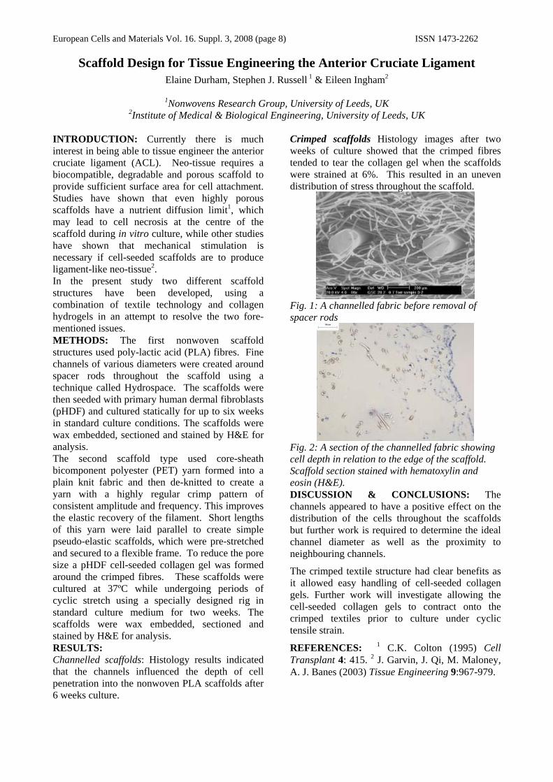
2Institute of Medical & Biological Engineering, University of Leeds, UK INTRODUCTION: Currently there is much interest in being able to tissue engineer the anterior cruciate ligament (ACL). Neo-tissue requires a biocompatible, degradable and porous scaffold to provide sufficient surface area for cell attachment. Studies have shown that even highly porous scaffolds have a nutrient diffusion limit1, which may lead to cell necrosis at the centre of the scaffold during in vitro culture, while other studies have shown that mechanical stimulation is necessary if cell-seeded scaffolds are to produce ligament-like neo-tissue2. In the present study two different scaffold structures have been developed, using a combination of textile technology and collagen hydrogels in an attempt to resolve the two fore-mentioned issues. METHODS: The first nonwoven scaffold structures used poly-lactic acid (PLA) fibres. Fine channels of various diameters were created around spacer rods throughout the scaffold using a technique called Hydrospace. The scaffolds were then seeded with primary human dermal fibroblasts (pHDF) and cultured statically for up to six weeks in standard culture conditions. The scaffolds were wax embedded, sectioned and stained by H&E for analysis. The second scaffold type used core-sheath bicomponent polyester (PET) yarn formed into a plain knit fabric and then de-knitted to create a yarn with a highly regular crimp pattern of consistent amplitude and frequency. This improves the elastic recovery of the filament. Short lengths of this yarn were laid parallel to create simple pseudo-elastic scaffolds, which were pre-stretched and secured to a flexible frame. To reduce the pore size a pHDF cell-seeded collagen gel was formed around the crimped fibres. These scaffolds were cultured at 37ºC while undergoing periods of cyclic stretch using a specially designed rig in standard culture medium for two weeks. The scaffolds were wax embedded, sectioned and stained by H&E for analysis. RESULTS: Channelled scaffolds: Histology results indicated that the channels influenced the depth of cell penetration into the nonwoven PLA scaffolds after 6 weeks culture.
Crimped scaffolds Histology images after two weeks of culture showed that the crimped fibres tended to tear the collagen gel when the scaffolds were strained at 6%. This resulted in an uneven distribution of stress throughout the scaffold.
Fig. 1: A channelled fabric before removal of spacer rods
Fig. 2: A section of the channelled fabric showing cell depth in relation to the edge of the scaffold. Scaffold section stained with hematoxylin and eosin (H&E). DISCUSSION & CONCLUSIONS: The channels appeared to have a positive effect on the distribution of the cells throughout the scaffolds but further work is required to determine the ideal channel diameter as well as the proximity to neighbouring channels.
The crimped textile structure had clear benefits as it allowed easy handling of cell-seeded collagen gels. Further work will investigate allowing the cell-seeded collagen gels to contract onto the crimped textiles prior to culture under cyclic tensile strain.
REFERENCES: 1 C.K. Colton (1995) Cell Transplant 4: 415. 2 J. Garvin, J. Qi, M. Maloney, A. J. Banes (2003) Tissue Engineering 9:967-979.

European Cells and Materials Vol. 16. Suppl. 3, 2008 (page 9) ISSN 1473-2262
TISSUE ENGINEERING OF THE ANTERIOR CRUCIATE LIGAMENT CHARACTERISING DEGRADABLE GLASS FIBRES
S Rathbone1 , D Healy2, S Cartmell1 1 Institute of Science & technology in Medicine, Keele University, Hartshill, Stoke-on-Trent.
2 Giltech Ltd, North Harbour Estate, Ayr, Scotland.
INTRODUCTION: Approximately 800,000 people worldwide need to obtain medical treatment annually to repair injured anterior cruciate ligaments (ACL’s).1 Of these, approximately 150,000 people need to undergo surgical treatment known as an ACL reconstruction.2 It is evident that surgical ACL reconstructions have limitations and do not give completely satisfactory long-term results, therefore tissue engineering is being considered as a possible alternative approach to repair or replace this damaged tissue. This study has looked at the efficacy of using a degradable phosphate based glass fibre to guide cell growth for ACL tissue engineering strategies.
METHODS:
Degradable phosphate based glass fibres
The glass fibres were produced by Giltech Ltd, Scotland and consist of NaO, CaO, P2O5, MnO, B2O3, Na-F and Fe2O3.
Tensile testing
The tensile mechanical properties of five different formulations of glass fibre have been tested. The glass fibres were prepared in 5cm long bundles of 0.02g and pulled to failure using a tensile testing machine (ELF 3200 Bose, USA). From the data, maximum force at failure, elongation, Young’s modulus and strain energy were calculated. Cytotoxicity tests are currently being performed using L929 murine fibroblast cells. These tests include assessing cell viability & proliferation at 4 time points using Live/dead stain (Molecular probes), picogreen DNA assay and imaging with scanning electron microscopy. Future work includes using a Bose dynamic chamber to apply cyclic tensile strains to glass fibre/polymer composites seeded with cells to stimulate cell functionality.
RESULTS: The mechanical properties of the glass fibres (table 1) indicate that they possess a high tensile strength, a high stiffness (stiffer than the ACL) and a high resistance to fracture (higher than the ACL). Cell proliferation was supported on the fibres samples (figure 1).
Table 1. Mechancical properties of the glass fibres.
Glass fibre
Max Stress (MPa)
Youngs Modulus (MPa)
Strain Energy (MPa)
1 215 252 129 2 354 286 334 8 320 291 258 10 391 387 273 11 351 387 204
Fig. 1: Cell adherence leading to proliferation on glass fibres: viable cells with live/dead stain (left) and with SEM (right).
DISCUSSION & CONCLUSIONS: The glass fibres show a more than adequate strength for a cell substrate for tissue engineering of an ACL. However, they are very stiff and producing a composite made from glass fibre and polymer to introduce some elasticity is favoured for the cyclic straining regime, which will follow the cytotoxicity tests. Current studies are being performed on glass/polycaprolactone composites in a dynamic loading culture regime. REFERENCES: 1 D. Doroski, D. Brink and J. Temenoff (2007) Techniques for biological characterization of tissue engineered tendon and ligament. J Biomaterials 28(2): 187-202. 2 J. Cooper, H. Lu, et al. (2005) Fiber-based tissue-engineered scaffold for ligament replacement: design considerations and in vitro evaluation. J Biomaterials 26(13): 1523-1532.
ACKNOWLEDGEMENTS: This work is supported by a BBSRC CASE studentship.

European Cells and Materials Vol. 16. Suppl. 3, 2008 (page 11) ISSN 1473-2262
Designing New Supermacroporous Cryogel Materials for Bioengineering Applications
Ashok Kumar
Department of Biological Sciences and Bioengineering, Indian Institute of Technology Kanpur, 208016-Kanpur, India; E-mail: [email protected]
INTRODUCTION: Polymeric biomaterials in recent years have shown increasing potential in biomedical applications. Recently a new type of supermacroprous cryogel materials have been developed which show great promise in various biological applications. Cryogels typically have interconnected macropores (or supermacropores) allowing unhindered diffusion of solutes of practically any size, as well as mass transport of nano- and even microparticles. The unique structure of cryogels, in combination with their osmotic, chemical and mechanical stability, makes them attractive matrices for chromatography of biological particles/cells and for tissue engineering applications. The sponge-type gels with inter-connected pores in the range of 10-100 μm are synthesized under cryostatic conditions from agarose, gelatin, acrylamide, polyvinyl alcohol or any other suitable polymeric materials.
METHODS: Cryogels are synthesized both from monomers and polymeric precursors using mostly water as a solvent in moderately frozen conditions. Under these conditions an optimum concentration (4–10% w/v) of monomers or polymeric pre-cursors are solubilised in water. The mixture is kept on ice to keep very low temperature of the solution and then appropriate polymerization reagents or cross-linkers are added to the above mixture. The solution is thoroughly mixed and added into the tubes or in glass moulds in the shape of discs or sheets. The mixture is immediately immersed in cryostat bath at -12°C to allow it to polymerize and also to form ice crystals. After about 12-16 h of incubation, the croygels are thoroughly washed with water and then dried and sterilized in ethanol for further use. Cryogels are modified by incorporating epoxy or other functional groups during polymerization. This allows further coupling of protein or other molecules for desired application [l].
RESULTS: Here, we present the state-of-the-art use of supermacroporous cryogels for various applications in bioengineering. By using protein A affinity monolithic cryogel column, a generic approach was developed for specifically separating different cell types like lymphocytes [1] and
CD34+ stem cells from cord blood. A novel approach of releasing the cells from the cryogel matrix was designed by mechanically squeezing the cryogels [2]. Other interesting application of the macroporous matrices have been the cultivation of the mammalian cells on the gelatin modified cryogels bioreactors. The cells grow, proliferate and secrete the protein therapeutics continuously in the circulating medium when allowed to culture on the cryogel matrices. These reactors also showed potential when used as extracorporeal device for cryogel scaffolds from agarose, alginate and chitosan with gelatine. Cryogel showed good properties for cartilage tissue engineering [3]. A bilayered cryogel system consisting of bottom layer of gelatin and top layer of povidon was constructed for skin tissue engineering.
Fiugure: SEM picture of crygel matrix (left) and fibroblast cells grown on the cryogel matrix(right).
DISCUSSION & CONCLUSIONS: New approach of designing biomaterials using cryogel technology showed great promise in tissue engineering. The macroporous nature, inter-connected pores and suitable mechanical properties has provided these biomaterials with unique properties. It is expected that the use of cryogels in tissue engineering will expand and new applications will emerge. REFERENCES: 1A. Kumar, F.M. Plieva, et al. (2003). J Immunol Methods 283,185. 2M. B. Dainiak, A. Kumar, et al. (2006). PNAS (USA) 103, 849. 3 A. Tripathi, N. Kathuria & A. Kumar (2008). J Biomed Mat Res A (in press). ACKNOWLEDGEMENTS: DBT and DST, Ministry of Science and Technology, India
European Cells and Materials Vol. 16. Suppl. 3, 2008 (page 12) ISSN 1473-2262
Use of a rabbit cornea model for the development of a cell transfer system for limbal epithelial cells
P Deshpande1, N Bullett2, M Notara3, JT Daniels3, D Haddow2 and S MacNeil1 1 Department of Engineering Materials, University of Sheffield, Sheffield, S3 7HQ
2 CellTran Ltd, Innovation Centre, 217 Portobello, Sheffield, S1 4DP 3Cells for Sight Transplantation and Research Programme, Ocular Repair and Regeneration Biology Unit, UCL
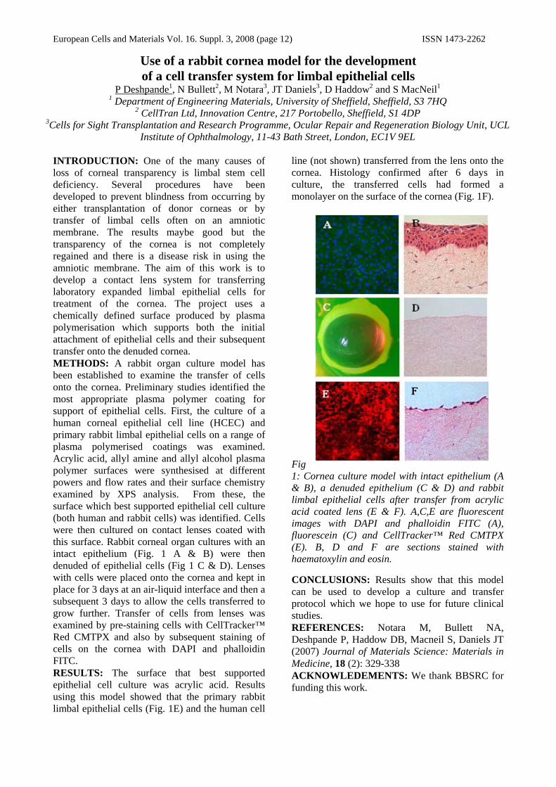
Institute of Ophthalmology, 11-43 Bath Street, London, EC1V 9EL INTRODUCTION: One of the many causes of loss of corneal transparency is limbal stem cell deficiency. Several procedures have been developed to prevent blindness from occurring by either transplantation of donor corneas or by transfer of limbal cells often on an amniotic membrane. The results maybe good but the transparency of the cornea is not completely regained and there is a disease risk in using the amniotic membrane. The aim of this work is to develop a contact lens system for transferring laboratory expanded limbal epithelial cells for treatment of the cornea. The project uses a chemically defined surface produced by plasma polymerisation which supports both the initial attachment of epithelial cells and their subsequent transfer onto the denuded cornea. METHODS: A rabbit organ culture model has been established to examine the transfer of cells onto the cornea. Preliminary studies identified the most appropriate plasma polymer coating for support of epithelial cells. First, the culture of a human corneal epithelial cell line (HCEC) and primary rabbit limbal epithelial cells on a range of plasma polymerised coatings was examined. Acrylic acid, allyl amine and allyl alcohol plasma polymer surfaces were synthesised at different powers and flow rates and their surface chemistry examined by XPS analysis. From these, the surface which best supported epithelial cell culture (both human and rabbit cells) was identified. Cells were then cultured on contact lenses coated with this surface. Rabbit corneal organ cultures with an intact epithelium (Fig. 1 A & B) were then denuded of epithelial cells (Fig 1 C & D). Lenses with cells were placed onto the cornea and kept in place for 3 days at an air-liquid interface and then a subsequent 3 days to allow the cells transferred to grow further. Transfer of cells from lenses was examined by pre-staining cells with CellTracker™ Red CMTPX and also by subsequent staining of cells on the cornea with DAPI and phalloidin FITC. RESULTS: The surface that best supported epithelial cell culture was acrylic acid. Results using this model showed that the primary rabbit limbal epithelial cells (Fig. 1E) and the human cell
line (not shown) transferred from the lens onto the cornea. Histology confirmed after 6 days in culture, the transferred cells had formed a monolayer on the surface of the cornea (Fig. 1F).
Fig 1: Cornea culture model with intact epithelium (A & B), a denuded epithelium (C & D) and rabbit limbal epithelial cells after transfer from acrylic acid coated lens (E & F). A,C,E are fluorescent images with DAPI and phalloidin FITC (A), fluorescein (C) and CellTracker™ Red CMTPX (E). B, D and F are sections stained with haematoxylin and eosin. CONCLUSIONS: Results show that this model can be used to develop a culture and transfer protocol which we hope to use for future clinical studies. REFERENCES: Notara M, Bullett NA, Deshpande P, Haddow DB, Macneil S, Daniels JT (2007) Journal of Materials Science: Materials in Medicine, 18 (2): 329-338 ACKNOWLEDEMENTS: We thank BBSRC for funding this work.

European Cells and Materials Vol. 16. Suppl. 3, 2008 (page 13) ISSN 1473-2262
0
20
40
60
80
100
120
0.1 1 10
mTHPC (μg/ml)
% c
ell d
eath
SatellitecellsNeurones
MCF-7
Assessing the Effect of Photodynamic Therapy on Peripheral Nerve and Cancer Cells Using a Thin Tissue Engineered Collagen Culture Model
K E Wright1, A J MacRobert2, & J B Phillips1
1Life Sciences Department, The Open University, Milton Keynes, U.K. 2 National Medical Laser Centre, UCL, London, U.K.
INTRODUCTION: This study used an innovative thin 3D collagen culture system, to evaluate the response of primary rat peripheral nerve cells to photodynamic therapy (PDT). PDT is a cancer therapy that involves the administration of a photosensitive drug which becomes activated following the focal application of light to tumour sites. The subsequent production of toxic singlet oxygen results in cell death. Clinically, nerve sparing has been observed after meta tetra hydroxyl phenyl chlorine (mTHPC) mediated PDT [1 & 2]. This study aims to simulate nerve PDT in culture with a thin tissue engineered collagen scaffold model in order to assess the cellular basis for the phenomenon of peripheral nerve sparing after PDT.
METHODS: Mixed cultures of neurones and satellite glial cells were propagated from the dorsal root ganglia of 250-300 g rats and the human breast adenocarcinoma cell line MCF-7 was used as a comparator. Cells were seeded within 200 µl type I collagen gels (1 mm think discs). Cell-seeded gels were cultured for 4 days in DMEM supplemented with 10 % foetal calf serum and 1 % penicillin & streptomycin before incubation with mTHPC of various doses for 4 h. Samples were exposed to 10 min white light with a low light fluence rate (0.518 mW/cm2 measured at 633 nm), and then maintained in culture for a further 24 h. Viability of cell populations was assessed using a propidium iodide (PI) exclusion assay. Neurones were distinguished from satellite cells using immunoreactivity for βIII-tubulin. RESULTS: This collagen model system supported the growth of neural and tumor cells and enabled PDT treatments to be applied in a consistent controllable manner. The collagen scaffold trapped live and dead cells throughout the staining procedures enabling microscopic analyses of treated samples (Fig 1). mTHPC-mediated PDT showed a cell population specific death response in the order: MCF-7 > satellite cells > neurones in a dose dependent manner (Fig 2).
Fig. 1: Confocal micrograph showing thin 3D collagen culture model seeded with dissociated DRG culture. (Red) dead PI stained cell nuclei & (Green) βIII-tubulin labelled axons.
Fig 2: Dose responses of cells in thin collagen model after exposure to mTHPC-mediated PDT.
DISCUSSION & CONCLUSIONS: This model has enabled the sensitivity of different cell populations to PDT to be established. Individual cell populations were identified, and cell death defined as having PI stained (red) nuclei. These experiments indicate the possibility of a clinical PDT dose that could be used without adversely affecting neurones at the treatment site.
REFERENCES: 1CS Betz, HR Jäger HR, et al. (2007) Lasers Surg Med 39(7):571-82. 2CM Moore, T.R. Nathan, W.R. Lees et al. (2006) Lasers Surg Med 38(5):356-63.

European Cells and Materials Vol. 16. Suppl. 3, 2008 (page 14) ISSN 1473-2262
Injectable Hyaluronan Gels Form Bone In-vivo J. Hilborn, D. Ossipov, K. Bergman, T. Bowden & T. Engstrand
Materials Chemistry Department, Uppsala University, 751 21 Uppsala, Sweden INTRODUCTION: Autologous bone grafts are routinely used to heal large bone defects but has the disadvantages limited graft quantity, donor site morbidity, pain for the patient, and cost of harvesting. A promising alternative to this autografting is the use of a growth factor loaded biomaterial where endogenous cells are recruited in-vivo by loading the gel with bone morphogenetic protein-2 (BMP-2) resulting in significant bone formation without causing inflammation or foreign body response. It has potential as an off-the-shelf injectable product to use where bone tissue is needed. METHODS: To tailor a non-toxic injectable system with rapid in vivo gel formation in water at 37°C, HA carboxyl groups were converted to aldehydes in a two-step procedure.1 An acetal derivative of HA was prepared by coupling with aminoacetaldehyde dimethylacetal, and in a second step hydrolyzed with hydrochloric acid to generate aldehyde-modified HA (HAA) (substitution degree of 5 %). PVA (16 kDa) was equipped with 5 % hydrazide functionality in a two-step reaction.2 The ability of the gels loaded with rhBMP-2 to induce bone formation was directly assessed in Sprague Dawley rats by co-injecting either HAA and PVAH with BMP-2 (0.2 mL, 30 μg rhBMP-2) into quadriceps muscles.
RESULTS: The two multi-functionalized polymers react spontaneously and selectively with each other in aqueous solutions to form a cross-linked network, scheme 1. Examination after 4 weeks show the formation of ectopic bone at the site where gels loaded rhBMP-2 were injected (figure 1).
Scheme 1
Bone formation at the site of injection, including mineralized tissue surrounding a bone marrow cavity, was visualized by histology.
Figure 1. A: Ectopic bone formation at the injection site (arrows, right pictures) in the BMP-group, gel without the growth factor had no effect (left pictures). Histology show active bone formation with osteoid and surrounding osteoblasts (C & D, lower-left arrow) and blood vessel formation (D, upper-right arrow). In the control, no residues of the gel was found with complete absence of inflammatory reaction (B).
Arteries and veins were found in adjacent soft tissues in BMP-2-hydrogel specimens, but not in the controls indicating the concurrent induction of vascular tissue by BMP-2 in the gel. Interestingly, the infiltration of inflammatory cells, including giant cells and lymphocytes, was completely absent in the BMP-2-hydrogel group and in the control. DISCUSSION & CONCLUSIONS: The benefit of not having to rely on transplantation of cells, in combination with the non invasive procedure, makes this approach towards guided bone repair particularly attractive in clinical use for the treatment of slow- or non-healing closed fractures. This system that diminish morbidity represents a simplified minimal invasive procedure with reduced risk of infection to patients. REFERENCES: (1) Bergman, K., Elvingson, C., Hilborn, J., Svensk, G. & Bowden, T., Biomacromolecules 8, 2190-2195 (2007). (2) Ossipov, D.A., Brännvall, K., Forsberg-Nilsson, K. & Hilborn, J., J. Appl. Polym. Sci. 106, 60-70 (2007).

European Cells and Materials Vol. 16. Suppl. 3, 2008 (page 15) ISSN 1473-2262
Peripheral nerve engineering using aligned polymer microfibres 1C Murray-Dunning, 2R McKean, 1SL McArthur, 2AJ Ryan & 1JW Haycock
Departments of 1Engineering Materials & 2Chemistry, University of Sheffield, UK.
INTRODUCTION: Injuries to the peripheral nervous system generally arise from acute trauma. Although regeneration of the injured nerve is sometimes seen naturally, often the axon damage is too significant for any reinnervation to take place. With a high level of cell death and incorrect orientation of regenerating axons the use of a device that acts as a physical guide for surviving axons is becoming a favoured technique over more common therapies. A nerve guidance conduit (NGC) is a hollow tubular device that bridges that the gap left following injury. NGCs are made from a variety of materials which can be used in conjunction with additional factors such as Schwann cells. The aim of the present study is to improve the NGC design by integrating Schwann cells and aligned electrospun degradable fibres within the lumen of the device, therefore increasing physical guidance and supporting Schwann cell growth.
METHODS: Aligned 100% poly-L-lactic acid (PLLA) fibres were electrospun and used to construct a planar scaffold in which 105 RN22 Schwann cells were seeded. Cells were analysed using a Syto-9 / propidium iodide stain to identify cell live/dead status while cell organisation and total cell number were observed using phalloidin TRITC/DAPI co-labelling, both visualised using confocal microscopy. A bioreactor was also used to study Schwann cell proliferation and organisation within experimental conduits in 3D culture (Figure 1) [1]. 100% aligned PLLA fibres were placed into silicone conduits, which were connected to a bioreactor, enabling fibre /conduit sterilisation and the introduction of 2.5x10⁵ cells per conduit. Cells were left to adhere under static culture for 2 hours and then cultured under flow conditions (0.8ml/hr) for different culture times (2 to 7 days). Fibre surface chemistry was also investigated. 100% PLLA fibres were coated with an acrylic acid coating by plasma polymer deposition and modified fibres were investigated in 2D and within the bioreactor as above. XPS was used to confirm the presence and stability of acid functional groups under culture conditions.
RESULTS: Use of aligned fibres as a planar scaffold device showed that the culture of Schwann cells for 96 hours was an optimum time
for cell organisation and adherence to individual fibres, where uniform cellular alignment and maximum live cell number was observed. This preliminary approach allowed us to confirm that fibres with an average diameter of 5μm were required for cellular adhesion and organisation, and provided a basis for studying scaffolds in 3D conduits. 100% PLLA fibres contained within experimental 3D conduits cultured using a closed looped bioreactor showed reasonable adhesion of Schwann cells to fibres, with approximately 50% viability. However this improved significantly by changing the surface chemistry of scaffold fibres. Deposition of an acrylic acid plasma polymer (<20nm) coating onto the fibres was shown to significantly increase cell number, organisation and viability in the planar model at each time point when compared to uncoated fibres as observed by Syto-9 / propidium iodide staining and phalloidin-TRITC/DAPI staining analysed by confocal microscopy. This trend was also observed when acrylic acid coated fibres were placed into the 3D bioreactor.
DISCUSSION & CONCLUSIONS: The use of a closed loop system allowed the study of Schwann cell adhesion, uniform organisation and viability on aligned polymer fibres contained within 3D experimental conduits. Further investigation into how different surface chemistries impact upon cell adherence and alignment is now underway using this approach [1], along with the study of different polymer fibre chemical and physical composition. This will form a basis for extending work to use of primary Schwann cells and stem cells.
REFERENCES: 1. Sun T, Norton D, Vickers N, McArthur SL, Mac Neil S, Ryan AJ, Haycock JW. Biotech Bioeng (2008) 99(5):1250-60.
ACKNOWLEDGEMENTS: EPSRC for financial support.
Figure 1. Closed nerve bioreactor for the controlled seeding and culture of Schwann cells within experimental

European Cells and Materials Vol. 16. Suppl. 3, 2008 (page 16) ISSN 1473-2262
Durotactic Control within a 3D Collagen Matrix Hadjipanayi, E., Mudera, V., Brown, R.A
University College London (UCL), Tissue Repair and Engineering Centre, Institute of Orthopaedics, Stanmore Campus, London, HA7 4LP, UK
INTRODUCTION: While matrix stiffness has been implicated in cell adhesion and migration, most studies have focused on the effects of substrate stiffness in 2D. This work describes a novel continuous stiffness gradient model for studying such processes in 3D. METHODS: Collagen scaffolds with a gradient of biomaterial matrix stiffness were prepared by casting 6 ml of collagen solution (embedded with agarose marker beads to mimic cell seeding) in moulds which were inclined at 15º. After setting, the wedge-shaped scaffolds were compressed vertically to produce sheets of (0.1mm) uniform thickness, but with increasing density along the length of the sheet. Dynamic mechanical analysis was carried out on 1 mm wide strips obtained from the two ends and the middle of each sheet, to measure changes in elastic modulus and mean agarose bead density was quantified in each region as a measure of the density gradient formed. Collagen scaffolds were seeded with growth-arrested HDFs and cultured for 3 and 6 days. Mean cell density in each of the three regions was measured to assess the effect of the matrix stiffness gradient on cell migration1.
RESULTS: The elastic moduli, 1057±487 KPa and 2305±693KPa at the soft and stiff end respectively and 1835±31 KPa in the middle, represented a near linear increase in modulus along the construct. Mean agarose bead density along the same gradient rose from 10±1 to 71±12 at the soft and stiff end respectively and was 19±5 in the middle. This indicates successful engineering of a density gradient (4% to 20% collagen from soft to stiff end respectively), corresponding to the stiffness gradient. Using agarose beads as an exemplar we have also successfully modelled the proposed formation of a cellular density gradient, that would be established in the direction of the stiffness gradient, if the construct was cell-seeded. Growth-arrested HDFs cultured within such constructs for 3 and 6 days, accumulated preferentially towards the stiff part of the gradient. Durotactic migration was significant after 6 days.
DISCUSSION & CONCLUSIONS: The ability to engineer a continuous 3D stiffness gradient together with precise control of both its absolute (by controlling the level of compression) and relative (by controlling the angle of inclination) properties, provides an effective model for studying cellular mechanotaxis, such as nerve or endothelial guidance in vitro and designing mechanically stable biomimetic material structures such as muscle-tendon interfaces for implantation. REFERENCES: 1 Gray DS, Tien J, Chen CS. (2003)Journal of Biomedical Materials Research Part A 1;66A(3):605-14.
ACKNOWLEDGEMENTS: This work was funded by BBSRC and EPSRC

European Cells and Materials Vol. 16. Suppl. 3, 2008 (page 17) ISSN 1473-2262
Directing Cell Behaviour Using Peptide Based ECM Mimics
Rein V Ulijn1
1 Manchester Interdisciplinary Biocentre and School of Materials, The University of Manchester
INTRODUCTION: Peptides and their derivatives are versatile structural and functional components for molecular biomaterials.1 We have adopted a ‘minimalist approach’ in designing peptide based bioactive hydrogels that allows for development of synthetic systems (i.e. simple, cost effective, reproducible) with the versatility of biology.
A number of examples will be discussed where peptides are exploited in biomaterials design. The role of the peptide components in these systems is either structural (nanosized fibres via self-assembly), functional (introducing bioactive epitopes) or both.
Fig. 1: Nanostructured hydrogels from aromatic short peptide derivatives containing RGD or RGE (control) peptides. A: schematic representation, B: cell proliferation, C: human dermal fibroblasts spread in Fmoc-RGD, D: rounded cells in Fmoc-RGE gel.
Self-assembling hydrogels: We will report on the use of aromatic short peptide derivatives that form nanostructured hydrogels via spontaneous, or enzyme-assisted,2,3 self-assembly. This approach affords highly tunable, cost-effective materials that may find applications in 3D cell culture. We have found that hydrogels based on aromatic short peptide derivatives support chondrocyte cell culture both in 2D and 3D.4 Peptide derivatives in these materials adopt a nanotubular architecture, that uniquely presents bioactive peptids at the surface of the nanostructure.5 Using the fibronectin derived tri-peptide RGD as an example, it is
demonstrated that very simple and cost-effective bioactive hydrogels can be created. These support 3D cell culture of human dermal fibroblasts (Fig 1).
Hydrogel particles for controlled release: We will report on design of peptide actuators that allow enzyme triggered swelling/collapse of PEG based hydrogel particles, with applications in targeted drug delivery.6 Specifically, we have designed actuators that match the specificity of a target enzyme, the charge propreties and the size of a to-be-released proteain payload (Fig 2).7
Fig. 2: Enzyme-responsive hydrogel particles for controlled release. A: schematic representation, B: hydrogel microparticles by ESEM, C/E: Release profiles as analysed by two-photon microscopy.
REFERENCES: 1 Ulijn and Smith, Chem. Soc. Rev., 2008, 37, 664-675. 2 Toledano, et al., J. Am. Chem. Soc., 2006, 128, 1070-1071.3 Das, et al., Small, 2008, 4, 279-287. 4 Jayawarna, et al., Adv. Mater., 2006, 18, 611-614. 5 Smith, et al., Adv. Mater., 2008, 20, 37-41. 6 Thornton, et al., Adv. Mater., 2007, 19, 1352-1256. 7Thornton, et al., Soft Matter, 2008, 4, 821-827.
ACKNOWLEDGEMENTS: The author would like to thank EPSRC, Johnson & Johnson, UMIP, HFSP and the Leverhulme Trust for funding.

European Cells and Materials Vol. 16. Suppl. 3, 2008 (page 18) ISSN 1473-2262
Gene expression changes in stem cells following targeted localisation in a flow system using magnetic particle technology.
H S Sura, J Magnay, K Attridge, N Zghoul, J Dobson & A J El Haj
Institute of Science and Technology in Medicine, Keele University, Thornburrow Drive, Hartshill, Stoke-on-Trent, Staffordshire, ST4 7QB, United Kingdom
INTRODUCTION: The use of stem cells as a strategy for tissue repair and regeneration is well documented and human mesenchymal stem cells (hMSCs) have been demonstrated to migrate to sites of inflammation within the body [1]. However, ensuring systemically delivered cells localise to and remain at the site of therapy is not always possible. We describe an in vitro model for manipulating cells around a circulating system using magnetic nanoparticles with the application of external magnetic forces and examine a remote targeting approach for controlled cell differentiation along different lineages.
METHODS: Stem cells were labelled with RGD-coated magnetic particles at a concentration of 2µg-50µg/2x105 cells (ø250µm, Nanomod). Cells with internalised particles were suspended in 2ml of complete DMEM medium, injected into silicone tubing then incubated at 37˚C and circulated in a specially designed flow system for 24 hours. Sintered, zinc-coated neodymium-iron-boron magnetic discs with a diameter of 7mm and a thickness of 3mm (Bmax = 310mT) were placed on the surface of the silicone tubing to trap circulating magnetically labelled cells. Three different flow rates were chosen [2] to establish if there was a relationship between the flow rate and efficiency in trapping cells. Confocal laser microscopy was used to identify particles on cells and establish the viability of the cells and LDH assays were carried out following treatment to confirm cell viability. Cells were also harvested in a lysis buffer, prepared for real-time PCR analysis and a bank of osteogenic and chondrogenic gene assays were performed. Superconducting quantum interference magnetometry (SQuID) analysis was used to quantify trapped cells.
RESULTS: Confocal microscopy (fig.1) demonstrated that the magnetic particles were attached to cells, which remained viable. Cells remained viable immediately after particle attachment and LDH assays showed that the cells continued to remain viable 24 hours after circulation in the flow system. Magnetometry data showed that magnetically labelled cells were most efficiently captured at particle concentrations of 50µg.
Fig. 1: hMSCs stained with cell tracker green and loaded with (a)10µg,(b)25µg and(c)50µg of RGD coated Nile red particles
Fig. 2: A Graph illustrating the changes in gene expression following circulation in the flow system for 24 hours.
DISCUSSION & CONCLUSIONS: This study has demonstrated that it is possible to manipulate stem cells labelled with magnetic nanoparticles remotely using a magnet. Trapping efficiency was affected by particle density, circulating media and fluid flow rate and cell viability remained unaffected by internalised particles. Gene expression analysis suggests that circulating within the flow system promotes differentiation along a connective tissue lineage when cells are labelled with magnetic nanoparticles [3]. The differences seen were significant for Cbfa-1, Coll-1 and SOX-9 (fig.2). These features may have future applications in tissue engineering and directed stem cell targeting for in vivo tissue repair.
REFERENCES:1M.Pittenger et al., Science, 1999,284.143. 2J. Lorenz, Am J Physiol Regul Integr Physiol. 2002, 282,R1565. 3S.Hughes, et al., Medical Engineering & Physics. 2005,25,754.
ACKNOWLEDGEMENTS: The authors would like to thank the BBSRC for funding. JD acknowledges the support of a Royal Society Wolfson Research Merit Award.
Flow Rate - 1.5ml/min
0
2
4
6
8
10
12
14
16
agg cbfa-1 coll 1 alp sox-9
cells onlycells labelled with particles
a b c
* *
*

European Cells and Materials Vol. 16. Suppl. 3, 2008 (page 19) ISSN 1473-2262
Enhanced Embryoid Body Formation and Augmentation of Osteogenesis via Chemically Engineered Embryonic Stem Cell Interaction
D. Gothard, K. M. Shakesheff, L. D. Buttery Wolfson Centre for Stem Cells Tissue Engineering and Modelling, Centre for Biomolecular Sciences, School
of Pharmacy, University of Nottingham, Nottingham, U.K. INTRODUCTION: It has long been recognised that the embryoid body (EB) stage is highly important in many procedures for the differentiation of embryonic stem (ES) cells. However, there is presently a lack of standardisation for the formation and growth of EBs. Consequently, comparability between ES cell studies is diminished which hinders the possibility of identifying trends between differentiation procedures. EBs provide a rudimentary means of in vitro recapitulation of natural 3D ES cell proliferation and development. This study investigates the possibility that control over early EB formation will exact control over subsequent differentiation of the constituent ES cells.
METHODS: A chemical alteration to the ES cell surface was employed to control ES cell-ES cell adhesion and resultant EB formation. Firstly, a reactive aldehyde group was created by washing the cells in sodium periodate. These groups were subsequently biotinylated in biotin hydrazide. Finally, a cross-linking protein called avidin was added to a cell suspension at 10μg/ml, inducing EB formation1. EBs were grown for up to 9 days in a standard culture medium. Measurements of EB diameters were taken every 2 days (Fig 1A). EBs were transferred at each time point into either osteo-inductive or control media, and cultured for 3 weeks in gelatin coated tissue-culture treated 6 well plates. After 3 weeks, the samples were fixed in formalin and stained for calcium deposition with alizarin red solution. Bone nodules were counted and equalized for DNA content between wells (Fig 1B). Osteo-inductive media contained dexamethasone, ascorbate-2-phosphate and β-glycerophosphate.
RESULTS: Chemically modified ES cells formed significantly larger EBs than unmodified ES cells (P<0.0001) (Fig 1A). This enhancement was observed at low seeding density (5 x 10⁴ cells/ml), but not at high seeding density (1 x 10⁶ cells/ml). Modified EBs demonstrated similar physical properties to unmodified EBs and exhibited the same myriad of cell morphologies. Fig 1B shows a significant difference in osteogenesis between
modified and unmodified EBs when cultured for 3 and 5 days prior to induction for 3 weeks.
Figure 1: Effect of modification on EB formation (A) and osteogenesis (B) when seeded at 5 x 10⁴ cells/ml, aggregated for up to 9 days then cultured in osteo-inductive media for 3 weeks
DISCUSSION&CONCLUSIONS: EB formation was enhanced by a novel non-cytotoxic chemical alteration made to the ES cells surface1. The modification provides control over inter-ES cell adhesion, and allows tuning of both resultant EB number and size. Modified EBs did not appear to exhibit any detrimental effect upon their physical properties, and did not display a diminished capacity for differentiation. Osteogenesis was greater when earlier stage EBs were used. Significant augmentation of osteogenesis was observed in modified EBs after 3 weeks of induction compared to unmodified EBs.
REFERENCES: 1De Bank, PA, Hou, Q, Warner, RM, Wood, IV, Ali, BE, Macneil, S, Kendall, DA, Kellam, B, Shakesheff, KM and Buttery LDK. Accelerated Formation of Multicellular 3-D Structures by Cell-to-Cell Cross-Linking. Biotechnology and Bioengineering. (97 (6): 1617-1625).
ACKNOWLEDGEMENTS: I would like to thank my supervisors and the University of Nottingham for making this research possible. I would also like to thank the BBSRC for funding.
European Cells and Materials Vol. 16. Suppl. 3, 2008 (page 20) ISSN 1473-2262
Topography-Induced Mechanotransduction: 3D Confocal and Proteomic Study LE McNamara1, 2, MO Riehle1, R Burchmore2 & MJ Dalby1
1 The Centre for Cell Engineering, University of Glasgow, UK 2 The Sir Henry Wellcome Functional Genomics Facility, University of Glasgow, UK
INTRODUCTION: The induction of contact guidance by microgrooved topography has a major impact on cell morphology and gene expression [1]. This makes such susbtrata useful tools for the study of mechanotransduction. Direct mechanotransduction (cytoskeletal tugging and deformation of the nucleus, leading to transcriptional effects) was examined in 3D using confocal reconstructions and proteomics to investigate the molecular and macromolecular results at the protein level.
METHODS: Microscopy: hTERT BJ-1 human fibroblasts were immunostained on microgrooved topography (2μm/5μm depth, 12.5μm pitch) and controls and examined as high-resolution 3D reconstructions by confocal microscopy. 2D-DiGE: Protein was extracted from fibroblasts on control and grooved substrates and labelled with Cy3 and Cy5 CyDye fluorescent saturation dyes. Proteins were resolved on 2D gels, and differences in expression quantitated using DeCyder v5.0 software (GE Healthcare).
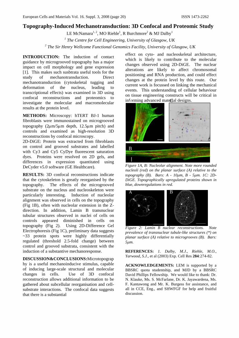
RESULTS: 3D confocal reconstructions indicate that the cytoskeleton is greatly reorganised by the topography. The effects of the microgrooved substrate on the nucleus and nucleoskeleton were particularly interesting. Induction of nucleolar alignment was observed in cells on the topography (Fig 1B), often with nucleolar extension in the Z-direction. In addition, Lamin B transnuclear tubular structures observed in nuclei of cells on controls appeared diminished in cells on topography (Fig 2). Using 2D-Difference Gel Electrophoresis (Fig 1C), preliminary data suggests ~33 protein spots were highly differentially regulated (threshold 2.5-fold change) between control and grooved substrata, consistent with the induction of a substantive mechanoresponse.
DISCUSSION&CONCLUSIONS:Microtopography is a useful mechanoinducive stimulus, capable of inducing large-scale structural and molecular changes in cells. Use of 3D confocal reconstruction allows additional information to be gathered about subcellular reorganisation and cell-substrate interactions. The confocal data suggests that there is a substantial
effect on cyto- and nucleoskeletal architecture, which is likely to contribute to the molecular changes observed using 2D-DiGE. The nuclear alterations are likely to affect chromosomal positioning and RNA production, and could effect changes at the protein level by this route. Our current work is focussed on linking the mechanical events. This understanding of cellular behaviour on tissue engineering constructs will be critical in informing advanced material designs.
Figure 1A, B: Nucleolar alignment. Note more rounded nucleoli (red) on the planar surface (A) relative to the topography (B). Bars: A - 10μm, B - 5μm. 1C: 2D-DiGE. Topographically upregulated proteins shown in blue, downregulations in red.
Figure 2: Lamin B nuclear reconstructions. Note prevalence of transnuclear tubule-like structures (*) on planar surface (A) relative to microgrooves (B). Bars: 5μm.
REFERENCES: 1. Dalby, M.J., Riehle, M.O., Yarwood, S.J., et al (2003) Exp. Cell Res 284:274-82.
ACKNOWLEDGEMENTS: LEM is supported by a BBSRC quota studentship, and MJD by a BBSRC David Phillips Fellowship. We would like to thank: Dr. N. Klauke, Ms. S. McFarlane, Dr. K. Jayawardena, Ms. F. Kantawong and Mr. K. Burgess for assistance, and all in CCE, Eng., and SHWFGF for help and fruitful discussion.
A C
B
A B

European Cells and Materials Vol. 16. Suppl. 3, 2008 (page 21) ISSN 1473-2262
Abnormal Endothelial Progenitor Cell Function in Diabetes Mellitus: Implications for Autologous Cell Therapy
Timothy O’Brien, Aaron Liew, Brian Kealy, Frank Barry.
Regenerative Medicine Institute and Department of Medicine, National University of Ireland, Galway.
INTRODUCTION: Coronary artery disease is the most common cause of death in patients with diabetes mellitus and endothelial progenitor cells (EPCs) have been implicated in the pathogenesis of this condition. EPCs are mononuclear cells isolated from blood or bone marrow with the capacity to differentiate into endothelial cells. They have a role in re-endothelialization, neovascularization and wound healing.
METHODS: EPCS were isolated from patients with poorly controlled diabetes mellitus and controls. EPC number and function were assessed. Expression of OPN in EPCs was assessed by RT PCR. The effect of OPN on EPC function was assessed. The role of OPN in angiogenesis was analyzed by inducing hindlimb ischemia in OPN knockout and control mice. Finally the effect of autologous EPC transplantation on wound healing in the alloxan-induced diabetic rabbit was assessed.
RESULTS: EPCs were reduced and dysfunctional in patients with diabetes mellitus. Reduced expression of osteopontin was detected in diabetic EPCs and culturing cells in the presence of osteopontin-containing media reversed cellular dysfunction. A crucial role for osteopontin in new vessel formation was identified by demonstrating impaired neovascularization in osteopontin knockout mice. The therapeutic potential of EPCs was demonstrated by improved wound healing following delivery of EPCs transduced with endothelial nitric oxide synthase in diabetes mellitus. Finally, detailed flow cytometric analysis of EPCs demonstrated that these cells represent a peripheral blood mononuclear cell population enriched in pro-angiogenic monocytes.
CONCLUSION: In summary, we show that EPCs are reduced in number and function in diabetes mellitus, demonstrate a previously unrecognized role for osteopontin expression in this cell type in neovascularization and demonstrate that this cell type represents an enriched peripheral blood mononuclear cell population expressing angiogenic receptors.

European Cells and Materials Vol. 16. Suppl. 3, 2008 (page 22) ISSN 1473-2262 DIFFERENTIATED MESENCHYMAL STEM CELLS FUNCTION AS SCHWANN CELLS
D Mahay, G Terenghi, S Shawcross
Tissue Injury and Repair, Blond McIndoe Laboratories, Stopford Building, University of Manchester, UK.
INTRODUCTION: Schwann cells (SCs) are essential facilitators for peripheral nerve regeneration following injury; they release supporting neurotrophic factors that provide both physical support and guidance. In vitro, these cells are slow growing, hence not well suited to a tissue engineering approach to nerve repair. The purpose of our study was to direct mesenchymal stem cells (MSCs) down a SC lineage. METHODS: Adult rat MSCs were differentiated (dMSCs) into SC-like cells using an established cocktail of growth factors [1-3]. The dMSCs, seeded on 1.0µm porous inserts, were co-cultured without contact with dissociated adult rat dorsal root ganglia (DRG) neurons pre-seeded onto a laminin substrate. The co-cultures were incubated for 24hr then fixed for βIII-tubulin immunostaining and analysed. Three parameters of neurite outgrowth were evaluated: percentage of neuron sprouting, length of longest neurite and total neurite density. The secretion of the neurotrophic factors was evaluated by an ELISA methodology and the use of secretion-blocking antibodies. The neurotrophic effect was further assessed by culturing the DRG neurons with pre-conditioned medium from the dMSCs in the above model. Further functional assessment was undertaken using poly-3-hydroxybutyrate (PHB) conduits seeded with GFP-transfected dMSCs in a rat sciatic nerve injury model. The conduits were harvested after two months and evaluated by electron microscopy. RESULTS: The neurite outgrowth of the DRG neurons was enhanced in co-culture with dMSCs compared to the undifferentiated MSCs. Like SCs, dMSCs were responsible for the stimulation of DRG neurons to produce longer, branched neurites in the co-culture model (Fig. 1). ELISA methodology and blocking antibodies showed that, for the main part, this effect resulted from the release of brain-derived neurotrophic factor (280 pg/ml) and nerve growth factor (111 pg/ml) by the dMSCs. Co-culture of the DRG neurons with pre-conditioned medium from dMSCs also stimulated neurite outgrowth. Using an in vivo nerve repair model system comprising transplanted dMSCs in PHB conduits, 30% of regenerated nerve fibres in the distal stump were myelinated post-transplantation (Fig. 2).
Fig. 1: βIII-tubulin stained DRG neuron co-cultured with dMSC (x10 mag) Fig. 2: Electron micrograph showing myelinated nerve fibres at the distal stump (x18500 mag)
DISCUSSION & CONCLUSIONS: Adult rat MSC were differentiated into SC-like cells. The results of our study further support the notion that MSC differentiated into SC-like cells display cellular, molecular and functional characteristics of SCs. The study provides further evidence for the use of stem cells as effective substitutes for SCs in nerve repair. REFERENCES: 1 M Tohill, C Mantovani, M Wiberg & G Terenghi (2004). Rat bone marrow mesenchymal stem cells express glial markers and stimulate nerve regeneration. Neuroscience Letters. 362, 200-203. 2 J Caddick, PJ Kingham, NJ Gardiner, M Wiberg & G Terenghi (2006). Phenotypical and functional characteristics of mesenchymal stem cells differentiated along a Schwann cell lineage. Glia. 54, 840-849. 3 Growth factors in mesenchymal stem cells following glial cell differentiation (2008). Biotechnology and Applied Biochemistry. doi:10.1042/BA20070212. ACKNOWLEDGEMENTS: This work has been supported by funding from the MRC and the Rosetrees Trust. The authors would like to thank Acorda Therapeutics (USA) for the generous supply of GGF-2 for the continuation of this work, Astra Tech (Sweden) for the supply of PHB and Faculty of Life Sciences Imaging Unit, University of Manchester for help with the electron microscopy.

European Cells and Materials Vol. 16. Suppl. 3, 2008 (page 23) ISSN 1473-2262
Strategies For The Vascularisation Of 3-D Tissues M V Sefton
Institute of Biomaterials and Biomedical Engineering, University of Toronto, Canada Addressing the consequences of cardiovascular disease presents many challenges for regenerative medicine. A critical issue associated with creating large tissues and whole organs is the difficulty of supplying nutrients to all the cells in a thick segment of tissue beyond the limits of Fick's law. Delivery of angiogenic growth factors from microencapsulated cells is one approach among many that are under consideration. A murine fibroblast cell line (L929) was genetically engineered to secrete recombinant human vascular endothelial growth factor (rhVEGF165). Transfected (L929VEGF) and non-transfected (L929) cells were microencapsulated in a 75:25 hydroxyethyl methacrylate-methyl methacrylate (HEMA-MMA) copolymer. The matrigel plug assay
was used to study the in vivo response to encapsulated cells in a nominally syngeneic mouse model. More new blood vessels were recruited to the edge of L929VEGF capsule implants versus L929 capsule implants. A second approach is to use endothelial cell seeding. We have created a three-phase construct containing endothelial cells (EC), hepatocyes (HepG2) and collagen using modular components. HepG2 cells are encapsulated within collagen cylindrical modules and seeded with endothelial cells. Packing modules within a larger tube results in a scaleable, blood perfusable bed of uniform cell density. A third approach is to exploit a novel biomaterial that induces vascularisation in its vicinity as a consequence of the peculiarities of the wound healing response. This angiogenic Theramer™ (a therapeutic polymer made by Rimon Therapeutics, Ltd) has an angiogenic effect due to material composition and without immobilised or other form of biomolecule incorporation. For example, a 45 mole % methacrylic acid copolymer (with
MMA) caused a significant increase in the number of vascular structures in a rat skin graft model that also lead to enhanced ‘take’ of the graft.
European Cells and Materials Vol. 16. Suppl. 3, 2008 (page 24) ISSN 1473-2262
DEFINING PHYSIOLOGICAL PARAMETERS FOR ENGINEERING A VASCULAR MEDIA MODEL
U.Cheema, E.A. Hadjipanayi, N.Tamimi, B.Alp, V.Mudera, R.A Brown
University College London (UCL), Tissue Repair and Engineering Centre, Institute of Orthopaedics, Stanmore Campus, London, HA7 4LP, UK
INTRODUCTION: Tissue engineering of a blood vessel structure requires an understanding of the parameters governing the survival of resident vascular smooth muscle cells. We have developed a collagen-based vascular media model to examine the correlation between cell density, O2 requirements and cell viability. METHODS: Collagen type I gels were cast in rectangular wells and were compressed to produce 100µm thin, dense collagen sheets1. These were subsequently spiraled around a mandrel to mimic tubular structures. Constructs were seeded with porcine Pulmonary Artery Smooth Muscle Cells (PASMCs) at different densities (11.6x10⁶ and 23.2 x10⁶ cells/ml) and were cultured for up to 6 days in vitro. O2 levels were measured within the core2 and correlated to cell viability, under both static and dynamic perfusion culture conditions.
RESULTS: Within 24 hours O2 levels in the core of constructs dropped to 20mmHg for the high cell density compared to 80mmHg for the low cell density, with no significant cell death associated with the lowest O2 levels. Increasing construct radial thickness by 33% resulted in a significant reduction in core O2 levels to 60mmHg by 24hrs (p<0.05). After 6 days of static culture, cell viability decreased in the core of high cell density constructs (58%), but remained high at the surface (75%). Dynamic perfusion of constructs maintained a significantly higher cell viability in the core (79%, p<0.05) after 6 days of culture. DISCUSSION & CONCLUSIONS: In this study we have examined how O2 is consumed by vascular smooth muscle cells seeded in dense collagen tubular constructs. While O2 diffused freely through dense collagen matrix, O2 consumption by cells effectively resulted in the formation of a transverse O2 gradient from the surface of the construct to its core. Our results indicate that spatial cell distribution, as well as cell density, are critical factors controlling the rate of O2 consumption within a 3D construct. Exposure of core cells to low O2 levels was accompanied by a progressive reduction in core cell viability
indicating that physiological hypoxia could be detrimental to the survival of PASMC’s. The native, layered architecture of the blood vessel wall3 makes the current model a promising tool for examining physiological cell responses within biomimetic tissue-engineered vascular constructs. REFERENCES: 1 R.A.Brown, M.Wiseman, C.Chuo, U.Cheema, S.Nazhat(2005) Adv. Funct. Mater., 15:1762-1770. 2 U. Cheema, R.A Brown, B.Alp, A.J MacRobert (2008) Cell.Mol.Life Sci. 65: 177-186. 3 S.M.Hall, A.Soueid, T.Smith, R.A.Brown, S.G.Haworth, V.Mudera (2007) J Tissue Eng Regen Med 1:287-295
ACKNOWLEDGEMENTS: We are grateful to BBSRC/EPSRC for financial support
European Cells and Materials Vol. 16. Suppl. 3, 2008 (page 26) ISSN 1473-2262
Co-Culture Systems for Tissue Regeneration S. Levenberg
Department of Biomedical Engineering, Technion, Haifa, Israel 32000 The rapidly increasing demand for organ and tissue transplantation has promoted tissue engineering and stem cell research as promising approaches. Tissue engineering combines cells, growth factors and 3D scaffolds for repair and regeneration of biological tissues. To advance tissue engineering research, scaffold properties must be optimized for a given application and cell type. This includes chemical and mechanical properties, shape, and structure and degradation rate. In addition co-culture approaches are required to allow organization of complex tissue structures. Endothelial cell co-cultures are important for inducing vascularization of engineered tissues. Our experiments in engineered skeletal and cardiac muscle tissue indicate that endothelial cells promoted differentiation and organization of the co-cultured myoblasts1,2. Endothelial 3D tubular networks were formed within the tissue and shown to promote vascularization upon implantation1,3. Given the attractive potential of human embryonic stem cells in tissue regeneration we evaluate the ability to differentiate the cells and induce their 3D organization toward formation of complex tissues4. Biodegradable, growth factor-eluting nano-fibers are used to study embryonic stem cells process in 3D models5. Differentiation of the cells is further studied in micro perfusion system to allow the precise localization of a growth factor, a few microns in width, both temporally and spatially using laminar flows6,7. The technique can provide a tool to investigate cell-cell signaling between adjacent embryonic stem cells by maintaining a constant gradient of growth factors in the surrounding culture medium. Understanding embryonic stem cells differentiation and 3D cellular communications can lead to advances in cell therapy and tissue engineering and facilitate organ and tissue regeneration.
REFERENCES: 1Levenberg, S., Rouwkema, J., Macdonald, M., Gerfein, E., Kohane, D., Darland D., Marini, R., van Blitterswijk, C.A., Mulligan, R., D’Amore, P. and Langer, R. Engineering Vascularized Skeletal Muscle Tissue (2005) Nature Biotechnology. 2 Caspi O. and Lesman A., Basevitch Y., Gepstein A., Arbel G., Habib M., Gepstein L., and Levenberg S. Tissue Engineering of Vascularized Cardiac Muscle from Human Embryonic Stem Cells. Circulation Research.
(2007) 100(2):263-72. 23:879-884, 3Levenberg, S., Golub, J. S., Amit, M., Itskovitz-Eldor, J. and Langer, R. Endothelial Cells Derived From Human Embryonic Stem Cells.(2002) PNAS 99: 4391-4396, 4Levenberg, S., Huang, N., Erin, L., Rogers, A., Itskovitz-Eldor, J. and Langer, R. Differentiation of Human Embryonic stem cells on Three Dimensional Polymer Scaffolds. (2003) PNAS. 100:12741-12746, 5Tzezana R. Zussman E., Levenberg S. A Layered Ultra-Porous Scaffold for Tissue Engineering, created via a Hydrospinning Method. Tissue Engineering (2008). In print, 6Korin N., Bransky A., Dinnar U. and Levenberg S. A Parametric Study of Human Fibroblasts Culture in a Microchannel Bioreactor. Lab on a Chip. (2007), 7, 611 – 617. 7 Bransky A., Korin N. and Levenberg S. Experimental and theoretical study of selective protein deposition using focused micro laminar flows. Biomedical Microdevices. (2008) 10:421–428.
ACKNOWLEDGEMENTS
The research was supported by funds from the Israel Science Foundation and Horovits Foundation.

European Cells and Materials Vol. 16. Suppl. 3, 2008 (page 27) ISSN 1473-2262 Enhanced Human Bone Marrow Stromal Cell Bone Regeneration following the
Temporal Release of Encapsulated Angiogenic and Osteogenic Factors from Biodegradable Polymer Scaffolds
JM Kanczler1, P Ginty2, L White2, SM Howdle3, KM Shakesheff2, and ROC Oreffo1. 1The Bone & Joint Research Group, Institute of Developmental Sciences, University of Southampton, UK.
2School of Pharmacy, University of Nottingham, UK. 3School of Chemistry, University of Nottingham, UK. INTRODUCTION: The regenerative process of bone tissue involves complex and coordinated signal cascades. Bone fracture defects can repair spontaneously with minimal treatment. However, in the clinical settings where the bone defect is too large for natural repair, a replacement material is required to enhance the osteogenic healing process. Bone morphogenic factors such as bone morphogenic protein-2 (BMP-2) and angiogenic factors such as vascular endothelial growth factor (VEGF165) are known to play a prominent role in bone formation and bone fracture repair. Orchestrating the presentation of these factors and different kinetic release profiles by the encapsulation of these scaffolds within intelligent scaffold structures will enhance the coordinated capacity of the regeneration of critical-sized bone defects. The aim of this study was to determine if the delivery of VEGF165 and rhBMP-2 from a biodegradable alginate/Poly D,L-lactic acid (PDLLA) composite together with the addition of human bone marrow stromal cells (HBMSC) could enhance the bone regenerative capability in a mouse-femur segmental defect.
METHODS: PDLLA/alginate composite scaffolds were loaded with VEGF165 (encapsulated within alginate) and rhBMP-2 (encapsulated within a PDLLA) using a supercritical CO2 mixing technique and an alginate entrapment protocol. This enabled the fabrication of a complex hybrid monolith scaffold of VEGF165 and BMP-2 (alginate-VEGF/PLA-BMP-2). The scaffolds were placed in culture for a period of 28 days to determine the release of VEGF165 and BMP-2. In addition, the scaffolds were seeded with or without HBMSC and implanted into a mouse (MF1nu/nu) femur segmental defect (5mm) for four weeks (n=4 mice per group). The femur-defect samples were analysed for bone regeneration using micro-computer tomography and histology. RESULTS: The dual loaded scaffolds after 7 days in culture showed an increase in the release of VEGF165 (750.4±596.8pg/ml) compared to that of BMP-2 (136.9±123.4pg/ml). Cumulative release over 28 days in culture of VEGF165 and BMP-2 from the PDLLA/alginate
composite scaffolds resulted in release of VEGF (2553±1932pg/ml) and BMP-2 (5773±1879pg/ml). The alginate-VEGF/PLA-BMP-2 + HBMSC group showed significant bone regeneration in the femur-segmental defects compared to the alginate/PLA and alginate-VEGF/PLA-BMP-2 groups by indices of increased bone volume, trabecular number and reduced trabecular separation in the segmental femur defect region. Additionally, trabecular thickness was reduced in the alginate-VEGF/PLA-BMP-2 + HBMSC compared to the alginate/PLA group (table 1).
Table 1. μCT analysis of MF-1 nu/nu mouse femur segmental defect model 28 days post scaffold implantation. BV = Bone Volume (mm3); BV/TV = Bone Volume/Total Volume; Tb.Th = Trabecular thickness (mm3); Tb.N = Trabecular number/mm; Tb.Sp = Trabecular spacing/mm. (Values = mean ± standard deviation). ***P, <0.001;** P, <0.01.
Alginate/ PDLLA
Alginate(VEGF)/ PDLLA (BMP-2)
Alginate (VEGF)/ PDLLA
(BMP-2) + HBMSC
BV 6.63±1.06 9.92±1.78 17.79±1.85**
* BV/TV 0.199±0.01 0.207± 0.03 0.395±0.04**
* Tb.Th 0.112± 0.02 0.082± 0.02 0.076 ±0.01* Tb. N 1.81±0.33 2.59±0.53 5.32±0.61*** Tb.Sp 0.45±0.07 0.32±0.072 0.12±0.014** DISCUSSION & CONCLUSIONS: In conclusion, these studies demonstrate the ability to deliver, temporally, a combination of HBMSC, angiogenic (VEGF165) and osteogenic (BMP-2) growth factors released from biodegradable PDLLA/alginate composite scaffold composites to sites of bone defects in a regulated manner and the necessity for cells and growth factor delivery, in combination, for the regeneration of large bone defects. Such cell based and tissue engineering strategies offer innovative approaches in orthopaedics and the wider tissue reparative arena.

European Cells and Materials Vol. 16. Suppl. 3, 2008 (page 28) ISSN 1473-2262
Phenomenological modelling tools for orthopaedic tissue engineering N. I. Nikolaev1, G. Lemon 2, B. Obradovic3, H. K.Versteeg1 and D. J. Williams 1
1 Wolfson School of Mechanical and Manufactural Engineering, Loughborough University, Loughborough LE11 3TU, UK
2 School of Mathematical Sciences, University of Nottingham, Nottingham NG7 2RD, UK 3Department of Chemical Engineering, Faculty of Technology and Metallurgy, University of Belgrade
Karnegijeva 4, 11000 Beograd, Serbia and Montenegro INTRODUCTION: Today tissue engineering has to solve challenging task - in vitro growth of different tissues. In specially constructed bioreactors in-vivo-like conditions have to be created. Many experiments will be needed first to identify key growth factors/growing conditions and subsequently optimise them. Because many processes are involved, mathematical models may be useful to estimate the impact of each of them and later to improve biological and mechanical functionality of tissue engineered constructs [1]. We analyze the role of the different processes for tissue growth: nutrient transport and consumption, cell population dynamics, extra-cellular matrix (ECM) secretion, stress and strain distribution, construct expansion, tissue degradation, and the role of mechanical stimulation. Our aim is to embed current understanding of processes controlling the seeding of a scaffold, cell and ECM growth and maintenance for engineered spinal disk tissue in to a modelling tool to predict tissue quality metrics and for potential use in the optimisation of processes to create such constructs with a bioreactor. METHODS: Multiphase theory [2] is used to describe tissue growth. We have incorporated a viscoelastic model [3] and approaches used for wound healing description [4]. Tissue is described as a mixture of four phases – cells, water, scaffold and ECM (at this stage we represent ECM in terms of a single component - GAG due to lack of suitable experimental data for other parts). Scaffold and ECM move together like in [4] and cells are attached to them. This joint movement is caused by cell traction and deformations associated with tissue growth. The scaffold is gradually degraded by hydrolysis. ECM synthesis is modelled as a two-stage process. Initially, cells produce proteoglycan (PG) molecules, which may bind to already existed GAG matrix or diffuse and leak out of the construct. The rate of PG synthesis is proportional to nutrient and cell concentration and is limited by the local concentration of GAG. To simulate expansion of the construct due to the growth a “thermoelastic” model is employed in conjunction with moving boundary conditions [2,3]. The experimental results in [6] have been used to calibrate the model. One of the current challenges is to match the temporal evolution of spatial cell distribution. In this paper we investigate the ability of different mechanisms to explain the experimental trends. Different models for description of the growth of the construct have been tested and compared.
Fig. 1: Comparison between experimental results from [6] (dots or triangles) with simulations (solid lines). (a) GAG distribution - 10th day (thin line), 42nd day thick line; (b) increase of the width of the construct.
RESULTS: The results obtained (Fig. 1) show the satisfactory agreement to those observed experimentally with respect to the time evolution of GAG distribution and increase the size of the construct. Further work is required to verify the model, a recently commissioned experimental system will enable this.
REFERENCES: [1] Sengers B G, Taylor M, Please C P, Oreffo R O C (2007) Computational modeling of cell spreading and tissue regeneration in porous scaffolds , Biomaterials 28: 1926-1940 [2] Lemon G and King JR. (2007) Multiphase modelling of cell behaviour on artificial scaffolds: effects of nutrient depletion and spatially nonuniform porosity Mathematical Medicine and Biology-a Journal of the Ima 24: 57-83. [3] Moon, A.G and Tranquillo R T. (1993) Fibroblast-Populated Collagen of Cell Traction Force: Part 1 Microsp here Assay Continuum Model, AIChE Journal, 39: 163-177. [4] Murray J. D. Mathematical biology – Spatial models and biomedical application, Chap. 10. Dermal Wound Healing. [5]Volokh, K.Y (2004) A simple phenomeno-logical theory of tissue growth, Mech Chem Biosyst. 1:147-160. [6] Obradovic B, Meldon JH, Freed LE, Vunjak-Novakovic G., Glycosaminoglycan deposition in engineered cartilage: experiments and mathematical model. AIChE J 2000, 46, 1860-1871. ACKNOWLEDGEMENTS: This work forms part of the UK EPSRC funded Innovative Manufacturing Grand Challenge in Regenerative Medicine - remedi. Remedi is a partnership of Loughborough, Nottingham, Cambridge, Birmingham, Ulster and Liverpool Universities, Industry and Agency stakeholders.

European Cells and Materials Vol. 16. Suppl. 3, 2008 (page 29) ISSN 1473-2262
EFFECT OF FLUID FLOW-INDUCED SHEAR STRESS ON HUMAN MESENCHYMAL STEM CELL GENE EXPRESSION
J R Glossop & S H Cartmell
Institute for Science and Technology in Medicine, Guy Hilton Research Centre, University of Keele, Stoke-on-Trent, Staffordshire, UK.
INTRODUCTION: Human bone marrow-derived mesenchymal stem cells (MSCs) are multipotent, unspecialized cells that have the ability to differentiate into numerous cell types including those from the osteocytic, chondrocytic and adipocytic lineages [1]. This makes them ideal for tissue engineering purposes. Mechanical forces and the process of mechanotransduction are known to be important factors influencing cell activity and responses [2]. However, there is limited data on this for MSCs. The objective of this work was to investigate the affect of fluid flow-induced shear stress exposure on MAPK signalling pathway gene expression in primary human MSCs.
METHODS: Bone marrow-derived human MSCs (Lonza) were cultured in monolayer on pronectin-coated glass slides. Fluid flow-induced shear stress of 1, 5 and 10 dyn/cm2 was applied to cells for 1 hour using a Streamer Fluid Shear Bioreactor. Samples were collected 0, 1, 2 and 24 hours after exposure to shear stress. Experiments were performed once and a static control was performed on each occasion. Isolated total RNA samples were used for DNA microarray analysis (2 hour time point only) using Affymetrix Human Genome U133 Plus 2.0 arrays and for quantitative real-time RT-PCR. Microarray data were analysed using GeneSpring GX to identify differential gene expression compared to static control, focusing on genes featuring in the MAPK signalling pathways. Real-time data on genes of interest from these pathways were normalised to 18S. Differential gene expression was considered as ≥2-fold compared to static control.
RESULTS: DNA microarray analysis identified between 440 and 600 genes that were differentially expressed in response to the different magnitudes of fluid flow-induced shear stress. For each of the magnitudes studied, a number of the differentially expressed genes also featured in the MAPK signaling pathways (Table 1), overlaps which were all statistically significant (p<0.005). In total, 24 genes were differentially expressed, of which 8 were consistently so across the different magnitudes (Figure 2). In particular, shear stress induced consistent and marked up-regulation of MAP3K8 and IL1B. Quantitative real-time RT-
PCR confirmed these data, with statistically significant increases in MAP3K8 and IL1B which were evident across the 24 hour period after shear stress exposure.
Table 1: Differential expression of genes featuring in the MAPK signalling pathways.
No. (%) Maximum fold change No. (%) Maximum
fold change1 18 15 (83.3) 24.2 3 (16.7) –2.35 10 10 (100) 15.5 0 (0) -10 16 13 (81.2) 11.6 3 (18.8) –2.7
Shear stress magnitude (dyn/cm2)
No. of genes differentially
expressed
Up-regulated genes Down-regulated genes
Figure 2: Up-regulation of 8 genes from the MAPK signalling pathway in response to different magnitudes of fluid flow-induced shear stress.
DISCUSSION & CONCLUSIONS: Our data demonstrate significant differential expression of numerous genes featuring in the MAPK signalling pathways in human MSCs exposed to different magnitudes of fluid flow-induced shear stress. Furthermore, they suggest that modulation of these pathways may, at least in part, be via an IL1B-MAP3K8-associated route.
REFERENCES: 1 M. Pittenger, M.F. Mackay, A.M. Beck, et al (1999) Science 284:143-7. 2 H. Huang, R.D. Kamm, and R.T. Lee (2004) Am. J. Physiol. Cell Physiol. 287:C1-11.
ACKNOWLEDGEMENTS: We would like to thank Mr. Brian King for designing and manufacturing the slide chambers for cell lysis, and the BBSRC for financial support (grant reference code BB/D000548/1).

European Cells and Materials Vol. 16. Suppl. 3, 2008 (page 30) ISSN 1473-2262 The Influence of Delivery Via Narrow-Bore Needles on Mesenchymal Stem Cell
Viability, Apoptosis and Morphology K Agashi, DYS Chau & KM Shakesheff
Division of Drug Delivery and Tissue Engineering, School of Pharmacy, Centre for Biomolecular Sciences, The University of Nottingham, University Park, Nottingham, NG7 2RD, UK
INTRODUCTION: The past few years have seen the development of cell therapy; a novel technique whereby new cells are directly introduced into a tissue or organ, with the hope of treating many degenerative diseases. Numerous studies [1] have investigated the regenerative capacity of cell suspensions following their direct injection into a target organ, but, at present, limited data exists as to whether cell characteristics are affected following their passage through narrow-bore needles. Here, we investigated the cellular response of murine mesenchymal stem cells (mMSC) following their manipulation within a narrow-bore syringe-based delivery system.
METHODS: Primary mMSC were isolated following the protocol previously described in [2]. The cells were expanded and then processed into a concentrated cell suspension (5×10⁷ cells/ml in either HBSS or HBSS with 60mM n-acetyl cysteine (nAC) (an anti-oxidant)). The suspension was drawn up into a 10µl Hamilton syringe with a 26s gauge needle and left to stand for pre-defined time periods at room temperature. Cells were ejected at a controlled rate of either 1µl/min or 5µl/min, then assessed for: (i) viability, using a trypan blue exclusion method, (ii) proliferation, using a CellTiter AQ One Solution Cell Proliferation® assay (Promega, Southampton), (iii) caspase-3 activity levels (apoptosis), using the CaspACE® Assay (Promega) (iv) cell attachment and spreading, using a dual cytoplasmic (May-Grunwald) and nuclear (Giemsa) staining technique
RESULTS: A significant decrease in cell viability was seen immediately upon ejection from the needle, regardless of ejection rate or the presence of nAC. A further decrease in viability and an increase in caspase-3 activity occurred when the cells were left within the syringe chamber for increasing amounts of time.
A significant decrease in attached cell number occurred post-ejection compared to control (fig 1), and an increase in cell debris was observed (fig 2).
Cells that, after being left within the syringe for up to 8 hours, were still viable post-ejection retained their ability to proliferate at rates comparable to control.
DISCUSSION & CONCLUSIONS: The decrease in cell viability may be attributed to the shear forces the cells experience when expelled from the syringe, causing lysis. This proposition is supported by the cellular debris (fig 1). These shear forces may have activated the apoptotic cascade, causing cell viability to decrease further.
The data stresses the importance of reducing the time period that researchers leave cells within a syringe chamber when performing cell-therapy studies, and questions whether the use of anti-oxidants in this setting is beneficial.
REFERENCES: 1 V. Schächinger, S. Erbs, A. Elsässer et al (2006), N Engl J Med 355:1210-1221. 2 Lda S. Meirelles, NB. Nardi (2003) Br J Haematol. 123:702-711.
ACKNOWLEDGEMENTS: The authors thank colleagues in the Tissue Engineering Group for advice and the BBSRC and Regentec® for funding.
200μm
200μ
m
Fig. 2: MSC spreading when drawn up into a syringe and expelled immediately (b) and after 2 hours left within the syringe (c) compared to control (a)
a b
c
Fig 1: Number of attached cells after they were left within syringe for varying time periods. Cells were ejected at a rate of 5µl/min (black), 1µl/min (grey) and 1µl/min with nAC (white).
0
20
40
60
80
100
120
140
160
Control 0 30 120 240Length of time within syringe chamber (min)
Rel
ativ
e C
ell N
umbe
r .
***
****

European Cells and Materials Vol. 16. Suppl. 3, 2008 (page 31) ISSN 1473-2262
Non-destructive monitoring of collagen degradation in tissue engineered corneal constructs
M Ahearne, E Ladva, KK Liu, A El Haj, Y Yang
Institute of Science and Technology in Medicine, Keele University, UK.
INTRODUCTION: Collagen has been under investigation for use in developing constructs for tissue engineering cornea. Several methods have previously been used to examine the degradation and restructuring of collagen constructs by cells including SEM, TEM and histological staining, however these approaches require destruction of the construct. We propose fluorescently labelling collagen with fluorescein isothiocyanate (FITC) to allow non-destructive online monitoring of collagen degradation by corneal fibroblasts.
METHODS: FITC was bound to rat-tail collagen type-1 (BD Bioscience) by adding it to the collagen solution prior to gelation. Unbound FITC was removed by repeated washing after gelation. Tissue engineered corneal equivalents were created using human corneal fibroblasts embedded in collagen as previously described [1]. Hydrogels without cells were also examined.Two non-destructive methods were used to examine the degradation of the collagen constructs. The fluorescence intensity of FITC in the constructs was measured using confocal microscopy. Three dimensional scans of the constructs were taken at different times over a nine week culture period. The degradation of the constructs was also examined by measuring the level of fluorescence in the culture medium using a spectrofluorimeter at wavelengths 495-525nm.
RESULTS: Images of the constructs captured by confocal microscopy showed how the cells degraded and restructured the constructs (Fig 1). After several weeks pores appeared in the fluorescent images of the constructs. These pores were not visible under light microscopy which suggested that they contain newly formed matrix. The peak fluorescent intensity of the constructs increased over the first few weeks in culture then subsequently decreased (Fig 2A). The increase in fluorescence was the result of contraction of the collagen causing the construct to become thinner and denser. The fluorescence intensities of culture medium taken from constructs with cells were higher than those without cells (Fig 2B).
Fig. 1: Images obtained by confocal microscope of FITC-collagen constructs seeded with corneal fibroblasts after (A) 13 and (B) 38 days in culture.
Fig. 2: (A) Peak fluorescence intensity of constructs measured by confocal microscopy; (B) mean fluorescence intensity in culture medium measured by spectrofluorimetry.
DISCUSSION & CONCLUSIONS: It has been demonstrated that fluorescent labelling of collagen by FITC can be used to non-destructively examine collagen degradation by corneal fibroblasts. Images obtained by confocal microscopy showed how the cells degrade and restructure the construct. The peak fluorescence intensity values demonstrated how the construct becomes denser while also undergoing degradation. Examination of culture medium showed that the cells increased the rate of degradation of the construct most likely through the release of MMPs. This technique will be a useful tool for non-destructively examining the degradation of collagen in engineered tissues.
REFERENCES: 1 M. Ahearne, Y.Yang, K.Y Then et al (2008) Br J Ophthalmol 92:268-271.
ACKNOWLEDGEMENTS: This research was funded by BBSRC (BB/F002866/1)
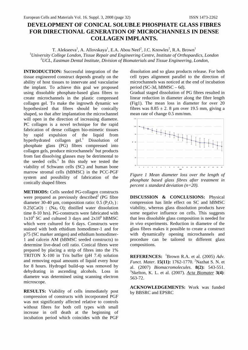
European Cells and Materials Vol. 16. Suppl. 3, 2008 (page 32) ISSN 1473-2262
DEVELOPMENT OF CONICAL SOLUBLE PHOSPHATE GLASS FIBRES FOR DIRECTIONAL GENERATION OF MICROCHANNELS IN DENSE
COLLAGEN IMPLANTS.
T. Alekseeva1, A. Allovskaya1, E.A. Abou Neel2, J.C. Knowles2, R.A. Brown1 1University College London, Tissue Repair and Engineering Centre, Institute of Orthopaedics, London
2UCL, Eastman Dental Institute, Division of Biomaterials and Tissue Engineering, London,
INTRODUCTION: Successful integration of the tissue engineered construct depends greatly on the ability of host tissues to innervate and vascularise the implant. To achieve this goal we proposed using dissoluble phosphate-based glass fibres to create microchannels in the plastic compressed collagen gel. To make the ingrowth dynamic we hypothesized that fibres should be conically shaped, so that after implantation the microchannel will open in the direction of increasing diametre. PC collagen is a novel technique for the rapid fabrication of dense collagen bio-mimetic tissues by rapid expulsion of the liquid from hyperhydrated collagen gel.1 Dissolution of phosphate glass (PG) fibres compressed into collagen gels, produce microchannels2 but products from fast dissolving glasses may be detrimental to the seeded cells.3 In this study we tested the viability of Schwann cells (SC) and human bone marrow stromal cells (hBMSC) in the PCC-PGF system and possibility of fabrication of the conically shaped fibres METHODS: Cells seeded PG-collagen constructs were prepared as previously described2 (PG fibre diameter 30-40 µm, composition ratio: 0.5 (P2O5 ) : 0.25(CaO) : (Na2 O); distilled water dissolution time 8-10 hrs). PG-constructs were fabricated with 1x10⁶ SC and cultured 3 days and 2x10⁶ hBMSC which were cultured for 6 days. Constructs were stained with both ethidium homodimer-1 and for p75 (SC marker antigen) and ethidium homodimer-1 and calcein AM (hBMSC seeded constructs) to determine live-dead cell ratio. Conical fibres were prepared by placing a strip of fibres into the 1% TRITON X-100 in Tris buffer (pH 7.4) solution and removing equal amounts of liquid every hour for 8 hours. Hydrogel build-up was removed by dehydrating in ascending alcohols. Loss in diametre was determined using scanning electron microscope. RESULTS: Viability of cells immediately post compression of constructs with incorporated PGF was not significantly affected relative to controls without fibres for both cell types with small increase in cell death at the beginning of incubation period which coincides with the PGF
dissolution and so glass products release. For both cell types alignment parallel to the direction of microchannels was noticed at the end of incubation period (SC-3d, hBMSC – 6d). Gradual staged dissolution of PG fibres resulted in linear reduction in diameter along the fibre length (Fig1). The mean loss in diameter for over 20 fibres was 8.85 ± 2. 8 µm over 19.5 mm, giving a mean rate of change 0.5 mm/mm.
50
60
70
80
90
100
110
1 2 3 4 5 6 7 8 9 10 11 12 13 14 15 16 17 18 19 20
mm
%
Figure 1 Mean diameter loss over the length of phosphate based glass fibres after treatment in percent ± standard deviation (n=20).
DISCUSSION & CONCLUSIONS: Physical compression has little effect on SC and hBMSC viability, whereas glass dissolution products have some negative influence on cells. This suggests that less dissoluble glass composition is needed for in vivo experiments. Reduction in diametre of the glass fibres makes it possible to create a construct with dynamically opening microchannels and procedure can be tailored to different glass compositions. REFERENCES: 1Brown R.A. et al. (2005) Adv. Funct. Mater. 15(11): 1762-1770. 2Nazhat S. N. et al. (2007) Biomacromolecules. 8(2): 543-551. 3Skelton, K. L. et al. (2007). Acta Biomater 3(4): 563-72. ACKNOWLEDGEMENTS: Work was funded by BBSRC and EPSRC

European Cells and Materials Vol. 16. Suppl. 3, 2008 (page 33) ISSN 1473-2262 Bioactive Sugar Gels For Liver Tissue Engineering
R.F. Ambury1, B.G. Cousins1, R.V.Ulijn1, C. Merry1
1. School of Materials, University of Manchester, Manchester, UK.
INTRODUCTION: The liver is one of few organs in the body able to heal itself, however under conditions of constant damage; the liver inevitably begins to fail. Despite 50 years of medical research in this area, organ transplantation remains the common remedy [1]. The primary objective of this study was to identify and develop a novel bioactive hydrogel capable of evaluating hepatocyte behaviour on a tissue realistic mimic. METHODS: The materials were selected to mimic the native tissue: 70-76 % hydration, E=30 kPa, ν=0.43-0.47 with β-galactose moieties to control cellular adhesion through the specific aisaloglycoprotein receptors (ASGP-R) found on hepatocytes. The gels were created by modifying a commercially available block co-polymer of polyethylene glycol and acrylamide, (PEGA) with galactose moieties found in lactobionic acid (LA), producing a unique bioactive sugar-based gel [2]. Monomers used are mono- and bis-acryloamido PEG (Mw=1900 gmol-1), dimethylacrylamide and LA. The pendant PEGA amine groups are used as ligands to which the LA attaches. Sugar modified PEGA was either spin coated or polymerised in bulk onto epoxy-functionalised glass substrates by exposing the material to a 365 nm UV light source The resultant gels were characterised using FTIR, water contact angle analysis, dansyl chloride labeling and swelling studies prior to cellular evaluation. Two forms of cellular evaluation were undertaken; extract testing with 3t3 fibroblasts and evaluation with hepatic cell lines looking at attachment, proliferation, morphology and degree of protein secretion. RESULTS: FTIR analysis of LA exhibits a broad OH absorption occurring in the region between 2800-3500cm-1, and a distinctive band at approximately 1740 cm-1 which is associated with carbonyl stretching (C=O) of carboxylic peaks, fig 1c). This unique peak disappears as the galactose moieties within the LA are incorporated into the PEGA gel, fig 1c.
Fig 1: FT-IR traces of a) LA, b) PEGA and c) PEGA +LA + crosslinkers In support of the FTIR data, static water contact angles decreased when free unbound galactose moieties were incorporated into PEGA. The presence of cross-linking agents resulted in increased in water contact angle. Cytotoxic eluent studies utilising 3t3 fibroblasts showed high doping levels, >50% v/v had no effect on cell viability. Initial hepatocyte evaluation illustrated excellent cellular attachment and proliferation, whilst the PEGA control showed no cellular activity. DISCUSSION & CONCLUSIONS: Galactose moieties have been successfully immobilised onto the pendant amine of PEGA which has been verified by FTIR, water contact angle measurements and dansyl chloride staining. Initial cellular evaluations show hepatic cells attach and proliferate on sugar modified PEGA surfaces. REFERENCES: 1) J. Allen, S. Bhatia. Engineering liver therapies for the future. Tissue Engineering, 2002, 8, 725-737. 2) M. Zourob, J.E. Gough, R.V. Ulijn. A micro-patterned hydrogel platform for chemical synthesis and biological analysis. Adv. Mater., 2006, 18, 655-659

European Cells and Materials Vol. 16. Suppl. 3, 2008 (page 34) ISSN 1473-2262
Spatial Layering of Cells in A Novel Poly(lactic acid-co-caprolactone)-Collagen Hybrid Construct
Michaël Ananta¹, Jöns Hilborn², Cecilia Aulin², Stephanie Houis³, Robert Brown¹, Vivek Mudera¹
¹UCL Tissue Repair & Engineering Centre, IOMS, Middlesex, HA7 4LP, United Kingdom
²Department of Materials Science, Uppsala University, SE -751 21 Uppsala, Sweden ³ITA, University of Technology Aachen, Eilfschornsteinstrasse 18, D -52062 Aachen, Germany
INTRODUCTION: Advantages of synthetic polymers as a scaffolding material include the ability to produce them on a large scale and the ability to tailor their physical and mechanical properties to exact specification. A continuing difficulty is the uncertainty of cell seeding, largely due to a non-ideal surface chemistry. Natural biopolymers on the other hand, provide a substrate for cell-adhesion, are highly organized, and have the ability to induce tissue growth. A main disadvantage, however, is that they are less readily available and their mechanical and physical properties are difficult to control. The concept of combining these two biomaterials and their advantages is attractive. In this study we have fabricated a hybrid construct consisting of a synthetic polymer mesh coated with hyperhydrated collagen gels, which were plastic compressed. METHODS: Hydrated collagen gels were plastic compressed onto a mesh of poly(lactic acid-co-caprolactone) (PLACL), resulting in two spaces and two surfaces for cell seeding. As the collagen and cell concentration depend on the amount of fluid leaving the collagen gels, the influence of varying load and the time of the plastic compression process was studied. The hybrid constructs were embedded and/or surface layered with human dermal fibroblasts (final concentration 2x10⁶ cells/ml) to mimic an (1) interstitial, an (2) epithelial and a (3) composite tissue, which were cultured for seven days under static conditions. Cell viability, directly after the plastic compression process and over the cultivation period, was qualitatively assessed with AlamarBlue. Quantitative analysis was performed at the end of the cultivation period to evaluate the short-term biocompatibility of the polymer collagen blend using fluorescence staining and confocal microscopy. RESULTS: We found that the duration rather than the weight of the load in the plastic compression process determines the final collagen and cell density. No significant cell death was observed after the plastic compression of the interstitial
equivalents, confirming previous reports of good cell viability retention [1]. The interstitial, epithelial and composite tissue equivalents showed no macroscopic signs of contraction and good cell proliferation with a two to three fold increase in cell number over 7 days. Quantitative analysis showed a homogenous cell distribution and good biocompatibility. DISCUSSION & CONCLUSIONS: The aim of this study was to test “in vitro” a novel design of 3D tissue engineered construct capable of spatially layering different cell phenotypes using a hybrid PLACL -collagen construct to enhance or promote tissue repair processes “in vivo”. Plastic compression of hydrated collagen gels onto the surface of the polymer mesh allows for seeding of up to four cell layers to histologically mimic stratified tissues. The slow degrading biocompatible PLACL backbone provides for the mechanical support of the hybrid construct. The plastic compression process allows for cell independent compaction of the matrix to mimic “in vivo” cell and collagen densities, which has implications for implant integration, angiogenesis and other cell responses. The PLACL-Collagen hybrid construct has potential applications in bladder wall, blood vessels and skin, which are currently being explored. REFERENCES: 1 R.A. Brown, M. Wiseman, C -B. Chuo, U. Cheema and S.N. Nazhat. Ultrarapid Engineering of Biomimetic Materials and Tissues. Adv. Funct. Mater., 2005, 15, 1762. ACKNOWLEDGEMENTS: This research was funded by EU Framework VI programme, 3G -Scaff reference: 01360 2-3G SCAFF

European Cells and Materials Vol. 16. Suppl. 3, 2008 (page 35) ISSN 1473-2262 Schwann cell-ECM molecule interactions: Towards the development of new
bioengineered nerve conduits
SJ Armstrong1, M Wiberg2, G Terenghi1 & PJ Kingham1
1 Blond McIndoe Laboratories, University of Manchester, UK 2 Department of Surgical & Perioperative Science, University Hospital Umea, Sweden
INTRODUCTION: Tissue engineering using a combination of biomaterials and cell based therapies represents a new approach to nerve repair. Previously we have shown that poly-3-hydroxybutyrate (PHB) is a suitable biomaterial for construction of artificial nerve conduits designed to enhance nerve repair (1). However, the regeneration that occurs is far from optimal. This study aims to investigate the interactions between PHB, ECM molecules and SCs, and use the findings to modify nerve conduits to further enhance nerve regeneration.
METHODS AND RESULTS: ECM molecules were coated onto PHB mats and the attachment of SCs measured. The attachment did not differ significantly between laminin, fibronectin and collagen with approximately 60% of SCs attached after 3h. Proliferation of SCs was however enhanced in the presence of the ECM molecules compared to uncoated PHB, with laminin having the greatest effect. Two different co-culture systems were employed to determine the effects of ECM molecules on SCs and the subsequent effect on neurite outgrowth from a motor neuron like cell line, NG108-15. SCs were seeded onto ECM coated tissue culture inserts suspended above a monolayer of NG108-15 cells to determine the effect of diffusible molecules released by SCs. The effect of direct contact between the two cell types plated on different ECM molecules was also determined. Compared with NG108-15 cells seeded alone, SCs enhanced the percentage of NG108-15 cells expressing neurites and the number of neurites per cell body in both systems – these effects were enhanced by all the ECM molecules (laminin>fibronectin>collagen). NG108-15 cells grown in direct contact with SCs had significantly longer neurites than those exposed to diffusible factors and this effect was also enhanced by the ECM molecules. SC proliferation was similar under control conditions (PDL) and when cultured on ECM molecules indicating that the increased neurite outgrowth was not simply due to an increase in cell number but rather an altered SC phenotype resulting from the interaction with the ECM molecules. The nuclear factor kappa B (NF-кB) signaling pathway has been shown to play a role in SC myelination but its
effects on SC neurotrophic activity are unknown (2). Immunocytochemistry and western blot analysis showed that laminin enhanced the phosphorylation of IκB and p65 NF-κB signalling proteins in SCs. Phosphorylated NF-κB p65 was localised to the nucleus indicating activation of NF-κB. Treatment of SCs with quinazoline (QNZ, 40 nM), an inhibitor of NF-κB activation, prevented the neurite elongation evoked by laminin activated SCs but had no effect on basal levels of SC mediated neurite outgrowth. Thus laminin appears to promote SC neurotrophic activity via the activation of NF-κB signaling pathways.
Fig. 1: Effect of laminin and Schwann cells on neurite outgrowth from NG108-15 cells
DISCUSSION & CONCLUSIONS: This study has shown that laminin provides an improved substrate for SC proliferation on PHB and increases the neurotrophic potential of SCs. The incorporation of laminin into nerve conduits may provide a more successful strategy for future peripheral nerve repair. To address this we have recently performed in vivo studies of sciatic nerve repair using PHB conduits seeded with SCs and we are currently assessing the effect of laminin treatment on axonal regeneration.
REFERENCES: 1.Mosahebi, A., Fuller, P., Wiberg, M., and Terenghi, G. Exp Neurol 173, 213, 2002. 2. J.C. Nickols, W. Valentine, S. Kanwal, B.D. Carter. Nature Neuroscience 6 (2003) 161-167.
ACKNOWLEDGEMENTS: This work was funded by a University of Manchester strategic studentship award (SJA), the Swedish Medical Research Council, the County of Vasterbotten and the Aners Foundation.

European Cells and Materials Vol. 16. Suppl. 3, 2008 (page 36) ISSN 1473-2262 The effect of pH on the growth of human ex vivo skin explants and keratinocyte
and fibroblast proliferation in vitro.
N.J. Bainbridge, K.L. Harris, N.R. Jordan & J.R. Sharpe
Blond McIndoe Research Foundation, Queen Victoria Hospital, East Grinstead, RH19 3DZ
INTRODUCTION: In acute wounds such as burns, epithelial closure in the fastest possible time is required to increase survival and reduce scarring. Skin grafts are used to achieve wound closure and skin graft take rate has previously been shown to be affected by wound pH. The optimal pH for graft take rate has varied between studies but is found to be between 7.2 and 8.2 [1]. Our aim is to determine the optimum pH for skin cell growth in vitro using an ex vivo skin explant model and cell assays. Information gained can be used to optimize skin grafting, wound dressings and the development of tissue engineered skin.
METHODS: Split thickness skin (STS) (200-250µm) was obtained using a dermatome from human skin discarded following routine surgery. Round 2mm diameter explants were created from the STS and cultured in growth media that had been adjusted with HCl and NaOH to give a range of pHs from 4.8-8.6. Explants were fixed, stained using Coomassie Brilliant Blue, photographed and analysed for cell sheet outgrowth by image analysis. The in vitro proliferation of primary human keratinocytes and fibroblasts in the range of pH media was measured using (3H) thymidine incorporation.
RESULTS: Outgrowth of keratinocyte cell sheets from explants continued to increase significantly at pHs above 7.72. At pH 8.43, which was the highest pH tested, outgrowth was over 2x higher than at any pH between 7 and 8. Keratinocyte proliferation was greatest between pH 7.21 and 8.33 and formed a plateau between these values where the amount of proliferation was relatively constant. In contrast Fibroblasts showed a peak in proliferation at pH 7.81 (Fig 1).
DISCUSSION & CONCLUSIONS: These preliminary results show that the optimal pH for both keratinocyte and fibroblast proliferation is much higher than previously reported [1, 2] and that keratinocytes are more tolerant to changes in pH as shown by their plateau in proliferation over a range of pHs. Ex vivo skin explants have optimal outgrowth at pH 8.43 which correlates with improved skin graft take at higher pHs. These results show that skin cells and explants are
capable of growing at higher pHs than previously reported and this knowledge could help in the development of engineered tissue and dressings to alter and maintain wound pH at a level which would optimise wound repair.
pH4.5 5.0 5.5 6.0 6.5 7.0 7.5 8.0 8.5 9.0
Mea
n C
PM
0
5000
10000
15000
20000
25000
30000
35000
40000
Keratinocyte
i)
pH4.5 5.0 5.5 6.0 6.5 7.0 7.5 8.0 8.5 9.0
Are
a of
cel
l she
et o
utgr
owth
(mm
2)
0102030405060708090
100110
STS Explant
ii)
pH4.5 5.0 5.5 6.0 6.5 7.0 7.5 8.0 8.5 9.0
Mea
n C
PM
0100002000030000400005000060000700008000090000
100000
Fibroblast
iii)
Fig. 1: Effect of pH on i) STS explant outgrowth ii) keratinocyte proliferation and iii) fibroblast proliferation.
REFERENCES: 1 L.A Schneider, A. Korber, S. Grabbe et al (2007) Arch of Dermatol Res 298:413-20. 2 V.K. Shukla, D. Shulka, S.K. Tiwary et al (2007) J Wound Care 16(7): 291-4.

European Cells and Materials Vol. 16. Suppl. 3, 2008 (page 37) ISSN 1473-2262 Controlled Cell Aggregation via Biotinylated Cell Cross-linking
Z Bayoussef, S Stolnik-Trenkic & KM Shakesheff
Division of Drug Delivery and Tissue Engineering, School of Pharmacy, University of Nottingham, NG7 2RD.
INTRODUCTION: Organisation of cells into three dimensional structures is an integral element in tissue engineering. These structures aim to model or replicate cell arrangement and interactions in vitro. One method is the generation of multicellular aggregates. Most of the current cell aggregation methods suffer from variability in aggregation outcome and efficiency of aggregation. Thus, synthetic methods and interventions are required especially that some cell types have limited tendency to aggregate naturally[1, 2].
METHODS: Two cell lines were aggregated. 3T3 fibroblast cell line, which has a low tendency to aggregate without intervention and C2C12 myoblast cell line which aggregates readily in suspension culture. Cells were treated with 1mM sodium periodate for 10min to create reactive aldehydes at the cell surface followed by coupling of biotin hydrazide resulting in the biotinylated cells. These engineered cells were suspended to the required density in nutrient media containing 10µg/ml avidin and allowed to aggregate on an orbital shaker at 100rpm for 24hours. Analysis of aggregate size and size distribution was performed using Accusizer® 780 (NICOMP PSS, US).
RESULTS: For 3T3 fibroblast (Fig.1) treatment induces cell aggregation and distinct aggregate distribution profiles can be obtained depending on the initial cell density. These distribution profiles show a bias to specific size ranges whereby 90% of aggregates formed in the following ranges, 50-110µm using 100,000cells/ml, 70-200µm for 500,000cells/ml and 110-250µm for 250,000 cells/ml. With C2C12 cells, aggregation occurs in both control (untreated) and treatment samples with aggregation efficiency being higher with treatment. As shown in Fig.2 using 100,000 cells/ml initial cell density, 35% of control sample remained unaggregated (0-25µm representing mainly single cells and very small aggregates) compared to 6.1% with treatment. Reproducibility of the size distribution profiles is also higher with treatment as seen from SEM. For this cell line the differences between the control and treatment is reduced with increasing the initial cell density.
0
2
4
6
8
10
0 100 200 300 400Aggregate size (um)
% V
ol
100,000250,000500,000
Fig. 1: Effect of cell density on 3T3 cell aggregate volume distribution. Data set representing the average of six repeats.
0
10
20
30
40
50
0-25 25-50 50-75 75-100 100-125 125-150Size (um)
% V
ol
Contro l aggregates
Engineered aggregates
Fig.2. Size distribution of C2C12 aggregates using 100,000 cells/ml.( Mean ± SEM, n = 6)
DISCUSSION&CONCLUSIONS: Biotinylated cell cross-linking provides a good alternative technique for cell aggregation. The aggregate size and size distribution can be controlled with initial cell density. With cells that have the ability to aggregate naturally this treatment can be used to enhance cell aggregation, increase efficiency and improve reproducibility of aggregation.
REFERENCES: 1P.A. De Bank, Q. Hou, R.M. Warner, et al. (2007) Biotechnol Bioeng 97:1617-25, 2P.A. De Bank, B. Kellam, D.A. Kendall, et al. (2003) Biotechnol Bioeng 81:800-8 ACKNOWLEDGEMENTS: This work is funded by the Algerian Government.

European Cells and Materials Vol. 16. Suppl. 3, 2008 (page 38) ISSN 1473-2262 Attachment of neural stem cells on PGLA microparticles
E. Bible1, D,Y.S. Chau2, J. Price1, K. M. Shakesheff2 and M. Modo1
1 CCBB, Institute of Psychiatry, Kings College London, UK and 2 Wolfson Centre for Stem Cells, Tissue Engineering and Modelling (STEM) Centre for Biomolecular Sciences, University of
Nottingham , UK.
INTRODUCTION: A number of studies in recent years have employed neural stem cells to promote functional recovery in a range of brain injury models (Bjorklund and Lindvall, 2000). However, neural stem cells (NSC) transplantation have not afforded the level of functional recovery that was initially anticipated. As NSCs do not replace lost tissue it is reasonable to assume that de novo tissue formation in the lesion cavity will be needed to provide a more complete recovery. We here describe the development of an injectable scaffold composed of either 50-100 μm or 100-200 μm diameter PGLA microparticles engineered to form a biodegradable macroporous network after administration into the stroke-damaged brain.
METHODS: Microparticles of PLGA [poly(DL lactic acid-co-glycolic acid)] were generated and size selected at 50-100 μm or 100-200 μm diameter. Microparticle surface chemistry and charge was modified via allylamine plasma polymerisation. After coating of microparticles with fibronectin MHP36 cells were seeded onto 20 mg of microparticles in 1 ml of media in a 24 well plate. Cells and particles were incubated with alternate periods of agitation on a shaker (25 mins) and static periods (5 mins) for the first 4 hours of incubation followed by overnight incubation with agitation. MHP 36 cells were seeded at increasing cell densities on microparticles and attachment was determined by DAPI staining cells on microparticles and capturing images on a Zeiss axioskop for cell counts. All cell counts were carried out in triplicate from 5 images per slide.
RESULTS: The majority of microparticles are evenly distributed with cells, as visualized by DAPI stained nuclei (Fig. 1). An optimal cell seeding density of 5X10⁵ was found for 100-200 μm microparticles resulting in a mean of 36.6 cells/particle and an optimal seeding density of 1X10⁶ cells/ml on 50-100 μm particles resulting in a mean of 43.8 cells/particle (Fig. 2) . At lower cell concentrations there are fewer cells attached to a smaller number of microparticles.
Fig. 1: DAPI stained nuclei attached to 50-100 μm microparticles at different cell seeding densities
Seeding Density
50 100 250 500 10000
5
10
15
20
25
30
35
40
45
50-100100-200
Cell Seeding Density (x1000)/ml
Fig. 2: Cell attachment to microparticles at different cell seeding densities.
DISCUSSION & CONCLUSIONS: Optimal cell seeding density differs between different sized microparticles. Larger 100-200 μm microparticles require a higher cell seeding density due to a larger surface area for attachment and maximum loading capacity is reached at 5X10⁵ cells /ml. Smaller 50-100 μm microparticles require a lower cell seeding density, but reach optimal levels at 1X10⁶ cells/ml as the smaller sized microparticles form cell-microparticle aggregates enhancing cell-cell interactions and increasing the number of cells indirectly attached to particles.
REFERENCES: Bjorklund and Lindvall, (2000) Cell replacement therapies for CNS disorders. Nat Neurosci. 3:537-44. ACKNOWLEDGEMENTS:
BBSRC (BB/D014808/1)

European Cells and Materials Vol. 16. Suppl. 3, 2008 (page 39) ISSN 1473-2262
Sourcing cells for gut tissue engineering – understanding and inducing embryonic stem cell differentiation to the intestinal cell lineage
D A Carter1 V Sottile2 FRAJ Rose1 1 Division of Drug Delivery & Tissue Engineering, School of Pharmacy. 2 School of Human Development,
Centre for Biomolecular Sciences, University of Nottingham, NG7 2RD, UK.
INTRODUCTION: When multipotent cells differentiate in vivo they are exposed to a wide range of signalling interactions from surrounding cells and tissues.
To differentiate embryonic stem (ES) cells towards the intestinal precursor fate in vitro these signals must be mimicked. The growth factor Activin-A (Act-A), a TGF-β superfamily member, has been shown to selectively differentiate ES cells towards the endodermal germ layer from which the intestinal cell lineages derive [1, 2, 3].
The purpose of this study was to direct the differentiation of murine ES (mES) cell line CCE towards the definitive endoderm fate by treatment with growth factors and differing serum levels.
METHODS: Naïve mES cells and mES cells that had been aggregated as embryoid bodies (EBs) for 3 days were cultured in a monolayer in the presence and absence of Activin-A (at 10ng/ul) in either 10% Foetal Bovine Serum (FBS) or 10% Serum Replacement (SR) in Dulbecco Modified Eagles Media (DMEM) for 168 hours. RNA samples were prepared after 48 hours and every 24 hours thereafter. Cells were fixed for immunohistochemical staining after 96 and 168 hours. Primers were designed for the cell surface receptor CXCR4 and the transcription factors FoxA2 and Sox17 (all of which are markers for the endodermal germ layer) and Oct4 (a marker of undifferentiated cells). These primers were used to investigate the changes in expression of these genes during the course of the experiment. Antibodies for the same markers were also used for immunohistochemical staining of their proteins in the fixed cell samples. .
RESULTS: PCR expression of all the marker gene fragments (fig1) was noticeably stronger in the conditions where Activin-A was present. Expression of FoxA2 in cells derived from aggregates and from monolayer derived cells cultured in the presence of Activin-A was strongly up-regulated in the later timepoints compared to the control conditions. In the monolayer derived
cells there was some expression of FoxA2 after 48 hours but this then disappeared at the 72 and 96 hour timepoints. Weak CXCR4 expression was observed in the later timepoints in the presence of Act-A with none in the conditions without Act-A. Sox17 expression followed a similar pattern to that of FoxA2 but the level of expression was lower (not shown). However, Oct4 expression was still in evidence in all conditions throughout the duration of the experiment (not shown). In the conditions where SR was used the cells grew slowly. Consequently the RNA samples obtained were of poor quality and yielded little data. FoxA2 CXCR4 48h72h96h120h144h168h 48h72h96h120h144h168h
A: EB derived cells in Serum + Act-A
B: Monolayer derived cells in Serum + Act-A
C: Monolayer derived cells in Serum Fig. 1: Expression of FoxA2 (left) and CXCR4 (right).
DISCUSSION & CONCLUSIONS: The presence of Activin-A produced increased expression of the endodermal marker genes in both EB and monolayer derived cells when compared to the control conditions. This suggests that the presence of Activin-A has the same endoderm inducing effect in the CCE cell line as those used in the literature. The continued expression of Oct4 throughout the experiment indicates that either a population of undifferentiated cells remained or that the cells were not fully differentiated. REFERENCES: 1 Kubo (2004) Development 131:1651-62. 2 Yasunaga (2005) Nature Biotechnology 23(12):1542-50. 3 D'Amour (2005) Nature Biotechnology 23(12):1534-41.
ACKNOWLEDGEMENTS: The authors wish to thank the BBSRC for funding this work.

European Cells and Materials Vol. 16. Suppl. 3, 2008 (page 40) ISSN 1473-2262 IMPROVING BRAIN REPAIR WITH NEURAL STEM CELLS AND
ENGINEERED SCAFFOLDS
DYS Chau1, E Bible2, K Agashi1, M Modo2, J Price2 and KM Shakesheff1
1STEM, Centre for Biomolecular Sciences, School of Pharmacy, University of Nottingham, University Park, NG7 2RD. 2CCBB, The James Black Centre, Institute of Psychiatry, King’s College London, De Crespigny
Park, SE5 8AF. INTRODUCTION: Following traumatic injury to the brain, lost brain tissue may be replaced with a fluid cavity. Although discrete stem cell transplantation has been shown to restore lost functions, little restoration of the lost or damaged tissue is observed. The aim of this project is to develop an injectable biocompatible scaffold which may be exploited to improve brain repair and its associated neurobiological functions. Specifically, we describe the development of a modified PLGA [poly(D,L lactic acid-co-glycolic acid)] microparticle-based scaffold combined with the appropriate neural stem cells and/or growth factors. METHODS: PLGA microparticles (50-100µm or 100-200µm sized) were fabricated using an optimised single oil-in water (O/W) emulsion technique. Surface chemistry was modified using allylamine plasma polymerisation coating (ppAAm) and surface adsorption with fibronectin. VEGF-incorporated microparticles were manufactured using a double W/O/W emulsion technique and a novel in-house developed biodegradable and biocompatible triblock copolymer. Cell characteristics: attachment, spreading, viability, proliferation and differentiation were optimised for Swiss 3T3 and MHP36 cell lines on the modified microparticles. Release kinetics of VEGF from incorporated microparticles were analysed using a controlled release system and assessed using the chick chorioallantoic membrane (CAM) assay. RESULTS: Enhanced cell attachment, spreading and proliferation was observed using microparticles treated with a 0.5KÅ layer of ppAAm followed with surface adsorption of 50µg/ml (w/v) fibronectin. The optimal coating protocol allowed >95% microparticle-surface coating and >80% total population coverage as confirmed by confocal microscopy and ELISA. Ejection of the modified microparticles from a syringe-based delivery device caused no significant effect on the surface coating/properties. VEGF-incorporated microparticles produced a
sustained controlled release profile which induced angiogenesis.
Fig. 1: Cell attachment and spreading on modified microparticles (A) x10 (B) x40 magnification. Samples stained with 0.25% (w/v) May-Grunwald and 0.4% (w/v) Giemsa stains
Fig. 2: CAM assay exploiting VEGF-incorporated microparticles for angiogenic response (A) x10 (B) x25 magnification
DISCUSSION & CONCLUSIONS: The surface chemistry of microparticles can be modified for the enhancement of many cell characteristics. Modifications, including the direct attachment of cells, can withstand the manipulation within delivery devices commonly used for cell therapy applications. Furthermore, the incorporation of growth factors may also be entrapped within these microparticles and provide a localised and defined release system. Taken together, these results suggest that a novel injectable scaffold may be exploited for brain tissue repair and regeneration.
ACKNOWLEDGEMENTS: Many thanks to Dr Morgan Alexander (plasma reactor), Dr Karin Kindle (confocal), Bryan Morris (controlled release) and Andrew Olaye (Triblock formulation).
A B
A B

European Cells and Materials Vol. 16. Suppl. 3, 2008 (page 41) ISSN 1473-2262 Delivering Sensing Nanoparticles to mESCs
H.Chesters L.Buttery J.Aylott
School of Pharmacy University of Nottingham, UK.
INTRODUCTION: Probes Encapsulated by Biologically Localised Embedding (PEBBLEs) are optical nanosensors which can be utilised for the assessment of cellular response to exogenous factors such as environmental stress or drug delivery. This work focuses upon the development of such fluorescent based oxygen and pH sensitive nanosensors and the optimisation of the delivery to murine Embryonic Stem Cells (mESCs). PEBBLEs address the need for non-toxic non-invasive intracellular measurements because the sensing fluorophores are isolated from the cellular environment by being encapsulated in the biocompatible yet analyte permeable matrix and offer fast reversible response times to the analyte of interest. PEBBLEs have been delivered to mESCs via liposomal incorporation where they have the capability of relaying chemical information obtained from a large number of cells simultaneously.
METHODS: Oxygen sensitive sol-gel PEBBLEs are prepared by modification of the Stöber method and incorporate two fluorophores; tris (1,10-phenanthroline) ruthenium(II) chloride (Ru(phen)3) which is readily quenched by oxygen so the fluorescence signal decreases with increasing oxygen concentration and Oregon Green Dextran which is insensitive to oxygen and acts as the reference dye.1 Polyacrylamide PEBBLEs for the measurement of pH are prepared using an inverse microemulsion polymerisation technique2 and incorporate the fluorophores FITC-dextran, which responds to changes in pH and TAMRA-dextran which is insensitive to pH as the reference dye. A commercially available liposomal transfection agent was utilized to deliver the nanosensors to mESCs. Flow Cytometry which can rapidly analyse a large number of single cells was employed for the assessment of nanosensor delivery and to ascertain the optimal delivery conditions. Real-time observation of nanosensors located in mESCs was carried out using Confocal Laser Scanning Microscopy (CLSM).
RESULTS: PEBBLEs responsive to oxygen and pH have been delivered to mESCs using a liposomal transfection agent for which the experimental conditions were optimized. Figure 1
displays the confocal images of mESCs containing oxygen sensitive PEBBLEs showing: (a)
transmission, (b) (Ru(phen)3) (c) Oregon Green Dextran and (d) the co-localisation of the (Ru(phen)3) and Oregon Green Dextran dyes.
Fig 1 Confocal images of oxygen sensitive PEBBLEs delivered to mESC’s.
DISCUSSION & CONCLUSIONS: Analyte responsive PEBBLEs have been prepared and can be delivered to mESCs using techniques such as liposomal transfection. Neither the nanosensors nor the delivery technique appear to cause any detrimental effect to the mESC. The delivery of PEBBLEs to the intracellular environment and co-localisation of the dye within the mESCs enable ratiometric measurements to be made. The nanosensors can be utilised to measure the analyte concentrations in the mESCs and silently monitor the response of the mESC to exogenous factors for example the response of the cell to hypoxia.
REFERENCES:
1. H. Xu, J. W. Aylott, R. Kopelman, T. J. Miller and M. A. Philbert, Analytical Chemistry, 2001, 73, 4124-4133.
2. C. Daubresse, C. Grandfils, R. Jerome and P. Teyssie, Journal of Colloid and Interface Science, 1994 168, 222-229.
ACKNOWLEDGEMENTS: The author would like to acknowledge the BBSRC for financial support.
a b
c d

European Cells and Materials Vol. 16. Suppl. 3, 2008 (page 42) ISSN 1473-2262 A 3D Skin Tissue-Engineered Model for Inflammatory and Toxicity Testing
1J Chunthapong, 1I Cantón, 2EH Kemp, 3AJ Ryan, 1S MacNeil & 1JW Haycock
1 Department of Engineering Materials, 2 Division of Clinical Sciences, Royal Hallamshire Hospital, 3 Department of Chemistry, University of Sheffield, Sheffield, UK.
INTRODUCTION: Pharmaceutical, chemical, and cosmetic products are routinely evaluated for safety reasons, prior to release into the market. The Draize rabbit skin and eye tests have traditionally been used for safety evaluation [1], however wide-spread scientific and ethical concerns have surrounded the use of these tests for a number of years [2]. In addition, European Council Directive 76/768/EEC states that the use of alternative models must replace animal test models where available from 2009 [3]. In vitro 3D culture models have been developed as alternatives for chemical testing; however they are typically expensive and largely confined to epidermal only models. Following cell injury, IL-1α is released from kertinocytes which in turn stimulates IL-6, IL-8, PLA2 and TNF-α release. In turn, these cytokines activate the NF-κB transcription factor, and therefore regulate inflammatory signalling within the skin thereafter. The aim of this work is therefore to develop a 3D tissue-engineered skin model for detecting inflammatory stress, using normal human keratinocytes (NHKs) and fibroblasts (HDFs) and the in situ detection of IL-1α, IL-6, IL-8 and NF-κB for testing purposes.
METHODS: NHKs and HDFs were isolated from human skin obtained from abdominoplasty or breast reduction operations (under ethically approved guidelines). The effect of sodium dodecyl sulphate (SDS) and potassium formamide (FORMI®) as model chemical irritants were studied on the release of pro-inflammatory cytokines from NHKs using ELISA, and on the activation of NF-κB in HDFs using p65 translocation and confocal microscopy. MTT-ESTA assay was also performed in parallel to determine the cytotoxic effect of both model chemical irritants. RESULTS: HDK cells were cultured in 2D and incubated with SDS at 10-4M and 10-5M and with FORMI at 10mM and 15mM for 2 hours and 24 hours. Importantly, these concentrations were found to be sub-toxic as determined by MTT measurement. However, elevation of IL-1α release from keratinocytes was detected in keratinocyte conditioned medium. Basal IL-1α was measured at 25pg/ml, whereas 10mM and 15mM FORMI elevated levels to 58pg/ml and 105pg/ml, respectively. SDS incubation at 10-4M
and 10-5M elevated IL-1α to 75pg/ml and 90pg/ml, respectively. Conditioned medium (where elevated IL-1α was detected) was then used to determine relative activation of NF-κB in fibroblast cells, by p65 translocation. Negative control conditioned medium (containing 25pg/ml IL-1α) was found not to activate fibroblast NF-κB, whereas FORMI and SDS-incubated keratinocyte medium activated NF-κB acutely. DISCUSSION & CONCLUSIONS: A 3D keratinocyte-fibroblast co-culture model is being developed to as an alternative for animal testing.
Data from the present work underpins the validity of the model whereby chemical test agents that come in to contact with keratinocyte cells stimulate the release of IL-1α. This is turn activates the release of other cytokines and also the activation of NF-κB in fibroblast cells. This work is now being developed using an NF-κB-promoter-driven GFP reporter plasmid transfected into HDFs, together with the integration of keratinocyte cells in a 3D tissue-engineered model using a non-woven viscose rayon scaffold for the purpose of inflammatory and chemical testing [4].
REFERENCES: 1. J.H. Draize et al (1944) J Pharmacol Exp Ther 82:377-90. 2. J.D. Harvell et al (2005) Textbook of Cosmetic Dermatology (R. Baran and H.I. Maibach) pp 65-70. 3. T. Welss et al (2004) Toxicol In Vitro 18(3):231-43. 4. I. Cantόn et al (2007) Tissue Eng 13(5):1013-24.
ACKNOWLEDGEMENTS: We acknowledge financial support from Thailand’s Strategic Scholarship for Frontier Research Network (to JC) and BBSRC (Grant no. BBC50322X/1).
0
20
40
60
80
100
120
140
Control Formi10mM
Formi15mM
SDS -5M SDS -4M
IL-1
alph
a (p
g/m
L)

European Cells and Materials Vol. 16. Suppl. 3, 2008 (page 43) ISSN 1473-2262
Toxic Effects of Curcumin (Diferuloylmethane) on Equine Articular Chondrocytes and Synoviocytes In Vitro
A.L. Clutterbuck1, D. Allaway2, P. Harris2 and A. Mobasheri1
1Division of Veterinary Medicine, School of Veterinary Medicine and Science, University of Nottingham, Sutton Bonington Campus, Leicestershire, LE12 5RD; 2WALTHAM Centre for Pet Nutrition, Waltham-on-
the-Wolds, Leicestershire, LE14 4RT
INTRODUCTION: Autologous chondrocyte transplantation (ACT) utilises patient derived chondrocytes to repair cartilage damage, with the goal of improving joint function. The procedure involves chondrocyte isolation, proliferation and expansion in vitro followed by implantation into the cartilage defect. Cell yield and viability together with maintenance of the unique chondrocyte phenotype are key determinants of success in ACT and cartilage tissue engineering applications. Curcumin, the principal polyphenolic curcuminoid of turmeric, is increasingly used in tissue engineering and banking applications [1, 2] as an antioxidant, and a cell protective and proliferative agent. It has also been added to biodegradable PLGA (polylactic acid-co-glycolic acid) polymers as an anticoagulation agent [3]. However, its effects on the viability of articular chondrocytes have not been extensively explored. Recent data from our laboratories have demonstrated that curcumin exerts anti-apoptotic and anti-catabolic effects on IL-1β-stimulated chondrocytes [4, 5]. Recent work from other groups however, has shown that curcumin is toxic to immortalized human C-28/I2 chondrocytes [6]. In this study we examined the effect of different concentrations of curcumin on the morphology of primary equine chondrocytes and synoviocytes.
METHODS: Articular cartilage was obtained from weight-bearing joints of horses euthanased for purposes other than research. Chondrocytes were isolated by collagenase digestion in Dulbecco’s Modified Eagle’s medium (DMEM) Chondrocytes were isolated by collagenase digestion in Dulbecco’s Modified Eagle’s medium (DMEM). Cells were then cultured in 6-well plates in DMEM containing 10% fetal bovine serum and 2% penicillin/streptomycin. Once cells were confluent, media was removed and the following treatments were added to the plates; Control consisted of DMEM containing 10% fetal bovine serum and 2% penicillin/streptomycin, which also formed the base media for the other treatments. Curcumin was made up into 25µM, 50µM, 75µM and 100µM solutions and added to the wells. After 24hr incubation, ice cold
methanol was added for 10mins to the methanol well as a positive control for inducing necrotic cell death. Cell morphology and viability was then examined using an inverted microscope.
RESULTS: Incubation over 24 hours with curcumin at concentrations over 50μM changed chondrocyte and synoviocyte morphology, causing cells to detach from their monolayers. Fig. 1: Primary equine chondrocytes after 24hr culture in DMEM containing 10% fetal bovine serum and 2% penicillin/ streptomycin (control) and the following curcumin concentrations; 25µM, 50µM, 75µM and 100µM, magnification x50 (A) and x400 (B)
Fig. 2: Primary equine synoviocytes after 24hr culture in DMEM containing 10% fetal bovine serum and 2% penicillin/ streptomycin (control) and the following curcumin concentrations; 25µM, 50µM, 75µM and 100µM, magnification x50 (A) and x400 (B)
DISCUSSION & CONCLUSIONS: Curcumin is cytotoxic to equine chondrocytes and synoviocytes at high doses. These results suggest that using curcumin as an antioxidant and proliferative agent in cultures of stem cells and progenitor cells should be approached with caution.
REFERENCES: [1] Biotechnol Prog 2006, 22, 38. [2] Life Sci 2008, 82, 182. [3] J Control Release 2006, 116, 42. [4] Ann N Y Acad Sci 2004, 1030, 578. [5] Ann Anat 2005, 187, 487. [6] Toegel et al., Osteoarthritis Cartilage 2008 (in press).
ACKNOWLEDGEMENTS: This work was supported by the BBSRC (BBS/S/M/2006/13141) and the WALTHAM Centre for Pet Nutrition.

European Cells and Materials Vol. 16. Suppl. 3, 2008 (page 44) ISSN 1473-2262
Cells response sensing durotaxis by varying mechanical rigidity B Cortese1, M Riehle 2 Roberto Cingolani and Giuseppe Gigli
1 National Nanotechnology Laboratories of CNR-INFM, Distretto Tecnologico, NNL, Università del Salento, Italy
2 Centre for Cell Engineering, Institute of Biomedical and Life Sciences, University of Glasgow, U.K.
INTRODUCTION: Cell movement plays an important role in many processes, such as immune response and wound healing[1]. Physical environment surrounding cells is a key factor in response of cells to surfaces. Recent studies have shown that cell movement is sensitive to the rigidity of the substrate, a phenomenon defined as “mechanotaxis or durotaxis”[2]. The purpose of this study is to investigate cell migration by fabricating uniform substrates imposing different mechanical stiffness with patterns of various shapes.
METHODS: The fabrication of the thin membrane device is illustrated in Fig. 1. Substrates with pillars of various shapes and sizes were fabricated by simply replicating a patterned SU-8 master with a mixture of PDMS 10:1, (Sylgard 184 Silicone Elastomer Kit, Dow Corning, Midland, MI), which were subsequently easily peeled from the mould. Meanwhile a 50:1 PDMS membrane, was spin-coated onto a glass cover slip on which a thin layer of Trehalose is pre-coated as a sacrificial layer. The sacrificial layer served to prevent the rupture of the thin PDMS membrane, when being peeled. Next, both the prepatterned PDMS substrate and thin PDMS membrane are treated by oxygen plasma, and brought into contact to form an irreversible bond. Finally, the resulting substrate was coated with a fibronectin..
Fig.1:Schematic steps of the fabrication of the membrane device
The cellular response was monitored with live microscopy and determined by immunostaining.
RESULTS: National Institutes of Health 3T3 and hTERT (human telomerase reverse transcriptase) fibroblasts were cultured on the membrane device,
and a directional locomotion to stiffer regions was clearly observed, as shown in Fig. 2. After 24 hours, it was clearly seen the accumulation of the cells following the underneath pattern, due to a preference for stiffer regions as no other physical or chemical changes had been introduced during cell migration.
Fig. 2: Composite immunofluorescence images of 3T3 Nih, stained with actin (red) and vinculin (green) on square pillars. Scale bar 20 μm.
DISCUSSION & CONCLUSIONS: This work presents the development of a PDMS device, consisting of a thin PDMS membrane bonded on top of a stiffer patterned PDMS substrate, with a stiffness gradient, without varying chemical properties or thickness of the substrate. By culturing cells within a uniform defined environment, we have observed a migration of cells towards stiffer regions. It is obvious that this behaviour is due to rigidity gradient of the substrate. This innovative approach suggests potential applications for studies of the mechanism of cell migration due to a rigidity gradient, for biomedical and tissue-engineering purposes.
REFERENCES: 1 Martin, P. (1997) Wound healing: aiming for perfect skin regeneration. Science. 276:75–81. 2Lo, C. M., H. B. Wang, M. Dembo, and Y. L. Wang (2000) Cell movement is guided by the rigidity of the substrate Biophys. J. 79:144–152.
ACKNOWLEDGEMENTS: The authors would like to thank Mrs Stefania D’Amone for her technical assistance

European Cells and Materials Vol. 16. Suppl. 3, 2008 (page 45) ISSN 1473-2262 Fluorescence Unravelling the Fate of Scaffolds: Real-time Monitoring of
Scaffold Degradation
1Cassilda Cunha-Reis, 1Alicia J. El Haj and 1Ying Yang
1 Institute for Science and Technology in Medicine, School of Medicine, Keele University, Thornburrow Drive, Hartshill, Stoke-on-Trent ST4 7QB UK
INTRODUCTION: To date, no real-time tool exists for assessing the in-situ fate of biomaterials for surgical implants or scaffolds in tissue engineering. The aim of this work is to establish the framework for a real-time monitoring system to follow the degradation of scaffolds in-vitro and in-vivo. The strategy is based on the covalently binding of a fluorescent probe into the backbone of the polymers used to produce scaffolds. As the polymer degrades, fluorescent probe molecules will be released and the fluorescence intensity of the scaffolds will decrease. Thus, at a given point the mass of the scaffold can be derived from intensity measurements.
METHODS: Production of labelled scaffolds: Chitosan scaffolds with 80µmx1.5cmx1.5cm were produced by casting a 1% chitosan solution in 2% acetic acid. After drying and neutralization the membranes were immersed in a 0.1 mg/ml solution of Tetramethylrhodamine Isothiocyanate (TRITC) in ethanol/water/bicarbonate buffer for 24 hours. The free dye was removed using an extraction system. Scaffolds’ degradation: Chitosan scaffolds were incubated for 10 days in a 0.2 mg/ml lipase solution (in Phosphate Buffer Saline (PBS)), pH 7.4 at 37 ºC. At days 3, 7 and 10 the scaffolds were rinsed with distilled water, dried to a constant weight and the weight loss was determined. Quantification of fluorescence intensity (FI): The FI of the scaffolds was assessed by Confocal Microscopy with a laser line at 543 nm. Measurements were done before and after a given incubation period. The samples FI was normalized with the FI of standards prepared from InSpeck Red and InSpeck Green (6μm) (Molecular Probes). The same settings were used for all measurements. Cell viability, morphology and extra-cellular matrix deposition: MG63 osteoblast-like cells were seeded on the scaffolds and cultured for 7 days. Cell viability and morphology were evaluated by confocal microscopy after Calcein AM staining. At the same time, collagen deposition was assed by light microscopy after Picro-sirius red staining. RESULTS:
There is a synchronized response of weight and FI losses in response to incubation time in lipase.
3 days 7 days 10 days0
5
10
15FIWeight
Incubation Time
Perc
enta
ge o
f Los
sFig. 1: Weight and FI losses during lipase mediated degradation.
During the period of study cell viability was high, cell morphology was typical of osteoblast-like cells and extra-cellular matrix was produced.
Fig. 2: MG63 cells after 7 days of culture in a scaffold, after: Calcein staining (left); Picro-sirius red staining with collagen stained in red (right).
DISCUSSION & CONCLUSIONS: Using confocal microscopy, we have demonstrated how the FI of scaffolds decreases with weight loss during degradation allowing us to define the extent of degradation. Cell viability is demonstrated on the scaffolds with no interference from collagen deposition on FI measurements (data not shown). This finding opens new avenues to in-vivo real-time monitoring of implants using quantitative 3D imaging techniques
ACKNOWLEDGEMENTS: Marie Curie Actions: Alea Jacta EST–MEST-CT-2004-008104.

European Cells and Materials Vol. 16. Suppl. 3, 2008 (page 46) ISSN 1473-2262
100μm 100μm Wound bed
Smart Matrix Collagen
100μm 100μm Wound bed
Smart Matrix Collagen
Fig 1. Vascularisation of Smart Matrix and collagen-based scaffold at 3 weeks. Capillary density shown by vWF staining (arrows) in the fibrin-based scaffold is greater than collagenous scaffold even when scaffold integration is completed, at 3 weeks. In the case of the collagenous scaffold, cell growth penetrates the scaffold and continues on the outer surface, and the vascularity of the wound bed (WB) is greater than within the scaffold.
capcap
myoFB100μm
cap
wound bed cap
myoFB
myoFB cap
100μm
Fig 2. α-smooth muscle actin distribution in either Smart Matrix or collagen-based scaffold-formed neodermis (3 weeks), showing the investment of capillaries with pericytes (arrows: cap) and formation of myofibroblasts (arrows: myoFB). The differentiation of myofibroblasts to α-SMA –ve fibroblasts is seen in both scaffolds.
In vivo evaluation of the integration and neovascularisation of a novel fibrin-based dermal scaffold
S Dann, FC Edwards, Y Di, EA Clayton & JF Dye. RAFT Institute of Plastic Surgery Research, Mount Vernon Hospital, Northwood, Middlesex, UK.
INTRODUCTION: Enhanced migration of endothelial cells in vitro has been demonstrated in fibrin gels when compared with collagen1. Based on this potential pro-angiogenic property, a fibrin-based dermal scaffold (‘Smart Matrix’) has been developed with a view to treating full-thickness skin loss. We aimed to establish in vivo evidence of integration between Smart Matrix and wound bed, through histological analysis of cellularisation and new vessel formation within the neodermis, compared to commercial collagen-based scaffold.
METHODS: Scaffold integration was evaluated in vivo using a porcine full-thickness wound chamber model. Punch biopsies of scaffolds were taken for histological and immunocytochemical analysis (CD31, vWF, VE-cadherin at days 3, 7, 14 and 21. Neovascularisation was assessed by examination, and neovessel profile counts per field at day 7. Stability of these structures was inferred by extent of associated pericytes (a-SM-actin +ve).
RESULTS: Cellular ingress from the wound bed occurred more rapidly into Smart Matrix than Collagen. The rate and extent of neovascularisation was distinct between Collagen and Smart Matrix. Whereas capillary ingress into Collagen was via angiogenesis, Smart Matrix displayed evidence of
vasculogenesis through
differentiation of granulation tissue, and the profile of endothelial differentiation marker expression. The vascular density in Collagen at day 21 remained relatively low (fig 1). The histological deposition of fibroblasts was distinct between the two scaffolds, but showed myofibroblast-to-fibroblast differentiation in both (fig 2). Evidence of α-SM-actin +ve cell investment of capillaries was also present in both scaffolds.
Ingress into Smart Matrix, but not Collagen was
associated with a variable inflammatory (predominantly neutrophil) infiltration. The degree was related to the concentration of calcium used in the scaffold manufacture process, being significant at >12mM.
DISCUSSION & CONCLUSIONS: Fibrin-based matrices appear to offer a more favourable environment for accelerated tissue re-growth compared to conventional collagen-based scaffolds. REFERENCES: 1 MJ. .Potter, C. Linge, P. Cussons, JF. Dye and R. Sanders (2006). Plast Reconstr Surg 117(6): 1876-85.
ACKNOWLEDGEMENTS: This project is supported by Dept. Health HTD LINK grant. We also thank the Rosetrees trust, The Gaynor Trust and RAFT for financial support.
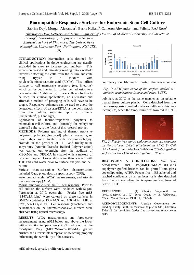
European Cells and Materials Vol. 16. Suppl. 3, 2008 (page 47) ISSN 1473-2262
Biocompatible Responsive Surfaces for Embryonic Stem Cell Culture Sabrina Dey1, Morgan Alexander2, Barrie Kellam3, Cameron Alexander1, and Felicity RAJ Rose1
Division of Drug Delivery and Tissue Engineering1,Division of Medicinal Chemistry and Structural Biology2, Laboratory of Biophysics and Surface
Analysis3, School of Pharmacy, The University of Nottingham, University Park, Nottingham, NG7 2RD,
UK INTRODUCTION: Mammalian cells destined for clinical applications in tissue engineering are usually expanded in vitro to increase cell numbers. This expansion period and ultimately seeding onto a scaffold involves detaching the cells from the culture substrate using trypsin in a mixture with ethylenediaminetetraacetic acid (EDTA). This inflicts damage to cell membrane receptors (e.g. integrins) which can be detrimental for further cell adhesion to a new substrate1. Additionally, if these cells are further to be used for clinical applications, an alternative and affordable method of passaging cells will have to be sought. Responsive polymers can be used to avoid the deleterious effects of trypsin/EDTA as the cells detach from the culture substrate upon a stimulus (temperature2, pH and light). Application of thermo-responsive polymers to mammalian cell culture, and ultimately for embryonic stem cell culture, is the focus of this research project. METHODS: Polymer grafting of thermo-responsive polymers: poly (allyl-alcohol) plasma coated glass cover slips were treated with 2-bromoisobutyryl bromide in the presence of THF and triethylamine anhydrous. (Atomic Transfer Radical Polymerisation) was carried out overnight after the addition of MEO2MA and OEGMA in ethanol with a mixture of Bpy and copper. Cover slips were then washed with THF and cold water prior to surface analysis and cell culture. Surface characterisation: Surface characterisation included X-ray photoelectron spectroscopy (XPS), water contact angle (WCA) measurements, and Atomic force microscopy (AFM). Mouse embryonic stem (mES) cell response: Prior to cell culture, the surfaces were incubated with 5ug/ml fibronectin at 37˚C overnight. Feeder free mES (E14Tg2A Line) were cultured on these surfaces in DMEM containing 15% FCS and 100 uL/ml LIF, at 37oC, 5% CO2 in air. Cell response (attachment and detachment) on the thermo-responsive surfaces were observed using optical microscopy.
RESULTS: WCA measurements and force-curve measurements using AFM below and above the lower critical solution temperatures (LCST) indicated that the copolymer Poly (MEO2MA-co-OEGMA) grafted brushes had a reversible temperature switching property influencing the wettability of the surfaces.
mES adhered, spread, proliferated, and reached
confluency on fibronectin coated thermo-responsive
polymers at 37°C in the same manner as on gelatine treated tissue culture plastic. Cells detached from the thermo-responsive grafted surfaces (although this was incomplete) when the temperature was lowered to 10ºC.
Fig. 2: Feeder free mouse embryonic stem cell response on the surfaces: 1-Cell attachment at 37°C. 2- Cell detachment from Poly(MEO2MA-co-OEGMA) grafted surfaces below LCST at 10°C. (μ bars: 100μm) DISCUSSION & CONCLUSIONS: We have demonstrated that Poly(MEO2MA-co-OEGMA) copolymer grafted brushes can be grafted onto glass coverslips using ATRP. Feeder free mES adhered and reached confluency on all surfaces; cells also detached from the surface when the temperature was lowered below LCST. REFERENCES: (1) Charity Waymouth. In vitro.1974;10;97-111 (2) Teruo Okano et al. Makromol. Chem., Rapid Commun.1990; 11, 571-576.
ACKNOWLEDGEMENTS: Algerian Government for Funding, Emily Smith for technical help with XPS, Christina Tuferalli for providing feeder free mouse embryonic stem cells.
1 2
Force(nN)
-500
0
500
1000
1500
2000
2500
3000
37 35 32 30 28 26 24 22 20 22 24 26 28 30 32 35 37
Temp (C)
Forc
e(nN
)
Force(nN)
Fig. 1: AFM force-curve of the surface studied at different temperatures (Above and below LCST).

European Cells and Materials Vol. 16. Suppl. 3, 2008 (page 48) ISSN 1473-2262
Oscillating magnet arrays for enhanced magnetic nanoparticle-based gene transfection
J Dobson 1, SC McBain1, N Farrow1, CD Batich2 1Institute forScience & Technology in Medicine, Keele University, Thornburrow Drive, Hartshill,
Stoke-on-Trent ST9 9EZ United Kingdom 2Dept. ofMaterials Science & Engineering, University ofFlorida, Gainesville, Florida, USA 32611
INTRODUCTION: Magnetic nanoparticle-based gene transfection has been shown to be effective both in combination with viral vectors and with
1-4 non-viral agents. In these systems, therapeutic or reporter genes are at tached to magnetic nanoparticles which are then focused to the target site/cells via high-field/high-gradient magnets. The technique has been shown to be efficient and rapid for in vitro transfection and compares well with cationic lipid-based reagents, producing good overall transfection levels with lower doses and shorter transfection times. In spite of its potential advantages, the overall transfection levels do not always exceed those of other non -viral agents. In order to improve the overall transfection levels while maintaining the advantages inherent in this technique, we have developed a novel, oscillating magnet array system which adds lateral motion to the particle/gene promoting enhanced transfection. METHODS: H292 human lung epithelial cells were transfected with OzBiosciences Polymag particles coated with pciLux luciferase reporter construct in response to static and oscillating magnetic fields. H292 cells were seeded in RPMI culture medium supplemented with 10% foetal calf serum, 1 00U/ml penicillin, 0.1 mg/ml streptomycin, 0.25ug/ml amphortericin B and 2mM L-glutamine at 2.5x10⁴ cells per well in 96 well plates and allowed to attach overnight. All transfections were performed in serum free RPMI 1640 media as follows; Polymag 0.1ug DNA/0.1ul PolyMag per well and LF2000 0.1ug DNA/0.3ul LF2000). Following the addition of reagents, the plates were transferred to an incubator at 37ûC 5% CO2 and placed above static/oscillating (Frequency 2Hz amplitude 200pm) magnetic fields. Magnetic fields were produced by pairs of 6x4mm NdFeB magnets per well for 2 hrs. At 2 hrs. post-transfection, the magnets were removed and the media was replaced with of RPMI 1640 culture media containing 10% foetal calf serum, 1 00U/ml penicillin, 0.1 mg/ml streptomycin, 0.25ug/ml amphortericin B and 2mM L-glutamine. At 48hr post-transfection, the media was removed from each well and the cells lysed by the addition of 30tl of cell reporter lysis buffer (Promega CCLR). Samples were assayed for Luciferase activity using a Luciferase assay reagent (Promega) and the total protein
concentration determined using a BCA assay reagent (Pierce, USA). The Halbach transfection system was compared to the cationic lipid transefction agent, Lipofactamine2000, GeneJuice and particle and DNA-only controls.
RESULTS: Experimental results indicate that the system significantly enhances overall in vitro transfection levels in several cell types compared to both static field techniques (p<0.005) and the cationic lipids (p<0.001) tested (Figure 1).
Fig. 1: Luciferase expression as measured by Relative Light Units per mg of protein. N= 72 per group.
DISCUSSION & CONCLUSIONS: The oscillating array system outperforms the best cationic lipid-based agents available. In addition, it has the previously demostrated advantages of magnetic transfection Ð rapid transfection times and requiring lower levels of DNA than cationic lipid-based agents Ð and shows little or no effect on cell viability. This method shows great potential for non-viral gene delivery both in vitro and in vivo. REFERENCES: 1C. Mah et al. (2000) Molec. Ther. 1:S239 2C. Mah et al. (2002) Molec. Ther. 6:106-1 12; 2J. Dobson (2006) Gene Ther. 13:283- 287. 3F. Shere et al. (2002) Gene Ther. 169: 1–10.
ACKNOWLEDGEMENTS: J Dobson is supported by a Royal Society Wolfson Merit Award.
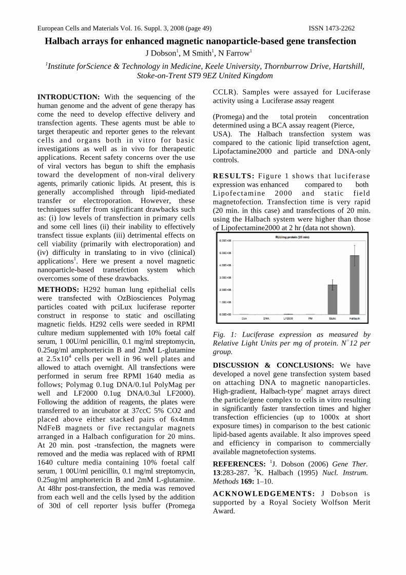
European Cells and Materials Vol. 16. Suppl. 3, 2008 (page 49) ISSN 1473-2262
Halbach arrays for enhanced magnetic nanoparticle-based gene transfection J Dobson1, M Smith1, N Farrow1
1Institute forScience & Technology in Medicine, Keele University, Thornburrow Drive, Hartshill, Stoke-on-Trent ST9 9EZ United Kingdom
INTRODUCTION: With the sequencing of the human genome and the advent of gene therapy has come the need to develop effective delivery and transfection agents. These agents must be able to target therapeutic and reporter genes to the relevant cel ls and organs both in vitro for basic investigations as well as in vivo for therapeutic applications. Recent safety concerns over the use of viral vectors has begun to shift the emphasis toward the development of non-viral delivery agents, primarily cationic lipids. At present, this is generally accomplished through lipid-mediated transfer or electroporation. However, these techniques suffer from significant drawbacks such as: (i) low levels of transfection in primary cells and some cell lines (ii) their inability to effectively transfect tissue explants (iii) detrimental effects on cell viability (primarily with electroporation) and (iv) difficulty in translating to in vivo (clinical) applications1. Here we present a novel magnetic nanoparticle-based transefction system which overcomes some of these drawbacks. METHODS: H292 human lung epithelial cells were transfected with OzBiosciences Polymag particles coated with pciLux luciferase reporter construct in response to static and oscillating magnetic fields. H292 cells were seeded in RPMI culture medium supplemented with 10% foetal calf serum, 1 00U/ml penicillin, 0.1 mg/ml streptomycin, 0.25ug/ml amphortericin B and 2mM L-glutamine at 2.5x10⁴ cells per well in 96 well plates and allowed to attach overnight. All transfections were performed in serum free RPMI 1640 media as follows; Polymag 0.1ug DNA/0.1ul PolyMag per well and LF2000 0.1ug DNA/0.3ul LF2000). Following the addition of reagents, the plates were transferred to an incubator at 37ccC 5% CO2 and placed above either stacked pairs of 6x4mm NdFeB magnets or five rectangular magnets arranged in a Halbach configuration for 20 mins. At 20 min. post -transfection, the magnets were removed and the media was replaced with of RPMI 1640 culture media containing 10% foetal calf serum, 1 00U/ml penicillin, 0.1 mg/ml streptomycin, 0.25ug/ml amphortericin B and 2mM L-glutamine. At 48hr post-transfection, the media was removed from each well and the cells lysed by the addition of 30tl of cell reporter lysis buffer (Promega
CCLR). Samples were assayed for Luciferase activity using a Luciferase assay reagent
(Promega) and the total protein concentration determined using a BCA assay reagent (Pierce, USA). The Halbach transfection system was compared to the cationic lipid transefction agent, Lipofactamine2000 and particle and DNA-only controls.
RESULTS: Figure 1 shows that luciferase expression was enhanced compared to both Lipofectamine 2000 and stat ic f ield magnetofection. Transfection time is very rapid (20 min. in this case) and transfections of 20 min. using the Halbach system were higher than those of Lipofectamine2000 at 2 hr (data not shown).
Fig. 1: Luciferase expression as measured by Relative Light Units per mg of protein. N=12 per group.
DISCUSSION & CONCLUSIONS: We have developed a novel gene transfection system based on attaching DNA to magnetic nanoparticles. High-gradient, Halbach-type2 magnet arrays direct the particle/gene complex to cells in vitro resulting in significantly faster transfection times and higher transfection efficiencies (up to 1000x at short exposure times) in comparison to the best cationic lipid-based agents available. It also improves speed and efficiency in comparison to commercially available magnetofection systems. REFERENCES: 1J. Dobson (2006) Gene Ther. 13:283-287. 3K. Halbach (1995) Nucl. Instrum. Methods 169: 1–10.
ACKNOWLEDGEMENTS: J Dobson is supported by a Royal Society Wolfson Merit Award.
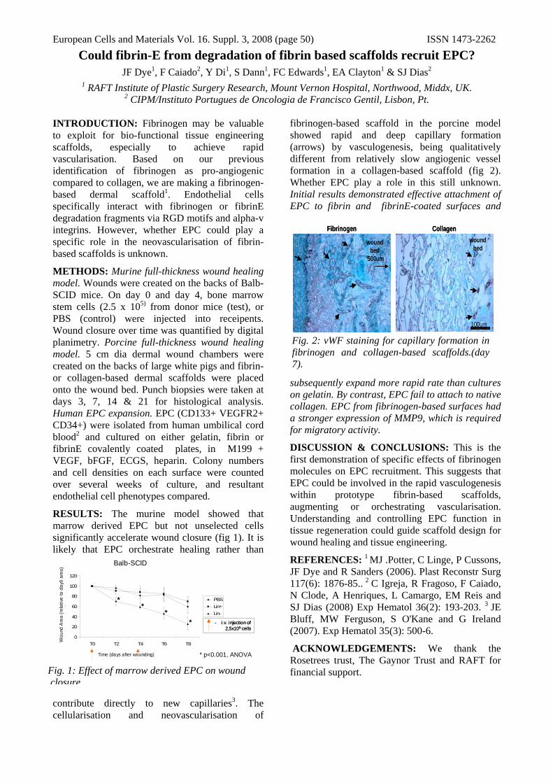
European Cells and Materials Vol. 16. Suppl. 3, 2008 (page 50) ISSN 1473-2262
Balb-SCID (2x injections)
0
20
40
60
80
100
120
T0 T2 T4 T6 T8
Time (days after wounding)
Wou
nd a
rea
(rel
ativ
e to
co
PBSLin+Lin-
Balb-SCID
Time (days after wounding)
Wou
nd A
rea
(rel
ativ
e to
day
0 ar
ea)
- i.v. injection of2,5x105 cells
- i.v. injection of2,5x105 cells
**
**
* p<0.001, ANOVA
Fig. 1: Effect of marrow derived EPC on wound closure.
100um
wound bed
500um
woundbed
Fibrinogen Collagen
100um
wound bed
500um
woundbed
100um
wound bed
500um
woundbed
Fibrinogen Collagen
Fig. 2: vWF staining for capillary formation in fibrinogen and collagen-based scaffolds.(day 7).
Could fibrin-E from degradation of fibrin based scaffolds recruit EPC? JF Dye1, F Caiado2, Y Di1, S Dann1, FC Edwards1, EA Clayton1 & SJ Dias2
1 RAFT Institute of Plastic Surgery Research, Mount Vernon Hospital, Northwood, Middx, UK. 2 CIPM/Instituto Portugues de Oncologia de Francisco Gentil, Lisbon, Pt.
INTRODUCTION: Fibrinogen may be valuable to exploit for bio-functional tissue engineering scaffolds, especially to achieve rapid vascularisation. Based on our previous identification of fibrinogen as pro-angiogenic compared to collagen, we are making a fibrinogen-based dermal scaffold1. Endothelial cells specifically interact with fibrinogen or fibrinE degradation fragments via RGD motifs and alpha-v integrins. However, whether EPC could play a specific role in the neovascularisation of fibrin-based scaffolds is unknown.
METHODS: Murine full-thickness wound healing model. Wounds were created on the backs of Balb-SCID mice. On day 0 and day 4, bone marrow stem cells (2.5 x 10⁵) from donor mice (test), or PBS (control) were injected into receipents. Wound closure over time was quantified by digital planimetry. Porcine full-thickness wound healing model. 5 cm dia dermal wound chambers were created on the backs of large white pigs and fibrin- or collagen-based dermal scaffolds were placed onto the wound bed. Punch biopsies were taken at days 3, 7, 14 & 21 for histological analysis. Human EPC expansion. EPC (CD133+ VEGFR2+ CD34+) were isolated from human umbilical cord blood2 and cultured on either gelatin, fibrin or fibrinE covalently coated plates, in M199 + VEGF, bFGF, ECGS, heparin. Colony numbers and cell densities on each surface were counted over several weeks of culture, and resultant endothelial cell phenotypes compared.
RESULTS: The murine model showed that marrow derived EPC but not unselected cells significantly accelerate wound closure (fig 1). It is likely that EPC orchestrate healing rather than
contribute directly to new capillaries3. The cellularisation and neovascularisation of
fibrinogen-based scaffold in the porcine model showed rapid and deep capillary formation (arrows) by vasculogenesis, being qualitatively different from relatively slow angiogenic vessel formation in a collagen-based scaffold (fig 2). Whether EPC play a role in this still unknown. Initial results demonstrated effective attachment of EPC to fibrin and fibrinE-coated surfaces and
subsequently expand more rapid rate than cultures on gelatin. By contrast, EPC fail to attach to native collagen. EPC from fibrinogen-based surfaces had a stronger expression of MMP9, which is required for migratory activity.
DISCUSSION & CONCLUSIONS: This is the first demonstration of specific effects of fibrinogen molecules on EPC recruitment. This suggests that EPC could be involved in the rapid vasculogenesis within prototype fibrin-based scaffolds, augmenting or orchestrating vascularisation. Understanding and controlling EPC function in tissue regeneration could guide scaffold design for wound healing and tissue engineering.
REFERENCES: 1 MJ .Potter, C Linge, P Cussons, JF Dye and R Sanders (2006). Plast Reconstr Surg 117(6): 1876-85.. 2 C Igreja, R Fragoso, F Caiado, N Clode, A Henriques, L Camargo, EM Reis and SJ Dias (2008) Exp Hematol 36(2): 193-203. 3 JE Bluff, MW Ferguson, S O'Kane and G Ireland (2007). Exp Hematol 35(3): 500-6.
ACKNOWLEDGEMENTS: We thank the Rosetrees trust, The Gaynor Trust and RAFT for financial support.

European Cells and Materials Vol. 16. Suppl. 3, 2008 (page 51) ISSN 1473-2262
Modelling the injured spinal cord using 3-dimensional cell cultures; strategies for improving tissue engineered repair
E East, J P Golding & J B Phillips
Department of Life Science, The Open University, Walton Hall, Milton Keynes, UK. INTRODUCTION: Traumatic injuries to the spinal cord are debilitating and often lead to paralysis and loss of sensation. CNS injury results in the formation of a glial scar that is largely composed of reactive, hypertrophic astrocytes which upregulate various markers including glial fibrillary acidic protein (GFAP) and sulphated protoglycans. The glial scar microenvironment is inhibitory for axon growth and regeneration A common finding of strategies aimed at bridging CNS lesions [1], in particular recent tissue engineered approaches using fibronectin [2], is that whilst axons readily enter and traverse the bridging graft they generally fail to exit the graft and re-enter the parenchyma due to the inhibitory glial scar at the graft-CNS interface. The aim of this work was to create a 3D cell culture model of CNS reactive gliosis, in which improved tissue-engineered repair strategies can be developed.
METHODS: Primary rat astrocytes were seeded onto collagen coated coverslips (2D) or into 3D collagen gels and the expression of markers associated with glial scar formation were analysed by immunocytochemistry (GFAP, chondroitin sulphate proteoglycan (CSPG), vimentin, aquaporin 4 (AqP4) and S100β). To stimulate reactive gliosis, 3D astrocyte cultures were treated with TGFβ1 (10ng/ml) every other day for 15 days [3]. Cultures without TGF β1 were used as controls. Expression of glial scar markers was assessed and measured using Volocity image analysis software (Improvision). Cell perimeter was measured as an indicator of astrocyte activation. Area of immunoreactivity was measured for CSPGs released from astrocytes.
RESULTS: Astrocytes in 3D cultures had significantly less immunoreactivity for GFAP, CSPG, vimentin, AqP4 and S100β, than astrocytes in 2D cultures indicating a lower level of activation in 3D (fig 1). However immunoreactivity for β-actin did not differ in 2D or 3D. Following treatment with TGFβ1, astrocytes in 3D became hypertrophic and reactive, with changes in the expression of gliosis markers over time relative to control cultures (fig 2).
Fig. 1: Comparison of astrocyte activation markers in 2D and 3D cultures in type I collagen. Bars represent mean ± SD, ***P<0.001 2D vs 3D.
Fig 2. Increased astrocyte activation following TGFβ1 treatment of 3D cultures.
DISCUSSION & CONCLUSIONS: Astrocytes in 3D are less reactive than their 2D counterparts, thus resembling the physiological in vivo situation and providing a useful system in which activation can be investigated. TGFβ1 treated astrocytes over 15 days take on characteristics of the mature glial scar, including their morphology and protein expression profile. This model will provide a useful test-bed for tissue engineered strategies to improve axonal growth through the glial scar environment.
REFERENCES: 1 Geller HM & Fawcett JW (2002) Exp Neurol 172:125-36. 2 Phillips JB et al, (2004) Biomaterials 25:2769-79. 3 DK Cullen, CM Simon & MC LaPlaca (2007) Brain Res 1158:103-115.
ACKNOWLEDGEMENTS: This research was funded by the Wellcome Trust.
GFA
PG
FAP
CS
PG
CS
PG
Vim
entin
Vim
entin
AqP
4 A
qP4 β
S10
0 βS
100
-Act
inβ -A
ctin
β
0
25
50
75
100
3D collagen2D collagen
***
******
*** ***
% p
ositi
ve c
ells
/fiel
d
0 5 10 15
1
2
3
4
56789
AqP4VimentinGFAPCSPG (area mm2)
(perimeter mm)
Time (days of TGFβ1 treatment)
Rat
io T
GF β
1/co
ntro
l(c
ell p
erim
eter
(mm
) or a
rea
of s
tain
ing
(mm
2 ))
2D CSPG
3D CSPG
Control 15d - GFAP
TGFβ1 15d - GFAP

European Cells and Materials Vol. 16. Suppl. 3, 2008 (page 52) ISSN 1473-2262
Fig. 1: Exposure of neutrophils to supra-physiological levels of calcium in PBS (but not HEPES) causes potent activation of CD18 (ED50 around 5 mM). There is a slight CD18 activation spontaneously over I hr from t=0 value, and a large increase by 5μM PMA (positive control).
Neutrophil activation in response to Calcium
Calcium concentration
2.5mM 3mM 4mM 5mM 10mM 20mM t=0 t=1 PMA
MC
F
0
200
400
600
800
Time
n=3
Calcium phosphate formation as a cause of inflammatory response to a prototype biomaterial scaffold.
FC Edwards, Y Di, S Dann, EA Clayton & JF Dye.
RAFT Institute of Plastic Surgery Research, Mount Vernon Hospital, Northwood, Middlesex, UK.
INTRODUCTION: Calcium ions are used for ionic cross-bridging in a variety of biomaterials, not only conferring structure and function to proteins but also organising hydrogels such as alginate. We are currently developing a fibrin/alginate material encorporating calcium ions, as a dermal replacement scaffold. In vivo evaluations of these scaffolds in a porcine full thickness wound model revealed a degree of inflammatory response. Endotoxin assay ruled this out as a contributory factor. The aim of this study was to identify and characterise the cause of this inflammation.
METHODS: Inflammatory potential of individual scaffold components and supernatants from whole scaffolds soaked for 24 hours in buffered saline solutions were assayed in vitro using neutrophil CD18 activation. Neutrophils from healthy volunteers were suspended in dulbecco’s PBS with Ca & Mg salts and glucose, and agents were tested for stimulation experiments in this buffer. Activated CD18 was measured by FACS. Supernatants were assayed for elastase (ELISA). Inflammatory responses to scaffolds were evaluated in vivo using a porcine full-thickness wound chamber model, from which punch biopsies were sampled over 3 weeks. Inflammatory infiltration was measured using a histological scoring system.
RESULTS: Of all the components and degradation products of the scaffolds, only calcium chloride added to PBS resulted in significant CD18 activation, suggesting a potential source of inflammatory stimulus (Fig 1). Further work showed this effect depends on phosphate and could be reduced by centrifugation. Activation increases progressively with time of exposure to the calcium phosphate up to 1 hr requires at least 10 min. Calcium addition to PBS also stimulated release of neutrophil elastase, and in whole blood cultures, the cytokines IL-8 and TNF-alpha, suggesting a self-stimulating mechanism driving the inflammatory response. In vivo histology showed progressive inflammation in scaffolds was related to the concentration of calcium used to make them. However, we were unable to demonstrate inflammatory effects in supernatants
from saline-soaked scaffolds made with up to 50mM calcium, suggesting that potentially released calcium phosphate remains associated with the scaffold microenvironment.
DISCUSSION & CONCLUSIONS: Although effects of various calcium phosphates in particulate form are known to regulate osteoblast and osteoclast function, and have been used as a model of arthritis, inflammatory effects of ‘fresh’ hydrated calcium phosphate precipitates on inflammation do not appear to have been studied. These studies show that amorphous calcium phosphate is a potent stimulus of neutrophil activation, as well as causing fibroblast cytotoxicity. Overall, these data suggest that noxious effects of calcium phosphate localised to the cell-biomaterial interface could drive an inflammatory response. Calcium phosphate deposition may cause inflammation and should be carefully monitored and controlled in biomaterials.
ACKNOWLEDGEMENTS: This project is supported by Dept. Health HTD LINK grant. We also thank the Rosetrees trust, The Gaynor Trust and RAFT for financial support.
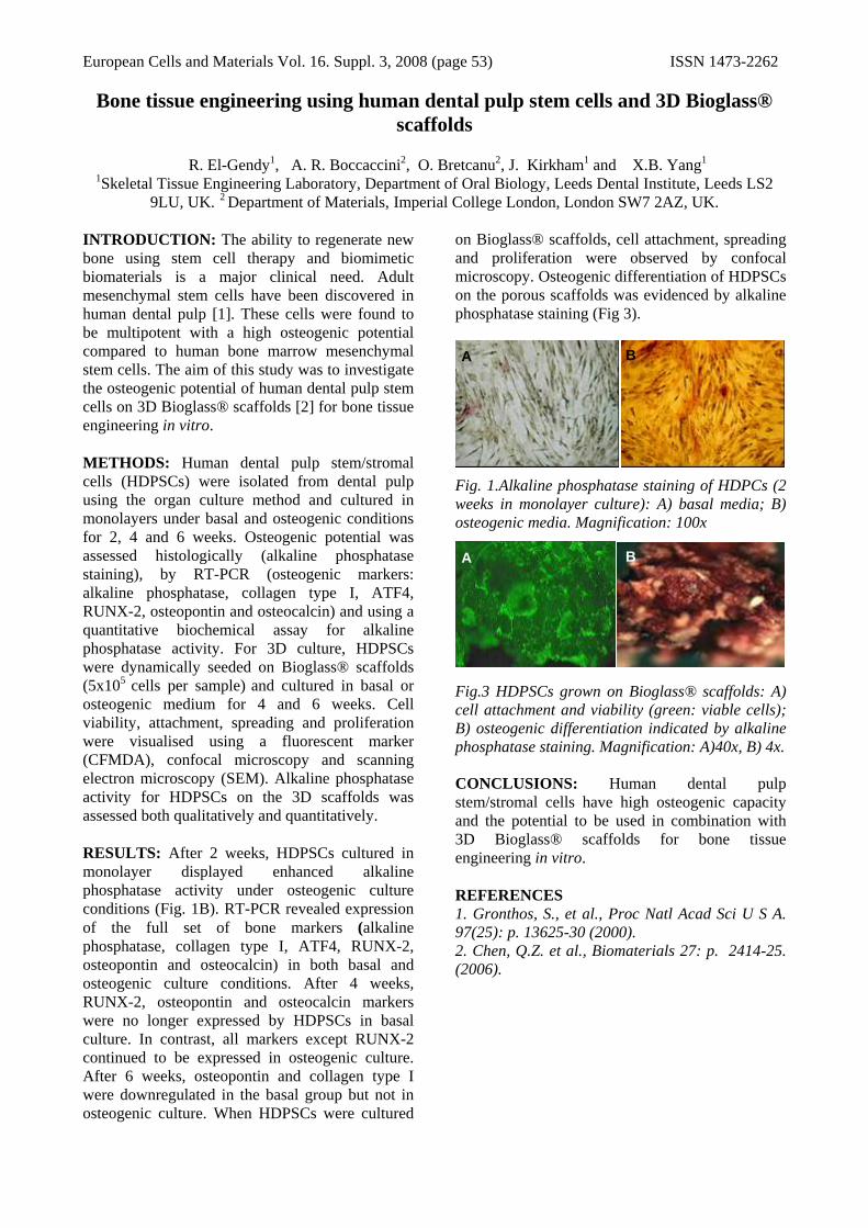
European Cells and Materials Vol. 16. Suppl. 3, 2008 (page 53) ISSN 1473-2262
Bone tissue engineering using human dental pulp stem cells and 3D Bioglass® scaffolds
R. El-Gendy1, A. R. Boccaccini2, O. Bretcanu2, J. Kirkham1 and X.B. Yang1
1Skeletal Tissue Engineering Laboratory, Department of Oral Biology, Leeds Dental Institute, Leeds LS2 9LU, UK. 2 Department of Materials, Imperial College London, London SW7 2AZ, UK.
INTRODUCTION: The ability to regenerate new bone using stem cell therapy and biomimetic biomaterials is a major clinical need. Adult mesenchymal stem cells have been discovered in human dental pulp [1]. These cells were found to be multipotent with a high osteogenic potential compared to human bone marrow mesenchymal stem cells. The aim of this study was to investigate the osteogenic potential of human dental pulp stem cells on 3D Bioglass® scaffolds [2] for bone tissue engineering in vitro. METHODS: Human dental pulp stem/stromal cells (HDPSCs) were isolated from dental pulp using the organ culture method and cultured in monolayers under basal and osteogenic conditions for 2, 4 and 6 weeks. Osteogenic potential was assessed histologically (alkaline phosphatase staining), by RT-PCR (osteogenic markers: alkaline phosphatase, collagen type I, ATF4, RUNX-2, osteopontin and osteocalcin) and using a quantitative biochemical assay for alkaline phosphatase activity. For 3D culture, HDPSCs were dynamically seeded on Bioglass® scaffolds (5x10⁵ cells per sample) and cultured in basal or osteogenic medium for 4 and 6 weeks. Cell viability, attachment, spreading and proliferation were visualised using a fluorescent marker (CFMDA), confocal microscopy and scanning electron microscopy (SEM). Alkaline phosphatase activity for HDPSCs on the 3D scaffolds was assessed both qualitatively and quantitatively. RESULTS: After 2 weeks, HDPSCs cultured in monolayer displayed enhanced alkaline phosphatase activity under osteogenic culture conditions (Fig. 1B). RT-PCR revealed expression of the full set of bone markers (alkaline phosphatase, collagen type I, ATF4, RUNX-2, osteopontin and osteocalcin) in both basal and osteogenic culture conditions. After 4 weeks, RUNX-2, osteopontin and osteocalcin markers were no longer expressed by HDPSCs in basal culture. In contrast, all markers except RUNX-2 continued to be expressed in osteogenic culture. After 6 weeks, osteopontin and collagen type I were downregulated in the basal group but not in osteogenic culture. When HDPSCs were cultured
on Bioglass® scaffolds, cell attachment, spreading and proliferation were observed by confocal microscopy. Osteogenic differentiation of HDPSCs on the porous scaffolds was evidenced by alkaline phosphatase staining (Fig 3).
Fig. 1.Alkaline phosphatase staining of HDPCs (2 weeks in monolayer culture): A) basal media; B) osteogenic media. Magnification: 100x
Fig.3 HDPSCs grown on Bioglass® scaffolds: A) cell attachment and viability (green: viable cells); B) osteogenic differentiation indicated by alkaline phosphatase staining. Magnification: A)40x, B) 4x.
CONCLUSIONS: Human dental pulp stem/stromal cells have high osteogenic capacity and the potential to be used in combination with 3D Bioglass® scaffolds for bone tissue engineering in vitro. REFERENCES 1. Gronthos, S., et al., Proc Natl Acad Sci U S A. 97(25): p. 13625-30 (2000). 2. Chen, Q.Z. et al., Biomaterials 27: p. 2414-25. (2006).
A B
A B

European Cells and Materials Vol. 16. Suppl. 3, 2008 (page 54) ISSN 1473-2262
Nanofuntionalized polymeric networks for tissue engineering applications.
MV Flores-Merino, G Reilly & G Battaglia. Biomaterials and Tissue Engineering Group, Department of Engineering Materials, The Kroto Research
Institute, University of Sheffield, Sheffield, UK.
INTRODUCTION: Polymeric networks are useful in tissue engineering applications acting as scaffolds for the delivery of molecules such as growth factors and other agents that induce cell differentiation. Studies have reported that nanoparticles embedded in polymers and polymeric gels can enhance their thermal and mechanical properties2. Here we report synthesis of a chemical hydrogel3 containing nanoparticles of amphiphilic copolymers that self-assemble into spherical micelles, cylindrical micelles, and vesicles according to the hydrophile/ hydrophobe ratio. The nanofuncionalized hydrogel exhibits improved mechanical properties and is able to encapsulate model pharmaceutical agents.
METHODS: Nanoparticle preparation. Amphiphilic Diblock copolymers were synthesized via living anionic polymerization. Block copolymers were placed in a glass vial and dissolved in chloroform (4mg/mL), followed by the evaporation of all the solvent in a vacuum oven to obtain a dry film was obtained. Hydratation of the film was performed by the addition of distilled water (10mg/mL). The dispersions were extruded (200nm pore size) and sonicated for 5 minutes. Synthesis of Nanofuncionalized Polymeric Networks. A solution of nanoparticles in water (1mg/mL) was mixed with the monomer of N-vynilpyrrolidinone (NVP) (Aldrich, UK), 1%wt of diethylene glycol bisallylcarbonate and 1%wt of 2,2-Azobis(2-methylpropionitrile) (Fluka, UK), polymerization was carry out a 60º C for 24hrs. Static Mechanical Testing. Compression testing was carry out in Hounsfield Universal Machine, with a 100 KN load cell, test speed 5mm /min, load range 500N, extension range 25mm and preload 10 N. Dynamic Mechanical Analysis. Experiments were performed on a Dynamic stress rheometer (Reometric Scientific SR-5000) with parallel plate geometry of 25mm diameter. Encapsulation and release studies: amphiphilic rhodamine and the anticancer drug 5-Fluorouracil (5-FU) were used as models to examine encapsulation into the nanofuncionalized networks. Separation of nanoparticles from free 5-FU was performed on a Sepharose 4B column
(0.7cm×10cm) (Sigma-Aldrich,UK) and measurements were carry out in a spectrophotometer.
RESULTS & DISCUSSION: Static mechanical measurements showed that the Young’s modulus (a measured by the slope of the strain stress curve) of the samples differed with the addition of either micelles, cylindrical micelles or vesicles (see Fig.1). Young’s modulus increases with the addition of vesicles and cylindrical micelles. The differences encounter in both systems may be attributed to the variant in the geometry and properties between polymersomes and polymeric micelles.
0 1 0 2 0 3 0 4 0 5 0 6 00
5 0 0 0 0
1 0 0 0 0 0
1 5 0 0 0 0
2 0 0 0 0 0
2 5 0 0 0 0St
ress
(N/m
2 )
S t r a in ( % )
N a n o f u n z io n a l iz e d H y d r o g e l N V P H y d r o g e l
Fig. 1: Stress-Strain curve of NVP polymeric network.
Dynamic analysis show similar results and differences between the materials. Encapsulation efficiency of 5-FU in polymersomes (polymeric vesicles) was 23%. Release of 5-FU vesicles incorporated in the hydrogel showed a delay in their release profile compared with the 5-FU encapsulated in only the NVP Hydrogel.
CONCLUSIONS: Addition of polymeric nanoparticles enhances the mechanical properties of the hydrogel such as Young’s modulus and maximum stress and allows encapsulation and release of pharmaceutical agents. These gels may have a wide range of applications in the biomedical area such as delivery of growth factors in tissue engineering scaffolds.
REFERENCES: 1 Jacob N. Israelachevili, Academia Press (1985) 2 S Bhattacharya, A Srivastava, and P. Asish (2006) 118, 303. 3LE Smith,, S Rimmer, and MacNeil S.(2006) Biomaterials, 27, 20.
ACKNOWLEDGEMENTS: The authors gratefully acknowledge Dr. Tony Ryan and the financial support provided by CONACyT.
εσ /=E

European Cells and Materials Vol. 16. Suppl. 3, 2008 (page 55) ISSN 1473-2262
INHIBITING CHONDROCYTE DEATH AT THE WOUND EDGE RESULTS IN SUCCESSFUL INTEGRATIVE CARTILAGE REPAIR IN VITRO.
SJ Gilbert, SK Singhrao, IM Khan, VC Duance & CW Archer Connective Tissue Biology Laboratories, School of Biosciences, Cardiff University, Museum Avenue,
Cardiff, CF10 3US, Wales, UK.
INTRODUCTION: Experimental wounding of articular cartilage results in necrotic and apoptotic chondrocyte death at the lesion edge [1]. Viable cells at the wound edge are critical for producing a repair tissue capable of integrating with the native cartilage [2,3]. The aims of this study were to investigate the effect of inhibiting wound-induced cell death on integrative cartilage repair.
METHODS: Full depth, articular cartilage discs (6mm) were harvested from the metacarpophalangeal joint of 7-day old bovine calves. Cartilage was pre-incubated in media containing inhibitors of necrosis (Necrostatin-1; 30µM) or apoptosis (ZVF, 20µM) prior to cutting a central 3mm inner core. This core was left in situ to create disc/ring composites in order to study cartilage-cartilage integration. Disc/ring composites were cultured for 2-weeks with the above inhibitors and analysed for necrotic (LDH release) and apoptotic (TUNEL) cell death, sGAG release (DMMB), tissue integration (histology, SEM, TEM), and matrix gene expression (qPCR).
RESULTS: Wounding of the cartilage resulted in a significant increase in necrotic cell death compared to unwounded cartilage (p<0.001). The pan-caspase inhibitor, ZVF significantly reduced both necrotic cell death (p<0.001) and apoptosis occurring at the wound edge. Necrostatin-1 also reduced necrotic cell death (p<0.01). The extent of sGAG lost into the media occurring as a result of wounding the cartilage was reduced by incubating the cartilage with either ZVF or Necrostatin-1. Toluidine blue staining (Figure 1), SEM and TEM of cartilage revealed significant integration of the wound edges in composites treated with ZVF. Necrostatin-1 improved integration but to a lesser extent. Quantitative PCR analysis revealed an increase in the expression of type II collagen mRNA in ZVF treated disc/ring composites (p<0.001).
Fig. 1: ZVF enhances integrative repair in cartilage disc/ring composites.
DISCUSSION & CONCLUSIONS: This study shows that treatment of articular cartilage with cell death inhibitors, in particular an inhibitor of apoptosis, prior to and during wound repair increases the number of viable cells at the wound edge, prevents matrix loss and results in a significant improvement in cartilage-cartilage integration in vitro. The use of caspase inhibitors in vivo may thus be a good candidate to promote cartilage integration, a process that remains problematical in clinical cartilage repair strategies.
REFERENCES: 1S.R. Tew, A.P. Kwan, A. Hann, et al (2000) Arthritis Rheum 43:215-25. 2J.S. Huntley, J.M. McBirnie, A.H. Simpson, A.C. Hall (2005) Osteoarthritis Cartilage 13:665-71. 3L.M. Janssen, C.D. In der Maur, P.K. Bos, J.A. Hardillo, G.J. van Osch (2006) Ann Otol Rhinol Laryngol 115:461-8.
ACKNOWLEDGEMENTS: This project was co-funded by the Technology Strategy Board's Collaborative Research and Development programme, following an open competition and Smith & Nephew.

European Cells and Materials Vol. 16. Suppl. 3, 2008 (page 56) ISSN 1473-2262
CELL/MICROPARTICLE AGGREGATES – A NOVEL METHOD TO ENGINEER LARGE EX VIVO NEO-TISSUE
C. Gribbon & P. De Bank
Centre for Regenerative Medicine, Department of Pharmacy & Pharmacology, University of Bath, Bath, BA2 7AY, UK
INTRODUCTION: A major problem to overcome in Tissue Engineering is how to achieve ex vivo tissue of sufficient size as to be practical for implantation into the patient; as tissue size increases, issues of mass transfer become increasingly important. Some methods for permitting large constructs are: using porous scaffold materials; using self-assembling materials that can be polymerised around cells in situ; and using suspension culture techniques such as microgravity bioreactors to assist with mass transfer.
The work described here used a novel combination of the above methods: porous PLGA microparticles were manufactured and functionalised with avidin. These microspheres were then cultured with biotinylated cells in microgravity conditions, forming large porous aggregates.
METHODS: Microparticles were prepared by a coacervation technique and sized by laser diffraction. These microparticles were then functionalised with avidin by standard peptide coupling reagents. Amount of avidin was determined by use of FITC-Avidin and FACS/MESF. NIH 3T3 cells were then biotinylated, using techniques previously described1 (biotinylation determined by staining with FITC-avidin, followed by FACS/MESF), and a mixed suspension of cells plus aggregates was incubated in a microgravity rotary cell culture bioreactor2. The cells and microparticles therefore cross-linked into large aggregates. After incubation, the aggregates were fixed and either sectioned and examined by standard histological methods, or examined by SEM.
RESULTS: Manufactured microparticles were of approximately cell-size (~10-20μm diameter), and could be controllably either porous or non-porous (Fig.1). After avidination, FACS/MESF analysis showed ~ 106 avidin molecules per microparticle and ~107 biotin molecules per cell (results not shown).
Fig.1: SEM of engineered (A) porous & (B) non-porous microparticles. Scale bar 1μm.
Histological examination of sectioned aggregates showed an even distribution of cells and microparticles throughout the structure, and no evidence of cell death in the centre at t=1 week (Fig.2A). SEM showed a smooth surface of cells on the aggregates, with occasional channels into the centre (Fig.2B)
Fig.2: (A) Section through a haematoxylin-stained aggregate; scale bar 100μm. (B) SEM of a cell + microparticle aggregate, scale bar 20μm
DISCUSSION & CONCLUSIONS: This novel method allowed the creation of porous tissue constructs in vitro, with cells and microparticles evenly distributed throughout. Incubation of these aggregates for time periods of up to 1 week showed no evidence of cell death in the constructs.
Future plans for the method include bone tissue engineering using mesenchymal stem cells. Also, encapsulation of growth factors in the PLGA, permitting controlled release directly into the vicinity of cells in the construct, providing pro-differentiation stimuli.
REFERENCES: 1 P. A. De Bank, et al (2007) Biotechnol. Bioeng. 97: 1617-1625. 2 B. J. Klement, et al (2004) Bone 34: 487-498
ACKNOWLEDGEMENTS: This work was supported by the BBSRC.

European Cells and Materials Vol. 16. Suppl. 3, 2008 (page 57) ISSN 1473-2262 Action of Simvastatin on Primary Human Osteoblasts:
Influences of Foetal Calf Serum on Mineralisation S. L. Griffiths & S. H. Cartmell
Institute of Science and Technology in Medicine, University of Keele, England, ST47QB
INTRODUCTION: Statins usually prescribed to lower cholesterol, can also enhance bone differentiation, maturation and matrix formation, with increased expression of BMP2 mRNA locally (1-2). Our initial studies in 2D and 3D in vitro cultures revealed the potential for enhancing mineralised matrix formation for the novel use in enhancing human primary bone tissue engineering (3).This study has looked at optimising Simvastatin’s bone forming effects in primary human osteoblasts (hOBs), by studying the effects of fetal calf serum (FCS) in media, on Simvastatin’s ability to enhance mineralisation. It has been shown that serum can have an inhibitory effect on BMP2 regulated osteoblastic gene expression in MSCs (4). As statin’s act specifically on BMP2, they may experience a similar effect as seen with BMP2 alone.
METHODS: An initial 14 day experiment used 4 groups, culturing hOBs in normal osteogenic and statin media (2.5µM Simvastatin), with and without FCS (Graph 1). A second expanded experiment increased the number of groups to 10 (A-J) (see Table 1), and cultured for a 7 day period (Graphs 2-4). Analysis was performed by Alkaline Phosphatase assay and Real-time RT-PCR assays for BMP2, Osteopontin (OPN) and Alkaline Phosphatase (ALP) gene expression. Table 1: FCS Media Experiment Groups A-J.
St = Simvastatin media, Ctrl = Osteogenic media
RESULTS: 14 day protein alkaline phosphatase assays showed that removing FCS or adding statin decreases enzyme activity. 7 day protein alkaline phosphatase for expanded groups (Table 1) showed that there was no significant difference compared to control group. Realtime RT-PCR results showed that there was no ALP expressed at 3 days, but BMP2 increased significantly with statin, regardless of FCS. 7 day gene expression showed that removing statin and FCS (group J) will increase ALP but decrease BMP2 and OPN expression. Group C shows significantly reduced ALP expression but not enzyme activity, potentially missed timing of expression. Group D
suggests a compromise, where ALP is increased by removing FCS after statin treatment, but re-administering statin after ALP is expressed, maintains BMP2 and OPN expression. Results for groups E-I are currently in progress.
GRAPH 1
GRAPH 2
GRAPH 3
GRAPH 4
DISCUSSION: After 7 days the effects of FCS on statins ability to stimulate BMP2 and thus it’s regulation of other osteogenic genes, doesn’t appear to be significant. Whilst statins do appear to reduce alkaline phosphatase activity in the long term, they may induce early expression of ALP when FCS is removed (graphs 2 and 4, group C). Prolonged loss of FCS (14 days) significantly reduces alkaline phosphatase activity and triggers de-differentiation. However there exists a potential for optimisation of FCS removal and statin addition to get maximal ALP protein production and BMP2 gene expression.
REFERENCES: (1) Mundy, et al. (1999) Science 286: 1946-1949. (2) Oxlund, et al. (2001) Calcif Tissue Int. 69: 299-304. (3) Griffiths and Cartmell, (2007) European Journal of Plastic Surgery, (Online) [in press]. (4) Osyczka and Leboy (2005) Endocrinology, 146(8): 3428–3437

European Cells and Materials Vol. 16. Suppl. 3, 2008 (page 58) ISSN 1473-2262
IMAGING OF POLYMERSOME PENETRATION INTO 3D TISSUE ENGINEERED MODELS OF ORAL MUCOSA AND HEAD AND NECK
CANCER V Hearnden1,2, G Battaglia1, C Murdoch2, M Thornhill2 & S MacNeil1
1Biomaterials and Tissue Engineering Group, Department of Engineering Materials, Kroto Research Institute and 2 Department of Oral & Maxillofacial Medicine and Surgery, School of Clinical Dentistry , University of Sheffield, UK.
INTRODUCTION: Tissue engineered models of oral mucosa (TEOM) and multi-cellular tumour spheroids (MCTS) accurately replicate in vivo oral mucosa and head and neck cancers respectively. These models enable us to study the behaviour and response of cells in a 3D situation in vitro. Our aim is to use these 3D models to develop new drug and gene delivery systems using nanopolymersomes. The first step towards this is to look at how to image the penetration of fluorescently labelled polymeric vesicles (polymersomes) into models of normal oral mucosa and into expanding MCTS.
Polymersomes are self assembling membrane enclosed structures made up of the amphiphilic block co-polymer PMPC-PDPA. The amphiphilic nature of the polymer allows both hydrophobic and hydrophilic compounds to be encapsulated into the membrane and core of the vesicles respectively. This, along with good biocompatibility and mechanical properties, means polymersomes have the potential to be used as delivery vectors for drugs and/or DNA [1].
METHODS: Full thickness tissue engineered oral mucosa is comprised of normal oral keratinocytes and normal oral fibroblasts grown on a de-epidermised dermis (DED) scaffold [2]. These are cultured for up to 2 weeks at an air liquid interface to encourage epithelial stratification. MCTS are grown from the head and neck carcinoma cell line FaDu. FaDu are cultured on agarose causing them to form MCTS which continue to expand over 2 weeks in culture. The models are incubated with rhodamine labelled polymersomes for up to 5 days.Imaging of the oral mucosa was examined using confocal laser scanning microscopy and imaging of the MCTS was examined using frozen sections and fluorescent microscopy.
RESULTS: Polymersomes penetrated the epithelial layers of the TEOM and MCTS in a time-dependent manner and were avidly internalized by the cells. Diffusion into the epithelium occurred within 6 hours.
Figure. 1: Confocal image of penetration of rhodamine labelled polymersomes (red) into TEOM after 48 hrs. Scale bar; 50µm.
Figure 2. Fluorescence image of rhodamine labelled polymersom penetration (red) of MCTS after 24 hrs. (blue = DAPI counter-stained nuclei).
DISCUSSION & CONCLUSIONS: Confocal and fluorescence microscopy demonstrate that rhodamine labeled polymersomes can penetrate deeply into the epithelium of TEOM models and tumour spheroids to deliver their load intracellularly. REFERENCES: 1 Lomas, H., et al.(2008), Non-cytotoxic polymervesicles for rapid and efficient intracellular delivery. Faraday Discussions. 139, 1-18. 2 Bhargava, S., et al.(2004), Tissue engineered buccal mucosa for substitution urethroplasty. BJU Int. 93 (6) 807-11. ACKNOWLEDGEMENTS: EPSRC for funding this project. Biocompatibles for providing the polymer.

European Cells and Materials Vol. 16. Suppl. 3, 2008 (page 59) ISSN 1473-2262
The effect of oxygen supply on chondrocyte metabolic phenotype during monolayer culture HK Heywood & DA Lee
School of Engineering and Materials Science, Queen Mary, University of London, UK.
INTRODUCTION: The expansion of chondrocytes by monolayer culture under atmospheric oxygen levels is used as part of the autologous chondrocyte implantation procedure and also for research purposes. Recent work has demonstrated enhanced oxidative capacity, oxidative phosphorylation and mitochondrial adaptations in expanded cell populations compared to freshly isolated cells [1]. However, the precise stimuli for these changes remain to be elucidated. This work tests the hypothesis that high oxygen levels experienced in standard incubator conditions stimulate the induction of an oxidative metabolic phenotype in articular chondrocytes.
METHODS: Bovine articular chondrocytes were seeded into flasks at 2.3 x10⁴ cells.cm-2. The flasks were distributed between 3 experimental groups: Standard incubator conditions (20% O2) and two reduced oxygen conditions (5% and 2% O2). Cell maintenance was performed under the specified O2 atmosphere using a BioSpherix (USA) workspace and pre-equilibrated reagents. After 0, 3, 7 and 14 days cells were recovered from flasks by trypsinisation and suspended in media with 200 μM O2. The O2 consumption rates of the cell samples were measured using an O2 biosensor (BD, Oxford, UK)[2]. Lactate production was determined from media and total cell protein measured using the BCA assay (Pierce, USA).
RESULTS: Cell proliferation appeared to be dependent on the incubator O2. Greatest cell yields were recorded with 20% oxygen and the lowest in 2% oxygen (fig 1). Total protein, indicative of cell size, also increased with culture, increasing ~4-fold over 14 days. However, no significant difference in total cell protein was observed as a result of altered O2 atmosphere (data not shown). O2 consumption rate, all measured at 200 µM O2, increased with culture duration at a rate proportional to the O2 in the culture atmosphere. Data recorded after 7 days culture is illustrated in fig 2. By contrast, lactate production remained stable throughout culture, when normalisation to cell protein.
Fig. 1: Effect of O2 supply on chondrocyte proliferation in monolayer culture. Data represent mean ± sd, n= 3.
Fig. 2: Effect of O2 supply during culture on chondrocyte O2 consumption and lactate production. Data represent means ± sd, n= 3.
DISCUSSION&CONCLUSIONS: Chondrocytes shift towards an oxidative metabolic phenotype during monolayer culture under atmospheric O2 levels [1]. Here we demonstrate that this shift is inhibited by culture in reduced oxygen atmosphere, but is not prevented entirely at the lowest O2 level tested, 2%.
REFERENCES: 1H.K. Heywood & D.A.Lee (2008) ORS transactions 33:0285. 2H.K. Heywood & D.A.Lee (2006) J. Cell. Phyiol. 206(2):402-410.
ACKNOWLEDGEMENTS: This work was funded by a Wellcome Trust project grant.

European Cells and Materials Vol. 16. Suppl. 3, 2008 (page 60) ISSN 1473-2262
MECHANICAL STIMULATION OF HL-1 CARDIOMYOCYTES CULTURED IN POLY-(1,8-OCTANEDIOL-CO-CITRIC ACID) [POC] SCAFFOLDS
L.A. Hidalgo-Bastida1, J.H. Ingram2, R. Majani1, F.H. Falcone3, LJ White1, E. Ingham2, W.C.
Claycomb4, L.D. Buttery1, F.R.A.J. Rose1, K.M. Shakesheff1
1 Division of Drug Delivery and Tissue Engineering. School of Pharmacy. University of Nottingham, UK. 2 Institute of Molecular and Cellular Biology. Faculty of Biological Sciences.
University of Leeds, UK. 3 Immune Modulation Research Group. School of Pharmacy. University of Nottingham, UK. 4 Department of Biochemistry & Molecular Biology. School of Medicine at New
Orleans. Louisiana State University, USA.
INTRODUCTION: Mechanical stimulation has been proved to have an effect in cardiomyocytes similar to that of growth factors on other cells, promoting protein expression, differentiation and survival [1]. In this study HL-1 cardiomyocytes were seeded in POC scaffolds and mechanically stimulated in the Tencell to assess whether there was any effect caused by the uni-axial stretching load applied to the constructs.
METHODS: HL-1 cardiomyocytes were cultured in 70% porous POC scaffolds previously coated with 400 µg/cm2 fibronectin [2-3]. Tencell, a stretching bioreactor, was used to provide the uni-axial mechanical load [4]. Constructs were cultured in a stretching regime of 10% amplitude and 1 Hz for 1 week. Mechanical stimulation was either continuous or discontinuous with 4 hours of mechanical stimulation per day. After culture, constructs were evaluated for survival (with Alamar Blue), morphology (using SEM and confocal after Live/Dead staining) and gene expression for actc1 and nppa with qRT-PCR.
RESULTS: Figure 1 presents the cell survival of the constructs after one week with no difference between static and continuously loaded but a significant difference with the discontinuous load regime. Both genes were up-regulated in all the cases. Although continuous stretching presented a greater expression of both genes when compared to the discontinuous regime, no statistically significant difference was found between these regimes. Figure 2 shows that after 1 week of culture, static culture promoted the formation of aligned myofibres whilst dynamic culture did not. Nevertheless, discontinuously stretched constructs did exhibit layers of HL-1 cells with normal cardiac morphology.
0
20
40
60
80
100
120
TENCELL static TENCELL continuous TENCELL discontinuous
Cell n
umbe
r [x 10 (3
) cells]
***
***
#
Fig 1: Survival of HL-1 cells after 1 week in the Tencell cultured at different stretching regimens. (mean ± SD, *** indicates p<0.001, # indicates p>0.05, n=3)
Fig. 2: SEM images of POC constructs seeded with HL-1 cells and cultured for a week in the Tencell (A) statically, (B) continuously and (C) discontinuously.
DISCUSSION & CONCLUSIONS: Mechano-stimulation of cardiac constructs encouraged cell survival in the discontinuous regime and up-regulated the expression of actc1 and nppa genes regardless of the treatments. Although mechanical stimulation had a positive effect on cell survival and gene expression, tissue formation was not promoted.
REFERENCES: 1 CA Powell et al (2002) Am J Physiol Cell Physiol 283 (5):C1557-C1565 2 WC Claycomb et al (1998) PNAS 95 (6):2979-84. 3 LA Hidalgo-Bastida et al (2007) Acta Biomater 3 :457-462.4 E Ingham E & J Fisher (2003) Eur Cell Mat 6 (2):5 ACKNOWLEDGEMENTS: This project was funded by the Mexican Science & Technology Council (CONACYT), the Mexican Ministry of Education (SEP) and the British Council.

European Cells and Materials Vol. 16. Suppl. 3, 2008 (page 61) ISSN 1473-2262
The Manipulation Of Live Mouse Embryonic Stem Cells Using Holographic Optical Tweezers
D. Howard1, J. Leach2, G. Gibson2, D. Gothard1, M.R.R. Self1, S.J. Roberts1, J.M. Cooper3, K.M. Shakesheff1, M.J. Padgett2 and L.D. Buttery1.
1Wolfson Centre for Stem Cells, Tissue Engineering & Modelling, Centre for Biomolecular Sciences School of Pharmacy, University of Nottingham, University Park, NG7 2RD. 2SUPA, Department of Physics and
Astronomy, 3Department of Electronics and Electrical Engineering, University of Glasgow, Glasgow, UK, G12 8QQ.
INTRODUCTION: Optical tweezers use a highly focused laser to exert a force on micron sized objects. Under the correct conditions, the force produced by the light is great enough to suppress low frequency Brownian motion and confine a cell into position. It has been shown that by using holographically generated optical traps it is possible to form arrangements of live bacterial cells and that these cells remain viable, as demonstrated by subsequent culture for at least 72 hours1. Applying this system to arrange stem cells into specific patterns may allow the determination of localised cues in cell interaction and development.
METHODS: We used a Ti:Sapphire laser operating at 830nm reflected off a spatial light modulator (Hamamatsu) to generate and control a number of separate trapping beams. Mouse embryonic stem cells were maintained on mitotically inactivated feeder layers supplemented with 109 U/ml leukaemia inhibitory factor (LIF; Chemicon, Watford, UK). Before manipulation the stem cells were detached into a single cell suspension using a two-minute incubation with trypsin enzyme solution with detachment verified using microscopy. A 100 µl aliquot of a dispersed cell suspension containing approximately 1000 cells was added to a chamber slide. Examination of cell survival used 0.001% Trypan Blue as live cells exclude this from their cytoplasm any dead/damaged cells stain dark-blue, allowing us to select for manipulation of only live cells and to assess cell integrity. Additional studies on cell viability were performed after manipulation by incubation at 37 ºC for 24 hr with the more sensitive fluoregenic Calcein AM with Ethidiuium Homodimer (Live/Dead stain, Molecular Probes, Paisley, UK) which produces green fluorescence in live cells and red fluorescence in dead cells.
RESULTS: Mouse embryonic stem cells could quickly and easily be positioned into straight lines, curves, circles, C shapes and the number patterns of dice (fig 1). In addition, cells could be moved
over one another indicating the potential to construct 3D cell configurations. The ability of the cells to exclude Trypan blue dye indicated that, despite comparatively high trapping powers, the cells remained viable. Cell viability after manipulation was further confirmed by Live/Dead staining and could be maintained for at least 18 hours (fig 1, No 4).
Fig 1.Laser moved artificial arrangements of live mouse embryonic stem cells (No 4 demonstrates live cells via florescent live/dead stain).
DISCUSSION & CONCLUSIONS: The experiments demonstrated that live embryonic stem cells can be manipulated into precise, pre-defined patterns using holographic optical trapping. We also demonstrated that cells could be cultured for several hours after manipulation. It is hoped that this system will allow investigation of the effects of arrangement and micro-environmental interactions upon stem cell growth and differentiation.
REFERENCES: 1P Jordan, J Leach, M Padgett, P Blackburn, N Isaacs, M Goksor, D Hanstorp, A Wright, J Girkin and J Cooper. Creating permanent 3D arrangements of isolated cells using holographic optical tweezers Lab Chip 5, 1224-1228 (2005).
ACKNOWLEDGEMENTS: The authors would like to thank Chris Denning and his team.

European Cells and Materials Vol. 16. Suppl. 3, 2008 (page 62) ISSN 1473-2262
The Use Of Rapid Prototype Pore Model Arrays To Assess Tissue Ingress In The CAM Assay.
D. Howard1, G. Lemon2, M.J. Tomlinson1, H. Yang3, R. Ronaldo3, L.D. Buttery1, S.L. Waters4, J.R. King2, K.M. Shakesheff1 and F.R.A.J. Rose1.
1 School of Pharmacy, 2School of Mathematical Sciences, 3Centre of Excellence in Customised Assembly, University of Nottingham, University Park, NG7 2RD. 4Oxford Centre for Industrial and Applied
Mathematics, Mathematical Institute, 24-29 St Giles, Oxford, OX1 3LB, UK.
INTRODUCTION: How host tissues ingress into polymeric tissue engineering type devices is a key stage in the regenerative process. The ability to understand and potentially manipulate these features would therefore be advantageous. The ability to isolate individual features of interest by creating an individual pore designed specifically for the development of mathematical and computational models could allow the understanding of relevant features to manage tissue ingress.
METHODS: A pore design based upon a sine curve rotated about a vertical axis (fig 1A,B) was generated and subsequently made using a Perfactory type III mini system (Envisiontec Inc, Germany) prototyping machine and photo-curable R11 acrylic resin. To provide a vascularised surface for the implant the pore arrays were added to a Chick chorioallantoic membrane (CAM) using a method modified from Ribatti et al1 (2006). In brief; at day 4 a window was cut into the top of a fertilised egg and the implant added to the CAM membrane. After incubation for 6 days the membrane and implant were excised and fixed in neutral buffered formalin. Tissues in the construct pores were visualized by epifluorescence microscopy using Propidium Iodide solution in PBS. Tissues were also located by staining with the heavy metal dye Osmium Tetroxide with subsequent X-ray µCT location (skyscan uCT 1174).
RESULTS: It was possible to produce a prototype scaffold with an array of defined individual pores of a scaffold for analysis (fig 1A,B). It was also possible to culture these devices on the CAM model without any observable adverse side effects to the membrane (fig 1C,D). Staining of the tissues clearly demonstrated tissues ingress into and through the pores (figure 1E) the tissues could also be removed from the scaffold and showed a change in the tissues in response to the construct (fig 1F).
Figure 1:(A,B)Rapid prototype construct of a 5mmx1mm array of 500 micron pores with a 100 micron restriction, (C) cultured upon the CAM membrane, (D) removed and fixed, (E) with tissues highlighted by staining with Propidium Iodide and with (F) the tissue separated, highlighting the morphological response of the membrane to the construct.
DISCUSSION & CONCLUSIONS: Tissues were able to penetrate the array of defined pores without any observable deleterious effects. With further work these devices may help to design mathematical and computational models to elucidate how tissues respond to some of the architectural cues present in many implants.
REFERENCES: 1 Ribatti D, Nico B, Vacca A, Presta M. The gelatin sponge-chorioallantoic membrane assay. Nat Protoc. 2006;1(1):85-91.
ACKNOWLEDGEMENTS: Many thanks to the BBSRC, MRC and EPSRC for funding. Thanks also to the Centre of Excellence in Customised Assembly (CECA) and the extended mathematical modelling group.

European Cells and Materials Vol. 16. Suppl. 3, 2008 (page 63) ISSN 1473-2262
The dose-dependant increase of ALP activity in human multipotent mesenchymal stromal cells after treatment with soybean extract.
M Illsley¹,², J.R. Sharpe1, S. E James2, S. James2 & M Santin2
1 The Blond McIndoe Centre, Queen Victoria Hospital, East Grinstead, RH19 3DZ, UK. 2 School of Pharmacy and Biomolecular Science, University of Brighton, Sussex, BN2 4GJ, UK
INTRODUCTION: Soybeans contain three major types of isoflavones, genistein, daidzein and glycitein. All three have been shown to have a powerful antioxidant effect and bind to oestrogen receptor β with high specificity. It has been demonstrated that dietary soybeans favourably impact bone health in postmenopausal women by lowering the incidence of osteoporosis. More recently soybean biomaterials (SB) have been developed which contain significant levels of isoflavones¹ and have shown bone regeneration potential in vivo. Here we demonstrate the effect of soy extract on stem cell differentiation into osteoblast-like cells and a potential use in regenerative medicine.
METHODS: MSCs were allowed to form confluent monolayers in 96 well plates, after a 24 hour attachment period the media was replaced with experimental conditions as described. DMEM +5% FCS or osteogenic media was spiked with soy flour extract, some experimental media was also spiked with 1x10⁻⁷M Fulvestrant, an oestrogen receptor inhibitor. Alkaline Phosphatase activity in cell lysates (ALP) was measured by a standard spectrophotometric method based on the enzymatic transformation of p-nitrophenol (Sigma-Aldrich UK, product code P7998). Protein concentration was determined by Bradford’s method. Data were expressed as ALP specific activity (IU/ mg protein). Soy isoflavones were extracted from de-fatted soy flour using an ethanol/water solvent mix, the extract was freeze dried to produce a stable and water soluble isoflavone concentrate. The efficacy of the extraction process was determined by HPLC analysis using internal forononetin standards and bookend standard runs.
RESULTS: ALP was significantly up-regulated in MSCs at all extract concentrations by day 6 (P=<0.05) (Fig 1). At shorter incubation times, there was no induction (day 2 concentrations <15μM) however similar activation was apparent in time points longer than day 6.
ALP activity in MSCs after culture in DMEM +5% and exctract
0
10
20
30
40
50
60
70
80
90
0 0.9375 1.875 3.75 7.5 15
µM genistein in extract
ALP
U/m
g pr
otei
n
Day 2Day 6Day 8Day 10
Fig. 1: Soy extract effect on MSC ALP activity. Error bars indicate SD.
When the experimental media was also supplemented with the competitive inhibitor Fulvestrant the up-regulation of ALP was retarded in a consistent manner (Fig 2) indicating that phytoestrogens in the soy extract are responsible for the demonstrated dose-dependant increase.
ALP activity in MSCs at Day 6, in osteogenic media and extract, with and without inhibitor
020406080
100120140160180
0 0.9375 1.875 3.75 7.5 15
µM genistein in extract
ALP
U/m
g pr
otei
n
Without inhibitorwith inhibitor
Fig. 2. ALP up-regulation with and without inhibitor. Error bars indicate SD.
CONCLUSIONS: SB bone regeneration potential may be attributed to the effect of the isoflavones on MSC differentiation into osteoblast-like cells. Treatments based on the use of SB or SB-derived tissue engineering constructs may lead to enhanced clinical outcomes in maxillo-facial, orthopaedic and periodontal surgery
REFERENCES: 1 Santin M. et al. (2001) PCTGB/01/03464

European Cells and Materials Vol. 16. Suppl. 3, 2008 (page 64) ISSN 1473-2262
Myocardial tissue engineering using human embryonic stem cell-derived cardiomyocytes and elastomeric polymers
H. Jawad1, H.Ishii2, A. Piegat3, M. El Fray3, S.E. Harding2, A. R. Boccaccini1, N.N. Ali2 1Department of Materials, Imperial College London, UK; 2Cardiac Medicine, NHLI Imperial College
London, UK; 3Szczecin University of Technology, Polymer Institute, Szczecin, Poland
INTRODUCTION: Heart failure (HF) results from adult heart tissue having limited regenerative capacity following a heart attack. Due to donor shortages at end stage HF, cell therapy is an alternative strategy. Previous attempts to repair heart involved the injection of a suspension of cells either via coronary arteries or directly into the heart muscle, but this is highly inefficient. Delivery of cells to the desired area can be better achieved by attaching a pre-formed patch of cardiac cells onto the infarct region1. Our overall aim is to develop engineered patches of myocardial cells attached to a biodegradable polymeric support using embryonic stem cells (ESC) as the cell source.
METHODS: The following biomaterials were used: (1) plain copolymer PET-DFA, (2) PET-DFA with 0.2 wt% TiO2 nanoparticle addition. These materials were produced by transesterification and polycondensation and were subjected to the following tests: surface examination (with and without added cells) by SEM, material ultrastructure by TEM; tensile strength and surface topography analysis (Zygo®). The D3 mouse ESC line (from ATCC) and human ESC H7 (from Geron Corp) were maintained in an undifferentiated state by culturing in the presence of either 1000U/mL LIF and feeder cells (D3 line) or 8ng/mL bFGF in MEF-conditioned medium (H7 cells). Both lines were induced via embryoid body (EB) formation after the removal of LIF and feeders (D3), or bFGF(H7). EBs were induced to form by putting 400 D3 cells/20ul hanging drop for 2days in 15% FCS, then placed into suspension culture (3-5days) after which time they were plated. The colonies of H7 line were mechanically broken into smaller pieces and cultured in 20% FCS. They were maintained in suspension culture for 4 days then plated out. ESC-CM were isolated and enriched, either after microdissection of spontaneously beating foci from EB outgrowths, or separation on discontinuous percoll gradients. Isolated cells were seeded onto 10mm sterile discs of the biomaterials with and without pre-gelatinisation. Biomaterials films were also sutured without cells onto rat heart ex-vivo to study the effect of patches on the heart.
RESULTS: The presence of TiO2 nanoparticles, although in low concentration (0.2 wt%), has an effect on the polymer mechanical properties and on surface morphology hence on cellular adhesion, spread and proliferation. Cell proliferation, tested with a fibroblastic cell line, on both biomaterials with and without gelatin coating proliferated. By day 6 proliferation was better on the new biomaterials than on a commercial polymer (Polyactive®, P<0.05) but not as efficient as on tissue culture plastic (p<0.01, n=3). Lactate dehydrogenase (LDH) release as a marker of cell death did not differ significantly between biomaterials and tissue culture plastic. TiO2 (10 ppm) had no significant effect on cardiac contractility of either freshly isolated adult rat heart cells (n=6) or hESC-CM (n=5). Ex-vivo results indicated suturing the patch onto rat hearts had no effect on developed pressure (mmHg).
Fig. 1: hESC-CM on PET/DLA (right) vs. PET/DLA-0.2wt%TiO2
DISCUSSION & CONCLUSIONS: PET/DLA-0.2wt%TiO2 nanocomposite biomaterial seems to support the adhesion, spread and proliferation of hESC-CM in vitro. Nanotoxicity results prove that there is no significant effect on cardiac contractility of cardiomyocytes at 10ppm. Ex-vivo results demonstrated no significant effect of suturing the patches on pressure of the heart.
REFERENCES: 1 Zimmermann et al., Biomaterials. 2004 9:1639
ACKNOWLEDGEMENTS: Financial support from the Biotechnology and Biological Sciences Research Council (BBSRC) is gratefully acknowledged.

European Cells and Materials Vol. 16. Suppl. 3, 2008 (page 65) ISSN 1473-2262
OSTEOBLAST: OSTEOCLAST CO-CULTURE ON VARIOUS BIOMATERIALS: AN INVESTIGATION OF CELL ACTIVITY AND
SCAFFOLD DEGRADATION.
G. L. Jones1, A. Motta 2, M. J. Marshall3, A. J. El Haj1* and S. H. Cartmell1
1. ISTM, Keele University, Thornburrow Drive, Hartshill, Stoke-on-Trent, ST4 7QB, UK 2. DIMTI, Univerity of Trento, Via Mesiano 77, 38050 Trento, Italy
3. Charles Salt Centre, RJAH Orthopaedic Hospital, Oswestry, Shropshire, SY10 7AG, UK INTRODUCTION: Bone, although capable of self-regeneration, may lose this ability after severe injury or disease. Tissue engineering aims to aide the repair of damaged bone, traditionally uses a single cell type usually of osteoblastic lineage cultured on 2D or 3D biomaterials before implantation into the body. Materials to be compared in this investigation include silk fibroin from Bombyx mori, which has been used for centuries as a suture material, and has excellent biological properties for tissue engineering applications. Chitosan is a natural polymer also with good potential for tissue engineering, and PLLA is a well documented material for tissue engineering as it is easily manipulated to suit its purpose. This investigation explores the use of co-cultures to utilise the synergy of both osteoblasts and osteoclasts to create a tissue engineered model that recreates the physiological process. The aim is to use various biomaterials to determine the ability of four materials to support an osteoblast: osteoclast co-culture in vitro and to reveal any effects on the degradation rate of the material during the culture period. METHODS: A murine model of MC3T3-E1 osteoblast like cells and primary murine monocytes1 differentiated towards osteoclasts (using a media containing 10-8Dexamethasone and 10-8 1-25 di-hydroxy vitamin D3 and 10ng/ml receptor activated NFκB ligand (RANKL)) was used. Four groups were examined 1) Osteoblast: osteoclast co-culture (OB: OC), 2) Osteoblast only (OB), 3) Osteoclast only (OC) and Material only (MO). Cells were seeded onto 5mm diameter 2D films of vapour and methanol stabilised fibroin2, chitosan and PLLA for 10 days. They were stained for tartrate resistant acid phosphatase (TRAP) activity, cell activity monitored by DNA assay and SEM. Degradation of the films was monitored using DSC, FTIR and GPC
RESULTS:
1A) B)
Figure 1 TRAP stain of OB:OC co-cultures on A) vapour stabilised fibroin and B) methanol stabilised fibroin. Scale bar = 200µm.
2A) B)
Figure 2) SEM images of OB:OC co-cultures on A) vapour and B) methanol treated fibroin. Scale bar = 75µm.
DISCUSSION & CONCLUSIONS: Having previously optimised a ratio for co-culture of 1 osteoblast to 100 osteoclasts and devising a media that supports growth and differentiation of both osteoblasts and osteoclasts, this was utilised to investigate usefulness of various materials. Figures 1 and 2 demonstrate that vapour and methanol stabilised fibroin support cell growth and differentiation. TRAP stained images (figure 1) show a layer of osteoblasts with TRAP positive osteoclasts dispersed throughout the sample. SEM demonstrates cell adhesion, and shows different cell morphology between A) vapour stabilised fibroin, and B) methanol stabilised fibroin. DNA concentrations show similar concentrations of DNA in vapour and methanol stabilised fibroin and chitosan groups, indicating the 3 materials support cell growth. The effect cells may have on degradation is still under investigation. Future work aims use a 3D sponge of silk fibroin, as a substrate for co-cultures and to investigate the potential benefits of using a rotatory bioreactor (cellon).
REFERENCES:1. Dugard et al. 2005. Clinica Chimica Acta. 356(1-2): 154-63 2. Motta et al 2002 Macromol Chem Phys. 203: 1658-65.
ACKNOWLEDGEMENTS: EXPERTISSUES EU Vth framework for funding. Lorenzo Moschini, University of Trento for help with GPC. Cassilda Reis Keele University for providing chitosan films.

European Cells and Materials Vol. 16. Suppl. 3, 2008 (page 66) ISSN 1473-2262
3D Silk Fibroin Sponge Co-culture in a Rotatory Bioreactor
G. L. Jones1, A. Motta 2, M. J. Marshall3, A. J. El Haj1* and S. H. Cartmell1
1. ISTM, Keele University, Thornburrow Drive, Hartshill, Stoke-on-Trent, ST4 7QB, UK 2. DIMTI, Univerity of Trento, Via Mesiano 77, 38050 Trento, Italy
3. Charles Salt Centre, RJAH Orthopaedic Hospital, Oswestry, Shropshire, SY10 7AG, UK INTRODUCTION: Bone although capable of self-regeneration, may lose this ability after severe injury or disease. Tissue engineering aims to aid the repair of damaged bone, traditionally with the use of single cell type of osteoblastic lineage cultured on 2D or 3D biomaterials before implantation into the body. In this investigation a 3D sponge of silk fibroin obtained from water solution 7% (w/V), has been cultured in both static culture and a rotatory bioreactor (Cellon). Silk fibroin from Bombyx mori, has been used for centuries as a suture material, and has excellent properties for tissue engineering which include excellent biocompatibility. This investigation explores the ability for co-cultures of osteoblasts and osteoclasts on 3D sponges to utilise the synergy created by cell-cell interactions between these two cell types to create a functional tissue engineered construct that attempts to recreates the physiological process. The aim of this experiment is to assess the ability of 3D silk fibroin sponges to support a co-culture of osteoblast: osteoclasts in vitro by monitoring cell activity and also monitor degradation of the sponge. In addition we will investigate the potential benefits of using a rotatory bioreactor to enhance the co-cultures. METHODS: A murine model of MC3T3-E1 osteoblast like cells and primary murine monocytes1 (differentiated towards osteoclasts using a media containing 10-8Dexamethasone and 10-8 1-25 di-hydroxy vitamin D3 10ng/ml receptor activated NFκB ligand (RANKL)) was used. Osteoblasts were seeded at 5x10⁴ and monocytes at 5x10⁵ onto appropriate groups of silk sponges of 5x5x5mm dimensions2. Four groups were examined 1) Osteoblast: osteoclast co-culture (OB: OC), 2) Osteoblast only (OB), 3) osteoclast only (OC) and material only (MO). The sponges were then cultured in static or bioreactor conditions for 10 days. Cell activity was monitored by DNA assay and morphology by SEM. Degradation of the films was monitored using DSC, FTIR and GPC. RESULTS:
1 A) B) Figure 1 SEM images of silk fibroin sponges after 10 days OB:OC co-culture. A) Represents sponge cultured statically, B) sponge cultured in the bioreactor. Scale bar =100µm. 2)
Figure 2. DNA assay results after 10 days culture on silk sponges in static or bioreactor conditions.
DISCUSSION & CONCLUSIONS: Results from this investigation indicate that silk fibroin sponges support cell growth and proliferation during a 10 day culture in static and bioreactor conditions. The SEM images (figure 1) reveal differences in the topography of the sponges and cells can be seen on the statically cultured sponge (figure 1A), but not on the sponge cultured in the bioreactor (figure 1B). SEM data was supported by the results of the DNA assay. Concentration of DNA was higher in samples cultured statically, demonstrating a higher cell adhesion, to sponges cultured in the bioreactor. Results demonstrating degradation are pending analysis. This investigation was only short and future work would include use of a longer culture period, as this will allow degradation to occur and also explore the ability of the sponges to support cell growth long term.
REFERENCES: 1. Dugard et al. 2005. Clinica Chimica Acta. 356(1-2): 154-63 2. Motta et al 2002. Macromol Chem Phys. 203: 1658-1665.
ACKNOWLEDGEMENTS: EXPERTISSUES EU Vth framework for funding. Lorenzo Moschini, University of Trento for help with GPC.

European Cells and Materials Vol. 16. Suppl. 3, 2008 (page 67) ISSN 1473-2262
Effect of matrix stiffness on cellular responses in 3D D. Karamichos, H. Hadgipanayi, R. Brown and V. Mudera
University College London, UCL, Tissue Repair and Engineering Centre, Institute of Orthopaedics, Stanmore Campus, London, HA7 4LP, UK
INTRODUCTION: Several cell types respond to mechanical forces, key to in-vitro remodeling. We have tested the differential response of human dermal fibroblasts (HDFs) and human bone marrow stem cells (HBMSCs) using increasingly stiffer collagen matrices with the mechano-molecular regulation of specific extracellular matrix genes, following ramp load regimes. We have also tested the effect of stiffness on cell proliferation.
METHODS: Strain was applied using a computer driven tensional-Culture Force Monitor (t-CFM). Cell seeded collagen constructs were subjected to pre-strain (0% and 5%) and kept under tension. Ramp load applied was: a) 10% ramp over 1h, b) 10% ramp over 12h pre-strain level (i.e 0% and 5%). Molecular outputs from constructs were also investigated. For proliferation studies dermal fibroblasts were seeded in collagen constructs and three models tested. Free floating unconstrained, attached constrained and plastic compressed constructs to produce increasingly stiffer constructs.
RESULTS: Both HBMSCs and HDFs showed no significant increase on force generation following ramp load stimulation, with either 10 and/or 20% FCS. ECM regulatory genes TIMP-2 and COL-3 showed up regulation, with 10% FCS, at 5% pre strain and ramp load over 1h. In contrast, genes MMP-2 and COL-1 showed down regulation at the same groups. At 20% FCS genes showed no regulation over 1h ramp loads, instead the slow ramp load 12h led to, again, MMP-2 down regulation and TIMP-2 up regulation, but up regulation of COL-1. HBMSCs showed no significant gene regulation at 10% FCS, and only minimal though significant down regulation of COL-1 with 20% FCS and slow ramp load regimes. Human dermal fibroblasts (HDFs) in free floating collagen matrices show minimal proliferation, this increases when the matrix is ‘under tension’. In stiffest PC matrices cell proliferation was rapid, seeding-density dependent, with a population doubling time of 2 days. In contrast compliant attached matrices showed a 4 day lag period, and doubling time of 6 days. HDF proliferation was directly related to matrix stiffness such that increasing stiffness using a range of compression levels (0 to 75% fluid removal)
resulted in increasing proliferation rates and doubling times and also increasing matrix elastic modulus. HDF quiescence in compliant matrices was reversible, such that increasing stiffness in situ by compression at 1 and 5 days initiated proliferation.
DISCUSSION & CONCLUSIONS: We conclude that collagen matrix stiffness regulates proliferation of fibroblasts (a duro-response) with important implications for both wound healing and the design and regulation of engineered connective tissues based on collagen and other hydrogel scaffolds. ECM remodeling gene-regulation and turnover is important when stiff constructs, ramp rate and FCS levels are combined in bioreactors. This has major implications in TE, specifically in tissues where load bearing is a major function. REFERENCES: D. Karamichos*, J. Skinner, R.A. Brown, V. Mudera (2008) Matrix stiffness and serum concentration effects matrix remodelling and ECM regulatory genes of human bone marrow stem cells. Journal of Tissue Engineering and Regenerative Medicine: Mar 14; [Epub ahead of print] PMID: 18338818. D. Karamichos*, R.A. Brown, V. Mudera (2007) Collagen stiffness regulates cellular contraction and matrix remodeling gene expression Journal of Biomedical Materials Research: 83A:3, ,887-894 Karamichos D., Brown RA. and Mudera V.(2006) Complex dependence of substrate stiffness and serum concentration on cell-force generation. J Biomed Mater Res A. Aug;78(2):407-15. Hadjipanayi, E., Mudera, V., Brown, R.A (2008) Effect of Stiffness on Fibroblast Proliferation within a 3D Matrix. Submitted for publication Experimental Cell Research
ACKNOWLEDGEMENTS: The authors are thankful to the BBSRC and EU Framework VI for funding this research
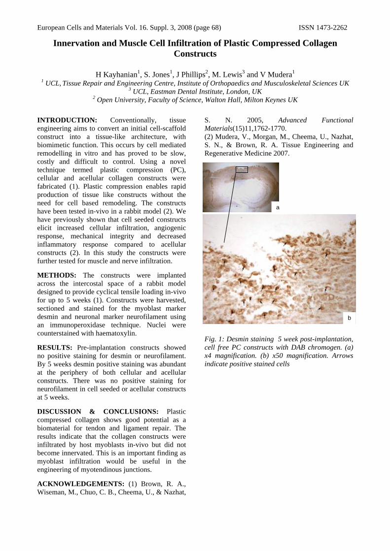
European Cells and Materials Vol. 16. Suppl. 3, 2008 (page 68) ISSN 1473-2262
Innervation and Muscle Cell Infiltration of Plastic Compressed Collagen Constructs
H Kayhanian1, S. Jones1, J Phillips2, M. Lewis3 and V Mudera1
1 UCL, Tissue Repair and Engineering Centre, Institute of Orthopaedics and Musculoskeletal Sciences UK 3 UCL, Eastman Dental Institute, London, UK
2 Open University, Faculty of Science, Walton Hall, Milton Keynes UK INTRODUCTION: Conventionally, tissue engineering aims to convert an initial cell-scaffold construct into a tissue-like architecture, with biomimetic function. This occurs by cell mediated remodelling in vitro and has proved to be slow, costly and difficult to control. Using a novel technique termed plastic compression (PC), cellular and acellular collagen constructs were fabricated (1). Plastic compression enables rapid production of tissue like constructs without the need for cell based remodeling. The constructs have been tested in-vivo in a rabbit model (2). We have previously shown that cell seeded constructs elicit increased cellular infiltration, angiogenic response, mechanical integrity and decreased inflammatory response compared to acellular constructs (2). In this study the constructs were further tested for muscle and nerve infiltration. METHODS: The constructs were implanted across the intercostal space of a rabbit model designed to provide cyclical tensile loading in-vivo for up to 5 weeks (1). Constructs were harvested, sectioned and stained for the myoblast marker desmin and neuronal marker neurofilament using an immunoperoxidase technique. Nuclei were counterstained with haematoxylin. RESULTS: Pre-implantation constructs showed no positive staining for desmin or neurofilament. By 5 weeks desmin positive staining was abundant at the periphery of both cellular and acellular constructs. There was no positive staining for neurofilament in cell seeded or acellular constructs at 5 weeks. DISCUSSION & CONCLUSIONS: Plastic compressed collagen shows good potential as a biomaterial for tendon and ligament repair. The results indicate that the collagen constructs were infiltrated by host myoblasts in-vivo but did not become innervated. This is an important finding as myoblast infiltration would be useful in the engineering of myotendinous junctions. ACKNOWLEDGEMENTS: (1) Brown, R. A., Wiseman, M., Chuo, C. B., Cheema, U., & Nazhat,
S. N. 2005, Advanced Functional Materials(15)11,1762-1770. (2) Mudera, V., Morgan, M., Cheema, U., Nazhat, S. N., & Brown, R. A. Tissue Engineering and Regenerative Medicine 2007.
Fig. 1: Desmin staining 5 week post-implantation, cell free PC constructs with DAB chromogen. (a) x4 magnification. (b) x50 magnification. Arrows indicate positive stained cells
a
b

European Cells and Materials Vol. 16. Suppl. 3, 2008 (page 69) ISSN 1473-2262 Characterising Osteogenesis in Mouse Embryonic Stem Cell Aggregates
G.R Kirkham, L.D.K Buttery
1 Wolfson Centre for Stem Cells, Tissue Engineering and Modelling (STEM), University of Nottingham, University Park, NG7 2RD
INTRODUCTION: Murine embryonic stem cells (mES) have the potential to differentiate to all fetal and adult cell types and represent a useful cell source for tissue engineering. It has been shown that mES can undergo osteogenesis in vitro but little is known about the aggregation stage.
METHODS: A mouse ES cell line denoted CCE was co-cultured on a feeder layer of inactivated (with mitomycin C) mouse embryonic fibroblasts (SNL cell line) as described previously [1]. Cells were aggregated using the method previously described [2]. Aggregates were then pooled together into non-tissue culture treated petri dishes at a density of ~20 aggregates per dish. Aggregates were cultured under two distinct conditions: osteogenic samples were cultured in osteogenic media consisting of ascorbic acid 2-phosphate, β-glycerophosphate and dexamethasone [1], control samples were allowed to differentiate spontaneously in basal media. Separate cultures were grown up for 0, 2, 4, 6, 8 and 9 days and fixed for 20 minutes in 3.7% paraformaldehyde (pH 7.4). Aggregates were then embedded in agarose, dehydrated in ethanol and embedded in paraffin wax. 2μm sections were then re-hydrated and permeablised for 5 minutes with 0.2% TritonX-100 at -20oC. Samples were then washed and incubated with the relevant primary antibody overnight at 4oC. A FITC conjugated secondary antibody was then incubated for 40 minutes and samples were imaged with a fluorescent microscope (Leica DMIRB).
RESULTS: Aggregates allowed to differentiate spontaneously in basal media developed numerous fluid filled cavities as little as a single cell in thickness. Osteogenic aggregates retained a more densely associated morphology with a smaller degree of growth (in terms of surface area) compared to spontaneous conditions. Sections were immunostained for cadherin-11. The 9 day time point showed positive staining, with the highest levels in localised regions of the outer sections of the aggregates. The same aggregates were negative for osteocalcin expression.
Fig. 1: Florescence micrograph of a 2μm mES cell aggregate section stained for Cadherin-11 by immunocytochemistry (FITC labelled secondary antibody).
Fig. 2: mES cell aggregate grown for 9 days. A –In basal media (spontaneous (control) conditions). B – In osteogenic media. DISCUSSION & CONCLUSIONS: The differences in morphology between the two aggregate culture conditions (osteogenic and spontaneous) indicate the that culture conditions have a marked influence of aggregate development. The presence of relatively high levels of cadherin-11 at 9 days indicated that osteogenesis may occur in localised regions within the peripheral areas of the aggregate. The lack of osteocalcin expression may be due to the early time points tested and therefore further markers of osteogenesis will need to be used to confirm osteogenic differentiation.
REFERENCES: 1 Buttery L.D.K et al (2001) Tissue Engineering 7:1:89-99. 2Kurosawa H et al (2003) J of Biosci and Bioeng 96:4: 409-411.
ACKNOWLEDGEMENTS: Members of the tissue engineering group at Nottingham University.

European Cells and Materials Vol. 16. Suppl. 3, 2008 (page 70) ISSN 1473-2262
THE SELF-ASSEMBLING PEPTIDE, P11-4 AS A SCAFFOLD FOR USE IN REGENERATIVE MEDICINE
S Kyle1, A Aggeli2, M J McPherson1 & E Ingham1
1 Institute of Molecular & Cellular Biology, University of Leeds UK. 2 Centre for Self-Organising Molecular Systems, University of Leeds UK.
INTRODUCTION: An alternative approach to current biomaterial scaffolds in tissue engineering and regenerative medicine is in the use of self-assembling peptide hydrogels. This project investigated a new type of self-assembling peptide polymer for the development of molecular scaffolds for regenerative medicine. The properties of the peptides such as bioactivity, and responsiveness to external triggers, can be controlled by rational design. A self assembling 11-mer peptide has been designed which forms stable hydrogels in physiological culture medium [1]. The aim of this study was to evaluate the capacity of this peptide hydrogel to support primary human fibroblasts in vitro with a view to developing a cell delivery system for the treatment of skin and soft tissue defects. METHODS: The peptide P11-4 (QQRFEWEFEQQ) was custom synthesised by Synpep, Dublin CA. The peptide was shown to be 95% (w/w) pure using HPLC analysis. The peptide was sterilized in the dry state using a Gammacell 1000 Elite γ-irradiator (2.5 MRad). Peptide gels were prepared from freeze dried peptide in transport medium (DMEM with 10% v/v FCS, HEPES and supplements). Primary human dermal fibroblasts were added to the peptide gels and 100 µl was aliquoted into wells of a flat bottomed 96 well plate at a concentration of 5,000 cells.well-1. Collagen gels containing cells were used as controls during the experiments. The gels were allowed to form for 3 hours at 37ºC in a 5% CO2 (v/v) atmosphere. Cell culture medium (150 µl) was then added on top of each well. Replicates (n=3) were cultured for 7, 14, 21 and 28 days. ATP levels (as an indicator of viable cell number) and live/dead staining were carried out at these time points. RESULTS: The peptide P11-4 formed a self-supporting gel at a concentration of 30 mg.ml-1 in cell culture medium. The peptide was found to support 3D cell proliferation over 28 days, as shown in Figure 1. The live/dead experiments showed that P11-4 maintained cell survival over 28 days when cells were within and in contact with the matrix (Figure 2). This was comparable to collagen.
1.0E+00
1.0E+01
1.0E+02
1.0E+03
1.0E+04
1.0E+05
1.0E+06
1.0E+07
0 7 14 21 28Time (days)
ATP
cou
nts/
seco
nd
Collagen P11-4 Fig. 1: Primary human dermal fibroblasts within peptide P11-4 gel matrix and collagen gel controls as determined by ATPLite assay. Results are presented as the mean log10 CPS ± 95% confidence limits.
7 days 14 days 21 days 28 days
Fig. 2: Live/dead staining of primary human dermal fibroblasts within and surrounding P11-4.
DISCUSSION & CONCLUSIONS: This study has shown that peptides can be rationally designed to form hydrogels in physiological culture medium and that the peptide hydrogels are compatible with fibroblasts. The peptide shows promise as a scaffold for regenerative medicine. REFERENCES: 1C.J. Bell, L.M. Carrick, J. Katta, et al (2006) J Biomed Mater Res A 78:236-46. ACKNOWLEDGEMENTS: S.K. was supported by funding from the Wellcome Trust.

European Cells and Materials Vol. 16. Suppl. 3, 2008 (page 71) ISSN 1473-2262
A NOVEL SELF-SINTERING MICROPARTICLE-BASED SYSTEM FOR REGENERATIVE MEDICINE
SY Lee, K Agashi, DYS Chau and KM Shakesheff
Division of Drug Delivery and Tissue Engineering, School of Pharmacy, Centre for Biomolecular Sciences, The University of Nottingham, University Park, Nottingham, NG7 2RD, UK
INTRODUCTION: The use of injectable scaffolds has raised great interest as they minimise the need for invasive surgery and its associated complications, costs and discomfort to the patient. Furthermore, they can fill cavities of any size or shape as well as being able to deliver a localised therapeutic agent [1]. The aim of this study was to develop an injectable scaffold using PLGA microparticles which may be able to (i) carry cells and/or drugs to a site of injury (ii) be delivered via a narrow bore needle, and (iii) form a scaffold in situ with sufficient mechanical properties. The investigated system exploits a novel in situ solidification mechanism (liquid sintering) whereby the injectable microparticle-based precursors solidify into 3D constructs in response to thermal changes [2]. Thus, we demonstrate that PLGA microparticles incorporated with Triton X-100 are thermally responsive at body temperature (37°C) and may be exploited in regenerative medical applications.
METHODS: PLGA microparticles (<50µm and 50-100µm diameter) were fabricated using the single emulsion (oil-in-water; O/W) technique. Triton X-100 incorporated microparticles were fabricated using the double emulsion (water-oil-water; W/O/W) technique. Mechanical, rheological and injectability (27-gauge needle) characteristics of these microparticles were also documented. Fabricated microparticles were left to sinter at 37°C and 60°C (temperature above Tg) for 24h before being evaluated using a Joel 6060LV variable pressure SEM operating at 10kV. The attachment, spreading and proliferation characteristics of Swiss 3T3 cells on the microparticles were also investigated. Moreover, the biocompatibility of these scaffolds was assessed using the chick chorioallantoic membrane (CAM) assay.
RESULTS: Injectability of these microparticles (both size fractions) through the 27-gauge needle was feasible; although injectability of the suspension decreased with an increase in microparticle concentration. The rheological and mechanical profiles of the microparticles also expressed favourable transition characteristics. PLGA microparticles with Triton X-100
incorporated were found to sinter into matrices at both 37°C and 60°C while microparticles without Triton X-100 incorporated did not undergo liquid sintering at 37°C (Fig 1). Triton X-100 incorporated microparticles did not sinter at room temperature.
Fig. 1: SEM micrographs of PLGA microparticles with 0%, 10% and 15% Triton X-100 incorporated after liquid sintering at 37°C (top row) and 60°C (bottom row)
DISCUSSION & CONCLUSIONS: Injectability of the microparticles improved with higher liquid-to-particle ratio by mass. Interestingly the size fractions had no significant effects on their injectability through a 27-gauge needle (p>0.05). The sintering effect at both 37°C and 60°C was induced by the presence of Triton X-100 and the consequent fusing between neighbouring microparticles into a 3D construct. Moreover, due to the low concentration of Triton X-100 exploited, incorporation efficiency and also leaching characteristics, initial cell and CAM assay analyses indicate that this novel self-sintering system may be biocompatible and not inhibit angiogenesis.
REFERENCES: [1] Q Hou, PA De Bank and KM Shakesheff (2004) J. Mater. Chem. 14(13):1915-1923. [2] C Bouissou, J Rouse, R Price and C Van der Walle (2006) Pharm Res. 23(6):1295-1305.
ACKNOWLEDGEMENTS: The authors would like to thank Catherine Rogers and Christine Grainger-Boultby for support using the SEM, and the BBSRC and Regentec® for funding.

European Cells and Materials Vol. 16. Suppl. 3, 2008 (page 72) ISSN 1473-2262
Interactions between fibrinogen and nanoporous bioactive glass
Sen Lin1, Simon Baker2, Julian R. Jones1
Department of Materials, Imperial College London, SW7 2BP, UK. NovaThera Ltd., Minerva Building, Babraham Research Campus, Cambridge CB22 3AT
Introduction: Protein adsorption is a general phenomenon associated with biomaterial implantation. It is dependent on surface properties and nanotopography of the material together with many other factors. It is essential to learn how the proteins interact with materials in regenerative medicine. This work compares the fibrinogen adsorption onto melt derived Bioglass® and nanoporous sol-gel derived bioactive glasses with different final stabilisation temperatures. The effects of pH value of protein adsorption medium are also studied. Protein release tests exhibit sustained ability of the glasses on protein delivery.
Materials and Methods: Sol-gel glasses were prepared in two compositions, 70S30C (70 mol% SiO2, 30 mol% CaO) and 100S (100 mol% SiO2) were synthesised following using TEOS, HNO3 and Ca(NO3)2 (Sigma)1. Sols are effectively solutions containing nanoparticles that self-assemble forming a gel. The gels were aged at 60°C, dried at 130°C and stabilised at 600°C, 700°C and 820°C. Fibrinogen (Sigma) adsorption tests were conducted by mixing fibrinogen PBS solution (0.05, 0.1, 0.2 and 0.4 mg/ml) with different bioactive glass powders (<50µm). Adsorbed protein was quantified by measuring protein concentration of centrifuge supernatant by using UV spectrometry. The pH values of fibrinogen solution were changed to study the effect of pH on protein adsorption. Release tests were performed by immersing 2 mg of glass powder (700°C stabilised 70S30C) within 10 ml of PBS and the particles were pre-loaded with fluorescent labelled fibrinogen (invitrogen).
Results and Discussion: Fibrinogen adsorption tests show that Bioglass® and 70S30C stabilised at 600ºC adsorb much smaller amount of proteins (non-detectable by UV spectrometry) compared to 70S30C stabilised at 700 and 820ºC (Fig. 1). This is believed to be due to the surface chemistry of the glasses. Higher stabilisation temperature decreases SiO- group density on the surface. This makes the surface more hydrophobic, which is preferential for protein adsorption as proposed by Norde2. 100S, with even less SiO- groups, adsorbs even more proteins, which confirms the mechanism. In addition, fibrinogen adsorption results shown in Fig. 1 also indicate adsorption can be enhanced by protein concentration, which is in agreement with literature2. Fibrinogen adsorption is also found to increase with decreasing pH value of medium. The pH value determines overall charges of protein molecules and glass surfaces and lower pH value decreases repulsive Coulomb force between glasses and protein molecules and that among protein themselves. Fig.2 shows sequential confocal images of fibrinogen within a nanoporous glass particle (each image taken at the same focal length), which has been immersed in PBS for two hours (imaging every 10 minutes). Since the fluorescent
intensity within the particle did not decrease during the 2 hour period, it indicates that sol-gel derived bioactive glass has the potential to provide sustained release of fibrinogen after implantation.
0.0
1.0
2.0
3.0
4.0
5.0
6.0
7.0
0.05 0.10 0.20 0.40Firbinogen Concentration mg/ml
Fibr
inog
en A
dsor
ptio
n pe
r uni
t mas
s of
Th
eraG
lass
mg/
g
700 °C Stabilised820 °C Stabilised100S
Fig. 1 Fibrinogen adsorption onto sol-gel derived bioactive glass in PBS.
Fig. 2 Confocal microscope images of sol-gel derived bioactive glass with pre-loaded fluorescent fibrinogen. Images taken every 10 minutes.
Conclusion: Fibrinogen adsorption tests were conducted to study the interactions between proteins and bioactive glasses. Sol-gel glasses produced with higher stabilisation temperature adsorb more fibrinogen than those with lower stabilisation temperature and more than Bioglass®. This is due to changes in surface chemistry. Fibrinogen adsorption increases with decreasing the pH of the medium. Sol-gel derived bioactive glasses also show ability to retain adsorbed proteins within their nanoporous network, which is promising for creating a device that provides sustained protein delivery.
References: 1. Saravanapavan, P. Jones, J.R., Pryce, R.S., Hench,
L.L. J. Biomed. Mater. Res., 66A (2003), 110-199. 2. Norde. W., Advances in Colloid and Interface
Science, 25 (1986), 267-340.
Acknowledgements: NovaThera Ltd. (Cambridge UK) for funding. J. R. Jones is a Royal Academy of Engineering/ EPSRC Research Fellow

European Cells and Materials Vol. 16. Suppl. 3, 2008 (page 73) ISSN 1473-2262
Collagen Gel Scaffold for Intervertebral Disc (IVD) Tissue Engineering: Perfusion Performance Characterisation
Chaozong Liu, Immanuel M. Sebastine, David J Williams
Healthcare Engineering Research Group, Wolfson School of Mechanical & Manufacturing Engineering, Loughborough University, Loughborough LE11 3TU
INTRODUCTION: The mechanical properties and structure of such gels play an important role in the regulation of cell growth, ECM secretion and consequent tissue formation. In this study, the viscoelastic performance, micro-structure and permeability of the resulting gels were determined with the aim of developing an appropriate scaffold for IVD NP tissue engineering that could be consistently processed. METHODS: The CHEMICON® 3D collagen cell culture system (collagen type I from rat tail, CHEMICON® International, UK) was used as model scaffold. The gelation process was monitored using a TA ARES Rheometer. A house made permeability tester was used to measure the permeability of the gel scaffolds. The tests were performed at 37 oC under different hydrostatic pressure using DMEM medium as test liquid. The flow volume (V, ml) of the medium perfusion through gels was calculated from the weight measured via an electronic scale, and the permeability (K, m4/Ns) was calculated using the following modified version of Darcy law:
112 1025.3 −×⋅⋅⋅
=PR
LVK (1)
Where R and L are radius and thickness of the sample, respectively; P is the height of the medium column (mm).
RESULTS: Rheological performance testing results indicated that property-consistent collagen gels could be fabricated under controlled gelling conditions(Figure1).
0.001
0.01
0.1
1
10
0 500 1000 1500 2000 2500
Time (s)
Mod
ulus
(Pa)
-1
1
3
5
7
9
11
Tan
delta
G'G"tan_delta
Fig. 1: Variation of mechanical property during gelation process. The gel become elastic dominated gels after 5 minutes incubation.
CryoSEM examination showed an inter-connected porous structure with average pore size of around 15 um, fibres with a diameter of about 1.2 um. TEM examination revealed the banding pattern typical of collagen indicating that it had not been denatured in the gelling process.
The volume of medium perfused through the gel samples is a function of hydrostatic pressure and perfusion time, and higher flow rate was observed under higher hydrostatic pressure for each time points (Figure 2). Two stages were observed over the test period of 300 second, a stead-state perfusion stage (linear stage) followed by a increased perfusion stage. The permeability of the gels are increased with hydrostatic pressure (Figure 2a) in the range of 2.9~4x10-14 m4/Ns for the initial stage for the test under pressure of 60 to 200 mm H2O. Higher the hydrostatic pressure, shorter the initial stage. The increased flow rate indicated the gels begin to be compacted, the gels were compacted into an about 0.5 mm layer after the test.
0
0.05
0.1
0.15
0.2
0.25
0.3
0.35
0.4
0.45
KINI KEND
Test stage
Perm
eabi
lity
(m4 /N
sx10
-13 )
P=60 mm H2OP=130 mm H2OP=200 mm H2O
0
0.05
0.1
0.15
0.2
0.25
0.3
0.35
0.4
0.45
P=60 mm H2O P=130 mm H2O P=200 mm H2O
Hydrostatic Pressure
Perm
eabi
lity
(m4 /N
sx10
-13 )
KINI KEND
Figure 2: Permeability of the gels as a function of hydrostatic pressure and perfusion duration.
DISCUSSION&CONCLUSIONS: Permeability of collage gels is a function of hydrostatic pressure and perfusion duration. With the perfusion, the binding-water was replaced with free-water and gels were compacted. This leading to the gels become less permeable. Further work is necessary to determine the suitability of candidate gels for tissue culture using cell types appropriate for IVD NP.
ACKNOWLEDGEMENTS: This work forms part of the UK EPSRC funded Innovative Manufacturing Grand Challenge in Regenerative Medicine - remedi. Remedi is a partnership of Loughborough, Nottingham, Cambridge, Birmingham, Ulster and Liverpool Universities, Industry and Agency stakeholders.

European Cells and Materials Vol. 16. Suppl. 3, 2008 (page 74) ISSN 1473-2262
Engineered Collagen-Hydroxyapatite Scaffolds For Bone Tissue Engineering Chaozong Liu1,2, Jan T Czernuszka2
1Healthcare Engineering Group, Wolfson School of Mechanical & Manufacturing Engineering, Loughborough University, Loughborough LE11 3TU, UK
2 Department of Materials, University of Oxford, Oxford OX1 3PH, UK
INTRODUCTION: A well defined and controlled architecture that facilitates cellular infiltration and transport of nutrients and waste products is essential. This paper reports the strategy for the fabrication of collagen-hydroxyapatite composite scaffolds using a 3D printing technique to achieve such a controlled architecture. The in vitro performance of the scaffold in terms of cells’ viability, migration and proliferation behaviour using human mesenchymal stem cells (hMSC) is reported.
METHODS: Collagen type I (Sigma-Aldrich) and stochiometric HA powder (Biotal, UK; Sigma-Aldrich) were used to make composite scaffold. The manufacturing fabrication process involves fabrication of a sacrificial negative mould, casting of the collagen/HA dispersion, remove the negative mould and dehydration process.
The scaffolds have been characterised by micro-CT, and associated mechanical properties were investigated by dynamic mechanical analysis (DMA). The cell viability/proliferation was evaluated in vitro using human mesenchymal stem cells (hMSCs).
Figure 1: micro-channels were well preserved after 13 weeks culture and cell preferably to migrate along these channel.
RESULTS: Micro-CT examination revealed the resultant scaffolds have a porosity of 92%, with pore sizes distributed in the range of 100~300 μm. The in vitro evaluation revealed that the micro-channels were well preserved during cell culture (Figure 1) the hMSCc cells can migrate into the scaffold, preferably through the micro-channels to proliferate and differentiate. A combination of Alizarin red and Alcian blue staining revealed bone and cartilage-like tissue formed after 8 weeks culture. This was confirmed by ALP staining (Figure 2).
Figure 2: New ECM layer was formed over collagen fiber. Bone and cartilage-like tissue observed after 8 weeks culture.
DISCUSSION & CONCLUSIONS: A collagen based scaffold, featured predefined internal architecture and interconnected pores network, has been fabricated using a 3D printing technique. In vitro evaluation demonstrated hMSCs can migrate deep into scaffold, and proliferate and differentiate there. As the manufacturing process is carried out under temperatures below physiological temperature, the technique has the potential to integrate growth factors into the scaffold fabrication.
ACKNOWLEDGEMENTS: This work was financially supported by The Wellcome Trust under University Translation Award Scheme (grant no: 074486).
Cells prefer to migrate along the micro-channels and survive there
ECM Sheet
HA
Alizarin Red for Calcium
SEM Observation
ALP for Osteoblast
Safranin O for Cartilage

European Cells and Materials Vol. 16. Suppl. 3, 2008 (page 75) ISSN 1473-2262
Biomimetic Gradient Collagen/nano-Hydroxyapatite (HA) Composite Scaffold
Chaozong Liu,1,2 Jan T Czernuszka2
1Wolfson School of Mechanical Engineering, Loughborough University, Loughborough LE11 3TU 2 Department of Materials, University of Oxford, Oxford OX1 3PH, UK
INTRODUCTION: This paper reports a biomimetic diffusion method for fabrication of gradient collagen/nano-hydroxyapatite (HA) composite in terms of compositional and structural properties. The micro-structure and chemical compositions of the resultant scaffolds have been examined. It was observed that prism needle-like nano-HA crystallites precipitated in the interior of collagen scaffold onto the fibrils of the collagen to form compositional and structural gradient composite by carefully control the precipitate parameters such as ions concentration and porosity of collagen matrix. This new gradient collagen/HA composite has potential applications for gradient tissue culture and co-culture of tissues. MATERIALS AND METHODS: The collagen scaffold, as template for nano-HA/collagen composite scaffold, was made by casting and frozen dring, followed by cross-link treatment in presence of lysine, as reported elsewhere 1. A modified diffusion method was used for the fabrication of the gradient nano-HA/Collagen composite scaffold, as reported elsewhere 2. The precipitation was continued for 24 hours, then the scaffold was removed from the bottle and freeze dried to obtain nano-HA/collagen gradient composite. The microstructure was examined by SEM/FIB and TEM. The crystalline phase was investigated by a X-ray diffraction (XRD) system, Surface chemistry of the samples was determined using XPS and FTIR system. RESULTS AND DISCUSSIONS: As a template for collagen/nano-HA composite, the collagen scaffold obtained from the 4% collagen dispersion has a porosity of 73% and featured a interconnected pore network, as revealed by micro-CT examination. It was observed that collagen scaffold have a broad range of pore sizes ranging from less than 30 μm to larger than 200 μm, with a mean pore size of 109.7 μm (SD=5.6 μm) (Error! Reference source not found.1). The composite has a narrower pore size distribution, compared to the collagen scaffold, in the range of 10 μm ~ 100 μm, peaked at 64 μm with a mean pore size of 59 ± 17.5 μm.
Figure 1: Pore size distributions of (a) collagen scaffold and (b) nano-HA/collagen composite. Nano-crystallites deposited onto the collagen fibrils, and filled part of void space of collagen scaffold. The precipitated nano-crystallites agglomerated to formed flower” morphology-like flakes and the “leaves” of the flakes are entangled together to formed a dense structure (Figure 2).
Figure 2: SEM image of the collagen/nano-HA composite showing (a) HA-deplete side and (b) HA rich side The phase composition of the precipitated nano-crystallite was identified as hydroxyapatite of low crystallinity with a crystallite size of 22.65 nm. This is in agreement with the TEM observation of the sample (Figure 3).
Figure 3: XRD and TEM examinations confirmed nano-HA crystallite formation within collagen scaffold. CONCLUSIONS: A gradient collagen/nano-HA composite scaffold, with a HA-rich side and HA-deplete side, has been developed by a biomimetic diffusion-precipitate method. Such gradient composite scaffold may be an appropriate substrate that facilitates formation of tissue for regions of tissue attached to each other, where each region differs in terms of its resident cell type and composite, and could lead to a better understanding of the cellular requirements for co-culture of tissues. REFERENCES: 1.C. Z. Liu and J. T. Czernuszka, Materials Science and Technology, 23, 379-392 (2007). 2.A. C. Lawsonand J. T. Czernuszka, PIME - Part H: 212, 413-426 (1998). ACKNOWLEDGEMENT: This work was financially supported by The Wellcome Trust under University Translation Award Scheme.

European Cells and Materials Vol. 16. Suppl. 3, 2008 (page 76) ISSN 1473-2262
Collagen Synthesis by Tenocytes in Fascicles Subjected to Intermittent Cyclic Strain
E Maeda, JC Shelton, DL Bader, DA Lee Medical Engineering Division and IRC Biomedical Materials,
School of Engineering and Materials Science, Queen Mary, University of London, London, UK
INTRODUCTION: It is well established that tenocytes respond to external mechanical stimuli via alterations to both anabolic and catabolic processing, ultimately leading to changes in the structural and mechanical integrity of the bulk tendon tissue [1]. In an isolated tendon fascicle model, collagen synthesis by tenocytes was upregulated by the application of the continuous cyclic strain [2], but this phenomenon was dependent on the cycle number such that small numbers of cycles inhibited collagen synthesis [3]. In the present study, we tested the hypothesis that the same number of cyclic tensile strain cycles provided in different intermittent patterns alters collagen synthesis of tenocytes.
METHODS: Fascicles from rat-tail tendons were subjected to cyclic strain in a culture medium containing DMEM + 10% FBS supplemented with 10 μCi ml-1 [3H]-proline at 37 ˚C/5% CO2. 43,200 cycles of tensile strain, with a 3% strain amplitude superimposed on a 2% static strain at a frequency of 1 Hz, was provided for periods of 10 min (I10m), 1 hr (I1hr) and 6 hrs (I6hrs) followed by an equivalent unstrained period, or for 12 hours continuously (C12hrs) for a total of 24 hours (Fig. 1). Total DNA content was assessed using a fluorometric technique based on Hoechst 33258 binding. The incorporation of [3H]-proline into newly synthesized collagen was determined with Sirius red precipitation both in the fascicles and the culture medium. Incorporation values were normalised to total DNA content. The incorporation value of each strained sample was normalised to the mean incorporation value of their respective unstrained control samples (defined as 100%).
RESULTS: The application of an equivalent number of duty cycles delivered with different durations of strain/unstrain periods did not induce significant changes in the amount of incorporated [3H]-proline into newly synthesised collagen (Fig. 2). The amount of incorporated proline retained in strained fascicles was approximately 80 to 100 % of that of unstrained samples regardless of the number of cycles provided in a single strain block. The ratio of the released incorporated proline in
all strained groups was approximately 80% of the level of unstrained controls.
Fig. 1: Mechanical strain regimes to apply a total of 43,200 cycles over the 24 hrs incubation period.
Fig. 2: The percentage changes from unstrained control values for [3H]-proline incorporation within the matrix of fascicles and culture media.
DISCUSSION & CONCLUSIONS: Enhanced cellular responses to intermittent cyclic strain, compared to those in response to the same strain cycle number provided in a continuous manner, have been demonstrated in other cell types, such as chondrocytes, [4]. It is believed that the cells become desensitised shortly after the initiation of stimulation, which is restored during the unstrained period. However, this mechanism may not be applicable to mechano-responsive collagen production by tenocytes. Thus, it is suggested that the total duty cycles is an important parameter for the upregulation of tenocyte anabolism.
REFERENCES: 1 J.H.C. Wang (2006) J Biomech 39:1563-1582. 2 H.R.C. Screen et al (2005) Biochem Biophys Res Commun 336:424-429. 3 E. Maeda et al (2007) Biochem Biophys Res Commun 362:399-404. 4
T.T. Chowdhury et al (2003) Arch Biochem Biophys 31:1-12.

European Cells and Materials Vol. 16. Suppl. 3, 2008 (page 77) ISSN 1473-2262
Manipulating Substrate Topography for Embryonic Stem Cell Culture MM Mahlstedt1,2, JS Sharp2, LD Buttery1, FRAJ Rose1
1 School of Pharmacy, University of Nottingham, Nottingham, NG7 2RD, UK 2 School of Physics and Astronomy, University of Nottingham, Nottingham, NG7 2RD, UK
INTRODUCTION: Human embryonic stem cells are seen as the future of tissue engineering and regenerative medicine. Current stem cell culture methods are relatively ill-defined and expensive. An advanced, inexpensive and robust method of culturing stem cells will therefore be essential if cell culture methods are to be scaled up for the controlled growth and differentiation of stem cells for future use in the clinic. The present study aims to explore the effects of topographically patterned substrates on the adhesion and growth of murine fibroblasts as an initial step towards transferring this technology to embryonic stem cell culture.
METHODS: Thin polystyrene films were patterned using a technique that exploits a thermo-mechanical instability in thin film metal-polymer bilayers and results in the formation of periodic 1D wrinkles on the substrate surfaces [1].
Polystyrene (PS) thin films were spin coated from toluene onto silicon wafers and annealed at 120°C. The film thicknesses were controlled by varying the concentration of PS in solution; resulting film thicknesses were measured using ellipsometry. PS films were capped with a layer of aluminium and annealed at 180°C to induce wrinkling instabilities in the metal-polymer bilayers. The aluminium layer was then dissolved using a sodium hydroxide solution.
Murine 3T3 fibroblasts were cultured in DMEM supplemented with 10% foetal bovine serum, 1% L-glutamine (2mM), and 1% antibiotic-antimycotics. 3T3s were cultured on different substrates at 37°C in a 5% carbon dioxide (CO2) enriched humidified atmosphere for 48 hours before fixation with 4% paraformaldehyde. Cells were stained using DAPI (cell nuclei) and phalloidin (F-actin stress fibres) and imaged using fluorescence microscopy.
RESULTS: On the wrinkled PS samples, cells showed a significantly more spread morphology with thick and organised stress fibres that extended to the cell periphery. Cells that were grown on planar (flat) surfaces exhibited a more elongated morphology (see Fig. 1).
The number of cells attached to a patterned substrate was shown to correlate with the aspect ratio of the topographical features (see Fig. 2), i.e.
an inverse relationship between the aspect ratio and the number of cells.
Fig. 1: Cells on planar polystyrene controls exhibited extended lamellipodia, diffused F-actin cytoskeleton and longer and narrower cell morphology (A). Cells on wrinkled substrates presented thick and organised F-actin structures with a spread cell morphology (B).
Aspect Ratio (nm)40 60 80 100 120 140 160
Num
ber o
f Cel
ls
0
200
400
600
800
1000
Fig2. The number of 3T3s attached to the surface decreases with increasing aspect ratio [wavelength(λ)/amplitude(a)].
DISCUSSION & CONCLUSIONS: The results of this study demonstrate the effect of changing culture surface topographical length scales on cell morphology and adhesion. 3T3 fibroblasts preferentially adhere and spread on nano- and micro-structured topographical surfaces, in comparison to planar surfaces, as indicated by adherent cell numbers and the organization of cytoskeletal stress fibres.
REFERENCES: 1 P.J. Yoo, K.Y. Suh, H. Kang, et al (2004) Phys Rev Lett 93(3):034301.
ACKNOWLEDGEMENT: The University of Nottingham for funding.
A B
a λ

European Cells and Materials Vol. 16. Suppl. 3, 2008 (page 78) ISSN 1473-2262 Improving the porosity of poly (D,L-lactic-co-glycolic acid) (PLGA) membranes through plasma etching.
Ruby Majani1,2, Morgan R Alexander1, Felicity RAJ Rose2 Laboratory of Biophysics and Surface Analysis1, Division of Drug Delivery and Tissue Engjneering2, School of Pharmacy ,
University of Nottingham NG7 2RD, UK. Statement of Purpose: Porous polycarbonate membranes can be used to mimic the intestinal basement membrane and study electrophysiology of the epitheliuma. Although useful for in-vitro testing, the membranes are not suitable for tissue engineering and subsequent implantation as they are not biodegradable. A plasma method was therefore employed to modify PLGA membranes; changes to surface chemistry, porosity and intestinal epithelial cell response were assessed.
Methods: A phase inversion technique was used to create PLGA membranes. Plasma etching with oxygen gas was applied to induce porosity on the surface of the films. Chemical analysis of the films was performed using XPS and topographical images were captured using a scanning electron microscope (SEM). Human intestinal Caco-2 cells were cultured on the scaffolds and their attachment (CellTracker staining) and viability (Alamar Blue Assay) measured following 5 days in culture.
Results: The films as prepared had a matt side and shiny side which were initially non porous (Fig 1 A). Plasma etching created surface porosity (Fig1B) which was dependent on the side of the membrane immediately facing the plasma and the length of etching. Comparatively fewer cells were observed on untreated samples (Fig 1C) in relation to the plasma etched samples (Fig1D).
Figure 1:. Representative SEM images show non porous untreated side (A) and porous oxygen etched side (B). Fewer live cells were seen on the untreated side (C) compared to the oxygen etched side (D). Scale bar in D & C is 100μm. All images are of the matt side. The shiny side showed a similar response (data not shown). It was observed that etching with the matt side face up appeared to produce porosity through to the shiny side.
XPS analysis of the surface revealed that plasma etched samples were chemically similar to the untreated samples. This was seen both in elemental composition as well as the individual functionalities (Table 1). Table 1: Changes to C1s functionalities on matt surface.
Surface C-C/H(%) C-O(X) (%) C=O(X) (%) Untreated Plasma Etched
33 30
32 33
34 35
An AlamarBlue® viability assay showed enhanced cellular metabolic activity on the plasma etched surfaces compared to untreated membranes (figure 3). This was true for both sides of the membrane and was probably due to an increase in cell numbers.
Day 3 Day 5 Day 70
25000
50000
75000Untreated Shiny
Oxygen etched Shiny
Tissue Culture PolystyreneOxygen etched Matt
Untreated Matt
Inte
nsity
(arb
itrar
y un
its)
Figure 3: Cellular metabolic activity over a period of 7 days. Error bars represent mean ± SEM for n=3. Conclusions: Oxygen plasma etching resulted in the formation of micro-pores on the surface of the PLGA membranes without affecting the surface chemistry. Porosity was observed on both sides of the membrane after plasma etching. Improved cell attachment and viability was observed on modified membranes in comparison to non-modified membranes. Due to its increased porosity and ability to support cell growth, this membrane will be used in future to coculture intestinal cells to assess its suitability for tissue engineering applications.
References: aBeltinger J, Am J Physiol Cell Physiol 277:271-279, 1999.
Acknowledgements: The Majani family and the University of Nottingham for funding.
D C
A B
shiny
Figure 2: SEM image of a plasma etched membrane. Etching was applied to the matt side face up.
Matt

European Cells and Materials Vol. 16. Suppl. 3, 2008 (page 79) ISSN 1473-2262
Hydrogel delivery of Ibuprofen and Capsaicin to reduce melanoma cell migration
CMG Marques, C Dibden, A Waddon, S MacNeil The Tissue Engineering Group, The Kroto Research Institute, North Campus, University of Sheffield, UK
INTRODUCTION: Several studies suggest that inflammation exacerbates tumour progression and invasion. Patients under long-term treatment with NSAIDs (Non Steroidal Anti-Inflammatory Drugs) are more protected against cancer[1]. In vitro studies show that NSAIDs such as aspirin and ibuprofen have deleterious effects on cancer cells. The aim of this work was to investigate the potential usefulness of Ibuprofen and of Capsaicin as candidate NSAID’s to reduce melanoma cell migration and survival. A hydrogel to deliver Ibuprofen was also examined. Three human cells lines of increasing metastatic potential, HBL, A375SM and C8161 cells were used. METHODS: Melanoma cells were seeded on a 12-well culture plate and incubated for 48 h. Once cells were confluent, a scratch wound was made and capsaicin or ibuprofen were added and incubated for 24h. Melanoma cell migration was assessed by measuring the width of the scratch at different time points. Melanoma cell viability was assessed using MTT and apoptosis assays. Pluronics (F-127) hydrogel was used for the release of ibuprofen sodium salt. RESULTS: Initial studies showed Ibuprofen reduced inflammation induced migration in all three cell lines. Accordingly sodium salt (NaIbu) was then incorporated into a hydrogel and found to reduce melanoma migration. The sensitivity of the three melanoma cell lines to Ibuprofen differed slightly but by 100µM and greater all cell lines showed a significantly slower rate of migration as shown in Figure 1. Capsaicin reduced viability and migration of melanoma cells and induced apoptosis. There was some suggestion (being investigated further) that capsaicin had a slightly greater cytotoxic effect on some tumour cells (HBL and A3575SM) where IC 50 values of 91 and 223µM were seen compared to values of 382 and 440 for adjacent
fibroblasts and keratinocytes) as indicated in Table 1. Apoptosis was also induced at 300µM in melanoma cells compared to 500 µM for fibroblasts. Migration of melanoma cells was reduced at 300-400 µM capsaicin.
Table 1. Effect of capsaicin on skin cells
Concentration reducing
migration (µM)
Reduction in cell viability - MTT
(IC50 µM)
Induction of
apoptosis (µM)
Keratinocytes not done 440 not done Fibroblasts not done 362 500 C8161 cells 400 408 300 A375SM cells 400 223 300 HBL cells 300 91 300 DISCUSSION & CONCLUSION: This work demonstrates the inhibitory effects of two anti-inflammatory drugs Ibuprofen and Capsaicin on melanoma cell migration. Capsaicin may also be more damaging to melanoma cells than surrounding skin cells but this needs further investigation. A hydrogel releasing Ibuprofen also reduced melanoma migration. The latter approach of topical delivery would be of value for treatment of excised primary melanoma tumours. REFERENCES: [1]. Jacobs EJ, Thun MJ, Bain EB et al. A large cohort study of long-term daily use of adult-strength aspirin and cancer incidence. J Natl Cancer Inst 2007; 99(8): 608-15.
ACKNOWLEDGEMENTS: CAPES Foundation; State University of Santa Catarina - UDESC.
Ibuprofen Sodium Salt HydrogelN = 6
0
200
400
600
800
1000
1200
1400
1600
1800
Mig
ratio
n (µ
m)
Control
131 µM
C8161 Cells A375SM Cells HBL Cells
*
**
Figure 1. Ibuprofen inhibits melanoma migration.

European Cells and Materials Vol. 16. Suppl. 3, 2008 (page 80) ISSN 1473-2262
INVESTIGATING THE ROLE OF COLLAGEN IN PERIPHERAL NERVE BIOMECHANICS
S. Mason1 J.Harle2 J.B. Phillips1 1Department of Life Sciences, The Open University, Milton Keynes.
2Department of Physics and Astronomy, The Open University, Milton Keynes.
INTRODUCTION Peripheral nerves bend and stretch around joints during normal movement. Previous studies have demonstrated that the joint and non-joint areas of rat peripheral nerve are biomechanically diverse, with areas of nerve at joints more compliant than those areas at non-joint sites1. The rat sciatic nerve consists of bundles of axons surrounded by layers of perineurium and epineurium. Collagen is the most abundant of the structural proteins in these layers2. The aim of this study was to quantify and compare the collagen in the joint and non-joint areas of rat sciatic nerve using electron microscopy and histological techniques. METHODS Joint and non-joint regions of rat sciatic nerve were resected and prepared for microscopic examination. Digital image analyses were performed on electron and light micrographs to quantify and evaluate the collagen in each nerve section. RESULTS The collagen ultrastructure of the connective tissue layers of the rat sciatic nerve was revealed by TEM (Fig 1). Martius/Scarlet/Blue (MSB) staining of epi - and perineurial collagen (Fig 2) showed no significant difference between joint and non-joint regions of rat sciatic nerve (Fig 3).
Fig 1: Transmission electron micrographs showing distinct collagen fibrils in endoneurium (left; x6000) and epi- and perineurium (right; x5000).
Fig 2: MSB staining showing blue collagen fibrils in the epi- and perineurium of a joint area (left) and a non-joint area of rat sciatic nerve (right).
Fig 3: Mean area of MSB collagen staining, joint and non-joint areas of rat sciatic nerve.
DISCUSSION & CONCLUSIONS There was no significant difference in the quantity of collagen in joint and non-joint areas of sciatic nerve. This implies that the mechanical heterogeneity of peripheral nerves is likely to be due either to other structural proteins, or ultrastructural features of the collagen architecture.
REFERENCES 1. Phillips JB, Smit X, De Zoysa N, Afoke A, Brown RA. (2004) J. Physiol, 557(3):879-87. 2. Sunderland, S. (1978) Nerves and nerve injuries. 2nd ed.Churchill Livingstone, Edinburgh.

European Cells and Materials Vol. 16. Suppl. 3, 2008 (page 81) ISSN 1473-2262
Mathematical Investigation of Population Variability in Neural Stem Cells M L Mather1, J A Crowe1, R J Thomas2, S L Waters3 & H M Byrne4
1 Applied Optics Group, School of Electrical & Electronic Engineering, University of Nottingham, UK, 2School of Mechanical & Manufacturing Engineering, Loughborough University, UK, 3Mathematical
Institute, University of Oxford, UK, 4Division of Applied Mathematics, School of Mathematical Sciences, University of Nottingham, UK
INTRODUCTION: There is growing interest in the therapeutic potential of neural stem cells for a range of central nervous system disorders1. The utility of neural stem cells depends on the scalability, safety and stability of cells used2. The stability of cell type across passages is of particular importance with variability in the cellular population adding to the complexity of the population growth kinetics. This work aims to utilise mathematical models to examine the effect mixed cell populations have on cell culture. Time-lapsed imaging is also used to visually assess the changes in cell behaviour during culture.
METHODS: Neural progenitor cells were obtained from ReNeuron (ReNeuron Ltd. Surrey, UK) and cultured in six well plates for a period of three days. Cells were imaged on day two and day three of culture using a phase contrast microscope with image capture facilities. In each instance images were taken at thirty minute intervals over a five hour period.
Mathematically a two type population model was constructed in which cells undergo proliferation, death and change in type (See Fig. 1.).
Fig. 1: Population model containing two cell types and five possible events.
Both deterministic and stochastic approaches were considered in a logistic growth framework. In the deterministic approach a set of differential equations were formulated to describe the system and solved numerically. For the stochastic approach a master equation was formed and studied numerically using the Gillespie algorithm3.
RESULTS: Figure 2 displays examples of images obtained during the cell culture process for two different time points. Based on these images cells were classified as either isolated or in colonies.
Mathematical modelling was used to assess the effect different growth
kinetics of these two cells types would have
on the population dynamics. Fig. 2: Neural progenitor cells in culture: Day 2 (left) vs Day 3 (right).
Findings from deterministic modelling highlighted the need for the cell culture protocol to be varied in the case of mixed cell populations, specifically with respect to the point of passage. Application of the stochastic framework was shown to have greatest utility in modelling the change in cell type.
DISCUSSION & CONCLUSIONS: This work provides insight into the utility of mathematical modelling of mixed cell populations. The frameworks used here have wider applications possibly including the modelling of cell differentiation.
REFERENCES: 1R. Donato, E.A. Miljan, S.J. Hines, et al (2007) BMC Neurosci.. 8: 36. 2K. Pollock, P. Stroemer, S. Patel, et al (2006) Exper. Neurosci. 199: 143-155. 3D.T. Gillespie, (2007) Annu. Rev. Phys. Chem. 58: 35-55
ACKNOWLEDGEMENTS: Dr Andrew Hope (ReNeuron Ltd) is acknowledged for valuable discussions and the supply of cells. This work is supported by the Engineering and Physical Sciences Research Council, UK (Grant no. EP/C534247/1) and the University of Nottingham “Bridging the Gaps” scheme.

European Cells and Materials Vol. 16. Suppl. 3, 2008 (page 82) ISSN 1473-2262
Effect of Polyvinyl alcohol and sodium hypochlorite on porosity and mechanical properties of PLGA hollow fibre membrane scaffolds
G Meneghello1,2 , B Ainsworth2, P De Bank1, M J Ellis2, J Chaudhuri2
1 Department of Pharmacy and Pharmacology, University of Bath, UK. 2 Department of Chemical Engineering, University of Bath, UK
INTRODUCTION: Transfer of nutrients across the scaffold is an important factor for tissue regeneration. Hollow fibre membranes have proved to be a good scaffold for cells compared to flat sheets, because they offer a large surface area to volume ratio in order to obtain large cell numbers and they allow a better mass transfer when incorporated into a hollow fibre perfusion bioreactor1. Polylactic-co-glycolic acid (PLGA) is a synthetic material widely used in tissue engineering as it is biodegradable, biocompatible and FDA approved2. However, its hydrophobicity doesn’t allow cell affinity and a good mass transfer of nutrients across the membrane. Polyvinyl alcohol (PVA) is a hydrophilic polymer which has already been used to improve the hydrophilicity of PLGA flat sheet scaffolds3. Here we present PVA-PLGA hollow fibre membranes treated with sodium hypochlorite in order to obtain a more evenly porous scaffold for tissue engineering applications. METHODS: Hollow fibre membranes were fabricated by phase inversion technique with N-methyl-2-pyrrolidinone as solvent and water as non-solvent. The polymer solution was 75:75 PLGA to which PVA was added in three different concentrations (1.25%, 2.5% and 5% w/v), in order to improve the hydrophilicity of the scaffolds. The hollow fibres were subsequently treated with 5% v/v sodium hypochlorite for 24 hours and their morphology and mechanical properties analysed. The membranes were compared with those treated with water for 24 hours and with standard 75:25 PLGA membranes to see whether sodium hypochlorite had any effect on the morphology and on the mechanical properties of the fibres. RESULTS: The addition of PVA to the polymer solution resulted in an increase in porosity and pore size both in the inner and outer surface of the fibres when they were subsequently treated with sodium hypochlorite. The mechanical test conducted on the fibres shows that by increasing the concentration of PVA added to the polymer solution, the Young’s modulus, the tensile strength and the yield strength of the fibres decreased, and this result was even more evident when the fibres were treated with sodium hypochlorite.
Fig.1: effect of sodium hypochlorite on membrane morphology: outer surface of 5%PVA-PLGA hollow fibre treated with sodium hypochlorite (right) and untreated (left)
DISCUSSION & CONCLUSIONS: The higher porosity showed by the sodium hypochlorite treated PVA-PLGA membranes indicates that PVA and sodium hypochlorite act synergistically as porogens. Sodium hypochlorite causes the oxidation of the hydroxyl groups of PVA, thus facilitating the biodegradation of the polymer4, resulting in more even and bigger pores. This result is very important because a more porous scaffold would allow a more appropriate transfer of nutrients across the membrane, making these membranes a promising scaffold for tissue engineering. The decreased mechanical strength of the PVA-PLGA membranes is in line with the increase of porosity and degradation of the membrane. However, further studies must be carried out to determine the most appropriate concentration of PVA and the incubation time with sodium hypochlorite in order to obtain an evenly porous surface without significant loss of its mechanical properties.
REFERENCES: 1 M.J. Ellis and J.B.Chaudhuri (2007) Biotechnology and Bioengineering 96 (1): 177-87. 2 L. Lu, S. J. Peter et al (2000) Biomaterials 21 (18): 1837-45. 3 S.H. Oh, S.G.Kang et al (2003): Biomaterials 24(22): 4011-21 4 R Jayasekara, I. Harding et al (2005) Journal of Polymers and the Environment 13 (3): 231-51 ACKNOWLEDGEMENTS: This project is funded by Marie Curie Actions. The authors would also like to thank the Centre for Electron Optical Studies for the use of the Scanning Electron Microscope and Richard Weston in the department of Mechanical Engineering for the use of Instron machine.

European Cells and Materials Vol. 16. Suppl. 3, 2008 (page 83) ISSN 1473-2262
Strategies for Image-based Characterisation of Tissue Scaffolds D.E. Morris1, M.L. Mather1, J.A. Crowe1, Z. Liu1, & S.P. Morgan1
1 Applied Optics Group, School of Electrical & Electronic Engineering, University of Nottingham, Nottingham, UK.
INTRODUCTION: The use of tissue scaffolds to provide suitable environments for tissue regeneration is becoming wide spread. Although progress has been made in terms of the biological efficacy of tissue scaffolds, there is still work to be done to determine the optimum scaffold parameters for tissue growth and to devise ways of accurately monitoring these parameters.
Scaffold structures are typically complex and as such their characterisation is far from trivial. A common approach to characterisation involves capturing images of scaffolds using a suitable imaging modality (e.g. scanning electron microscopy1, micro x-ray computed tomography (μ x-ray CT)2) and subsequently processing these images to extract key scaffold parameters. Care must be taken in this process as it is known that the method of image processing can heavily influence the results of characterisation. The aim of this work is to highlight the effect image processing methods can have on scaffold characterisation through analysis of both computer simulated images whose characteristics are well defined and images obtained from experiment.
METHODS: Simulated scaffold images were generated using algorithms based in Matlab® (The Mathworks Inc.). The algorithms were used to describe pore nucleation, growth and collapse. The scaffolds were synthetically sectioned to provide a series of 2D images for analysis. For all images generated the contrast, resolution and signal to noise ratio of the images were adjusted. Quantifiable data from these images was obtained for scaffold characterisation following the principles outlined in the ASTM guideline for interpreting images of polymeric tissue scaffolds3. To this end scaffold porosity, mean pore size (based on pore effective diameter) and pore aspect ratio were determined as a function of image contrast, resolution and signal to noise ratio. Synthetic images were compared with those obtained from μ x-ray CT of foamed scaffolds.
RESULTS: Figure 1 compares a synthetically generated 2D image of an ideal scaffold with a 2D image slice obtained using μ x-ray CT. The histograms of the corresponding images are also shown. The two images differ noticeably in image
contrast which contributes to the broadening of peaks in the corresponding histograms. Differences in image resolution and signal to noise ratio are all shown to add to the complexity of pore identification and overall scaffold characterisation.
Fig. 1: (a) computer generated scaffold image & histogram (left), µ x-ray CT scaffold image & histogram (right).
DISCUSSION & CONCLUSIONS: This work highlights the effect image processing techniques have on results of image based scaffold characterisation. In particular, the image contrast, resolution and signal to noise ratio can significantly affect the results of scaffold characterisation. Future work will involve the characterisation of scaffold interconnectivity.
REFERENCES: 1J.R. Jones, G. Poologasundarampillai, R.C. Atwood, et al, (2007) Biomaterials 28:1404-13, 2V. Maquet, S. Blacher, R. Pirard, et al, (2000) Langmuir 16: 10463-70, 3ASTM standard guide (2007) no. F 2603-06.
ACKNOWLEDGEMENTS: Dr Lisa J White (School of Pharmacy, University of Nottingham) is acknowledged for supplying μ x-ray CT images. This work is supported by the Engineering and Physical Sciences Research Council, UK under the ‘remedi’ project, Grant no. EP/C534247/1.

European Cells and Materials Vol. 16. Suppl. 3, 2008 (page 84) ISSN 1473-2262
The Role of Mechanotransduction in Bone Tissue Engineering. H. L. Morris, J. W. Haycock, G. C. Reilly
Biomaterials and Tissue Engineering Group, Kroto Research Inst, University of Sheffield, Broad Lane, Sheffield, UK, S37HQ
Matrix production by tissue engineered bone is enhanced when the growing tissue is subjected to mechanical forces in bioreactor culture. The cells deposit collagen and mineral dependent upon the mechanical loading regimen they receive. For this to occur cells must be able to sense mechanical force and translate this to a matrix producing response. In bone, in vivo, osteocytes are believed to be the cells which are sensitive to mechanical stimulation [1]. However in tissue engineered bone it is the matrix synthesising cells that respond directly to mechanical forces. In vivo mechanosensitivity is proposed to result from the application of shear forces from the fluid movement on the cell membrane. Mechanosensory mechanisms can vary greatly throughout the body. Here we aim to investigate mechanosensory mechanisms in matrix synthesising osteoblasts (MLO-A5) by looking at several key modulators of signal transduction: 1) The pericellular glycosaminoglycan (GAG) and proteoglycan (PG) layer (glycocalyx), 2) the stress fibres of the actin cytoskeleton and 3) intracellular calcium release in responses to fluid shear. It has been suggested that the bone cell glycocalyx, may play a sensory role similar to that of the endothelial glycocalyx [2]. Hyaluronan (HA) has been described as a key component of the osteocyte pericelluler matrix and potentially acts as a buffering layer from shear forces [3]. The HA glycocalyx of the MLO-A5 osteoblast cell line was fluorescently labelled using hyaluronic acid binding protein (HABP) and visualized under confocal microscopy. (HA) appears over the majority of the cell surface, including the processes. HA can be degraded with hyaluronidase pre-treatment, whilst leaving the cell attached to the surface. Intracellular calcium release in response to fluid flow was examined using the calcium indicator fluo-4 (see fig. 1). Cells were grown for three days in static culture on the base of a closed parallel plate flow chamber, cells were then loaded with the indicator and subjected to pulsatile fluid flow (0.32Pa). Calcium responses were observed by a marked increase in fluorescence intensity after commencement of flow. As fluid flow experiments are usually conducted in a 2D environment but tissue engineering requires a 3D scaffold we compared cell shape and attachment on the base of a flow chamber and a polyurethane open foam scaffold using phalloidin-TRITC staining and confocal microscopy (see fig. 2). As expected there was a marked difference in cytoskeletal organisation and cell morphology between 2D and 3D environments. In conclusion, we have established MLO-A5 will be a good model cell type with which to study the mechanobiology of matrix synthesising osteoblasts.
Fig. 1. Selected MLO-A5 cells loaded with fluo-4. Image shows Ca2+ response to mean flow of 0.32 Pa. Flow started at 2s timepoint, images captured every 2 s. Image analysis software (Openlab v 4.0, Improvision, Coventry, UK) was used to process the images, the ratio of fluorescence output (flow:preflow) was calculated using ‘colour fitting’ software (captured in greyscale, top right). Fig. 2. MLO-A5 cells stained with DAPI (nucleus) and Phalloidin-TRITC (actin) [Bar = 10μm]. Left: Cells
grown in 3D scaffold, note cytoskeleton in close proximity to nuclear region and focal adhesions covering the cell surface Right: Cells grown in 2D monolayer, note cell cytoskeleton spreads far from the nuclear region, focal adhesions localised to the base and periphery of the cell Acknowledgements: Hayley Morris is funded by the EPSRC White Rose Doctoral Training Centre at the Life Sciences Interface. References: 1. L. E. Lanyon. Calcif Tissue Int. 53(1993)S102-6. S102-7. 2. S. Weinbaum, S. C. Cowin, Y. Zeng. J BioMech. 27::3(1994)339-360. 3. J. M. Tarbell, M. Y, Pahakis. J. Int. Med 259(2006)339-350.
Greyscale 0s
Application of flow

European Cells and Materials Vol. 16. Suppl. 3, 2008 (page 85) ISSN 1473-2262
Self-assembling peptide systems for 2D Tissue Cell Culture A Mujeeb1, A Miller 2 J Gough 1 & A Saiani 1
1 School of Materials, The University of Manchester, Grosvenor Street, Manchester, M1 7HS, UK. 2 School of Chemical Engineering and Analytical Science, The University of Manchester, Sackville Street, PO Box 88,
Manchester, M60 1QD, UK. INTRODUCTION: This project exploits octapeptides as building blocks for the self-assembly of fibrous networks that mimic extra cellular matrix1 (ECM) in living organisms. Scaffolds that mimic the ECM of tissues should be capable of supporting three-dimensional cell culture to promote cell attachment, cell proliferation and differentiation. By varying peptide concentration and type we aim to design and tailor hydrogel stiffness, porosity, viscosity, water content, fiber density and mechanical properties to control cell interactions and subsequent tissue growth. Here we will present our initial work on two peptides that self-assemble to form transparent hydrogels at room temperature; FEFEFKFK and FEFKFEFK.
METHODS: The peptides were synthesised using a solid phase peptide synthesiser (ChemTech ACT 90). The peptides were characterised using matrix-assisted laser desorption/ionisation-time of flight spectrometry (MALDI-TOF) and high pressure liquid chromatography (HPLC). The fiber morphology of the two hydrogels was analysed using TEM. The mechanical properties of the hydrogels were determined using oscillatory rheology. The ability of the hydrogels to support cell proliferation was tested under 2D cell culture conditions using chondrocytes. Live/dead staining was carried out to investigate the presence of living chondrocytes in the scaffolds. LDH (lactate dehydrogenase) assays and MTS ([3-(4, 5-dimethylthiazol-2-yl)-5(3carboxymethoxyphenyl)-2-(4-sulfophenyl)-2H-tetrazolium) assays were performed to assess the metabolic activity of the cells.
RESULTS & DISCUSSION: The purity of the peptides was found to be ~ 87%. Fiber morphology was examined by TEM. Figure 1a shows that fibrillar architectures were present in the gel matrix in an elongated fashion with a diameter of ~4.5nm. There was also an indication from the regular stain accumulation around the fiber, that the fibers were organised with a helical repeat (i.e. repeat =4.5nm and pitch =55º). Oscillatory rheology showed that above a critical concentration, the two hydrogels exhibited solid-like behaviour. Frequency sweeps between 0.5-50Hz were carried out on gels at 20ºC. The hydrogels revealed a low elastic
modulus (G') of ~100Pa. It was noted that the higher the concentration of peptide used, the higher the elastic modulus (G value).
To assess the stability of hydrogel scaffolds to support cell attachment and cell growth, optical micrographs revealed chondrocytes spreading throughout the gel-matrix (Figure 1b). Live/dead staining confirmed that the chondrocytes attached to the gel-matrix after day 3 in culture were living cells (Figure 1c). The cell proliferation assays results demonstrated the hydrogel scaffolds to be biocompatible with the cells showing varying metabolic activity after day 7 in culture.
Figure: 1. (A) TEM micrographs of hydrogel FEFEFKFK. The elongated structures in the micrographs are thin fibers present in the gel matrix. Scale bar represents 100nm.(B) optical micrograph of cell-seeded hydrogel FEFKFEFK with chondrocytes spread throughout the gel matrix.(C-D) Fluorescence microscopy images of FEFEFKFK and FEFKFEFK showing living cells in green attached to the gel matrix. Scale bar of 50 microns at 20X magnifications.
CONCLUSIONS: We demonstrated the spontaneous self-assembly of octapeptides into fibrous hydrogels which formed stable scaffolds that supported 2D cell culture conditions in vitro.
REFERENCES: 1 Zhang S., Gelain F., Zhao X. Seminar in cancer biology 2005, 15: 413-420.
A B
C D

European Cells and Materials Vol. 16. Suppl. 3, 2008 (page 86) ISSN 1473-2262
DETERMINING THE DIFFERENTIATION FATE OF EMBRYONIC STEM CELLS THROUGH LOCALISED GROWTH FACTOR DELIVERY.
A.E.Olaye, L. D. K Buttery, F. R. A. J Rose, and K. M. Shakesheff.
Division of Drug Delivery and Tissue Engineering, School of Pharmacy, University of Nottingham.
INTRODUCTION: The pluripotency of embryonic stem cells can be exploited for use in development of tissue composites through their induced differentiation into different cell types1. Murine ES (mES) cells have been successfully differentiated into osteoblasts, chondrocytes, and adipocytes for tissue engineering using growth factor supplemented media1, 2. PLGA microspheres have been extensively used for the sustained delivery of growth factors for embryonic stem cell differentiation. In this study, we aimed to control the differentiation fate of mES cells through the localised delivery of differentiation factors, by modifying the release kinetics of PLGA microsphere-based scaffolds.
METHODS: A release modifier (RM) was synthesized and blended with PLGA in various proportions. Dexamethasone (Dex) loaded microspheres were manufactured from the PLGA/RM blends. The effect of RM on the release kinetics of Dex was investigated using controlled release experiments. Chondrogenic and osteogenic differentiation of ES cells, was carried out using microspheres based scaffolds loaded with TGFβ1, and Dex plus ascorbate-2-phosphate (Asc) respectively. Microspheres were fabricated using PLGA + 30%RM blends. Differentiation involved the culturing of mES cell seeded scaffolds in basal media for 28 days. Successful differentiation was determined by histochemical and immunocytochemical staining of scaffolds.
RESULTS: The modification of PLGA microspheres through the incorporation of the release modifier accelerated Dex release and resulted in a zero order release profile (fig 1). Differentiation studies using mES cells showed that PLGA + 30% RM was successfully used to deliver TGF-β1, Asc and Dex to mES cells in concentrations suitable for inducing differentiation into chondrocytes and osteoblasts. Osteocalcin immunostaining, alizarin red staining (fig 2A-D) indicated the presence of mineralized matrix rich in osteocalcin, suggesting the presence of mature osteoblast-like cells. Alcian blue staining scaffolds indicated the presence of glycosaminoglycan (GAG) rich matrix, suggesting the presence of mature chondrocytes (fig 2E, F).
Figure 1: The presence of release modifier (RM) accelerated Dexamethasone release from PLGA microspheres.
Figure 2: mES cells cultured on Dex /Asc and TGFβ1 loaded scaffold resulted in increased deposition of osteocalcin rich mineralized matrix (A-D), and GAG production (E,F) as detected by (A, B) Alizarin Red staining for calcium, (C, D) Immunocytochemical staining for osteocalcin and (E, F) Alcian blue staining for GAG. (A, C & E) Control Samples. Scale bar is 500µm
DISCUSSION & CONCLUSIONS: PLGA microsphere based scaffolds were successfully used in delivering Asc, Dex, and TGFβ1 for differentiation of mES cells into osteoblast and chondrocyte-like cells. These results indicate the potential for use of these scaffolds in determining the differentiation fate of embryonic stem cells through localised delivery of growth factors. Current research is aimed at using these growth factor loaded scaffolds in generating tissue composites3.
REFERENCES: (1) Buttery, L et al. Tissue Eng. 2001 7: 89 - 99 (2) Wei, G et al. J. Controlled Release 2006 112: 103–110. (3) Suciati, T, et al. J Mater Sci: Mater Med 2006 17:1049–1056.
ACKNOWLEDGEMENTS: Special thanks to BBSRC and RegenTech for funding this work.
25
50
75
100
% E
ntra
pped
Dex
amet
haso
ne R
elea
sed
PLGAPLGA + 10% RMPLGA + 30% RM
A
C
E
B
D
F
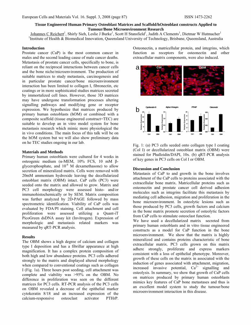
European Cells and Materials Vol. 16. Suppl. 3, 2008 (page 87) ISSN 1473-2262
Tissue Engineered Human Primary Osteoblast Matrices and Scaffold/hOsteoblast constructs Applied to Tumour/Bone Microenvironment Research
Johannes C Reichert1, Shirly Sieh, Leslie J Burke1, Scott H Stansfield1, Judith A Clements1, Dietmar W Hutmacher1 1Institute of Health & Biomedical Innovation, Queensland University of Technology, Brisbane, Queensland, Australia
Introduction Prostate cancer (CaP) is the most common cancer in males and the second leading cause of male cancer deaths. Metastasis of prostate cancer cells, specifically to bone, is reliant on the reciprocal interactions between cancer cells and the bone niche/microenvironment. The production of suitable matrices to study metastasis, carcinogenesis and in particular prostate cancer/bone microenvironment interaction has been limited to collagen I, fibronectin, etc coatings or in more sophisticated studies matrices secreted by immortalized cell lines. However, those 3D matrices may have undergone transformation processes altering signalling pathways and modifying gene or receptor expression. We hypothesize that matrices produced by primary human osteoblasts (hOM) or combined with a composite scaffold (tissue engineered construct=TEC) are suitable to develop an in vitro model system for bone metastasis research which mimic more physiological the in vivo conditions. The main focus of this talk will be on the hOM system but we will also show preliminary data on he TEC studies ongoing in our lab. Materials and Methods Primary human osteoblasts were cultured for 4 weeks in osteogenic medium (α-MEM, 10% FCS, 10 mM β-glycerophosphate, and 10-8 M dexamethasone) to allow secretion of mineralized matrix. Cells were removed with 20mM ammonium hydroxide leaving the decellularized osteoblast matrix (OBM) intact. CaP cells (PC3) were seeded onto the matrix and allowed to grow. Matrix and PC3 cell morphology were assessed histo- and/or immunohistochemically or by SEM. Matrix composition was further analyzed by 2D-PAGE followed by mass spectrometric identification. Viability of CaP cells was evaluated by FDA/PI staining. Cell attachment and cell proliferation were assessed utilizing a Quant-iT PicoGreen dsDNA assay kit (Invitrogen). Expression of morphologic and metastasis related markers was measured by qRT-PCR analysis. Results The OBM shows a high degree of calcium and collagen type I deposition and has a fibrillar appearance at high magnification. It has a complex protein composition of both high and low abundance proteins. PC3 cells adhered strongly to the matrix and displayed altered morphology when compared to conventional coatings such as collagen I (Fig. 1a). Three hours post seeding, cell attachment was complete and viability was >95% on the OBM. No difference in proliferation was seen on the different matrices for PC3 cells. RT-PCR analysis of the PC3 cells on OBM revealed a decrease of the epithelial marker cytokeratin 8/18 and an increased expression of the calcium-responsive osteoclast activator PTHrP.
Osteonectin, a matricellular protein, and integrins, which function as receptors for osteonectin and other extracellular matrix components, were also induced.
Fig. 1: (a) PC3 cells seeded onto collagen type I coating (Col I) or decellularized osteoblast matrix (OBM) were stained for Phalloidin/DAPI, 10x. (b) qRT-PCR analysis of key genes in PC3 cells on Col I or OBM. Discussion and Conclusion Metastasis of CaP to and growth in the bone involves attachment of the CaP cells to proteins associated with the extracellular bone matrix. Matricellular proteins such as osteonectin and prostate cancer cell derived adhesion molecules such as integrins facilitate this metastasis by mediating cell adhesion, migration and proliferation in the bone microenvironment. In osteolytic lesions such as those produced by PC3 cells, growth factors and calcium in the bone matrix promote secretion of osteolytic factors from CaP cells to stimulate osteoclast function. We have used a decellularized matrix secreted from primary human osteoblasts and in vitro tissue engineered constructs as a model for CaP function in the bone microenvironment. We show that the matrix is highly mineralized and contains proteins characteristic of bone extracellular matrix. PC3 cells grown on this matrix adhere strongly, proliferate and express markers consistent with a loss of epithelial phenotype. Moreover, growth of these cells on the matrix in associated with the induction of genes associated with attachment, migration, increased invasive potential, Ca2+ signalling and osteolysis. In summary, we show that growth of CaP cells on matrices produced by primary human osteoblasts mimics key features of CaP bone metastases and thus is an excellent model system to study the tumour/bone microenvironment interaction in this disease.
PTHrP
0
2
4
6
8
10
Col I OBM
Rel
ativ
e Ex
pres
sion
Cytokeratin 8
0
5
10
15
20
Col I OBM
Rel
ativ
e Ex
pres
sion
Alpha V Integrin
0.0
0.5
1.0
1.5
Col I OBM
Rel
ativ
e Ex
pres
sion
Osteonectin
0.00
0.01
0.02
0.03
Col I OBM
Rel
ativ
e Ex
pres
sion
Col OBM a
b

European Cells and Materials Vol. 16. Suppl. 3, 2008 (page 88) ISSN 1473-2262
Optimisation of electrospinning conditions for PDLLA and morphological characterisation of nanofibres
Y. Reinwald, L.J. White, K.M. Shakesheff, FRAJ Rose Division of Drug Delivery and Tissue Engineering, School of Pharmacy, Wolfson Centre for Stem Cells, Tissue Engineering, and Modelling (STEM), Centre for Biomolecular Sciences, University of Nottingham,
University Park, NG7 2RD, UK INTRODUCTION: Electrospinning is a straight-forward, cost effective and versatile technique to fabricate fibres of micro-or nanometre scale which mimic the extracellular matrix (ECM) in architecture [1]. ECM provides anchorage for cells and supplies support and structure to tissues. Therefore, electrospinning provides a means of producing tissue engineering scaffolds that mimic elements of the native tissue. The aim of this study was the optimisation of electrospinning conditions for PDLLA and the fabrication of nanometre sized fibres. Therefore the effect of process and solution parameters on fibre morphology was investigated. The applicability of electrospun fibres for cell culture experiments was examined by seeding fibroblast cells onto PDLLA nano-sized fibres. METHODS: PDLLA (52 kDa) was dissolved in dichloromethane and electrospun changing polymer concentration, voltage, flow rate, collecting distance and age of polymer solution. The conductivity of the polymer solution was altered by adding pyridinium formiate in order to obtain nanofibres [2]. After collecting, electrospun fibre sheets were morphologically examined using scanning electron microscopy (SEM). Plasma polymerised allyl amine (ppAAm) was deposited onto fibrous scaffolds and mouse 3T3 fibroblasts were seeded onto both surface modified and non-modified meshes. Cell viability was assessed using the Alamar Blue assay and Live/Dead staining. RESULTS: The alteration of process and solution parameters resulted in the electrospinning of morphologically uniform PDLLA fibres as shown in Figure 1 A and B. Fibre diameter (Figure 1 C) decreased with addition of pyridinium formiate and led to the fabrication of nanofibres. Fibres were spun from salt-containing and no-salt-containing polymer solutions at a voltage of 20 kV, 3ml/h flow rate and 14 cm collecting distance. Figure 1 D shows mouse 3T3 fibroblasts adhered onto salt-containing surface modified scaffolds 6 days after seeding. Cell shapes were rounded which suggests that they did not adhere strongly. During the next few days in culture, cells secreted ECM onto the scaffolds and by day 6, cells adhered more fully and started to spread.
The Alamar Blue assay indicated an increase in viability at day 6 (possibly due to an increase in cell number) in all samples seeded with cells. A
B
C
0
10
20
30
40
50
60
70
<0.5 <1 <1.5 <2.0 <2.5 <3.0 <3.5 <4.0 <4.5 <5.0
diameter [um]
fibre
s [%
]no salt salt
D
Figure 1: SEM micrographs of electrospun PDLLA fibres (polymer solution 20% w/v) A: no salt-containing and B: salt-containing PDLLA solution was spun at a flow rate of 3m/h, collecting distance of 14 cm and 20kV voltage. . C: Diameter distributions of both fibre types D: Cells adhered onto salt-containing ppAAm coated fibres after 6 days. DISCUSSION & CONCLUSIONS: By changing the process conditions uniform fibres in the 100 um range were reproducibly fabricated. Changing the composition of the polymer solution by adding pyridinium formiate resulted in the electrospinning of nanofibres (500-2000 nm). Cells adhered to these scaffolds following surface chemistry modification with ppAAm. Future experiments will investigate the ability to generate fibre sheets with fibre morphologies that mimic the basement membrane. REFERENCES: 1 R. Murugan, S. Ramakrishna (2006), 12 (3) pp. 435-447 2 Z. Jun, H. Hou, A. Schaper, J. Wendorff, A. Greiner (2003), e-Polymers, no.009 ACKNOWLEDGEMENTS: Authors would like to thank the German Academic Exchange Office for financial support.

European Cells and Materials Vol. 16. Suppl. 3, 2008 (page 89) ISSN 1473-2262
ADIPOSE TISSUE FORMATION IN VITRO AND IN VIVO USING NATURAL SEA SPONGE AND HUMAN BONE MARROW STROMAL CELLS P Roberts1, EA Horner1, DW Green2, J Kirkham1, ROC Oreffo2 and XB Yang1*
1Skeletal Tissue Engineering Laboratory, Department of Oral Biology, Leeds Dental Institute, University of
Leeds, LS2 9LU, UK. 2Bone and Joint Research Group, Centre for Human Development, Stem Cells and Regeneration, Institute of Developmental Sciences, University of Southampton, SO16 6YD, UK
INTRODUCTION: Treatment of adipose tissue defects caused by congenital factors, burns, surgical mastectomy or lumpectomy often involves the use of different filler materials inside a silicone elastomer shell. However, there are significant health risks involving such a therapies following shell disintegration. Therefore, novel therapies, including tissue engineering strategies are ongoing. The aim of this study was to investigate the efficacy of a combination of human marrow stromal cells (HBMSCs) and naturally occurring sea sponge as a scaffold for adipose tissue formation. METHODS: HBMSCs enriched for MSCs (Lonza) were seeded (5x10⁴ cells) onto 2mm3 scaffolds (Spongia officinalis) and cultured in basal or adipogenic media in vitro for up to three weeks. Cell growth was examined by scanning electron microscopy (SEM), Live/Dead®
fluorescent markers and confocal microscopy. Adipogenesis was analysed by Oil Red-O staining and RT-PCR (PPAR-γ). Osteogenic (RUNX2) and myogenic (MyoD) markers were also investigated. For in vivo study, the scaffolds seeded with/without cells were implanted subcutaneously in six MF1 Nu/Nu mice. After three weeks, the samples were removed for assessment. RESULTS: The results indicate that natural sea sponge scaffolds can support extensive cell adhesion and proliferation as confirmed by live/dead analysis and SEM. Enhanced adipogenic differentiation (lipid droplets formation. Fig. 1A) and Oil Red-O staining was observed following adipogenic culture (Fig. 1B). In addition, RT-PCR showed a time-dependent increase in PPARγ (adipogenic marker) expression in HBMScs on sea sponge scaffold under adipogenic conditions. In contrast, negligible expression of RUNX-2 and MyoD were observed. Following three weeks of subcutaneous implantation in Nu/Nu mice,, the scaffolds were observed to maintain their original shape (Fig. 2A). Macro-dissection displayed fat pad formation with typical vascular pedicle supply to the new adipose tissue, necessary for the exchange of metabolites & adipocyte proliferation and function (Fig. 2B).
Fig. 1: Adipocytes growth on HBMSCs seeded sea sponge scaffolds in vitro: A) Brown fat cells containing numerous lipid droplets and mitochondria; B) characteristic Oil Red-O stain for adipocyte lipids on sea sponge scaffolds after 3 weeks.
Fig. 2: In vivo subcutaneous implantationof sea sponge scaffolds ± cells: A)Maintenance of the scaffold shape beneath the skin; B) Fat pad formation on the scaffold alone (left); scaffolds with cells cultured in basal (middle) or adipogenic media (right).
DISCUSSION & CONCLUSIONS: This study has demonstrated the potential of using natural sea sponge as a scaffold support for HBMSCs grown under adipogenic conditions to form adipose-like tissue in vitro and in vivo. The sea sponge scaffolds implanted in vivo not only maintained their shape but also permitted vascular invasion to the regenerated adipose tissue. This is essential for metabolite exchange and provision of optimum conditions for adipocyte function. Experiments are on-going to examine the immunogeneicity associated with such a strategy.
ACKNOWLEDGEMENTS: Mrs Jackie Hudson for technical support. PR was a MSc student.
A B
A
B

European Cells and Materials Vol. 16. Suppl. 3, 2008 (page 90) ISSN 1473-2262
Barriers To The Commercialization Of Regenerative Medicine Products E Rowley1 & A Zimmermann2
1 Institute for Science & Society, University of Nottingham, UK. 2 Faculty of Social & Political Sciences, University of Cambridge, UK.
INTRODUCTION: Regenerative medicine (RM) has been hailed as a future revolution in medical care. However, as yet, relatively few products are commercially available and utilized in patient care. In this paper, we examine the reasons why so many promising products have failed or appear to have taken a long time to reach the market. In doing so, we ask whether the barriers to commercialization are any different in RM than in other health care industries, or whether the RM industry is merely suffering from the same problems that are experienced by other innovative technologies. In light of our examination of the barriers to the commercialization of RM, policies targeting the promotion and development of this emerging industry are suggested. METHODS: Representatives of RM companies were interviewed about the current state of RM, product development, market barriers, collaborations, future directions and policy interventions. RESULTS: The overwhelming consensus among respondents was that while UK RM science is first rank, it lags behind the USA and other countries when translating and commercializing the outcomes of that science. In some areas, such as hESCs, the UK appears to have a scientific lead of several months over the USA. However, scientific leads like this may quickly disappear if the translation and commercialization of therapies is hindered. Barriers to the commercialization of RM products include: 1) Lack of funding. Interviewees reported the struggle to raise sufficient funds for translation, with private equity investors unwilling to fund high risk, early stage RM ventures. 2) Organizational culture. The structure of the National Health Service and the demands of the National Institute for Health and Clinical Excellence were seen as a huge burden for industry to overcome, mostly because of the considerable amount of clinical evidence required. RM companies argue that they are unable to gain such data given their limited resources. Yet without such evidence, it is unlikely that RM products will be approved for use in UK public healthcare. 3) Regulatory muddle. Interviewees argued that their ability to commercialize products had been
greatly hindered by a lack of regulatory clarity. Respondents repeatedly mentioned uncertainty about regulatory requirements and definitions, and the expected size and protocols of clinical trials, all of which created confusion regarding what information was needed to gain product approval and subsequent entry into the market. It was however anticipated that the regulatory space would be improved with the implementation of EU ATMP legislation. DISCUSSION & CONCLUSIONS: RM is thriving in the UK with respect to the underlying science. Unfortunately, lack of access to capital, regulatory hurdles, lack of clinical evidence, as well as the NHS culture result in a number of barriers to the commercialization of RM products. Policy interventions, such as greater translational Government funding, a restructuring of the NHS and NICE, and regulatory clarity are likely to improve the general outcomes for the industry in the UK.
ACKNOWLEDGEMENTS: This paper would not have been possible without the support of remedi, EPSRC and our study participants. www.remedigc.org www.nottingham.ac.uk/iss/regenmed

European Cells and Materials Vol. 16. Suppl. 3, 2008 (page 91) ISSN 1473-2262
TGFβ1 AFFECTS THE BIOLOGICAL RESPONSES OF PRIMARY CHONDROCYTES GROWN ON POLY(ETHYLENE
TEREPHTHALATE)/POLY (ε−CAPROLACTONE) CO-POLYMER FLAT SCAFFOLDS
SK Singhrao, SJ Gilbert, I Khan, VC Duance, & CW Archer
Connective Tissue Biology Laboratories, School of Biosciences, Cardiff University, Museum Avenue, Cardiff, CF10 3US, Wales, UK.
INTRODUCTION: The effect of transforming growth factor beta 1 (TGFβ1) on chondrocytes in in vitro cultures is well documented1. However, methods for in vitro culture of chondrocytes are not standardized. Chondrocytes can be cultured in monolayers, or as pellet cultures and in three dimensions within biomaterials. The effect of TGFβ1 is dependent on the physical environment
of the chondrocyte culture2. This study was conducted to examine the suitability of flat polyethylene terepthalate/polycaprolactone (PET/PCL) scaffolds with a view to therapeutic strategies for cartilage tissue engineering in the presence of TGFβ1. The main objectives being whether articular cartilage chondrocytes grown on PET/PCL scaffolds proliferated, maintained phenotype and were able to deposit an increased extracellular matrix representative of hyaline-like cartilage.
METHODS: Full depth bovine articular cartilage chondrocytes were isolated and seeded at a density of 2.5x10⁶ cells/scaffold. Scaffold cultures were maintained for 14 days in media supplemented with ITS+/-TGFβ1 (10ng/ml). Scaffolds were evaluated for cellularity (SEM), proliferation (BrdU), glycosaminoglycan (GAG) content by histology and DMMB, collagen (histology, SEM, hydroxyproline content and immunoblotting) and matrix gene expression (q-PCR).
RESULTS: It was found that chondrocytes grown on scaffolds cultured in TGFβ1 containing medium allowed greater bioactivity in terms of cell survival and proliferation. There was an increase in the amount of total GAG and collagen deposited on the scaffolds cultured in medium supplemented with TGFβ1. Cartilage genes for collagen II, Sox 9 and aggrecan were upregulated. Immunoblotting revealed that type II collagen was the most abundant collagen type. The expression levels of type I collagen gene were too low for quantitation by q-PCR. Evidence of type VI collagen deposition in the chondrocyte pericellular matrix was confirmed by immunofluorescence.
DISCUSSION & CONCLUSIONS: The primary articular chondrocytes were able to grow, proliferate and carry out their biochemical functions on PET/PCL scaffolds in the TGFβ1 supplemented medium. Furthermore enhanced matrix synthesis and deposition that was more hyaline in composition than that produced by the ITS supplemented cultures was observed. The overall enhanced matrix deposition in the TGFβ1 medium was due to both an increase in cell population and the up-regulation of matrix synthesis genes. These results indicate ITS+TGFβ1 together synthesise and assemble a hyaline-like matrix suitable for use in cartilage repair strategies.
REFERENCES: 1 F. Redini, P.Galera, A Mauviel, et al (1988) FEBS Lett 234(1):172-175. 2Y.M. Bastiaansen-jenniskens, W.Koevoet, A.C.W. De Bart et al (2008) Tissue Engineering 14(6):1-8
ACKNOWLEDGEMENTS: Smith & Nephew for the scaffold materials. This project was co-funded by the Technology Strategy Board’s Collaborative Research and Development programme, following an open competition.

European Cells and Materials Vol. 16. Suppl. 3, 2008 (page 92) ISSN 1473-2262
Interactions of CO2 with Poly (DL-lactic acid) and Poly (lactic acid-co-glycolic acid) Copolymers
H Tai1,2, KM Shakesheff2 and SM Howdle1
1 School of Chemistry & 2School of Pharmacy, University of Nottingham, University Park, Nottingham, UK, NG7 2RD, Email:[email protected]
INTRODUCTION: Amorphous poly (DL-lactic acid) (PDLLA) and poly (lactic acid-co-glycolic acid) (PLGA) polymers have been used to fabricate porous scaffolds for tissue engineering applications via a supercritical foaming technique.1,2,3 The chemical composition of the polymers, as well as the morphology (pore size, porosity and interconnectivity) of the scaffolds are crucial in terms of their biomedical and biological applications, such as cell filtration, migration, nutrient exchange, degradation and drug release rate. To control the morphology of supercritical foamed scaffolds, it is essential to study the interactions of polymers with CO2 and the consequent solubility of CO2 in the polymers, as well as the viscosity of the plasticized polymers.
MATERIALS AND METHODS: A high pressure attenuated total reflection Fourier transform infrared (ATR-IR) was used to investigate the interactions of CO2 and PLGA polymers with the glycolic acid (GA) content in the copolymers as 0, 15, 25, 35 and 50 % respectively. A novel high pressure parallel plate rheometer was also developed for the shear viscosity measurements of the CO2-plastisized polymers at a temperature below their glass transition temperatures.
RESULTS AND DISCUSSION: Shifts and intensity changes of IR absorption bands of the polymers in the carbonyl region (~1750 cm-1) are indicative of the interaction on a qualitative level. The viscosity of CO2-plasticized polymers has been measured directly using high pressure parallel plate rheometer at 35 oC and 100 bar and the results were compared with the viscosity of the polymer melts at 140 oC (Fig. 1).
CONCLUSIONS: The data obtained from both high pressure ATR-IR and high pressure rheology indicated that the interactions of CO2 with PDLLA and PLGA polymers decreased with the increase of GA contents in the copolymers. This investigation provided further fundamental understandings on the control of foaming process for the fabrication of PLA and PLGA porous scaffolds and is in a good agreement with our previous findings on manipulating scaffold fabrications by altering the fabrication conditions and polymer compositions.3
REFERENCES: 1 D.J. Mooney, D.F. Baldwin, N.P. Suh, et al. (1996) Biomaterials 14:1417-1422. 2 S.M. Howdle, M.S. Watson, M.J. Whitaker, et al. (2001) Chem. Commun. 1:109-110. 3 H. Tai, M.L. Mather, D. Howard et al. (2007) European Cells and Materials Journal 14: 64-77.
ACKNOWLEDGEMENTS: We gratefully acknowledge EPSRC for funding.
Fig. 1: Comparison of the shear viscosities for the polymers at 140 ºC and atmosphere (in white) with those for the polymers at 35 ºC and 100 bar CO2 pressure (in grey).

European Cells and Materials Vol. 16. Suppl. 3, 2008 (page 93) ISSN 1473-2262
CHARACTERISATION OF A NOVEL HYDROGEL FORMED IN PHYSIOLOGICAL CONDITIONS BY AN FMOC-LINKED AMINO ACID
K. Thornton1, Andrew Smith, 1,2 Rein V. Ulijn *1,2 & Catherine L. R. Merry *1
1 Material Science Centre, University of Manchester, Grosvenor Street, Manchester, M1 7HS 2 Manchester Interdisciplinary Biocentre, University of Manchester, 131 Princess Street, Manchester, M1
7DN * Corresponding authors
INTRODUCTION: Self-assembling peptide hydrogels are believed to have great potential as the next generation of biomaterials1. An application of particular interest is three dimensional culture of cells, as the self-assembled peptide structures can mimic the structure of the extracellular matrix 2. This places limitations on the peptides as they must be able to self-assemble under physiological conditions3.
Within literature the molecular weights of the peptides varied with a novel system developed recently by Yang and Xu4. Within this system an enzyme assisted hydrogel formed from a single amino acid (tyrosine-phosphate) that was linked at its N terminal by fluorenylmethoxycarbonyl (Fmoc)4. Its potential applications within tissue engineering were limited as the reaction conditions fell outside physiological ranges. However in this current study, through careful optimisation of the processing parameters, it has been possible to form a hydrogel within physiological conditions (0.15M, 37 °C and pH 7). This paper discusses the production, structure and mechanical properties if the hydrogel. METHODS: The amino acid, Fmoc-tyrosine-phosphate, was dissolved in an alkaline phosphate buffer. The pH was adjusted to neutral by the addition of concentrated hydrochloric acid or sodium hydroxide. A hydrogel formed on addition of alkaline phosphatase. The hydrogel produced was characterised by HPLC, TEM, Cryo-ESEM, rheology, FTIR, CD and fluorescence spectroscopy. RESULTS: A basic characterisation of the mechanical and structural properties of the hydrogel has been completed. Fluoresence spectroscopy and visual inspection have confirmed that a hydrogel did form within physiological conditions. A range of enzyme and amino acid concentrations were investigated. A hydrogel would only form on addition of the alkaline phosphatise due to the cleavage of the phosphate
group, confirmed by HPLC. The fibres within the hydrogel formed an interconnecting network with an average fibre size of less than 1 µm (fig 1).
Fig. 1: Cryo-ESEM of Fmoc-Tyrosine (40mM) hydrogel formed on addition of alkaline phosphatase
DISCUSSION & CONCLUSIONS: The formation of a hydrogel from a single amino acid in physiological conditions has not yet been investigated. As the fibres formed are smaller than 1 µm the hydrogels may structurally mimic the ECM. The suitability of these hydrogels for three dimensional cell culture will be investigated using embryonic stem cells to monitor the effect on cell fate decision.
REFERENCES: 1 Mart, R.J. et al Peptide-Based Stimuli-Responsive Biomaterials. Soft Matter, 2006. 2(10).2 Zhang, S. et al, Designer Self-Assembling Peptide Nanofiber Scaffolds for 3d Tissue Cell Cultures. Seminars in Cancer Biology, 2005. 15(5).3 Jayawarna, V. et al Nanostructured Hydrogels for Three-Dimensional Cell Culture through Self-Assembly of Fluorenylmethoxycarbonyl-Dipeptides. Ad. Materials, 2006. 18(5).4 Yang, Z.M. and Xu, B., A Simple Visual Assay Based on Small Molecule Hydrogels for Detecting Inhibitors of Enzymes. Chem. Comm. 2004. (21).
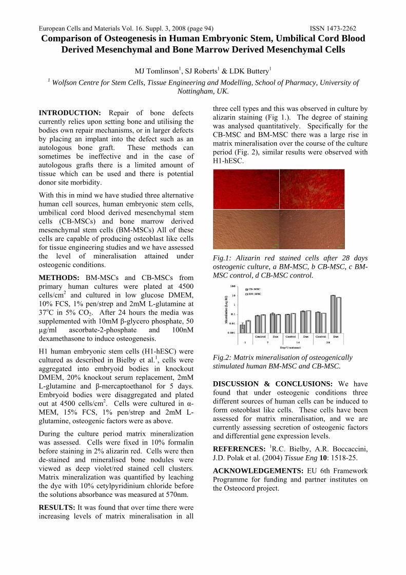
European Cells and Materials Vol. 16. Suppl. 3, 2008 (page 94) ISSN 1473-2262 Comparison of Osteogenesis in Human Embryonic Stem, Umbilical Cord Blood
Derived Mesenchymal and Bone Marrow Derived Mesenchymal Cells
MJ Tomlinson1, SJ Roberts1 & LDK Buttery1
1 Wolfson Centre for Stem Cells, Tissue Engineering and Modelling, School of Pharmacy, University of Nottingham, UK.
INTRODUCTION: Repair of bone defects currently relies upon setting bone and utilising the bodies own repair mechanisms, or in larger defects by placing an implant into the defect such as an autologous bone graft. These methods can sometimes be ineffective and in the case of autologous grafts there is a limited amount of tissue which can be used and there is potential donor site morbidity.
With this in mind we have studied three alternative human cell sources, human embryonic stem cells, umbilical cord blood derived mesenchymal stem cells (CB-MSCs) and bone marrow derived mesenchymal stem cells (BM-MSCs) All of these cells are capable of producing osteoblast like cells for tissue engineering studies and we have assessed the level of mineralisation attained under osteogenic conditions.
METHODS: BM-MSCs and CB-MSCs from primary human cultures were plated at 4500 cells/cm2 and cultured in low glucose DMEM, 10% FCS, 1% pen/strep and 2mM L-glutamine at 37oC in 5% CO2. After 24 hours the media was supplemented with 10mM β-glycero phosphate, 50 µg/ml ascorbate-2-phosphate and 100nM dexamethasone to induce osteogenesis.
H1 human embryonic stem cells (H1-hESC) were cultured as described in Bielby et al.1, cells were aggregated into embryoid bodies in knockout DMEM, 20% knockout serum replacement, 2mM L-glutamine and β-mercaptoethanol for 5 days. Embryoid bodies were disaggregated and plated out at 4500 cells/cm2. Cells were cultured in α-MEM, 15% FCS, 1% pen/strep and 2mM L-glutamine, osteogenic factors were as above.
During the culture period matrix mineralization was assessed. Cells were fixed in 10% formalin before staining in 2% alizarin red. Cells were then de-stained and mineralised bone nodules were viewed as deep violet/red stained cell clusters. Matrix mineralization was quantified by leaching the dye with 10% cetylpyridinium chloride before the solutions absorbance was measured at 570nm.
RESULTS: It was found that over time there were increasing levels of matrix mineralisation in all
three cell types and this was observed in culture by alizarin staining (Fig 1.). The degree of staining was analysed quantitatively. Specifically for the CB-MSC and BM-MSC there was a large rise in matrix mineralisation over the course of the culture period (Fig. 2), similar results were observed with H1-hESC.
Fig.1: Alizarin red stained cells after 28 days osteogenic culture, a BM-MSC, b CB-MSC, c BM-MSC control, d CB-MSC control.
Fig.2: Matrix mineralisation of osteogenically stimulated human BM-MSC and CB-MSC.
DISCUSSION & CONCLUSIONS: We have found that under osteogenic conditions three different sources of human cells can be induced to form osteoblast like cells. These cells have been assessed for matrix mineralisation, and we are currently assessing secretion of osteogenic factors and differential gene expression levels.
REFERENCES: 1R.C. Bielby, A.R. Boccaccini, J.D. Polak et al. (2004) Tissue Eng 10: 1518-25.
ACKNOWLEDGEMENTS: EU 6th Framework Programme for funding and partner institutes on the Osteocord project.
European Cells and Materials Vol. 16. Suppl. 3, 2008 (page 95) ISSN 1473-2262
MECHANICAL AND MORPHOLOGICAL STUDIES OF SUPERCRITICAL FLUID FOAMED POLY(D,L-LACTIC ACID) SCAFFOLDS
LJ White1, SM Howdle2 and KM Shakesheff1
1STEM, Centre for Biomolecular Sciences, School of Pharmacy, University of Nottingham, University Park, NG7 2RD. 2School of Chemistry, University of Nottingham, University Park, NG7 2RD. INTRODUCTION: Supercritical CO2 scaffold formation is a solvent free, low temperature process which produces open cell, inter-connected foamed structures. Drug molecules and proteins can be encapsulated within these scaffolds as protein structure and activity are retained during processing. Successful applications of this technique include the controlled release of proteins [1], promotion of bone formation in vitro and in vivo [2] and the induction of angiogenesis in vitro [3]. Supercritical CO2 scaffold fabrication can produce scaffolds of divergent pore size and structure. Hence, this study sought to elucidate the effects of processing conditions on the porosity, pore size distribution and mechanical properties of the scaffolds. METHODS: Three molecular weights of poly(D,L-lactic acid) (PDLLA) (15kDa, 24kDa and 57kDa) were used to form scaffolds under different depressurization rates. The morphology of the resultant scaffolds was characterized by micro X-ray computed tomography (μCT) and mechanical properties were tested using a TA.HD Texture Analyser (Stable Micro Systems Ltd., Surrey, UK) RESULTS: Scaffolds created from different molecular weights showed varying morphologies (Figure 1). Upon mechanical testing 57kDa PDLLA scaffolds displayed a typical stress-strain curve for elastomeric open cell foams, comprising a linear elastic region, a collapse plateau and densification, as shown in Figure 2. The elastic collapse stress, Young’s modulus and ultimate stress were considerably increased by using shorter vent times in scaffold fabrication; the ultimate stress ranged from 5.1 MPa using a 10 minute vent to 2.8 MPa for a 45 minute vent. The failure behaviour of 15kDa and 24kDa PDLLA scaffolds was however more typical of porous brittle materials [4]. The porosity of all scaffolds significantly increased with decreased depressurization rates (increased vent times).
Fig. 1: Varying morphology of scCO2 foamed scaffolds created with a 60 minute vent.
Fig. 2: Stress strain curve for 57k MW foamed scCO2 scaffold produced with a 60 minute vent.
DISCUSSION & CONCLUSIONS: This study conclusively shows that the architecture and mechanical properties of PDLLA scaffolds can be tailored by adjusting the processing conditions and the molecular weight of the polymer. REFERENCES: 1S. M. Howdle, M.S. Watson, M.J. Whitaker, et al., (2001) Supercritical fluid mixing: preparation of thermally sensitive polymer composites containing bioactive materials. Chemical Communications. (01): p. 109-110. 2 X.B.B. Yang, M.J. Whitaker, W. Sebald, et al., (2004) Human osteoprogenitor bone formation using encapsulated bone morphogenetic protein 2 in porous polymer scaffolds. Tissue Engineering. 10(7-8): p. 1037-1045. 3 J. M. Kanczler, J. Barry, P. Ginty, et al., (2007) Supercritical carbon dioxide generated vascular endothelial growth factor encapsulated poly(dl-lactic acid) scaffolds induce angiogenesis in vitro. 352(1): p. 135. 4L. J. Gibson, (2005) Biomechanics of cellular solids. Journal of Biomechanics. 38(3): p. 377-399
Strain (%)
Stress (MPa)
52 kDa
15 kDa

European Cells and Materials Vol. 16. Suppl. 3, 2008 (page 96) ISSN 1473-2262
Identification of a cell population from human articular cartilage displaying progenitor characteristics
R Williams1, L Nelson1, I M Khan1, J Dudhia2, K Richardson1 & C W Archer1
1 Connective Tissue Research Labs, Cardiff University, UK. 2 The Royal Vet School, London, UK.
INTRODUCTION: Repair of cartilage defects requires the use of chondrocytes for tissue engineering procedures. However, the cells used often do not retain their chondrogenic phenotype in vivo leading to poor integration and subsequently repair of the defect is not maintained. Work presented here describes the detection and isolation of a chondroprogenitor population that can be expanded in culture whilst maintaining the cartilage phenotype, making it an ideal candidate for use in tissue engineering procedures.
METHODS: Human cartilage (age 10-57 years) was obtained with the relevant ethical approval. Isolated cells were plated in monolayer cultures (non-adhered population). Clonal cell populations were isolated using the fibronectin adhesion assay1. All cells were expanded in chondrogenic growth medium [DMEM+10% FCS, TGF-β2 (1ng/ml) & FGF-2 (5ng/ml)]. Expanded cells were subjected to FACS analysis. Cells were then pelleted, cultured in either chondrogenic differentiation medium [DMEM+2% FCS, 1% ITS, & TGF-β2 (5ng/ml) & 10-7 dexamethasone] or osteogenic differentiation medium [DMEM+10% FCS, 10mM β-glycerophosphate & 10nM dexamethasone]2 for 21 days. Histological and immunohistochemical procedures were undertaken to determine presence of chondrogenic and osteogenic components. mRNA analysis of chondrogenic and osteogenic markers was also performed. RESULTS: FACS analysis for the stem cell markers CD105 and CD166 showed labelling in over 75% of the cells within the non-adhered population whereas CD49e (α5 integrin) labelled a sub-population of only 1.4% of the cells.
Fig. 1: CD166 (A), CD105 (B) and CD49e expression in non-adhered populations.
Stem cell markers Notch 1and CD90 could be
detected in clonal monolayer populations before cells were expanded and pelleted (Fig.2).
Fig. 2: Notch1 (A) and CD90 (B) expression in clonal monolayers.
Toludine blue stained pellets showed GAG content when subjected to 21 days in chondrogenic media whereas pellets grown in osteogenic differentiation media showed the presence of calcium deposits as demonstrated with alizarin red staining (Fig.3).
Fig. 3: Toludine blue stained pellet cultured in chondrogenic media (A) and alizarin red stained pellet cultured in osteogenic differentiation media (B).
DISCUSSION & CONCLUSIONS: These data suggest, due to the presence of several stem cell markers, that a chondroprogenitor resides in human cartilage. Isolating the chondroprogenitor cells using the fibronectin adhesion assay allows expansion of these cells in monolayer whilst they still retain their expression of stem cell markers. It is interesting to note that a sub-population of cells within the non-adhered population show CD49e expression which could highlight a strong affinity of these cells to fibronectin. In the 3D pellet cultures, the cells show a chondrogenic phenotype yet also show plasticity, as demonstrated by osteogenic differentiation. Further characterisation of the cells will allow us to determine their suitability for tissue engineering procedures.
REFERENCES: 1G.P Dowthwaite, J.C Bishop et al. (2004) J Cell Science 29:889-97. 2 A Barbero, S Ploegert, et al. (2003) Arthritis & Rheumatism 48 1315-1325.
A B
A B C

European Cells and Materials Vol. 16. Suppl. 3, 2008 (page 97) ISSN 1473-2262
In vivo assessment of the immunogenicity of self-assembling peptides for use in regenerative applications
Wilshaw, S P.1, Aggeli, A.2, Fisher, J.1, Ingham, E.1
1Institute of Medical and Biological Engineering, University of Leeds, Leeds, West Yorkshire, UK.
2Centre for Self-Assembling Systems, University of Leeds, Leeds, West Yorkshire, UK.
Introduction Previous work has established criteria for the formation of hydro-gels from chemically synthesised self-assembling β-sheet peptides under physiological conditions. This has enabled the rational design of peptides that spontaneously self-assemble into scaffolds of potential utility in tissue engineering and regenerative applications. The aim of this study was to determine the biocompatibility and immunogenicity of two such peptides (P11-4 and P11-8). Methods Biocompatibility was determined using primary human fibroblasts and L929 cells in vitro. Immunogenicity of P11-4 or P11-8 was determined in a) naive BALBC mice b) mice immunised with peptide c) mice immunised with peptide conjugated to keyhole limpet hemocyanin (KLH). Antibodies to the peptides were determined by ELISA. Peptide specific lymphocyte responsiveness was determined in lymphocyte
proliferation assays. Serum antibodies to the peptides and peripheral blood lymphocyte proliferation in response to the peptides was also determined in six human volunteers. Results and Conclusions Following culture with human and murine cells P11-4 or P11-8 demonstrated no cytotoxicity. Naive mice possessed no antibodies against P11-4 or P11-8 and no lymphocyte response was detected. Mice immunised with peptide alone did not demonstrate peptide specific lymphocyte responsiveness and antibodies to P11-4 or P11-8 were not detected in serum. Mice immunised with KLH conjugated peptide did not develop peptide specific lymphocyte responses, however they produced peptide specific antibodies (titres of 1/64). Human volunteers demonstrated no prior sensitisation to the peptides. These studies suggested that the peptides are not immunogenic in mice but may act as haptens when conjugated to a carrier protein.
European Cells and Materials Vol. 16. Suppl. 3, 2008 (page 98) ISSN 1473-2262
Optical Manipulation of Stem Cells Hu Zhang, Kuo-Kang Liu & Alicia El Haj
Institute for Science and Technology in Medicine, Keele University, Stoke-on-Trent, ST4 7QB, UK
INTRODUCTION: Mechanical microenvironment has been shown to have a great impact on proliferation and differentiation of stem cells. To quantitatively determine the effect of fluid mechanical forces on stem cells, it is essential to apply and maintain a well-defined exterior force on a single stem cell. Optical tweezers have been widely used for manipulating single biological cells and performing sophisticated biophysical/biomechanical characterizations. Advantages of using tweezers include non-contact force for cell manipulation, force resolution as accurate as 100 aN, and amiability to liquid medium environments. This study aims to develop a novel optical-mechanical system which can be employed to generate a range of shear forces on mesenchymal stem cells (MSCs) and quantitatively characterize the resulting effects on MSCs including deformation, viability and gene expression profiles of MSCs. This understanding helps designing new means of delivering stem cells for regenerating tissues or optimizing current delivery systems.
METHODS:. Stem cells of passage 5 were used. The cells were attached to RGD (Arg-Gly-Asp, Sigma-Aldrich, UK) coated microparticles (averaged 3.19 um, Spheotech Inc., USA). The commercial available optical tweezers (Cell Robotic, Inc, USA) which is driven by a Windows XP based software is used in this present experimental work. The experimental setup was shown in Figure 1. The source of laser is Nd:YAG at a wavelength of 1064nm pumped by a 1.5W diode. Laser is then reflected through dichroic mirrors and focused by an inverted microscope (Nikon optical microsystem) before it reaches the objects. The cells were pumped into a microfluidic device or in-house chamber which is placed on the invert microscopy platform. The flow rate can be varied to exert different shear stress on the stem cells.
RESULTS: A bead-attached stem cell can be manipulated under this optical-mechanical system in three different ways when it is exposed to fluid flow field while trapped by laser beam: it may flow with fluid as the cell has not attached to the bottom; it may rotate under certain fluid forces (partially attached to the surface bottom); and the bead may be pulled away but the cell is not moving
or rotating (fully attached to the surface bottom). The fluid velocity of 40μm/s is found to be the escape velocity of the bead-attached cells, and equivalent shear force is calculated to be about 7.5 pN.
Fig. 1:Optical tweezers drag the microparticle-attached stem cell in a microfluidic channel at different positions from (a) to (d).
DISCUSSION & CONCLUSIONS: Several new enabling techniques have been developed for optical-mechanically manipulating and characterizing stem cells. Bead-attached stem cells have been successfully manipulated at various speeds in physiological medium. Quantitative analyses of the nanomechanical forces on the cells during cell manipulation/delivery have been performed. REFERENCES: 1Kuipinski et al. (2006) PNAS, 103:16095-16100. 2Engelmayr Jr et al. (2006) Biomaterials 27: 6083-6095. 3H. Zhang & Liu, K.K (2008) J Roy Soc Interface, in press
ACKNOWLEDGEMENTS: We are gratefully acknowledged BBSRC and EPSRC (BB/D014786/1) for financial support and Dr Fisher at Cambridge University for providing microfluidic chambers.
a b
c d