register now for cms electronic - west virginia state medical
TRANSCRIPT


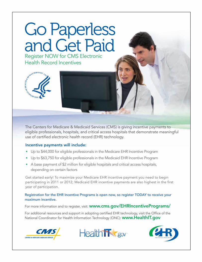
The Centers for Medicare & Medicaid Services (CMS) is giving incentive payments to eligible professionals, hospitals, and critical access hospitals that demonstrate meaningful use of certified electronic health record (EHR) technology.
Incentive payments will include:
• Upto$44,000foreligibleprofessionalsintheMedicareEHRIncentiveProgram
• Upto$63,750foreligibleprofessionalsintheMedicaidEHRIncentiveProgram
• Abasepaymentof$2millionforeligiblehospitalsandcriticalaccesshospitals,dependingoncertainfactors
Getstartedearly!TomaximizeyourMedicareEHRincentivepaymentyouneedtobeginparticipatingin2011or2012;MedicaidEHRincentivepaymentsarealsohighestinthefirstyearofparticipation.
Registration for the EHR Incentive Programs is open now, so register TODAY to receive your maximum incentive.
For more information and to register, visit: www.cms.gov/EHRIncentivePrograms/For additional resources and support in adopting certified EHR technology, visit the Office of the National Coordinator for Health Information Technology (ONC): www.HealthIT.gov
Register NOW for CMS Electronic Health Record Incentives
Go Paperless and Get Paid
EHR_Ad_2ppl_WestVA_7.5x10.indd 1 10/25/11 1:58 PM

contentsAnnual Business Meeting/
Physician Practice ConferenceFridAy, FeBruAry 3 - SAturdAy, FeBruAry 4
at the Charleston Marriott
Friday, February 3Health Care in the 2012 Session •Special Legislative Leadership Briefing
Physician Practice Conference —Compliance is NOT Optional - What Your •Practice Must Know
2012 Payor Update - Latest Policies •and Procedures of All the Major Health Insurance Carriers
2012 Medicare Update•
Saturday, February 4Dealing with Difficult and Disruptive •Behavior in Health Care
Special Risk Management program hosted by the WV Mutual Insurance Company
Eligible for WV Mutual 2% Risk Management Credit!
WVSMA 2012 Annual Business Meeting• » House of Delegates
» 2012-2013 WVSMA Officer Elections
» Policy Resolutions
For more information visit wvsma.com or call 304.925.0342, ext. 12See reverse side for 2012 Coding Update Course Schedule and Registration Form.

contents
EditorF. Thomas Sporck, MD, FACSCharleston
Managing Editor/ Director of CommunicationsAngela L. Lanham, Dunbar
Executive DirectorEvan H. Jenkins, Huntington
January/February 2012, Volume 108, No. 1
The West Virginia Medical Journal is published bimonthly by the West Virginia State Medical Association, 4307 MacCorkle Ave., SE, Charleston, WV 25304, under the direction of the Publication Committee. The views expressed in the Journal are those of the individual authors and do not necessarily reflect the policies or opinions of the Journal’s editor, associate editors, the WVSMA and affiliate organizations and their staff.
WVSMA Info: PO Box 4106, Charleston, WV 25364 | 1-800-257-4747 or 304-925-0342
features 4 President’s Message
6 Our Editor Speaks
39 WESPAC Contributors
41 WVSMA Alliance News
42 West Virginia University Healthcare and Health Sciences News
43 West Virginia Bureau for Public Health News
44 Marshall University Joan C. Edwards School of Medicine News
45 New Members
45 Obituaries
46 West Virginia Medical Insurance Agency News
48 Professional Directory
50 Classified Ads
52 Manuscript Guidelines/Advertisers
Martha D. Mullett, MD, MorgantownLouis C. Palmer, MD, ClarksburgRichard A. Vaughan, MD, FACS, MorgantownStanley Zaslau, MD, Morgantown
Associate EditorsJames D. Felsen, MD, MPH, CharlestonCollin John, MD, MPH, MorgantownDouglas L. Jones, MD, White Sulphur SpringsSteven J. Jubelirer, MD, CharlestonRoberto Kusminsky, MD, MPH, FACS, Charleston
Scientific Articles
8 The Relationship Between Gamma-Glutamyl Transferase Levels and Chronic Kidney Disease Among Appalachian Adult
15 Ectopic Production of HCG by a Benign Ovarian Mature Cystic Teratoma Simulating an Extra-uterine Pregnancy: a Case Report
18 Gender and Geographic Differences in CAD Risk Factors and CHADS2 Scores in Atrial Fibrillation Patients
23 Identification of Genes Contributing to Cardiovascular Disease in Overweight and Obese Individuals from West Virginia
32 Strongyloides Hyperinfection Syndrome Complications: A Case Report and Review of the Literature
In this issue…
U P C O M I N G E V E N T S
2012 Coding And MediCAre UpdAte3 Locations ~ 3 dates ~ 3 CeUsJanuary 10, 11 & 12
WVSMA AnnualMidwinter Business MeetingFebruary 3-4, 2012(see highlights on the opposite page)
Cover photo courtesy ofMorehead Photography, Inc.
www.moreheadphotography.com
C O R R E C T I O NIn the article, "The Integration of Clerkships," published in the West Virginia Medical Journal November/December 2011 on page 20, Table 1, the Charleston OB/GYN mean score was published incorrectly.The score should have been 73.7 as published by the NBME over the same time period.—Submitted by R. Todd DePond, MD, FACOG, Assistant Professor OB/GYN Department, Medical Student Clerkship Director, West Virginia University-Charleston.
register today!See page 51

4 West Virginia Medical Journal
President’s Message
We call it the art and science of medicine. This is true for those of us who realize the practice of medicine is not merely writing prescriptions and ordering tests. The analysis of all the facts, gleaned from a carefully done history and physical examination and appropriately ordered tests are used by a physician, utilizing his/her vast knowledge of medicine and scientifically based medical evidence, to appropriately treat a disease process. The art is knowing when to treat and when to observe, when to hold the hand of a frightened patient, and how to present sometimes bad news in a comforting way. This kind of knowledge is learned in medical school! Years and years of studying and training are needed in order to be a physician.
Medical conditions are not always “right out of the textbook.” Things happen during procedures and surgery that require the ability to quickly analyze the situation and often act immediately. If these kinds of decisions can be made
by an allied health professional, then why send our young men and women to medical school followed by years of postgraduate training? Why require documented continuing medical education?
My fellow colleagues, health care delivery is changing before our eyes. We are losing control of the practice of medicine. This is not a turf war! Health care extenders are demanding and gaining privileges to practice medicine—often with little or no physician supervision. More and more, patients are calling extenders who have no Allopathic or Osteopathic degree …“Doctor”. We have no one to blame for this but ourselves.
For many years we have used physician assistants, nurse practitioners and nurse anesthetists to ASSIST the physician. Now these extenders want to practice medicine independently. Extending the scope of practice has been the battle cry. Who is granting these privileges? Did all of this start from greed? Do health care payment models
encourage seeing more patients per day, so physician can make more?
Teaching a technique to an extender can work well until that one time when things do not go smoothly. Without extensive training, deep knowledge of physiology and pharmacology, this is a disaster waiting to happen!
Do not go away from this article blaming nurse practitioners, physician assistants, nurse anesthetists and the like. We physicians need to look in the mirror. We as physicians are the ones who need to wake up! Get involved in organized medicine and its efforts to limit any scope of practice expansion that jeopardizes patient safety. This is not a fight for the dollar; this is a fight for the welfare of our patients.
There is no more noble profession than that of a physician. We need to encourage those who want to truly practice the art and science of medicine to go to medical school.
MaryAnn N. Cater, DOWVSMA President
Look in the Mirror

Like vast, sparkling waters, brilliance can be discovered in West Virginia’s largest, private insurer. Highmark Blue Cross Blue Shield West Virginia provides:
•Morephysicianandhospitalchoices•Superiorclaimsandcustomerservice•Support,24-7,withBluesOnCalland mybenefitshome.com•Wellnessprogramsforcustomers
Contact your agent today, and find out why more West Virginians carry a Blue Cross Blue Shield card than any other insurance carrier in the state. You’ll like what you see – the brilliance of blue.
Highmark Blue Cross Blue Shield West Virginia is an Independent Licensee of the Blue Cross and Blue Shield Association. The Blue Cross and Blue Shield are Registered Marks of the Blue Cross and Blue Shield Association, an Association of Independent Blue Cross and Blue Shield Plans.
1-888-644-BLUE(1-888-644-2583)

6 West Virginia Medical Journal
A few months ago we received notice from one of the insurance companies that we deal with that they would no longer allow us to use our in office CT scanner for their patients. They based their decision on the fact that we did not do five different types of scans and that we did not have a radiologist on our staff with advance cardiac life support. We only do sinus scans and do not use contrast of any kind so these restrictions seemed a bit unreasonable.
Before we purchased our scanner about six years ago we did an audit of the number of sinus scans we were sending out. In every year since, we have done roughly the same number of scans as we did
prior to having our own scanner.Prior to having our own CT
a patient with sinus complaints often had to make three visits: a new patient visit with us, a CT on another day somewhere else and a return visit with us to review CT and establish a treatment plan. That often meant three days away from work for many of our patients.
Using our scanner, a typical visit for sinusitis including CT if indicated, can be accomplished in about a half hour. The scan itself takes 45 seconds. This is a huge savings for both patient and employer. The scanner uses an extremely small radiation exposure.
We are accredited by a national accreditation organization and have regular inspections.
Several other ENT offices in
the state have also been affected
by this decision. A few weeks ago
we were given the opportunity
to have a conference call with the
company's Medical Director. On
the call with us were three other
offices, a representative of our
national academy and the CEO of
our accrediting organization. The
call lasted about an hour and we
presented our case. The medical
director kept repeating that this
was simply a business decision.
It certainly is not a wise choice for
our patients and their employers.
F. Thomas Sporck, MD, FACSEditor
It's Just a Business Decision
Our Editor Speaks

Presenting
Critical Training, Information and Educationat your fingertips
AT YOUR CONVENIENCE24 Hours a Day
1.5 CEUs per webinar
ToTalAccesspractice Management experts
WVSMa Member DiscountsLow monthly pricing
Hear sample sessions now:www.pmiMD.com/audio/totalaccess.asp
— Two formats to choose from —
Weekly Live, Interactive Webinars/Audio ConferencesIncluding 24-hour access to TOTAL ACCESS Audio Library
with more than 100 hours of pre-recorded training sessions.
For questions, additional information or to enroll, contact Paige Moskaitis at Practice Management Institute (PMI ) 800.259.5562, ext . 242, or [email protected]

8 West Virginia Medical Journal
Scientific Article |
The Relationship Between Gamma-Glutamyl Transferase Levels and chronic Kidney Disease Among Appalachian Adults
Loretta R. Cain, MPH
Alan M. Ducatman, MD, MS
Anoop Shankar, MD, MPH, PhDDepartment of Community Medicine, West Virginia University School of Medicine, Morgantown
AbstractBackground: Serum gamma-glutamyl
transferase (GGT), a marker of oxidative stress has been associated with diabetes and hypertension, which are risk factors for chronic kidney disease (CKD). However, it is unclear whether serum GGT is independently associated with CKD.
Methods: We analyzed data from a population-based study of Appalachian adults residing in six communities in Ohio and West Virginia, who were aged ≥18 years (n=55,187, 52% women). Serum GGT was examined as gender-specific quintiles (quintiles 1-5 in women: 0-11 U/L, 12-14 U/L, 15-19 U/L, 20-29 U/L and >29 U/L; quintiles 1-5 in men: 0-17 U/L, 18-23 U/L, 24-30 U/L, 31-45 U/L, and >45 U/L). The main outcome of interest was CKD (n=4482), defined as an estimated glomerular filtration rate of <60 mL/min/1.73 m2 from serum creatinine.
Results: Higher serum GGT levels were not found to be associated with CKD after adjusting for age, education, smoking, alcohol intake, body mass index (BMI), diabetes, hypertension and total cholesterol. In women, compared to quintile 1 of GGT, the odds ratio (OR) (95% confidence interval[CI]) of CKD associated with quintile 5 was 0.93 (0.82-1.06); p-trend=0.3102. Similarly, in men, compared to quintile 1 of GGT, the odds ratio (OR) (95% confidence interval[CI]) of CKD associated with quintile 5 was 0.94 (0.80-1.10); p-trend=0.4372. Subgroup analyses that examined the relation between GGT and CKD by alcohol intake and BMI categories also showed a consistent null association.
Conclusion: In a community-based sample of Appalachian adults, higher serum GGT was not found to be independently associated with CKD.
IntroductionSerum gamma-glutamyl
transferase (GGT), an enzyme responsible for extracellular catabolism of glutathione and a marker of oxidative stress1 has been shown to be associated with diabetes mellitus2 and hypertension,3 which are considered to be independent risk factors for chronic kidney disease (CKD). However, few epidemiological studies have explored the independent relationship between CKD and GGT.4,5 In this context, we examined the association between gamma-glutamyl transferase and chronic kidney disease in a population-based study of Appalachian adults after controlling for the effect of major confounding factors such as body mass index (BMI), diabetes mellitus, and hypertension.
Subjects and Methods
Study PopulationThe C8 Health Study is a
population-based study of Appalachian individuals residing in six communities in West Virginia and Ohio. The study was primarily aimed at examining the health effects of environmental exposures among community residents. The study subjects were examined and blood samples collected between August 2005 and August 2006. Written informed consent was obtained from each subject at the examination and the study was approved by the Institutional Review Board
of the West Virginia University Medical School, Morgantown.
We estimated the participation rate among adults aged ≥20 years using the 2005 census data. The overall study participation rate among adults aged ≥20 years was 81% and this ranged from 70.2% to 94.8% in these six communities. In the current paper, out of 69,030 subjects who were examined for the C8 Health Study, we excluded subjects who were <18 years of age (n=10,543), those with missing data for variables included in the multivariable analysis, including missing serum gamma-glutamyl transferase(n=2430), missing serum creatinine (n=2429), smoking status (n=148), years of school completed (n=1346), BMI (n=13,069) and alcohol intake (n= 2860), that could confound the association between serum GGT and CKD, resulting in 55,187 eligible subjects. In subsequent analyses, those who had cardiovascular disease (n=5,814) were also excluded to further eliminate possible confounding, resulting in 49,373 eligible subjects.
Exposure measurementThe study examination included
administering a standardized questionnaire that collected information regarding participants’ demographic characteristics, details regarding cigarette smoking, alcohol intake, medical histories and medications taken, including diagnosis of diabetes, hypertension or CVD by a physician. Blood specimens were obtained for measurement of plasma glucose, serum total cholesterol, high density lipoprotein

January/February 2012 | Vol. 108 9
| Scientific Article
(HDL) cholesterol and triglycerides. A detailed medical chart review was also performed to verify the accuracy of self-reported diagnoses.
Age was defined as the participants’ age at the time of examination. Education was categorized as below high school, high school, or above high school. BMI was defined as the participants’ weight in kilograms divided by the height in meter squared. Hypertension was defined as self-reported hypertension diagnosis by a physician and use of antihypertensive medications. Persons were defined as having diabetes mellitus if they had a history of diabetes diagnosis by a physician and were treated with insulin, oral hypoglycemic agents or diet, or were newly classified as having diabetes based on the presence of a casual blood sugar value ≥200 mg/dL (11.1 mmol/L)
or fasting glucose ≥126 mg/dL (7.0 mmol/L); fasting blood samples were available only on a subset of subjects (30.1% of the whole sample).
Outcome of Interest: Chronic Kidney Disease
Serum creatinine was measured using a kinetic rate Jaffe method consistent with the current National Kidney Disease Education Program (NKEDP) recommendations for serum creatinine measurement.6 Glomerular filtration rate (GFR) was estimated from serum creatinine using the re-expressed 4-variable Modification of Diet in Renal Disease (MDRD) equation defined as follows: eGFR = 175 X (serum creatinine in mg/ dL)-1.154 X age-0.203 X 0.742 (if the individual is female) or X 1.212 (if the individual is black.7 CKD was defined as an eGFR of <60 mL/min/1.73 m2, based on the
US National Kidney Foundation Kidney Disease Outcome Quality Initiative working group definition8 and the Kidney Disease Improving Global Outcomes (KDIGO).9
Statistical AnalysisWe examined serum GGT as
gender-specific quintiles (quintiles 1-5 in women: 0-11 U/L, 12-14 U/L, 15-19 U/L, 20-29 U/L and >29 U/L; quintiles 1-5 in men: 0-17 U/L, 18-23 U/L, 24-30 U/L, 31-45 U/L, and > 45 U/L). We also analyzed GGT as a continuous variable after logarithmic transformation due to its skewed distribution. The prevalence ratio (PR) [95% confidence interval (CI)] of CKD was calculated for each serum gamma-glutamyl tranferase quintile, with the lowest quartile as the reference, employing a log-linear model using PROC GENMOD in SAS, as in cross-sectional studies,
10 West Virginia Medical Journal
Scientific Article |
the PR is a more unbiased estimate of the magnitude of association than the odds ratio when the outcome is not rare. We used two models: the unadjusted model and the multivariable model adjusted for age (years), race-ethnicity (non-Hispanic whites, non-Hispanic blacks, all others), education categories (<high school, high school, >high school), smoking (never, former, current), alcohol intake (never, former, current), body mass index (normal, overweight, obese), diabetes mellitus (absent, present), hypertension (absent, present) and serum cholesterol (mg/dL). Trends in the PR of CKD across increasing GGT quintiles were determined by modeling GGT quintile as an ordinal variable. To examine the consistency of the association between GGT and CKD, we performed subgroup analyses by gender (women, men), BMI categories (<25 kg/m2, ≥25 kg/m2), alcohol intake categories (never drinker, current/former drinker). The observed association between GGT and CKD may be confounded by the presence of cardiovascular disease, as both high GGT levels and CKD may occur secondary to cardiovascular disease. Therefore, we repeated the analysis among study subjects who were free of cardiovascular disease (n=49,373). All analyses were performed in SAS version 9.2 (SAS Institute, Cary).
ResultsAmong 55,187, Appalachian adults
≥18 years of age, there were 28815 women and 26372 men. Overall, there were 4482 subjects with CKD (8.1%), including 2793 women and 1729 men.
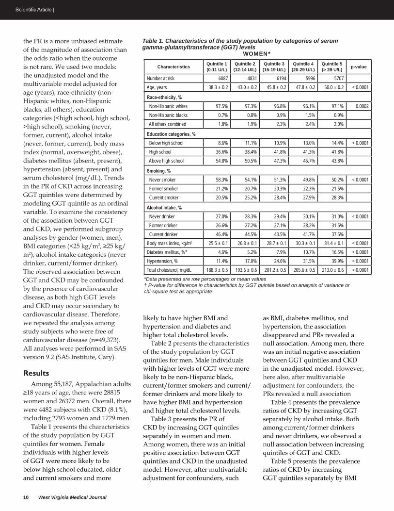
Table 1 presents the characteristics of the study population by GGT quintiles for women. Female individuals with higher levels of GGT were more likely to be below high school educated, older and current smokers and more
likely to have higher BMI and hypertension and diabetes and higher total cholesterol levels.
Table 2 presents the characteristics of the study population by GGT quintiles for men. Male individuals with higher levels of GGT were more likely to be non-Hispanic black, current/former smokers and current/former drinkers and more likely to have higher BMI and hypertension and higher total cholesterol levels.
Table 3 presents the PR of CKD by increasing GGT quintiles separately in women and men. Among women, there was an initial positive association between GGT quintiles and CKD in the unadjusted model. However, after multivariable adjustment for confounders, such
as BMI, diabetes mellitus, and hypertension, the association disappeared and PRs revealed a null association. Among men, there was an initial negative association between GGT quintiles and CKD in the unadjusted model. However, here also, after multivariable adjustment for confounders, the PRs revealed a null association
Table 4 presents the prevalence ratios of CKD by increasing GGT separately by alcohol intake. Both among current/former drinkers and never drinkers, we observed a null association between increasing quintiles of GGT and CKD.
Table 5 presents the prevalence ratios of CKD by increasing GGT quintiles separately by BMI
Characteristics Quintile 1(0-11 U/L)
Quintile 2(12-14 U/L)
Quintile 3(15-19 U/L)
Quintile 4(20-29 U/L)
Quintile 5(> 29 U/L) p-value
Number at risk 6087 4831 6194 5996 5707Age, years 38.3 ± 0.2 43.0 ± 0.2 45.8 ± 0.2 47.8 ± 0.2 50.0 ± 0.2 < 0.0001
race-ethnicity, % Non-Hispanic whites 97.5% 97.3% 96.8% 96.1% 97.1% 0.0002 Non-Hispanic blacks 0.7% 0.8% 0.9% 1.5% 0.9% All others combined 1.8% 1.9% 2.3% 2.4% 2.0%
education categories, % Below high school 8.6% 11.1% 10.9% 13.0% 14.4% < 0.0001 High school 36.6% 38.4% 41.8% 41.3% 41.8% Above high school 54.8% 50.5% 47.3% 45.7% 43.8%
Smoking, % Never smoker 58.3% 54.1% 51.3% 49.8% 50.2% < 0.0001 Former smoker 21.2% 20.7% 20.3% 22.3% 21.5% Current smoker 20.5% 25.2% 28.4% 27.9% 28.3%
Alcohol intake, % Never drinker 27.0% 28.3% 29.4% 30.1% 31.0% < 0.0001 Former drinker 26.6% 27.2% 27.1% 28.2% 31.5% Current drinker 46.4% 44.5% 43.5% 41.7% 37.5%Body mass index, kg/m2 25.5 ± 0.1 26.8 ± 0.1 28.7 ± 0.1 30.3 ± 0.1 31.4 ± 0.1 < 0.0001Diabetes mellitus, %* 4.6% 5.2% 7.9% 10.7% 16.5% < 0.0001Hypertension, % 11.4% 17.0% 24.6% 31.5% 39.9% < 0.0001Total cholesterol, mg/dL 188.3 ± 0.5 193.6 ± 0.6 201.2 ± 0.5 205.6 ± 0.5 213.0 ± 0.6 < 0.0001
*Data presented are row percentages or mean values† P-value for difference in characteristics by GGT quintile based on analysis of variance or chi-square test as appropriate
Table 1. Characteristics of the study population by categories of serum gamma‑glutamyltransferace (GGT) levels
WOMEN*

January/February 2012 | Vol. 108 11
| Scientific Article
category. Both normal weight and overweight/obese subjects, we observed a null association between increasing quintiles of GGT and CKD.
We conducted a supplementary analysis excluding those with cardiovascular disease to examine the robustness of our findings. Overall consistent with the main results, we observed a null association between GGT and CKD among either gender among subjects without cardiovascular disease. For women, compared to serum GGT quintile 1 (referent), the HR (95% CI) of CKD was 0.86 (0.73 - 1.02) in quintile 2, 0.98 (0.85- 1.14) in quintile 3, 0.88 (0.75 - 1.02) in quintile 4 and 0.90 (0.78 - 1.05) in quintile 5; p-trend=<0.0001. For men, compared
to serum GGT quintile 1 (referent), the HR (95% CI) of CKD was 1.00 (0.83, 1.21) in quintile 2, 1.06 (0.87, 1.28) in quintile 3, 0.99 (0.81, 1.20) in quintile 4 and 0.89 (0.72, 1.11) in quintile 5; p-trend=<0.0001.
DiscussionIn a population-based study of
Appalachian adults, serum gamma-glutamyl transferase levels were not found to be associated with CKD, independent of age, gender, smoking status, alcohol intake, education, diabetes mellitus, hypertension, body mass index and total cholesterol. The observed null association between serum gamma-glutamyl transferase levels and CKD remained consistent in subgroup analyses by
gender, alcohol intake, BMI and in a supplementary analysis excluding subjects with cardiovascular disease.
Our finding of no association between higher gamma-glutamyl transferase levels and CKD shows high internal validity, as shown by the magnitude of the association, independence from confounding factors such as age, smoking, alcohol intake, and the consistency of this association in subgroup analyses by gender, drinking status and BMI. When we examined the association between gamma-glutamyl transferase levels and CKD among study subjects free of cardiovascular disease, the results were found to be consistent with the main multivariable findings, suggesting the findings to be independent of cardiovascular disease.
Our results of a null association between gamma-glutamyl transferase levels and CKD are inconsistent with previous studies conducted in an occupational cohort of Korean men.4,5 The reason for the difference in our findings— from a US Appalachian general population sample—to that from the Korean occupational study is not fully clear. Reasons may include the fact that 1) we studied both men and women, as opposed to the Korean study that studied only men, 2) our study was a general community-based sample, as opposed to the Korean study which was an occupational cohort of workers from a semiconductor manufacturing plant, and 3) as GGT levels are influenced by alcohol and diet, findings from Asian populations may not be directly applicable to Western populations with different alcohol intake and dietary patterns. Furthermore, our findings are consistent with a recent study based on the US National Health and Nutritional Examination Survey that reported a similar null finding as ours.10
Table 2. Characteristics of the study population by categories of serum gamma‑glutamyltransferace (GGT) levels
MEN*
*Data presented are row percentages or mean values† P-value for difference in characteristics by GGT quintile based on analysis of variance or chi-square test as appropriate
Characteristics Quintile 1(0-17 U/L)
Quintile 2(18-23 U/L)
Quintile 3(24-30 U/L)
Quintile 4(31-45 U/L)
Quintile 5(> 45 U/L) p-value
Number at risk 5413 5329 4788 5421 5241Age, years 43.5 ± 0.2 46.4 ± 0.2 46.5 ± 0.2 45.9 ± 0.2 45.9 ± 0.2 < 0.0001
race-ethnicity, % Non-Hispanic whites 96.7% 97.2% 96.7% 96.8% 96.1% 0.0017 Non-Hispanic blacks 1.0% 0.9% 1.6% 1.2% 1.6% All others combined 2.3% 1.9% 1.6% 2.0% 2.3%
education categories, % Below high school 13.8% 12.6% 11.5% 11.4% 13.2% 0.0008 High school 43.6% 45.0% 44.9% 45.4% 45.9% Above high school 42.6% 42.4% 43.6% 43.2% 40.9%
Smoking, % Never smoker 46.3% 42.8% 42.9% 40.7% 38.5% < 0.0001 Former smoker 28.7% 32.7% 32.8% 32.4% 30.4% Current smoker 25.0% 24.6% 24.3% 26.9% 31.0%
Alcohol intake, % Never drinker 22.2% 19.6% 16.9% 15.0% 13.0% < 0.0001 Former drinker 29.3% 30.2% 29.5% 27.7% 26.1% Current drinker 48.6% 50.3% 53.6% 57.4% 60.9%Body mass index, kg/m2 25.9 ± 0.1 27.8 ± 0.1 29.1 ± 0.1 30.1 ± 0.1 30.3 ± 0.1 < 0.0001Diabetes mellitus, %* 9.6% 9.4% 9.5% 10.0% 10.4% 0.3622Hypertension, % 19.5% 25.5% 27.2% 29.8% 34.2% < 0.0001Total cholesterol, mg/dL 175.1 ± 0.6 188.3 ± 0.6 195.6 ± 0.6 201.2 ± 0.6 210.7 ± 0.6 < 0.0001

12 West Virginia Medical Journal
Scientific Article |
The major strengths of our study are its large sample size, standardized methods of data collection, detailed measurement of biomarkers that represent potential confounding pathways such as serum cholesterol, and the validation of medical diagnoses by chart review. Internal validity of our results are high as we stratified by gender, alcohol intake, BMI, and cardiovascular disease and performed multivariable adjustment of confounders. The first major limitation of this study is its cross-sectional design. Therefore, we are unable to draw any conclusions about the temporal association between serum gamma-glutamyl transferase and chronic kidney disease. Second, the large representative sample is primarily comprised of whites, reflecting the typical Appalachian community. Therefore, our results may not be generalized to other populations.
In conclusion, in a population-based sample of Appalachian adults, we found that increasing gamma-glutamyl transferase levels were not associated with CKD. Future prospective studies should be conducted to explore the temporal association between gamma-glutamyl transferase and CKD and to confirm the null association.
AcknowledgmentsThis research is supported in part
by the C8 Class Action Settlement Agreement (Circuit Court of Wood County, West Virginia) between DuPont and Plaintiffs, an American Heart Association National Clinical Research Program grant (AS), and a Predoctoral Fellowship (LC) from the Department of Community Medicine, West Virginia University School of Medicine.
Author Disclosure Statement“No competing financial
interests exist.”
Table 3. Association between serum gamma‑glutamyltransferace (GGT) levels and chronic kidney disease (CKD), by gender
Table 4. Association between serum gamma‑glutamyltransferace (GGT) levels and chronic kidney disease (CKD), by alcohol intake status
*Adjusted for age (years), sex (men, women), race-ethnicity (non-Hispanic whites, non-Hispanic blacks, others), education categories (<high school, high school, >high school), smoking (never, former, current), body mass index (normal, overweight, obese), diabetes (absent, present), hypertension (absent, present) and serum cholesterol (mg/dL
never drinker (n=12959) Current/former drinker (n=42043)
Serum ggt quintiles no. at risk(CKd cases)
Multivariable prevalence ratio (95% confidence interval)*
no. at risk(CKd cases)
Multivariable prevalence ratio (95% confidence
interval)*
Women Quintile 1(0-11 U/L) 1645 (176) 1 (referent) 4442 (184) 1 (referent) Quintile 2 (11-14 U/L) 1366 (191) 0.90 (0.73, 1.10) 3465 (196) 0.95 (0.78 01.16) Quintile 3 (14-19 U/L) 1825 (346) 1.04 (0.86, 1.25) 4369 (301) 0.94 0(.78, 1.13) Quintile 4 (19-29 U/L) 1805 (346) 0.96 (0.80, 1.15) 4191 (308) 0.86 (0.71, 1.04) Quintile 5 (>29 U/L) 1770 (322) 0.89 (0.74, 1.07) 3937 (383) 0.96 (0.79, 01.15) p-trend 0.3461 0.5215
Men Quintile 1(0-17 U/L) 1200 (120) 1 (referent) 4213 (280) 1 (referent) Quintile 2 (17-23 U/L) 1044 (119) 1.11 (0.86, 1.44) 4285 (269) 0.87 (0.74, 1.03) Quintile 3 (23-30 U/L) 809 (88) 1.14 (0.86, 1.51) 3979 (236) 0.92 (0.77, 1.09) Quintile 4 (30-45 U/L) 812 (73) 0.97 (0.72, 1.31) 4609 (250) 0.91 (0.77, 1.09) Quintile 5 (> 45 U/L) 683 (68) 1.09 (0.80, 1.48) 4558 (226) 0.88 (0.73, 1.06) p-trend 0.8715 0.3102
Serum ggt quintiles no. atrisk
no. ofcases
Unadjusted prevalence ratio(95% confidence interval)
Multivariable-adjusted prevalence
ratio (95% confidence interval)
Women (n=29390) Quintile 1(0-11 U/L) 6087 360 1 (referent) 1 (referent) Quintile 2 (12-14 U/L) 4831 387 1.35 (1.1y, 1.56) 0.92 (0.80, 1.06) Quintile 3 (15-19 U/L) 6194 647 1.77 (1.55, 2.01) 0.99 (0.87, 1.12) Quintile 4 (20-29 U/L) 5996 654 1.84 (1.62, 2.10) 0.91 (0.80, 1.04) Quintile 5 (>29 U/L) 5707 705 2.09 (1.84, 2.37) 0.93 (0.82, 1.06) p-trend < 0.0001 0.3102 Log-transformed serum GGT, U/L 1.40 (1.33, 1.48) 1.00 (0.94, 1.06)
Men (n=23383) Quintile 1(0-17 U/L) 5413 400 1 (referent) 1 (referent) Quintile 2 (18-23 U/L) 5329 388 0.99 (0.86, 1.13) 0.94 (0.82, 1.09) Quintile 3 (24-30 U/L) 4788 324 0.92 (0.79, 1.06) 0.98 (0.84, 1.13) Quintile 4 (31-45 U/L) 5421 323 0.81 (0.70, 0.93) 0.94 (0.81, 1.09) Quintile 5 (>45 U/L) 5241 294 0.76 (0.65, 0.88) 0.94 (0.80, 1.10) p-trend < 0.0001 0.4372 Log-transformed serum GGT, U/L 0.86 (0.79l 0.92) 0.97 (0.89. 1.05)
*Adjusted for age (years), race-ethnicity (non-Hispanic whites, non-Hispanic blacks, others), education categories (<high school, high school, >high school), smoking (never, former, current), alcohol intake (never, former, current), body mass index (normal, overweight, obese), diabetes (absent, present), hypertension (absent, present) and serum cholesterol (mg/dL)
January/February 2012 | Vol. 108 13
| Scientific Article
References1. Lee D, Jacobs D, Blomhoff R. Is serum gamma
glutamytransferase a marker of oxidative stress? Free Radical Research. 2004;38:535-39.
2. Jimba S, Nakagami T, Dya J, Wasada T, Endo Y, Iwamoto Y. Increase in gamma-Glutamyltransferase Level and Development of Established Cardiovascular Risk Factors and Diabetes in Japanese Adults. Metabolic Related Disorders. 2009.
3. Shankar A, Li J. Association between serum gamma-glutamyltransferase level and prehypertension among US adults. Circulation Journal. 2007;71:1567-72.
4. Chang Y, Ryu S, Sung E, Woo H, Oh E, Cha K et al. Nonalcoholic fatty liver disease predicts chronic kidney disease in nonhypertensive and nondiabetic Korean men. Metabolism. 2008;57:569-79.
5. Ryu S, Chang Y, Kim D, Kim W, Suh B. Gamma-Glutamyltransferase as a predictor of chronic kidney disease in nonhypertensive and nondiabetic Korean men. Clinical Chemistry. 2007;53:71-77.
6. Myers G, Miller W, Coresh J, Fleming J, Greenberg N, Greene T et al. Recommendations for improving serum creatinine measurement: a report from the Laboratory Working Group of the National Kidney Disease Education Program. Clinical Chemistry. 2006;52:5-18.
7. Levey A, Coresh J, Greene T, Stevens L, Zhang Y, Hendriksen S et al. Using standardized serum creatinine values in the modification of diet in renal disease study equation for estimating
glomerular filtration rate. Annals of Internal Medicine. 2006;145:247-54.
8. National Kidney Foundation. K/DOQI clinical practice guidelines for chronic kidney disease: evaluation, classification, and stratification. American Journal of Kidney Diseases. 2002;39:S1-266.
9. Levey A, Eckardt K, Tsukamoto Y, Levin A, Coresh J, Rossert J et al. Definition and classification of chronic kidney disease: a position statement from Kidney Disease: Improving Global Outcomes (KDIGO). Kidney International. 2005;67:2089-100.
10. Teppala, S, Shankar, A, Jialiang, L, Wong, T, and Ducatman, A. Association between Serum Gamma-Glutamyltransferase and Chronic Kidney Disease among US Adults. Kidney and Blood Pressure Research 33(1), 1-6. 2010.
*Adjusted for age (years), sex (men, women), race-ethnicity (non-Hispanic whites, non-Hispanic blacks, others), education categories (<high school, high school, >high school), smoking (never, former, current), body mass index (normal, overweight, obese), diabetes (absent, present), hypertension (absent, present) and serum cholesterol (mg/dL).
Table 5. Association between serum gamma‑glutamyltransferace (GGT) levels and chronic kidney disease (CKD), by body mass index
Normal weight (n=16886) Overweight/ obese (n=38121)
Serum GGT quintiles No. at risk(CKD cases)
Multivariable Prevalence Ratio (95%
confidence interval)
No. at risk(CKD cases)
Multivariable Prevalence Ratio (95% confidence
interval)
Women Quintile 1(0-11 U/L) 3433(156) 1 (referent) 2654(204) 1 (referent) Quintile 2 (11-14 U/L) 2219(149) 0.97 (0.77, 1.22) 2612(238) 0.89 (0.74, 1.07) Quintile 3 (14-19 U/L) 2127(188) 1.03 (0.83, 1.28) 4067(459) 0.97 (0.82, 1.14) Quintile 4 (19-29 U/L) 1549(154) 0.93 (0.74, 1.17) 4447(500) 0.90 (0.76, 1.06) Quintile 5 (>29 U/L) 1127(144) 0.97 (0.77, 1.23) 4580(561) 0.92 (0.78, 1.08) p-trend 0.7049 0.3934
Men Quintile 1(0-17 U/L) 2455(111) 1 (referent) 2958(289) 1 (referent) Quintile 2 (17-23 U/L) 1530(78) 1.03 (0.77, 1.38) 3799(310) 0.91 (0.78, 1.07) Quintile 3 (23-30 U/L) 929(52) 1.13 (0.81, 1.58) 3859(272) 0.93 (0.79, 1.10) Quintile 4 (30-45 U/L) 813(42) 1.02 (0.71, 1.47) 4608 (281) 0.90 (0.76, 1.07) Quintile 5 (> 45 U/L) 704(42) 1.04 (0.72, 1.51) 4537(252) 0.90 (0.75, 1.07) p-trend 0.7666 0.2664

Contact Us Now! ehrsg.com | 304.720.3300 | [email protected]
BeNefitsserviCes
OnClaim billing and account services
EHRsolutions is expanding OnClaim services! OnClaim allows you to focus on providing better patient care using Greenway’s PrimeSUITE while we focus on your everyday billing needs.
electronic Claims scrub, submission, and tracking
secondary Claims Processing
Charge and Payment Posting
Patient statement submission
insurance Claims follow-Up
electronic remittance Advice (erA) Management
Medical Credentialing
Billing Management Consulting
Monthly financial reporting
Monthly financial Meeting
improve Productivity and Profitability
reduce Administrative Costs
increase focus on Patient Care
Utilize Greenway’s PrimesUite (ONC-Certified)

January/February 2012 | Vol. 108 15
| Scientific Article
Brenda Dawley, MDAssociate Professor Department Ob/Gyn
Andreia Acuna, MDOB/Gyn Resident
Beatrice Grasu, MD Joan C. Edwards School of Medicine, Huntington
AbstractPhysicians should consider a benign
mature cystic teratoma in their differential diagnosis of a patient with an elevated serum human chorionic gonadotropin concentration.
BACKGROUND: Following tubal ligation, a woman with amenorrhea and elevated serum human chorionic gonadotropin (HCG) concentrations may be experiencing either an ectopic or an intrauterine pregnancy. Other sources of HCG production can include ovarian germ cell tumors or gestational trophoblastic disease such as a complete or partial molar pregnancy. A rare source of HCG production is a benign mature ovarian teratoma.
CASE: A 31-year old Gravida 2 para 2 presented with a positive home pregnancy test three years after she had experienced a Pomeroy tubal ligation. Her serum HCG was 57,914 mIU/mL but a transvaginal ultrasound did not find an intrauterine pregnancy. Laparoscopy was performed due to a suspicion of an ectopic pregnancy and an 11-cm benign mature cystic teratoma (dermoid cyst) within the right ovary was removed. An ectopic pregnancy was not visualized. Post-operatively, her serum HCG levels decreased and were negative within four weeks.
CONCLUSION: Mature ovarian cystic teratomas have rarely been reported to secrete HCG. They can be an infrequent source of HCG production and may lead to emergency surgery to treat a suspected extra-uterine pregnancy.
IntroductionThe incidence of an ectopic
pregnancy is one in fifty pregnancies
in the United States; however, pregnancy in a woman post tubal ligation is more likely to be an ectopic pregnancy compared to the general population. One third of post-sterilization failures are ectopic pregnancies due to the disruption of normal tubal anatomy.1 Pregnancy following a tubal ligation by the Pomeroy method is as high as 16%; pregnancy can occur because of spontaneous re-anastomosis of the Fallopian tubes or fistula formation.
An ectopic pregnancy mimics a normal intrauterine pregnancy until significant hemorrhage occurs. Breast tenderness, nausea, frequent urination, and amenorrhea are common symptoms of both intra- and extra-uterine pregnancies. Fifty percent of women with an ectopic pregnancy are asymptomatic until a fallopian tube ruptures.2 They may present with vaginal bleeding, abdominal cramping and pain. A tubal rupture may result in severe internal bleeding which can lead to shock and death. Ectopic pregnancies may resolve by spontaneous tubal abortion expelling the pregnancy without complications, but frequently, pain, bleeding, and hemorrhagic shock are associated. Ectopic pregnancies must be identified and removed early to avoid these life-threatening complications.
Transvaginal ultrasound can detect an intrauterine pregnancy with a Beta-hCG level of 1500 mIU/ml and detect an extra-uterine pregnancy in up to 80 percent of women. An ectopic pregnancy is treated with either medical management such as Methotrexate
or surgical management such as laparoscopy. Afterwards, HCG concentrations are monitored weekly until levels drop below 5 mIU/mL.
Trophoblastic tissue secretes beta-hCG and the differential diagnosis for an elevated serum HCG concentration includes intra-uterine and extra-uterine pregnancy, gestational trophoblastic neoplasia, and ovarian germ cell tumors. Forty percent of complete moles are associated with an HCG concentration greater than 100,000 mIU/mL.3 Sequellae of a molar pregnancy can be persistent gestational trophoblastic neoplasia (GTN) or choriocarcinoma. An abnormally large uterus, advanced maternal age, and abnormally elevated HCG levels raise suspicions for malignant GTN. Metastasis to the lung, liver, bone, etc. most often occurs from a choriocarcinoma within the ovary. Choriocarinomas develop from extra-embryonic differentiation of malignant germ cells of the placenta as opposed to molar pregnancies which are benign trophoblastic cells generated from abnormal fertilization. Ultrasound may reveal the abnormality, but histological examination is necessary to confirm the diagnosis.
Serum pregnancy markers such as alpha-fetoprotein (AFP) and HCG are useful when diagnosing a particular ovarian germ cell tumor. Granulosa cell tumors elevate inhibin levels while embryonal carcinomas increase AFP and HCG. The most common ovarian tumor is the mature cystic teratoma (dermoid cyst) which is usually
ectopic production of HcG by a Benign Ovarian Mature cystic Teratoma simulating an extra-uterine pregnancy: a case Report
16 West Virginia Medical Journal
Scientific Article |
hormonally inactive. Ultrasound is used to make a diagnosis and ovarian cystectomy is used to confirm the diagnosis, preserve ovarian tissue, and allow for removal.
Case ReportA 31-year old gravida 2 para
0-2-0-2 complained of amenorrhea, abdominal discomfort, nausea, and breast tenderness and had a positive home pregnancy test. She had a Pomeroy tubal ligation three years ago and a known right dermoid cyst. An office quantitative beta- HCG was 41,428 mIU/mL. A transvaginal ultrasound was performed and did not find an intrauterine pregnancy. An ectopic pregnancy was considered the most likely cause of her positive HCG.
The patient’s past medical history consisted of chronic abdominal pain and chronic hypertension. She had undergone two cesarean deliveries due to preterm severe preeclampsia and had a pomeroy tubal ligation with her second. Three days post partum; she developed a low grade fever and underwent computed tomogram of the abdomen revealing a 6x10 cm right dermoid cyst. She was asymptomatic and was discharged but did not follow-up with gynecology until this admission. She had been evaluated by surgery for her chronic abdominal pain and had a second abdominal computed tomogram scan in 2008 revealing the 6 x 10 cm midline dermoid lesion, nonobstructing right nephrolithiasis, and cholelithiasis. She underwent a laparoscopic cholecystectomy in 2008. Other prior surgeries included a total thryroidectomy for hyperthyroidism. She was currently on thyroid replacement and an antidepressant. Her family history was remarkable for diabetes and hypertension. She did not desire future childbearing. Once she discovered the positive home pregnancy test, she went to the emergency room for evaluation. The transvaginal ultrasound or emergency room pelvic exam did not demonstrate an ovarian mass and
the previous computed tomogram scan report was unavailable.
The plan was a diagnostic laparoscopy with possible oopherectomy or salpingectomy to identify and remove the presumed ectopic pregnancy. Laparoscopic findings included a modestly enlarged anteverted uterus with a large right ovary containing an 11 x 9 x 4.5 cm cyst. The left ovary appeared normal and both fallopian tubes displayed prior tubal ligation and did not demonstrate a tubal pregnancy. She underwent a laparoscopic right salpingo-oopherectomy and endometrial curettage. The specimen required a minilaparotomy for removal. Pathology reported that the 11 cm grey-tan specimen was a benign mature cystic teratoma. Sectioning of the cyst found caseous material, hair, and partially calcified areas. The endometrial pathology was also evaluated and demonstrated a benign hypersecretory endometrium without atypia. There was no evidence of chorionic villi on either specimen.
Following the operation, the patient’s HCG concentration was halved to 25,427 mIU/mL and was monitored weekly. One week post-operatively, her HCG was reported at 12,953 mIU/mL; three weeks post-operatively her HCG was 35.4 mIU/mL; and four weeks it was 5.9 mIU/mL. The patient continued to have her HCG levels monitored for two more negative results.
DiscussionThe differential diagnosis
for elevated human chorionic gonadotropin levels is pregnancy, gestational trophoblastic neoplasia (GTN), and ovarian tumors. This patient’s presenting symptoms were suspicious for an extra-uterine pregnancy following tubal ligation. However, the negative pelvic ultrasound and endometrial curretage made GTN and ectopic pregnancy unlikely.
Dermoid cysts or mature cystic teratomas are usually an inactive, benign combination of all three
embryonic germ cell layers, containing sebaceous fluid, hair, and even teeth. Common in the second and third decades, these cysts may present with symptoms depending on their size and susceptibility for torsion. Approximately one percent of dermoid cysts contain malignant cells that develop into squamous cell carcinoma later in life and the patient’s extremely high HCG level warranted serious concern. Choriocarcinoma was a possible source of her elevated HCG but the negative endometrial curettage with decreasing HCG levels was reassuring.
This patient’s dermoid cyst was particularly unusual for several reasons. It was missed at the time of her repeat cesarean section and again at the time of her cholecystectomy. The higher fat content of a larger dermoid cyst typically provides easier visualization on CT scan; conversely, its greater buoyancy may make the ovary high in the peritoneal cavity and not palpable by pelvic exam. She had undergone two prior surgeries and the large dermoid cyst was not visualized. In the case of her cesarean section, the ovary was pushed high in the upper abdomen by the gravid uterus and was missed by the operating surgeon. During her cholecystectomy, she was placed in reverse Trendelenburg resulting in the dermoid cyst returning to a pelvic location and was missed by the general surgeon. Second, the cyst’s reported size in 2007was large, but it continued to grow in the subsequent three years. On average, a dermoid cyst grows 1.8 mm each year.5
Finally, this cyst appeared to have secreted significant amounts of HCG and rather suddenly since the patient never complained of amenorrhea or pregnancy-like symptoms in the past two years. This benign dermoid’s HCG production simulating an ectopic pregnancy resembles two similar published cases.5,6 Downey et. al. published the first case in London in 1989 and very few cases have been reported since then.7 (See Table 1 for a comparison between this
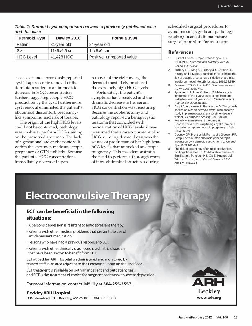
January/February 2012 | Vol. 108 17
| Scientific Article
case’s cyst and a previously reported cyst.) Laparoscopic removal of the dermoid resulted in an immediate decrease in HCG concentration further suggesting ectopic HCG production by the cyst. Furthermore, cyst removal eliminated the patient’s abdominal discomfort, pregnancy-like symptoms, and risk of torsion.
The origin of the high HCG levels could not be confirmed; pathology was unable to perform HCG staining on the preserved specimen. The lack of a gestational sac or chorionic villi within the specimen made an ectopic pregnancy or GTN unlikely. Because the patient’s HCG concentrations immediately decreased upon
removal of the right ovary, the dermoid most likely produced the extremely high HCG levels.
Fortunately, the patient’s symptoms have resolved and the dramatic decrease in her serum HCG concentration was reassuring. Because the oopherectomy and pathology reported a benign cystic teratoma that coincided with normalization of HCG levels, it was presumed that a rare occurrence of an HCG secreting dermoid cyst was the source of production of her high beta-hCG levels that mimicked an ectopic pregnancy. This case demonstrates the need to perform a thorough exam of intra-abdominal structures during
scheduled surgical procedures to avoid missing significant pathology resulting in an additional future surgical procedure for treatment.
References1. Current Trends Ectopic Pregnancy – U.S.,
1990-1992. Morbidity and Mortality Weekly Report 1995;44:46.
2. Buckley RG, King KJ, Disney JD, Gorman JD. History and physical examination to estimate the risk of ectopic pregnancy: validation of a clinical prediction model. Ann.Emer. Med. 1999;34:589.
3. Berkowitz RS, Goldstein DP. Chorionic tumors. NEJM 1996;335:1740.
4. Ayhan A, Bukulmez O, Genc C. Mature cystic teratomas of the ovary: case series from one institution over 34 years. Eur J Obstet Gynecol Reprod Biol 2000;88:153.
5. Caspi B, Appelman Z, Rabinerson D. The growth pattern of ovarian dermoid cysts: a prospective study in premenopausal and postmenopausal women. Fertility and Sterility 1997;68:501.
6. Pothula V, Matseoane S, Godfrey H. Gonadotropin-producing benign cystic teratoma simulating a ruptured ectopic pregnancy. JAMA 1994;86:221.
7. Downey GP, Prentice M, Penna LK, Gleeson RP. Ectopic beta-human chorionic gonadotropin production by a dermoid cyst. Amer J of Ob and Gyn 1989;160:449.
8. The risk of pregnancy after tubal sterilization. Findings from the U.S. Collaborative Review of Sterilization, Peterson HB, Xia Z.,Hughes JM, Wilcox LS, et al, Am J Obstet Gynecol 1996 Apr;174(4):1161-8.
Table 1: Dermoid cyst comparison between a previously published case and this case
Dermoid Cyst Dawley 2010 Pothula 1994Patient 31-year old 24-year oldSize 11x9x4.5 cm 14x8x6 cmHCG Level 41,428 HCG Positive, unreported value
Electroconvulsive TherapyECT can be beneficial in the followingsituations:• A person’s depression is resistant to antidepressant therapy.
• Patients with othermedical problems that prevent the use ofantidepressantmedication.
• Persons who have had a previous response to ECT.
• Patients with other clinically diagnosed psychiatric disordersthat have been shown to benefit from ECT.
ECT at Beckley ARHHospital is administered andmonitored bytrained staff in an area adjacent to the Operating Roomon the 2nd floor.
ECT treatment is available on both an inpatient and outpatient basis,and ECT is the treatment of choice for pregnant patients with severe depression.
Formore information,contact Jeff Lilly at 304-255-3557.
Beckley ARH Hospital306 Stanaford Rd | Beckley,WV 25801 | 304-255-3000
Beckleywww.arh.org

18 West Virginia Medical Journal
Scientific Article |
Ravindra Bhardwaj, MD, MPH1
Puneet Sharma, MD1
Mitchell S. Finkel, MD1-5
Sumesh Jain, MD1
Ahmad Arham, MD2
Tooba Kazmi, MD2
Robert J. Beto, MD1
Wissam Gharib, MD1
Bradford E. Warden, MD1
Abnash C. Jain, MD1
1Section of Cardiology, WVU Heart Institute, Departments of 2Medicine, 3Psychiatry,4 Physiology and Pharmacology West Virginia University, School of Medicine, 5Louis A. Johnson VA Medical Center, Morgantown and Clarksburg, WV
AbstractAtrial fibrillation (AF) is a cardiac
arrhythmia associated with a wide range of other co-morbid medical conditions. The state of West Virginia has a higher prevalence of coronary artery disease (CAD) and CAD risk factors compared to the national average. We hypothesized that West Virginians with atrial fibrillation would also have a higher prevalence of CAD risk factors and higher CHADS2 stroke risk scores. This is particularly important since Louisiana is the only high CAD risk southern state included in the original verification of the CHADS2 risk scoring system (i.e. California, Connecticut, Louisiana, Maine, Missouri, New Hampshire, and Vermont). Accordingly, we performed a retrospective analysis of the association between AF and CAD, CAD risk factors and CHADS2 scores in a cohort of men and women in the West Virginia University Hospital population. We report a greater positive association between AF and hypertension, diabetes mellitus and obesity than the national average. AF was seen more commonly among men. But, CHADS2 scores were higher among women as a result of a higher prevalence of diabetes mellitus. This study indicates that AF is associated with a greater prevalence of CAD risk factors and higher CHADS2 scores among West Virginians in comparison with the rest of the nation.
IntroductionAtrial fibrillation (AF) is an
arrhythmia associated with a wide range of cardiac conditions and is an independent predictor of heart failure and stroke.1,2 Hypertension (HTN) and coronary artery disease (CAD) are the most common co-morbidities associated with AF.3,4,5 According to the 2007 Behavioral Risk Factor Surveillance System survey results, the state of West Virginia has a higher prevalence of CAD and CAD risk factors compared to the national average (HTN- 33.3% vs 27.8%, diabetes mellitus (DM)- 10.8% vs 8.0%, Obesity- 68% vs 62.9%, smoking- 26.9% vs 19.8%).6 We hypothesized that West Virginians with atrial fibrillation would also have a higher prevalence of CAD risk factors and higher CHADS2 stroke risk scores. This is particularly important since Louisiana is the only high CAD risk southern state included in the original verification of the CHADS2 risk scoring system (i.e. California, Connecticut, Louisiana, Maine, Missouri, New Hampshire, and Vermont).7 Accordingly, we performed a retrospective analysis of the association between AF and CAD, CAD risk factors and CHADS2 scores in a cohort of men and women in the West Virginia University Hospital population.
MethodsThis retrospective, observational
study was conducted at West Virginia University Hospital after receiving the approval of the Institutional Review Board for the Protection of Human Subjects. A total of 8,986
consecutive EKGs were analyzed and screened for the diagnosis of atrial fibrillation (AF) between October 2009 and March 2010. After excluding duplicates, 541 patients with the diagnosis of AF on EKG tracing formed our study population. The patients’ demographic characteristics (age, gender, weight, height, clinical risk factors and medications) were assessed from the medical records of each of the 541 patients.
The stroke risk index was calculated based on the risk factors identified from chart review using the CHADS2 score.7 We calculated CHADS2 scores by adding 1 point for each of the following: recent
CHF, hypertension, age 75 years or older, and DM—and 2 points for a history of stroke or TIA. Therefore, CHADS2 is an acronym for the major risk factors and their scoring. For instance, a 78-year-old (+1) patient who had diabetes mellitus (+1) and hypertension (+1) would have a CHADS2 score of 3. A score of 0 identified patients at “low risk”, 1 to 2 at “moderate risk” and 3 to 6 at “high risk” for stroke. CHADS2 is commonly used by physicians to estimate the risk of stroke in patients with AF and decide about antithrombotic therapy based on patient-specific risk of stroke.
Body Mass Index of < 25 kg/m2 was considered “normal”, 25 to < 30 kg/m2 as “overweight” and ≥ 30 kg/m2 as “obese”.8 HTN was defined as blood pressure greater than 140/90 mm Hg or a history of hypertension and being on antihypertensive medication. The differences between men and women were evaluated
Gender and Geographic Differences in cAD Risk Factors and cHADs
2 scores in Atrial Fibrillation patients
January/February 2012 | Vol. 108 19
| Scientific Article
using Student’s t test for continuous variables and chi-square test for categorical variables. The results were considered statistically significant at a p value <0.05.
ResultsFive hundred forty one patients’
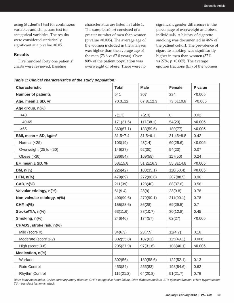
charts were reviewed. Baseline
characteristics are listed in Table 1. The sample cohort consisted of a greater number of men than women (p value <0.005). The average age of the women included in the analyses was higher than the average age of the men (73.6 vs 67.8 years). Over 80% of the patient population was overweight or obese. There were no
significant gender differences in the percentage of overweight and obese individuals. A history of cigarette smoking was documented in 46% of the patient cohort. The prevalence of cigarette smoking was significantly higher in men than women (57% vs 27%, p <0.005). The average ejection fractions (EF) of the women
Table 1: Clinical characteristics of the study population:
Characteristic Total Male Female P value
Number of patients 541 307 234 <0.005
Age, mean ± SD, yr 70.3±12 67.8±12.3 73.6±10.8 <0.005
Age group, n(%)
<40 7(1.3) 7(2.3) 0 0.02
40-65 171(31.6) 117(38.1) 54(23) <0.005
>65 363(67.1) 183(59.6) 180(77) <0.005
BMI, mean ± SD, kg/m2 31.5±7.4 31.5±6.1 31.45±8.8 0.42
Normal (<25) 103(19) 43(14) 60(25.6) <0.005
Overweight (25 to <30) 146(27) 92(30) 54(23) 0.07
Obese (>30) 286(54) 169(55) 117(50) 0.24
EF, mean ± SD, % 53±15.8 51.2±16.3 55.3±14.8 <0.005
DM, n(%) 226(42) 108(35.1) 118(50.4) <0.005
HTN, n(%) 479(89) 272(88.6) 207(88.5) 0.96
CAD, n(%) 211(39) 123(40) 88(37.6) 0.56
Valvular etiology, n(%) 51(9.4) 28(9) 23(9.8) 0.78
Non-valvular etiology, n(%) 490(90.6) 279(90.1) 211(90.1) 0.78
CHF, n(%) 155(28.6) 86(28) 69(29.5) 0.7
Stroke/TIA, n(%) 63(11.6) 33(10.7) 30(12.8) 0.45
Smoking, n(%) 246(46) 174(57) 62(27) <0.005
CHADS2 stroke risk, n(%)
Mild (score 0) 34(6.3) 23(7.5) 11(4.7) 0.18
Moderate (score 1-2) 302(55.8) 187(61) 115(49.1) 0.006
High (score 3-6) 205(37.9) 97(31.6) 108(46.1) <0.005
Medication, n(%)
Warfarin 302(56) 180(58.6) 122(52.1) 0.13
Rate Control 453(84) 255(83) 198(84.6) 0.62
Rhythm Control 115(21.2) 64(20.8) 51(21.7) 0.79BMI= body mass index, CAD= coronary artery disease, CHF= congestive heart failure, DM= diabetes mellitus, EF= ejection fraction, HTN= hypertension, TIA= transient ischemic attack
20 West Virginia Medical Journal
Scientific Article |
was significantly higher than that of the men (51.2±16.3 vs 55.3±14.8, p <0.005). DM was more common in women than men (p <0.005). Hypertension was diagnosed in 90%, coronary artery disease (CAD) in 39% and TIA/CVA in 11.6% of
the cohort. A little over half the patients (56%) received warfarin therapy. Women had the greatest number and percentage of high risk CHADS2 scores (p <0.005). Moderate risk CHADS2 scores were more common in men. The etiology of the
AF was identified as non-valvular in 90% of the patient population,
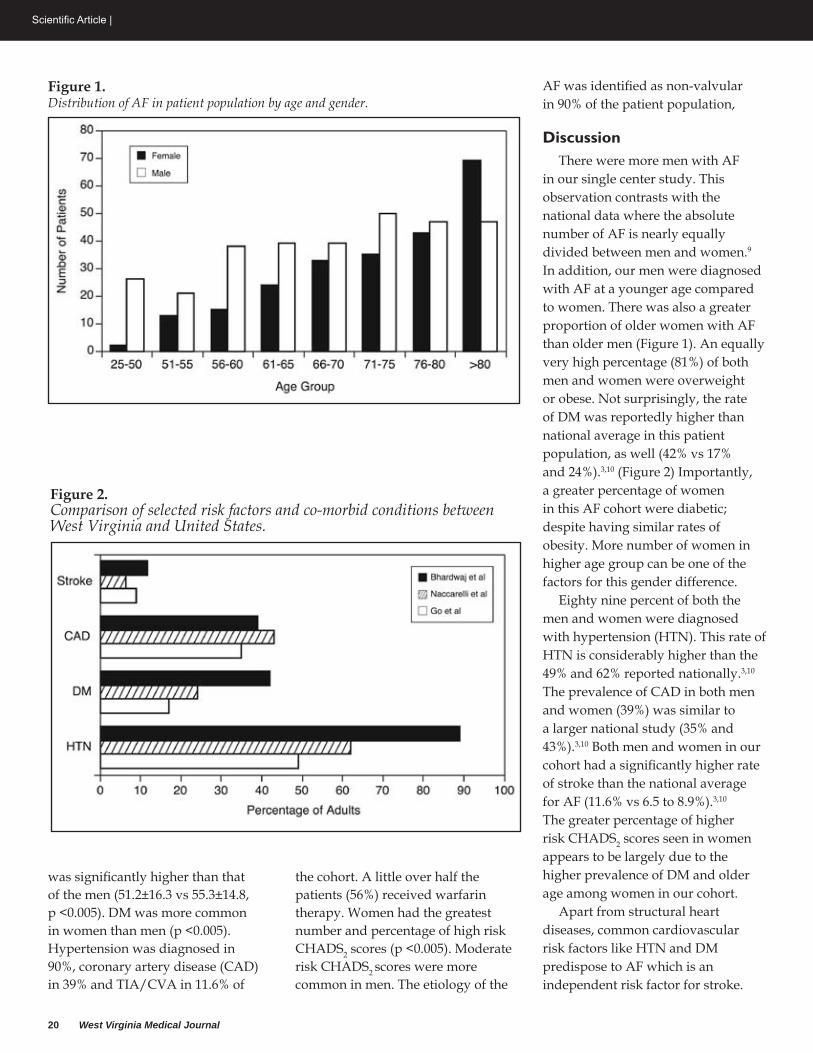
DiscussionThere were more men with AF
in our single center study. This observation contrasts with the national data where the absolute number of AF is nearly equally divided between men and women.9 In addition, our men were diagnosed with AF at a younger age compared to women. There was also a greater proportion of older women with AF than older men (Figure 1). An equally very high percentage (81%) of both men and women were overweight or obese. Not surprisingly, the rate of DM was reportedly higher than national average in this patient population, as well (42% vs 17% and 24%).3,10 (Figure 2) Importantly, a greater percentage of women in this AF cohort were diabetic; despite having similar rates of obesity. More number of women in higher age group can be one of the factors for this gender difference.
Eighty nine percent of both the men and women were diagnosed with hypertension (HTN). This rate of HTN is considerably higher than the 49% and 62% reported nationally.3,10 The prevalence of CAD in both men and women (39%) was similar to a larger national study (35% and 43%).3,10 Both men and women in our cohort had a significantly higher rate of stroke than the national average for AF (11.6% vs 6.5 to 8.9%).3,10 The greater percentage of higher risk CHADS2 scores seen in women appears to be largely due to the higher prevalence of DM and older age among women in our cohort.
Apart from structural heart diseases, common cardiovascular risk factors like HTN and DM predispose to AF which is an independent risk factor for stroke.
Figure 1.Distribution of AF in patient population by age and gender.
Figure 2.Comparison of selected risk factors and co-morbid conditions between West Virginia and United States.
January/February 2012 | Vol. 108 21
| Scientific Article
Although antithrombotic and antiarrthymic therapy is helpful in reducing the risk of stroke, however it is frequently contraindicated in elderly due to significant associated morbidity. There is no causative relationship of AF with HTN and DM, but it is hypothesized that aggressive management of these modifiable risk factors may be the safest and most effective approach to prevent the occurrence of AF and associated morbidity and mortality.
ConclusionThe association of AF with CAD
risk factors (e.g. HTN, DM and obesity) and the incidence of TIA/CVA was significantly greater in our single WV cohort than the previously reported national average.7,9,10 The association of AF with CAD, however, is still similar
to the study using the larger national sample.10 Further study is warranted to identify potential gender and regional differences in outcomes for patients with atrial fibrillation.
References1. Wolf PA, Abbott RD, Kannel WB. Atrial fibrillation
as an independent risk factor for stroke: the Framingham Study. Stroke. 1991; 22: 983–988.
2. Stewart S, Hart CL, Hole DJ, McMurray JJ. A population-based study of the long-term risks associated with atrial fibrillation: 20-year follow-up of the Renfrew/Paisley study. Am J Med. 2002;113(5):359-64.
3. Go AS, Hylek EM, Phillips KA, Chang Y, Henault LE, Selby JV, Singer DE. Prevalence of diagnosed atrial fibrillation in adults: national implications for rhythm management and stroke prevention: the Anticoagulation and Risk Factors in Atrial Fibrillation (ATRIA) Study. JAMA. 2001;285(18):2370-5.
4. Benjamin EJ, Levy D, Vaziri SM, et al. Independent risk factors for atrial fibrillation in a population-based cohort: the Framingham Heart Study. JAMA. 1994;271:840-844.
5. Miyasaka Y, Barnes ME, Gersh BJ, Cha SS, Bailey KR, Abhayaratna WP, Seward JB, Tsang TS. Secular trends in incidence of atrial fibrillation in Olmsted County, Minnesota, 1980 to 2000,
and implications on the projections for future prevalence. Circulation. 2006;114(2):119-25.
6. CDC. Division for Heart Disease and Stroke Prevention. Available at: http://www.cdc.gov/dhdsp/state_program/wv_text.htm, Last modified: February 8, 2010
7. Gage BF, Waterman AD, Shannon W, Boechler M, Rich MW, Radford MJ. Validation of clinical classification schemes for predicting stroke: results from the National Registry of Atrial Fibrillation. JAMA. 2001;285(22):2864-70.
8. ClinicalGuidelinesontheIdentification,Evaluation, and Treatment of Overweight and Obesity in Adults. Bethesda, Md: National Heart, Lung, and Blood Institute; 1998.
9. Feinberg WM, Blackshear JL, Laupacis A, Kronmal R, Hart RG. Prevalence, age distribution, and gender of patients with atrial fibrillation. Analysis and implications. Arch Intern Med. 1995;155(5):469-73.
10. Naccarelli GV, Varker H, Lin J, Schulman KL. Increasing prevalence of atrial fibrillation and flutter in the United States. Am J Cardiol. 2009;104(11):1534-9.
306 Stanaford Rd | Beckley,WV 25801304-255-3000 | www.arh.org
Teenagers face numerous challengestoday that can sometimes cause them tohave serious issues.Beckley ARH’s Behavioral Science Center can intervene to help themcope and prevent a problem frombecomingworse as they enteradulthood.The Unit allows for an excellent opportunity to identify,diagnose and treat problems at an early stage so they can get back toschool andwhat should be some of the happiest times of their lives.
Beckley ARHAdolescent Behavioral Science Center
Beckley

Physicians Insuring Physicians(304) 343-3000
www.wvmic.com
“This 2012 premium reduction continues our efforts
to provide responsible, sustainable premium
relief for our policyholders.”
R. Austin Wallace, M.D. • Chairman and CEO
January/February 2012 | Vol. 108 23
| Scientific Article
The Appalachian Cardiovascular Research Network*
Individual author listing and affiliation appear at the end of the paper.
AbstractExcess weight is a known risk factor
for coronary artery disease (CAD) and a large percentage of overweight and obese individuals ultimately develop CAD. The objective of this study was to identify human genes associated with CAD in a subgroup of overweight and obese individuals using population-based association methods. Logistic regression analyses were used to test the association between single nucleotide polymorphisms (SNPs) in 34 candidate genes and the CAD phenotype with age, gender, and BMI as covariates. Two SNPs in the Apolipoprotein B (Apo B) gene [rs1042031 and rs1800479], one in the Cholesterol Ester Transfer Protein (CETP) gene [rs5880], and one in the Low Density Lipoprotein Receptor (LDLR) gene [rs2569538] met the 0.01 significance level for association with CAD. Based on these findings, we conclude that variants within the CETP and Apo B genes conferred susceptibility to CAD in overweight individuals and that a variant with the LDLR gene conferred susceptibility in an obese group.
IntroductionCoronary artery disease (CAD)
is the leading cause of death and premature disability in the United States.1, 2,3 In its most recent update on heart disease, the American Heart Association reported that 16.8 million US adults are afflicted with CAD and that the estimated CAD patient care costs for 2009 are $165 billion.3 The primary cause of CAD is atherosclerosis, a progressive multifactorial disease characterized by the accumulation of lipids and fibrous elements in the large arteries.4 Evidence from animal studies, epidemiological
studies and clinical correlations strongly suggest a role for dietary cholesterol as an environmental promoter of atherosclerosis.2,3 A genetic etiology for atherosclerosis is indicated by familial aggregation of the trait and heritability estimates of contributing factors (e.g. LDL- and HDL-cholesterol levels) that range from 20% to 80%.2,5
Several significant causal genes have been identified by studying Mendelian disorders that either cause or confer susceptibility to CAD.5 Genome-wide linkage scans have revealed CAD susceptibility loci on chromosomes 1p34-36, 2q, 3q13, 13q12-13, 14q32, 16p13, and 17.(6, 7 and reviewed in 8) Several genome-wide association studies for CAD have been performed based on single nucleotide polymorphisms (SNPs).6 Although some identified regions were replicated (3q12-13, 9p21), most of the known findings of regions associated with CAD could not be replicated. A recent replication study on 85 previously identified variants in genetic risk factors for acute coronary syndrome found that only one of these variants (in the β-fibrinogen promoter) was validated as a risk factor.9 Additional candidate gene association studies in different subpopulations are needed in order to confirm the associations with these SNPs and identify new SNPs associated with CAD.
Overweight and obesity have become global epidemics in both children and adults10 and are known to contribute to CAD, heart failure, and sudden cardiac death.4,5,11 Since a significant number of overweight individuals go on to develop CAD, we hypothesized that one or more genes could cause or confer susceptibility to the development
of CAD in this group. We sought to identify genes associated with CAD in overweight and obese individuals by screening for SNPs in a set of cardiovascular disease candidate genes using case control association methods. We identified SNPs within three genes [Apo B (apolipoprotein B), LDLR (low density lipoprotein receptor) and CETP (cholesterol ester transfer protein)] that were associated with CAD phenotype. The Apo B and CETP SNPs have been identified in previous CAD association studies,12,13 while the LDLR SNP is a novel association.
Methods
Human Subjects and CAD Phenotype
Human subjects were enrolled according to Institutional Review Board protocols approved by the following West Virginia institutions: Marshall University Department of Cardiovascular Services, Charleston Area Medical Center, Lincoln Primary Care Center, Tri-County Clinic, Tug River Clinic, and Valley Health Systems.
The CAD phenotype was predicated on known indicators of the disease: angina, myocardial infarction, cardiac artery stenosis, and acute coronary syndrome. Individuals were classified as affected with CAD if one or more of the following conditions were present: (1) at least one vessel with at least 50% stenosis as determined by cardiac catheterization, (2) acute myocardial infarction diagnosed either by elevation of creatine phosphokinase myocardial band or by diagnostic electrocardiogram, (3) coronary artery bypass graft,
Identification of Genes contributing to cardiovascular Disease in Overweight and Obese Individuals from West Virginia

24 West Virginia Medical Journal
Scientific Article |
(4) percutaneous coronary intervention, (5) documented cerebral vascular disease, or (6) documented peripheral arterial disease. Individuals without any of these symptoms or interventions who did not report any history of CAD and who were at least 40 years of age were classified as controls.
Participants with Body Mass Index (BMI) ≥ 25 were placed in the overweight category while those with BMI ≥ 30 were considered obese. Of the 261 Caucasian participants, we identified 243 who were overweight. In this group of individuals, 104 had evidence of CAD and 92 were
controls. Forty seven individuals did not meet our criteria for cases or controls and were excluded from analysis. Of these 47 exclusions, 33 clinically normal individuals were younger than age forty and 14 individuals had an indeterminate disease phenotype. Obese (BMI ≥ 30) individuals accounted for 141 of the 196 overweight individuals.
Candidate Genes and SNP SelectionThirty four candidate genes
for CAD were selected from three different sources of cardiovascular disease susceptibility genes: Online Mendelian Inheritance in Man (www.ncbi.nlm.nih.gov/omim), CardioGenomics (cardiogenomics.med.harvard.edu), and Comparative Genomic Resources for Cardiovascular Research (Berkeley Programs for Genomic Applications; pga.jgi-psf.org/) (Table 1).
Selection of candidate gene SNPs was performed in a two phase approach. In the first phase, we selected a minimal set of tagging SNPs within each candidate gene using the HapMap Genome Browser and Haploview software.14,15 Only SNPs with a minor allele frequency (MAF) ≥ 5% were selected for analysis. When MAFs were not available from the SNP databases, we calculated the frequency based on our study population. SNPs for which a TaqMan Allelic Discrimination assays were available were pre-selected prior to application of the aggressive tagging protocol. Haplotype blocks were identified by Haploview analysis of HapMap data. In the second phase of the SNP selection, additional SNPs were genotyped for genes in which statistically significant associations were identified in the first phase. These additional SNPs were genotyped in order to maximize the coverage of the gene and better define the area of interest. One hundred thirty six SNPs from thirty-four selected candidate genes were tested for an association with CAD as described below.
Table 1: Candidate genes for CAD with corresponding number of genotyped SNPs
Candidate Gene Number of SNPs
ABCA1 (ATP-binding cassette) 6AGT (angiotensinogen preprotein) 8ALOX5A (arachidonate 5-lipoxygenase-activating protein) protein) 1Apo B (Apolipoprotein B) 8Apo C2 (Apolipoprotein C-II ) 1Apo E (Apolipoprotein E) 1BHMT (betaine-homocysteine methyltransferase) 3CETP (cholesteryl ester transfer protein) 14F5 (coagulation factor V) 5HDLBP ( high density lipoprotein binding protein) 3HNF4A (hepatocyte nuclear factor 4, alpha) 2LDLR (low density lipoprotein receptor) 7LIPA (lipase A, lysosomal acid, cholesterol esterase) 11LIPG (lipase, endothelial) 2LRP1 (low density lipoprotein-related protein 1) 3LRP1B (low density lipoprotein-related protein 1B) 7LRP5 (low density lipoprotein receptor-related protein 5) 8LPR8 (low density lipoprotein receptor-related protein 8) 7MPO (myeloperoxidase) 1MSR1 (macrophage scavenger receptor 1) 2MTHFR (5,10-methylenetetrahydrofolate reductase) 5MTR (5-methyltetrahydrofolate-homocysteine) 1
MTRR (5-methyltetrahydrofolate-homocysteine) 1
NOS3 (nitric oxide synthase 3) 3PLTP (phospholipid transfer protein) 2PPARA (peroxisome proliferative activated receptor) 6SHMT1 (serine hydroxymethyltransferase 1) 2SCARB1 (scavenger receptor class B, member 1) 5SERPINB2 (serpin peptidase inhibitor, clade B) 1TCN2 (transcobalamin II) 4
TNFSF4 (tumor necrosis factor superfamily, member 4) 2
USF1 (upstream stimulatory factor 1) 1USF2 (upstream stimulatory factor 2) 1VWF (von Willebrand factor) 2Total: 34 136

January/February 2012 | Vol. 108 25
| Scientific Article
Genotyping Methods Genomic DNA was purified from
patient whole blood samples using Qiagen Blood and Cell Culture DNA Maxi Kits. SNPs were genotyped using either pyrosequencing or TaqMan Allelic Discrimination methods. In the pyrosequencing method, specific loci were amplified from 25 nanograms of genomic DNA by the polymerase chain reaction (PCR). Pyrosequencing reactions were carried out as described by Nyren et al.16 using a Biotage PSQ96MA Pyrosequencing System. Allelic Discrimination analyses were performed on either an ABI Model 7000 or Model 7500 Sequence Detection Systems as described by Livak et al.17 ABI Sequence Detection System Absolute and Allelic Discrimination software was used to determine the genotype.
Statistical Methods A series of conditional logistic
regression analyses with age,
gender, and BMI as covariates were performed to identify SNPs associated with the CAD phenotype. Statistical analyses were performed using statistical analysis software (SAS) release 9.2. p-values less than or equal to 0.01 were considered to be statistically significant. A chi-
square test was used to determine if SNP alleles were in Hardy-Weinberg Equilibrium (HWE). Three of the 136 genotyped SNPs (rs289714, rs4823613, and rs891512) were found to be not in HWE at significance level of 0.05, and were excluded from further statistical analyses.
Table 2: Mean age (in years), BMI, and gender differences between CAD and control subjects. Data are given as means ± standard deviation (SD). NS stands for not significant.
CAD Subjects Control Subjects p-value
Total number of subjects (n) 104 92
Mean age (in years) 63.5 ± 10.7 56.3 ± 9.8 < 0.001
Mean BMI 33.4 ± 6.2 35.6 ± 7.5 < 0.05
Number of males/females 70/34 24/68 < 0.001
Mean age for males 63.4 ± 9.8 57.1 ± 9.7 < 0.01
Mean age for females 63.8 ± 12.7 55.9 ± 9.9 < 0.01
Mean BMI for males 32.3 ± 5.4 32.5 ± 4.6 NS
Mean BMI for females 35.7 ± 7.2 36.7 ± 8.1 NS
Preventing the spread of flu protects patients and saves lives.
100% flu vaccination begins with you! When you get the flu, you expose your family, patients
and co-workers to infection. That is why it’s so important for you to get vaccinated.
Get VaccinatedProtect yourself. Protect our patients.
www.immunizenow.org
The Center for Rural Health Development, Inc. is the lead agency for the West Virginia Immunization Network
WIN043_AD_WV State Medical Journal Ad half page.indd 1 8/25/11 4:02 PM

26 West Virginia Medical Journal
Scientific Article |
The remaining 133 SNPs passed the HWE significance test at 0.05.
ResultsDemographic data of the
individuals used in this study are given by age, gender, and BMI in Table 2. Out of 196 overweight individuals, 94 (48%) were males and 102 (52%) were females. The average age of the patients entering the study was 60.1 years with a standard deviation of 10.9 years. Both case and control groups were composed of non-Hispanic, Caucasian populations from West Virginia. Since there were significant differences in age, gender, and BMI between cases and controls, logistic regression analyses were performed with age, gender, and BMI as covariates.
Using the allelic model, we identified two SNPs (rs1042031 and rs1800479) within the Apo B gene that were associated with CAD at significance level below 0.01 (Table 3). SNP rs1042031 is located within Apo B exon 29 and substitution of the reference allele G by the minor allele A results in a substitution of the wild type glutamine (GLU) with lysine (LYS) at amino acid
4181. For overweight individuals, each occurrence of A at rs1042031 conferred an increased likelihood of 2.44 fold for developing CAD compared to individuals with a G substitution. Apo B SNP rs1800479 is an intronic SNP about 2kb up stream of rs1042031 SNP. For overweight individuals, each occurrence of the minor allele, G, at rs1800479 conferred an increased likelihood of 2.33 fold for developing CAD compared to individuals with a C substitution. Six other SNPs analyzed within the Apo B gene region did not show significant association with CAD (Table 4).
SNP rs2569538 in the LDLR gene was significantly associated with CAD in a subgroup of obese individuals under both the allelic (p-value = 0.005, Table 3) and the genotypic model (p-value = 0.007, Table 5). Obese individuals who are A/G heterozygotes at rs2569538 were four times more likely to develop CAD than G/G homozygotes. This SNP is located inside intron 15 within the second haplotype block of LDLR gene. Six other LDLR SNPs were genotyped but did not show significant relationship with CAD (Table 4).
Using the genotypic model in the overweight group, we identified a SNP within the CETP gene on chromosome 16 (rs5880) that was associated with CAD at a significance level of 0.008 (Table 5). SNP rs5880 represents a nonsynonymous change which results in substitution of proline for the normally occurring alanine at position 373 in the processed protein (position 390 in the precursor protein). Overweight individuals who were G/G homozygotes at rs5880 were 5.56 times more likely to develop CAD than C/G heterozygotes. SNP rs5880 was also found to be associated (p-value = 0.006) with CAD in a sample of overweight individuals in which participants with type II diabetes were excluded (data not shown). None of twelve additional CETP SNPs were associated with the CAD phenotype in overweight and obese groups (Table 4).
DiscussionTwo Apo B SNPs, rs1042031 and
rs1800479, met our criterion for association with CAD. Based on their physical map location both SNPs are located in the first haplotype
BMI ≥ 25
SNP GeneAllele Frequencies
p-value OR [95% CI]Cases (n = 96) Controls (n = 91)
A G A G
rs1042031 Apo B 0.30 0.70 0.13 0.87 0.0054 A 2.44 [1.30, 4.57]Cases (n = 100) Controls (n = 90)
C G C G
rs1800479 Apo B 0.71 0.29 0.87 0.13 0.0079 G 2.33 [1.25, 4.34]BMI ≥ 30
Cases (n = 65) Controls (n = 65)
A G A G
rs2569538 LDLR 0.18 0.82 0.06 0.94 0.0049 A 4.29 [1.56, 11.8]
Table 3: Association with Atherosclerosis (Allelic Model). The at‑risk allele is given in bold. CI stands for confidence interval, OR stands for odds ratio.

January/February 2012 | Vol. 108 27
| Scientific Article
28 West Virginia Medical Journal
Scientific Article |
block of Apo B.14,15,18 Two other SNPs (rs1042034 and rs693) within the first haplotype block of Apo B were not associated with CAD.
Apo B is the principal protein component of several low density lipoproteins (LDLs) and is the main apolipoprotein of chylomicrons. Defects in the Apo B gene are the cause of familial ligand-defective
apolipoprotein b-100 (fdb), an autosomal dominant disorder that leads to hypercholesterolemia and increased susceptibility to coronary artery disease. Causal mutations map to exon 26 of Apo B and lead to substitution of glutamine for arginine 3500 or substitution of cysteine for arginine 3532.19 In contrast to the fdb genotype-phenotype
correlation, a meta-analysis of 30 case control studies failed to find an association between CAD and the exon 26 XbaI restriction fragment length polymorphism (RFLP).12 This study did report a significant association with an EcoR1 RFLP located in exon 29 which is identical to SNP rs1042031. Based on the location of the two associated Apo
Table 4: Apo B (Chromosome 2), LDLR (Chromosome 19), and CETP (Chromosome 16) SNPs. Location of each SNP was identified through the HapMap project Database. Minor Allele Frequencies (MAFs) were either found through the HapMap project or the dbSNP web site. SNPs in bold were genotyped in the second phase of SNPs selection.Apo B SNP Position (bp) MAF p-value SNP type Location Substitution
rs1367117 21,175,542 0.267 NS nonsynonymous exon 4 T98Irs520354 21,171,254 0.475 NS intron 6rs679899 21,162,556 0.490 NS nonsynonymous exon 14 A618Vrs11676704 21,156,000 0.158 NS intron 18rs693 21,143,837 0.492 NS synonymous exon 26 T2515Trs1800479 21,139,025 0.174 0.008 intron 27rs1042031 21,137,395 0.208 0.005 nonsynonymous exon 29 E4181Krs1042034 21,136,923 0.192 NS nonsynonymous exon 29 S4338N
LDLR SNP Position (bp) MAF p-value SNP type Location Substitution
rs2228671 11,071,902 0.100 NS synonymous exon 2 C27Crs1799898 11,088,544 0.150 NS synonymous exon 12 L575Lrs5927 11,094,931 0.275 NS synonymous exon 15 R744Rrs2738456 11,097,794 0.250 NS intron 15 rs2738459 11,099,463 0.431 NS intron 15 rs2569538 11,099,538 0.108 0.0049 intron 15 rs1433099 11,103,648 0.300 NS 3’UTR
CETP SNP Position (bp) MAF p-value SNP type Location Substitution
rs1800775 55,552,727 0.425 NS promoter rs9926440 55,560,154 0.250 NS intron 2 rs1532625 55,562,792 0.380 NS intron 7 rs12708974 55,563,041 0.133 NS intron 7 rs289717 55,566,879 0.333 NS intron 10 rs289718 55,567,423 0.370 NS intron 10 rs289719 55,567,432 0.279 NS intron 10 rs4784744 55,568,676 0.333 NS intron 10 rs5880 55,572,582 0.050 0.008 nonsynonymous exon 12 A373Prs5882 55,573,583 0.305 NS nonsynonymous exon 14 V405Irs9923854 55,574,493 0.125 NS intron 14 rs289741 55,574,965 0.288 NS intron 15 rs1801706 55,575,153 0.200 NS 3’UTR
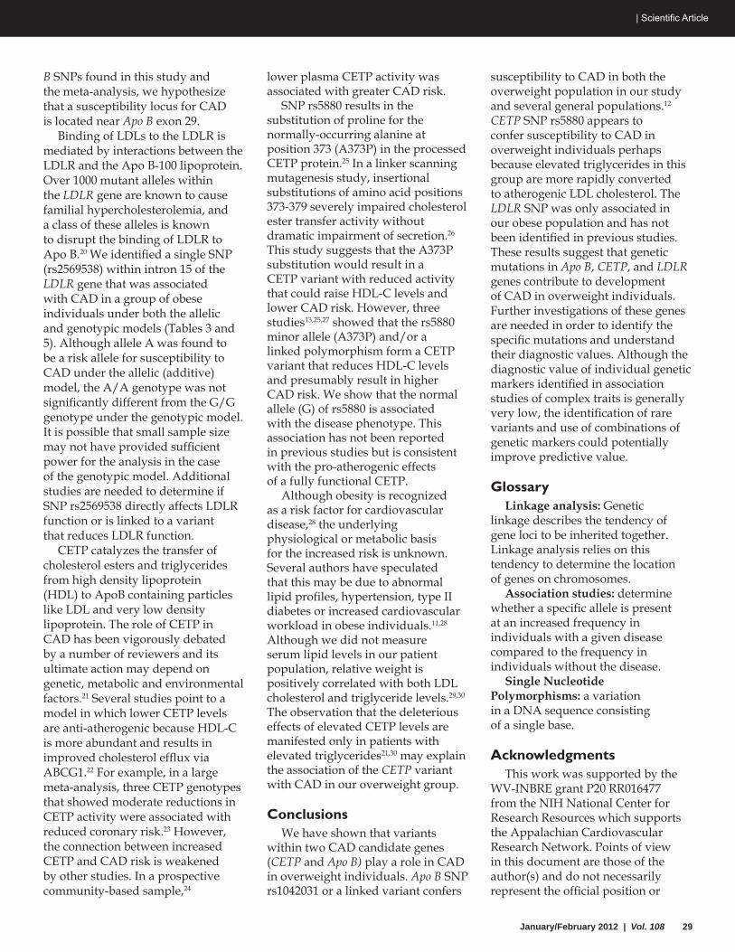
January/February 2012 | Vol. 108 29
| Scientific Article
B SNPs found in this study and the meta-analysis, we hypothesize that a susceptibility locus for CAD is located near Apo B exon 29.
Binding of LDLs to the LDLR is mediated by interactions between the LDLR and the Apo B-100 lipoprotein. Over 1000 mutant alleles within the LDLR gene are known to cause familial hypercholesterolemia, and a class of these alleles is known to disrupt the binding of LDLR to Apo B.20 We identified a single SNP (rs2569538) within intron 15 of the LDLR gene that was associated with CAD in a group of obese individuals under both the allelic and genotypic models (Tables 3 and 5). Although allele A was found to be a risk allele for susceptibility to CAD under the allelic (additive) model, the A/A genotype was not significantly different from the G/G genotype under the genotypic model. It is possible that small sample size may not have provided sufficient power for the analysis in the case of the genotypic model. Additional studies are needed to determine if SNP rs2569538 directly affects LDLR function or is linked to a variant that reduces LDLR function.
CETP catalyzes the transfer of cholesterol esters and triglycerides from high density lipoprotein (HDL) to ApoB containing particles like LDL and very low density lipoprotein. The role of CETP in CAD has been vigorously debated by a number of reviewers and its ultimate action may depend on genetic, metabolic and environmental factors.21 Several studies point to a model in which lower CETP levels are anti-atherogenic because HDL-C is more abundant and results in improved cholesterol efflux via ABCG1.22 For example, in a large meta-analysis, three CETP genotypes that showed moderate reductions in CETP activity were associated with reduced coronary risk.23 However, the connection between increased CETP and CAD risk is weakened by other studies. In a prospective community-based sample,24
lower plasma CETP activity was associated with greater CAD risk.
SNP rs5880 results in the substitution of proline for the normally-occurring alanine at position 373 (A373P) in the processed CETP protein.25 In a linker scanning mutagenesis study, insertional substitutions of amino acid positions 373-379 severely impaired cholesterol ester transfer activity without dramatic impairment of secretion.26 This study suggests that the A373P substitution would result in a CETP variant with reduced activity that could raise HDL-C levels and lower CAD risk. However, three studies13,25,27 showed that the rs5880 minor allele (A373P) and/or a linked polymorphism form a CETP variant that reduces HDL-C levels and presumably result in higher CAD risk. We show that the normal allele (G) of rs5880 is associated with the disease phenotype. This association has not been reported in previous studies but is consistent with the pro-atherogenic effects of a fully functional CETP.
Although obesity is recognized as a risk factor for cardiovascular disease,28 the underlying physiological or metabolic basis for the increased risk is unknown. Several authors have speculated that this may be due to abnormal lipid profiles, hypertension, type II diabetes or increased cardiovascular workload in obese individuals.11,28 Although we did not measure serum lipid levels in our patient population, relative weight is positively correlated with both LDL cholesterol and triglyceride levels.29,30 The observation that the deleterious effects of elevated CETP levels are manifested only in patients with elevated triglycerides21,30 may explain the association of the CETP variant with CAD in our overweight group.
Conclusions We have shown that variants
within two CAD candidate genes (CETP and Apo B) play a role in CAD in overweight individuals. Apo B SNP rs1042031 or a linked variant confers
susceptibility to CAD in both the overweight population in our study and several general populations.12 CETP SNP rs5880 appears to confer susceptibility to CAD in overweight individuals perhaps because elevated triglycerides in this group are more rapidly converted to atherogenic LDL cholesterol. The LDLR SNP was only associated in our obese population and has not been identified in previous studies. These results suggest that genetic mutations in Apo B, CETP, and LDLR genes contribute to development of CAD in overweight individuals. Further investigations of these genes are needed in order to identify the specific mutations and understand their diagnostic values. Although the diagnostic value of individual genetic markers identified in association studies of complex traits is generally very low, the identification of rare variants and use of combinations of genetic markers could potentially improve predictive value.
GlossaryLinkage analysis: Genetic
linkage describes the tendency of gene loci to be inherited together. Linkage analysis relies on this tendency to determine the location of genes on chromosomes.
Association studies: determine whether a specific allele is present at an increased frequency in individuals with a given disease compared to the frequency in individuals without the disease.
Single Nucleotide Polymorphisms: a variation in a DNA sequence consisting of a single base.
AcknowledgmentsThis work was supported by the
WV-INBRE grant P20 RR016477 from the NIH National Center for Research Resources which supports the Appalachian Cardiovascular Research Network. Points of view in this document are those of the author(s) and do not necessarily represent the official position or
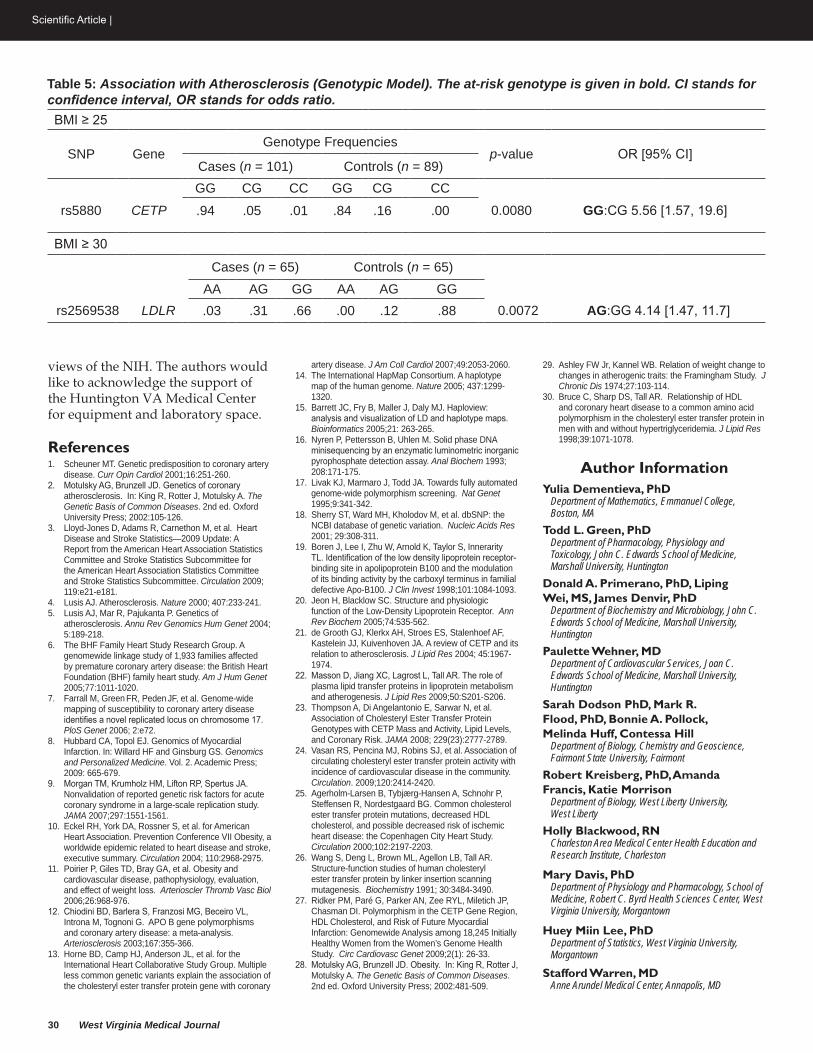
30 West Virginia Medical Journal
Scientific Article |
views of the NIH. The authors would like to acknowledge the support of the Huntington VA Medical Center for equipment and laboratory space.
References1. Scheuner MT. Genetic predisposition to coronary artery
disease. Curr Opin Cardiol 2001;16:251-260.2. Motulsky AG, Brunzell JD. Genetics of coronary
atherosclerosis. In: King R, Rotter J, Motulsky A. The Genetic Basis of Common Diseases. 2nd ed. Oxford University Press; 2002:105-126.
3. Lloyd-Jones D, Adams R, Carnethon M, et al. Heart Disease and Stroke Statistics—2009 Update: A Report from the American Heart Association Statistics Committee and Stroke Statistics Subcommittee for the American Heart Association Statistics Committee and Stroke Statistics Subcommittee. Circulation 2009; 119:e21-e181.
4. Lusis AJ. Atherosclerosis. Nature 2000; 407:233-241. 5. Lusis AJ, Mar R, Pajukanta P. Genetics of
atherosclerosis. Annu Rev Genomics Hum Genet 2004; 5:189-218.
6. The BHF Family Heart Study Research Group. A genomewide linkage study of 1,933 families affected by premature coronary artery disease: the British Heart Foundation (BHF) family heart study. Am J Hum Genet 2005;77:1011-1020.
7. Farrall M, Green FR, Peden JF, et al. Genome-wide mapping of susceptibility to coronary artery disease identifies a novel replicated locus on chromosome 17. PloS Genet 2006; 2:e72.
8. Hubbard CA, Topol EJ. Genomics of Myocardial Infarction. In: Willard HF and Ginsburg GS. Genomics and Personalized Medicine. Vol. 2. Academic Press; 2009: 665-679.
9. Morgan TM, Krumholz HM, Lifton RP, Spertus JA. Nonvalidation of reported genetic risk factors for acute coronary syndrome in a large-scale replication study. JAMA 2007;297:1551-1561.
10. Eckel RH, York DA, Rossner S, et al. for American Heart Association. Prevention Conference VII Obesity, a worldwide epidemic related to heart disease and stroke, executive summary. Circulation 2004; 110:2968-2975.
11. Poirier P, Giles TD, Bray GA, et al. Obesity and cardiovascular disease, pathophysiology, evaluation, and effect of weight loss. Arterioscler Thromb Vasc Biol 2006;26:968-976.
12. Chiodini BD, Barlera S, Franzosi MG, Beceiro VL, Introna M, Tognoni G. APO B gene polymorphisms and coronary artery disease: a meta-analysis. Arteriosclerosis 2003;167:355-366.
13. Horne BD, Camp HJ, Anderson JL, et al. for the International Heart Collaborative Study Group. Multiple less common genetic variants explain the association of the cholesteryl ester transfer protein gene with coronary
artery disease. J Am Coll Cardiol 2007;49:2053-2060. 14. The International HapMap Consortium. A haplotype
map of the human genome. Nature 2005; 437:1299-1320.
15. Barrett JC, Fry B, Maller J, Daly MJ. Haploview: analysis and visualization of LD and haplotype maps. Bioinformatics 2005;21: 263-265.
16. Nyren P, Pettersson B, Uhlen M. Solid phase DNA minisequencing by an enzymatic luminometric inorganic pyrophosphate detection assay. Anal Biochem 1993; 208:171-175.
17. Livak KJ, Marmaro J, Todd JA. Towards fully automated genome-wide polymorphism screening. Nat Genet 1995;9:341-342.
18. Sherry ST, Ward MH, Kholodov M, et al. dbSNP: the NCBI database of genetic variation. Nucleic Acids Res 2001; 29:308-311.
19. Boren J, Lee I, Zhu W, Arnold K, Taylor S, Innerarity TL. Identification of the low density lipoprotein receptor-binding site in apolipoprotein B100 and the modulation of its binding activity by the carboxyl terminus in familial defective Apo-B100. J Clin Invest 1998;101:1084-1093.
20. Jeon H, Blacklow SC. Structure and physiologic function of the Low-Density Lipoprotein Receptor. Ann Rev Biochem 2005;74:535-562.
21. de Grooth GJ, Klerkx AH, Stroes ES, Stalenhoef AF, Kastelein JJ, Kuivenhoven JA. A review of CETP and its relation to atherosclerosis. J Lipid Res 2004; 45:1967-1974.
22. Masson D, Jiang XC, Lagrost L, Tall AR. The role of plasma lipid transfer proteins in lipoprotein metabolism and atherogenesis. J Lipid Res 2009;50:S201-S206.
23. Thompson A, Di Angelantonio E, Sarwar N, et al. Association of Cholesteryl Ester Transfer Protein Genotypes with CETP Mass and Activity, Lipid Levels, and Coronary Risk. JAMA 2008; 229(23):2777-2789.
24. Vasan RS, Pencina MJ, Robins SJ, et al. Association of circulating cholesteryl ester transfer protein activity with incidence of cardiovascular disease in the community. Circulation. 2009;120:2414-2420.
25. Agerholm-Larsen B, Tybjærg-Hansen A, Schnohr P, Steffensen R, Nordestgaard BG. Common cholesterol ester transfer protein mutations, decreased HDL cholesterol, and possible decreased risk of ischemic heart disease: the Copenhagen City Heart Study. Circulation 2000;102:2197-2203.
26. Wang S, Deng L, Brown ML, Agellon LB, Tall AR. Structure-function studies of human cholesteryl ester transfer protein by linker insertion scanning mutagenesis. Biochemistry 1991; 30:3484-3490.
27. Ridker PM, Paré G, Parker AN, Zee RYL, Miletich JP, Chasman DI. Polymorphism in the CETP Gene Region, HDL Cholesterol, and Risk of Future Myocardial Infarction: Genomewide Analysis among 18,245 Initially Healthy Women from the Women’s Genome Health Study. Circ Cardiovasc Genet 2009;2(1): 26-33.
28. Motulsky AG, Brunzell JD. Obesity. In: King R, Rotter J, Motulsky A. The Genetic Basis of Common Diseases. 2nd ed. Oxford University Press; 2002:481-509.
29. Ashley FW Jr, Kannel WB. Relation of weight change to changes in atherogenic traits: the Framingham Study. J Chronic Dis 1974;27:103-114.
30. Bruce C, Sharp DS, Tall AR. Relationship of HDL and coronary heart disease to a common amino acid polymorphism in the cholesteryl ester transfer protein in men with and without hypertriglyceridemia. J Lipid Res 1998;39:1071-1078.
Author InformationYulia Dementieva, PhD
Department of Mathematics, Emmanuel College, Boston, MA
Todd L. Green, PhDDepartment of Pharmacology, Physiology and Toxicology, John C. Edwards School of Medicine, Marshall University, Huntington
Donald A. Primerano, PhD, Liping Wei, MS, James Denvir, PhD
Department of Biochemistry and Microbiology, John C. Edwards School of Medicine, Marshall University, Huntington
Paulette Wehner, MDDepartment of Cardiovascular Services, Joan C. Edwards School of Medicine, Marshall University, Huntington
Sarah Dodson PhD, Mark R. Flood, PhD, Bonnie A. Pollock, Melinda Huff, Contessa Hill
Department of Biology, Chemistry and Geoscience, Fairmont State University, Fairmont
Robert Kreisberg, PhD, Amanda Francis, Katie Morrison
Department of Biology, West Liberty University, West Liberty
Holly Blackwood, RNCharleston Area Medical Center Health Education and Research Institute, Charleston
Mary Davis, PhDDepartment of Physiology and Pharmacology, School of Medicine, Robert C. Byrd Health Sciences Center, West Virginia University, Morgantown
Huey Miin Lee, PhDDepartment of Statistics, West Virginia University, Morgantown
Stafford Warren, MDAnne Arundel Medical Center, Annapolis, MD
Table 5: Association with Atherosclerosis (Genotypic Model). The at‑risk genotype is given in bold. CI stands for confidence interval, OR stands for odds ratio.BMI ≥ 25
SNP GeneGenotype Frequencies
p-value OR [95% CI]Cases (n = 101) Controls (n = 89)GG CG CC GG CG CC
rs5880 CETP .94 .05 .01 .84 .16 .00 0.0080 GG:CG 5.56 [1.57, 19.6]
BMI ≥ 30
Cases (n = 65) Controls (n = 65)AA AG GG AA AG GG
rs2569538 LDLR .03 .31 .66 .00 .12 .88 0.0072 AG:GG 4.14 [1.47, 11.7]

entchas.com
Audiological Testing
Inhalant Allergy Testing & Treatment
Hearing Aid Evaluation & Placement Services
Computed Tomography (CT) for Sinuses & Ears
Complete Comprehensive Services
Board Certified Specialists
Ear, Nose and Throat Medical and Surgical Care
Michael R. Goins, MD G. Stephen Dawson, MD
F. Thomas Sporck, MD, FACSD. Richard Lough, MD
P. Todd Nichols, MD
500 Donnally Street Charleston, WV Suite 200
Hearing Aid Center 304.340.2222Appointments 304.340.2200

32 West Virginia Medical Journal
Scientific Article |
Asif Najmuddin, MD PGY II, Internal Medicine, WVU Hospitals, Morgantown
Sarah Hadique, MDFellow, Pulmonary and Critical Care Medicine, WVU Hospitals, Morgantown
John Parker, MDProfessor and Section Chief, Pulmonary and Critical Care Medicine, WVU Hospitals, Morgantown
AbstractStrongyloidiasis is a major global
health challenge that is often underestimated in many countries. In immuno-compromised hosts, an autoinfection can go unchecked with large numbers of invasive Strongyloides larvae disseminating widely and causing hyperinfection, with fatal consequences. This review will highlight a case of gram negative bacteremia complicated by meningitis and Adult Respiratory Distress Syndrome (ARDS) as a fatal outcome of Strongyloides hyperinfection, commonly known to occur in the setting of immunosuppression.
A middle aged female with chronic lymphocytic leukemia and hypogammaglobulinemia presented with severe respiratory distress requiring intubation. She had been on intermittent corticosteroids and multiple courses of antibiotics for the past six months for COPD exacerbations. Bronchoalveolar lavage showed Strongyloides stercoralis. Blood cultures grew Vancomycin Resistant Enterococci and a few days later Vancomycin Sensitive Enterococcus was found in the CSF. Afterwards, she grew Vancomycin Sensitive Enterococcus in the blood. She was treated with Ivermectin and Albendazole for Strongyloides and Linezolid and Vancomycin for these two different strains of E. faecium. After initial resolution of bacteremia and meningitis, she relapsed three weeks later with the same organism growing in the CSF. The clinical course continued to deteriorate with the development of significant neurological dysfunction. Poor nutritional state and ventilator associated pneumonia contributed to this downward trend. After a detailed discussion with the family, life support was discontinued and the patient succumbed to her illness.
IntroductionStrongyloides stercoralis is a soil
transmitted nematode, endemic in tropical and subtropical areas. Due to increasing immigration from countries endemic with strongyloides, physicians in developed countries may also encounter this condition. Infection usually results in asymptomatic chronic disease of the gut, which can go undetected for decades. Eosinophilia and larvae in stools are often the only indicatiors of infection. The disease is classified as ‘acute’, ‘chronic’ and ‘severe’ strongyloidiasis and commonly manifests with cutaneous, pulmonary and intestinal features. Common clinical features of acute strongyloidiasis include maculopapular rash involving the feet, epigastric discomfort, diarrhea, occasional nausea and vomiting. Other manifestations include cough, dyspnea, wheezing, low grade fever or constipation.
Strongyloides stercoralis is also capable of causing autoinfection in the host. Strongyloides Hyperinfection (SH) is a life threatening complication of chronic strongyloidiases and usually occurs in the setting of immunocompromised state (patients with malignancy, organ transplantation or concurrent human T-cell-lymphocytic virus 1 infection or those on corticosteroid therapy). Mortality is largely due to gram negative bacteremia leading to adult respiratory distress syndrome and rarely, meningitis. We present a unique case of a patient with SH complicated by simultaneous occurrence of vancomycin resistant enteroccus bacteremia and
vancomycin susceptible enterococcus bacteremia and meningitis.
Case ReportA 68-year-old diabetic female
with chronic lymphocytic leukemia was transferred from an outside facility with severe respiratory distress. She was admitted to the outside hospital three days prior to transfer, with complaints of fever, shortness of breath, cough with yellow sputum, wheezing, and generalized weakness. She had been on intermittent corticosteroids and multiple courses of antibiotics for the past six months for suspected COPD exacerbations. Her recent antibiotics included cephalosporins, fluoroquinolones and macrolides. Her initial white cell count was 38.0 (thou/ul) and hemoglobin was 7.7 g/dl for which she received 2 units of packed red blood cells. Her chest x-ray showed bilateral infiltrates.
On presentation to our facility, her temperature was 38.4 C, pulse was 111 beats per minute, respirations were 22/min, blood pressure was 100/60 mmHg. The oxygen saturation was 88% on 2 liters nasal cannula. On physical examination, the patient appeared tired and in mild distress. She had a diffuse maculopapular rash. Heart rhythm was irregularly irregular. Lungs had bilateral crackles and wheezes. The remainder of the physical examination including head and neck, abdominal, musculoskeletal and neurologic examination was normal.
The initial work up at our institution showed a WBC count of 57.7 (thou/ul) with neutrophils 33%, lymphocytes 64% (Table 1). The ABG results showed pH 7.45,
strongyloides Hyperinfection syndrome complications: A case Report and Review of the Literature

January/February 2012 | Vol. 108 33
| Scientific Article
PCO2 78, PO2 50, Bicarbonate 45, PaO2/FiO2 ratio of 114. Chest X-ray revealed increased interstitial opacities and mild bronchial wall thickening with suggestion of bronchiectasis (Figure 1). CT head showed no acute intracranial process. CT chest demonstrated bilateral areas of ground-glass opacity with reticular and nodular thickening. Interlobular septal thickening with areas of geographic sparing and traction bronchiectasis were also noted (Figure 2). A transthoracic echo showed left ventricular ejection fraction in the range of 65 % to 70 %. Features were consistent with mild diastolic dysfunction. There was very mild aortic valve stenosis with a mean gradient of 11mm Hg. Estimated peak right ventricular systolic pressure was in the range of 45 mmHg to 50 mmHg.
Due to progressive respiratory failure, the patient was intubated
and transferred to our medical intensive care unit. She was started on broad spectrum antibiotics including vancomycin, ciprofloxacin, tobramycin and caspofungin. Bronchoscopy was performed the next day and bronchoalveolar lavage showed a worm which was later identified as Strongyloides stercoralis (Figure 3). The patient continued to remain unresponsive and a spinal tap was performed, that showed a pyogenic CSF picture and grew Vancomycin Sensitive Enterococci.
Her blood culture drawn on the day of admission grew Vancomycin Resistant Enterococci and a few days later, she grew a strain of Vancomycin Sensitive Enterococci in her CSF. She was treated with Albendazole and Ivermectin for SH for two weeks, Linezolid for vancomycin resistant Enterococcus (VRE) bacteremia and vancomycin for vancomycin Sensitive
Enterococcus (VSE) meningitis. She was also started on IVIG due to low levels of immunoglobulins.
A few days after she had completed a two week course of antiparasitic drugs, she developed peripheral blood eosinophilia but without any redemonstration of Strongyloides in respiratory secretions. However, she was still treated with one more week of Ivermectin, after which the eosinophilia resolved.
The patient started to show a clinical response and was neurologically stable with clearing of CSF (Table 2). However, she decompensated again due to respiratory failure. She was found to have reoccurence of VSE bacteremia (Table 3) and CSF analysis showed a pyogenic process that again grew VSE.
A tracheostomy was performed for her long term ventilation. A
Edward C. Martin, Responsible Attorney | [email protected] | www.fsblaw.com | (304) 345-0200 | (800) 416-3225
…in litigation, privacy and security compliance, certificate of need, medical staff and professional disciplinary matters, credentialing concerns, complex regulatory matters and business transactions.
health care practice group
Charleston
Ryan A. BrownRobert L. CoffieldAlaina N. CrislipJ. Dustin Dillard
Sam FoxMichele GrinbergJohn D. Hoffman
Amy R. Humphreys
Justin D. JackRichard D. JonesEdward C. MartinMark A. RobinsonAmy L. Rothman
Don R. Sensabaugh, Jr.Salem C. Smith
Morgantown
Stephen R. BrooksStacie D. Honaker
WheelingDavid S. GivensPhillip T. GlyptisRobert C. James
Helping West Virginia pHysicians take the right path…
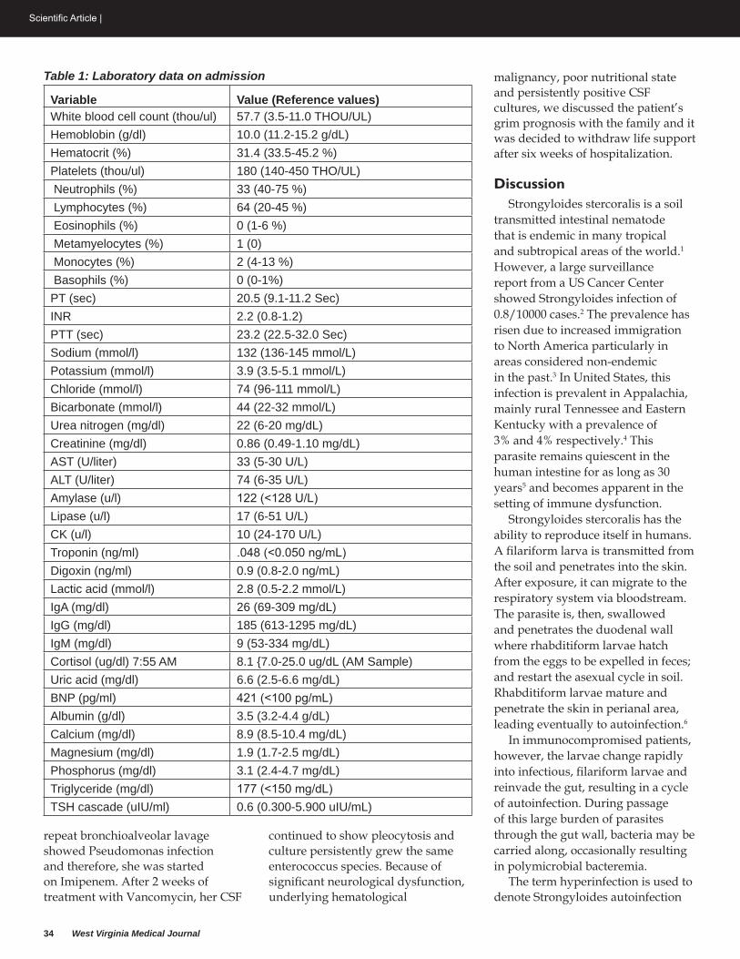
34 West Virginia Medical Journal
Scientific Article |
repeat bronchioalveolar lavage showed Pseudomonas infection and therefore, she was started on Imipenem. After 2 weeks of treatment with Vancomycin, her CSF
continued to show pleocytosis and culture persistently grew the same enterococcus species. Because of significant neurological dysfunction, underlying hematological
malignancy, poor nutritional state and persistently positive CSF cultures, we discussed the patient’s grim prognosis with the family and it was decided to withdraw life support after six weeks of hospitalization.
DiscussionStrongyloides stercoralis is a soil
transmitted intestinal nematode that is endemic in many tropical and subtropical areas of the world.1 However, a large surveillance report from a US Cancer Center showed Strongyloides infection of 0.8/10000 cases.2 The prevalence has risen due to increased immigration to North America particularly in areas considered non-endemic in the past.3 In United States, this infection is prevalent in Appalachia, mainly rural Tennessee and Eastern Kentucky with a prevalence of 3% and 4% respectively.4 This parasite remains quiescent in the human intestine for as long as 30 years5 and becomes apparent in the setting of immune dysfunction.
Strongyloides stercoralis has the ability to reproduce itself in humans. A filariform larva is transmitted from the soil and penetrates into the skin. After exposure, it can migrate to the respiratory system via bloodstream. The parasite is, then, swallowed and penetrates the duodenal wall where rhabditiform larvae hatch from the eggs to be expelled in feces; and restart the asexual cycle in soil. Rhabditiform larvae mature and penetrate the skin in perianal area, leading eventually to autoinfection.6
In immunocompromised patients, however, the larvae change rapidly into infectious, filariform larvae and reinvade the gut, resulting in a cycle of autoinfection. During passage of this large burden of parasites through the gut wall, bacteria may be carried along, occasionally resulting in polymicrobial bacteremia.
The term hyperinfection is used to denote Strongyloides autoinfection
Table 1: Laboratory data on admission
Variable Value (Reference values)White blood cell count (thou/ul) 57.7 (3.5-11.0 THOU/UL)Hemoblobin (g/dl) 10.0 (11.2-15.2 g/dL)Hematocrit (%) 31.4 (33.5-45.2 %)Platelets (thou/ul) 180 (140-450 THO/UL) Neutrophils (%) 33 (40-75 %) Lymphocytes (%) 64 (20-45 %) Eosinophils (%) 0 (1-6 %) Metamyelocytes (%) 1 (0) Monocytes (%) 2 (4-13 %) Basophils (%) 0 (0-1%)PT (sec) 20.5 (9.1-11.2 Sec)INR 2.2 (0.8-1.2)PTT (sec) 23.2 (22.5-32.0 Sec)Sodium (mmol/l) 132 (136-145 mmol/L)Potassium (mmol/l) 3.9 (3.5-5.1 mmol/L)Chloride (mmol/l) 74 (96-111 mmol/L)Bicarbonate (mmol/l) 44 (22-32 mmol/L)Urea nitrogen (mg/dl) 22 (6-20 mg/dL)Creatinine (mg/dl) 0.86 (0.49-1.10 mg/dL)AST (U/liter) 33 (5-30 U/L)ALT (U/liter) 74 (6-35 U/L)Amylase (u/l) 122 (<128 U/L)Lipase (u/l) 17 (6-51 U/L)CK (u/l) 10 (24-170 U/L)Troponin (ng/ml) .048 (<0.050 ng/mL)Digoxin (ng/ml) 0.9 (0.8-2.0 ng/mL)Lactic acid (mmol/l) 2.8 (0.5-2.2 mmol/L)IgA (mg/dl) 26 (69-309 mg/dL)IgG (mg/dl) 185 (613-1295 mg/dL)IgM (mg/dl) 9 (53-334 mg/dL)Cortisol (ug/dl) 7:55 AM 8.1 {7.0-25.0 ug/dL (AM Sample)Uric acid (mg/dl) 6.6 (2.5-6.6 mg/dL)BNP (pg/ml) 421 (<100 pg/mL)Albumin (g/dl) 3.5 (3.2-4.4 g/dL)Calcium (mg/dl) 8.9 (8.5-10.4 mg/dL)Magnesium (mg/dl) 1.9 (1.7-2.5 mg/dL)Phosphorus (mg/dl) 3.1 (2.4-4.7 mg/dL)Triglyceride (mg/dl) 177 (<150 mg/dL)TSH cascade (uIU/ml) 0.6 (0.300-5.900 uIU/mL)
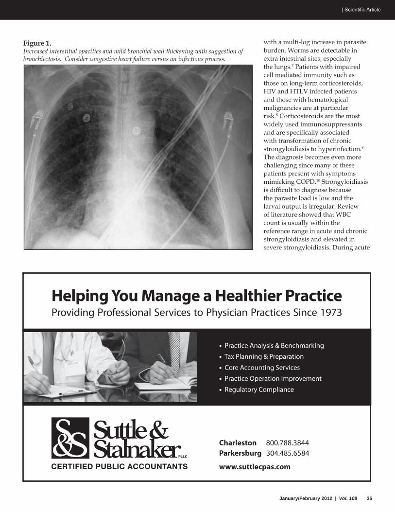
January/February 2012 | Vol. 108 35
| Scientific Article
with a multi-log increase in parasite burden. Worms are detectable in extra intestinal sites, especially the lungs.7 Patients with impaired cell mediated immunity such as those on long-term corticosteroids, HIV and HTLV infected patients and those with hematological malignancies are at particular risk.8 Corticosteroids are the most widely used immunosuppressants and are specifically associated with transformation of chronic strongyloidiasis to hyperinfection.9 The diagnosis becomes even more challenging since many of these patients present with symptoms mimicking COPD.10 Strongyloidiasis is difficult to diagnose because the parasite load is low and the larval output is irregular. Review of literature showed that WBC count is usually within the reference range in acute and chronic strongyloidiasis and elevated in severe strongyloidiasis. During acute
Helping You Manage a Healthier PracticeProviding Professional Services to Physician Practices Since 1973
Charleston 800.788.3844 Parkersburg 304.485.6584
www.suttlecpas.com
• Practice Analysis & Benchmarking
• Tax Planning & Preparation
• Core Accounting Services
• Practice Operation Improvement
• Regulatory Compliance
Figure 1.Increased interstitial opacities and mild bronchial wall thickening with suggestion of bronchiectasis. Consider congestive heart failure versus an infectious process.

36 West Virginia Medical Journal
Scientific Article |
infection eosinophilia is common, intermittent during chronic infection, and frequently absent in severe strongyloidiasis. Microscopically identifying S. stercoralis larvae in stool is a definitive diagnostic test. However, results of a single stool examination by use of conventional techniques fail to detect larvae in up to 70% of the cases due to fluctuating larval exretion and, therefore,
consecutive stool examinations have been recommended to increase the yield.9 Blood cultures in strongyloidiasis often yield growth of enteric pathogens, most commonly Escherichia coli and/or Klebsiella species. Strongyloides serology (enzyme immunoassay, indirect fluorescent antibody) has 88-95% sensitivity. Sensitivity may be lower in severely
immunocompromised patients. This test however, cannot differentiate between past and present infection.8
Strongyloides stercoralis is a unique parasite that can cause fatal disease years after the exposure, often in the setting of SH, with mortality as high as 87%.6 Lam et al. carried out a retrospective study and evaluated the characteristic features of seven patients. All the patients left the endemic area more than 20 years ago.11 Mortality is mainly due to gram negative, often polymicrobial bacteremia and meningitis because of leakage of gut flora from the ulcerated bowel mucosa or bacteria carried on the surface of the larvae as they migrate to extra intestinal sites. A review published in 1999 identified 38 case reports of bacterial sepsis complicating strongyloidiasis, 68% of those patients had bacteremia out of which 12% were polymicrobial.12
Gram negative bacterial meningitis has been reported in Strongyloides hyperinfection especially in association with immunosuppression.13 Our patient had active chronic lymphocytic leukemia with hypogammaglobulinemia and was on intermittent steroids for six months, which explains her predisposition to SH. In addition, she received multiple courses of antibiotics to treat COPD exacerbation including cephalosporins, flouroquinolones and macrolides, which predisposed her to VRE bacteremia. Vancomycin resistance is found to be an independent predictor of mortality in severe enterococcal bacteremia. 14 Furthermore, patients infected with Enterococcal faecium have a worse prognosis than those with E. faecalis.15 Our case is unique for its rarity. Although association of Enterococcus faecium meningitis with SH has been reported,16 to our knowledge there are no reported cases of concomitant VSE and VRE bacteremia in a patient with SH in literature.
Figure 2. Bilateral areas of ground-glass opacity, reticular and nodular thickening, and an interlobular septal thickening with areas of geographic sparing and traction bronchiectasis.
Figure 3.Strongyloides stercoralis in the BAL.
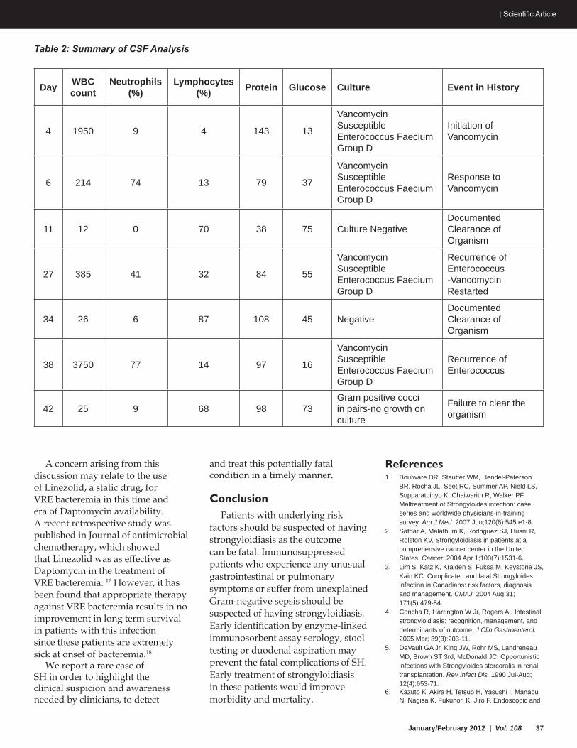
January/February 2012 | Vol. 108 37
| Scientific Article
A concern arising from this discussion may relate to the use of Linezolid, a static drug, for VRE bacteremia in this time and era of Daptomycin availability. A recent retrospective study was published in Journal of antimicrobial chemotherapy, which showed that Linezolid was as effective as Daptomycin in the treatment of VRE bacteremia. 17 However, it has been found that appropriate therapy against VRE bacteremia results in no improvement in long term survival in patients with this infection since these patients are extremely sick at onset of bacteremia.18
We report a rare case of SH in order to highlight the clinical suspicion and awareness needed by clinicians, to detect
and treat this potentially fatal condition in a timely manner.
ConclusionPatients with underlying risk
factors should be suspected of having strongyloidiasis as the outcome can be fatal. Immunosuppressed patients who experience any unusual gastrointestinal or pulmonary symptoms or suffer from unexplained Gram-negative sepsis should be suspected of having strongyloidiasis. Early identification by enzyme-linked immunosorbent assay serology, stool testing or duodenal aspiration may prevent the fatal complications of SH. Early treatment of strongyloidiasis in these patients would improve morbidity and mortality.
References1. Boulware DR, Stauffer WM, Hendel-Paterson
BR, Rocha JL, Seet RC, Summer AP, Nield LS, Supparatpinyo K, Chaiwarith R, Walker PF. Maltreatment of Strongyloides infection: case series and worldwide physicians-in-training survey. Am J Med. 2007 Jun;120(6):545.e1-8.
2. Safdar A, Malathum K, Rodriguez SJ, Husni R, Rolston KV. Strongyloidiasis in patients at a comprehensive cancer center in the United States. Cancer. 2004 Apr 1;100(7):1531-6.
3. Lim S, Katz K, Krajden S, Fuksa M, Keystone JS, Kain KC. Complicated and fatal Strongyloides infection in Canadians: risk factors, diagnosis and management. CMAJ. 2004 Aug 31; 171(5):479-84.
4. Concha R, Harrington W Jr, Rogers AI. Intestinal strongyloidiasis: recognition, management, and determinants of outcome. J Clin Gastroenterol. 2005 Mar; 39(3):203-11.
5. DeVault GA Jr, King JW, Rohr MS, Landreneau MD, Brown ST 3rd, McDonald JC. Opportunistic infections with Strongyloides stercoralis in renal transplantation. Rev Infect Dis. 1990 Jul-Aug; 12(4):653-71.
6. Kazuto K, Akira H, Tetsuo H, Yasushi I, Manabu N, Nagisa K, Fukunori K, Jiro F. Endoscopic and
Table 2: Summary of CSF Analysis
Day WBCcount
Neutrophils(%)
Lymphocytes(%) Protein Glucose Culture Event in History
4 1950 9 4 143 13
Vancomycin Susceptible Enterococcus Faecium Group D
Initiation of Vancomycin
6 214 74 13 79 37
Vancomycin Susceptible Enterococcus Faecium Group D
Response to Vancomycin
11 12 0 70 38 75 Culture NegativeDocumented Clearance of Organism
27 385 41 32 84 55
Vancomycin Susceptible Enterococcus Faecium Group D
Recurrence of Enterococcus -Vancomycin Restarted
34 26 6 87 108 45 NegativeDocumented Clearance of Organism
38 3750 77 14 97 16
Vancomycin Susceptible Enterococcus Faecium Group D
Recurrence of Enterococcus
42 25 9 68 98 73Gram positive cocci in pairs-no growth on culture
Failure to clear the organism

38 West Virginia Medical Journal
Scientific Article |
histopathological study on the duodenum of Strongyloides stercoralis hyperinfection. World J Gastroenterol. 2008;14(11):1768–1773.)
7. Siddiqui AA, Berk SL. Diagnosis of Strongyloides stercoralis infection. Clin Infect Dis. 2001 Oct 1;33(7):1040-7. Epub 2001 Sep 5.
8. Keiser PB, Nutman TB. Strongyloides stercoralis in the Immunocompromised Population. Clin Microbiol Rev. 2004 Jan; 17(1):208-17.
9. Asdamongkol N, Pornsuriyasak P, Sungkanuparph S. Risk factors for strongyloidiasis hyperinfection and clinical outcomes. Southeast Asian J Trop Med Public Health. 2006 Sep; 37(5):875-84.
10. Liu HC, Hsu JY, Chang KM. Strongyloides stercoralis hyperinfection presenting with symptoms mimicking acute exacerbation of chronic obstructive pulmonary disease. J Chin Med Assoc. 2009 Aug; 72(8):442-5.
11. Lam CS, Tong MK, Chan KM, Siu YP. Disseminated strongyloidiasis: a retrospective study of clinical course and outcome. Eur J Clin Microbiol Infect Dis. 2006;25(1):14–8. doi: 10.1007/s10096-005-0070-2.
12. Link K, Orenstein R. Bacterial complications of strongyloidiasis: Streptococcus bovis meningitis. South Med J. 1999 Jul; 92(7):728-31.
13. Newberry AM, Williams DN, Stauffer WM, Boulware DR, Hendel-Paterson BR, Walker PF. Strongyloides hyperinfection presenting as acute respiratory failure and gram-negative sepsis. Chest. 2005 Nov;128(5):3681-4.
14. DiazGranados CA, Zimmer SM, Klein M, Jernigan JA. Comparison of mortality associated with vancomycin-resistant and vancomycin-susceptible enterococcal bloodstream infections: a meta-analysis. Clin Infect Dis. 2005 Aug 1;41(3):327-33. Epub 2005 Jun 28.
15. Chou YY, Lin TY, Lin JC, Wang NC, Peng MY, Chang FY. Vancomycin-resistant enterococcal bacteremia: comparison of clinical features and outcome between Enterococcus faecium and Enterococcus faecalis. J Microbiol Immunol Infect. 2008 Apr;41(2):124-9.
16. Zeana C, Kubin CJ, Della-Latta P, Hammer SM. Vancomycin-resistant Enterococcus faecium meningitis successfully managed with linezolid: case report and review of the literature. Clin Infect Dis. 2001 Aug 15;33(4):477-82. Epub 2001 Jul 11.
17. Mave V, Garcia-Diaz J, Islam T, Hasbun R Vancomycin-resistant enterococcal bacteraemia: is daptomycin as effective as linezolid .J Antimicrob Chemother. 2009 Jul;64(1):175-80. Epub 2009 May 7.
18. Han SH, Chin BS, Lee HS, Jeong SJ, Choi HK, Kim CO, Yong D, Choi JY, Song YG, Lee K, Kim JM Vancomycin-resistant enterococci bacteremia: risk factors for mortality and influence of antimicrobial therapy on clinical outcome. J Infect. 2009 Mar;58(3):182-90. Epub 2009 Feb 23.
Table 3: Summary of Blood CulturesDay Blood culture result1 Vancomycin resistence Enterococcus Faecium Group D3 No growth5 No growth7 Vancomycin Susceptible Enterococcus Faecium Group D10 No growth21 No growth27 No growth35 No growth40 No growth42 No growth
304.343.EECP(3327) | eecpwv.com
The sensible choicefor specialized care.
Providing comprehensive pediatric and adult ear, nose and throat care, Eye & Ear Clinic Physicians also offers complete hearing aid services and allergy testing.
Featuring an extensive selection oF digital hearing aid devices and accessories From

January/February 2012 | Vol. 108 39
| WESPAC Contributors
2011 WespAc contributors
2012 WespAc contributors
The WVSMA would like to thank the following physicians, residents, medical students and Alliance members for their contributions to WESPAC. These contributions were received as of December 13, 2011:
Chairman’s Club ($1000)M. Barry Louden Jr., MDFriday G. Simpson, MDPhillip R. Stevens, MDCharles F. Whitaker III, MD
Extra Miler ($500)MaryAnn Nicolas Cater, DOJames L. Comerci, MDGeneroso D. Duremdes, MDCarlos C. Jimenez, MDMichael A. Kelly, MDKenneth C. Nanners, MDFrank A. Scattaregia, MDMichael A. Stewart, MD
Dollar-A-Day ($365)Greenbrier D. Almond, MDEdward F. Arnett, MDR. Mark Hatfield, MDJohn D. Holloway, MDMichael A. Istfan, MDSushil K. Mehrotra, MDJoseph B. Selby, MDJohn G. Tellers, MDL. Blair Thrush, MDJohn A. Wade, Jr., MD
Campaigner Plus (> $100)Finbar G. Powderly, MDRichard M. Fulks, MDManuel A. Gomez, MD
Campaigner ($100)James M. Carrier, MDStephen P. Cassis, MDRobert A. Caveney, MDPatsy P. Cipoletti, MDSerge Cormier, MDThomas O. Dickey, Jr., MDRuperto D. Dumapit, Jr., MDKenneth F. McNeil, MDStephen K. Milroy, MDBrian D. Powderly, MDMichael M. Rezaian, MDOphas Vongxaiburana, MD
Resident/Student ($20)Kyle T. Kutrovac, MD
DonorLynn ComerciPedro F. Lo, MD
Extra Miler ($500)James P. Clark II, MDAhmed D. Faheem, MDMichael A. Kelly, MDMichael A. Stewart, MD
Dollar-A-Day ($365)Edward F. Arnett, MDGina R. Busch, MDSamuel R. Davis, MDDavid E. Hess, MDJohn D. Holloway, MDTheodore A. Jackson, MDSushil K. Mehrotra, MDBradley J. Richardson, MD
Campaigner ($100)John A. Adeniyi, MD
Derek H. Andreini, MDMichael M. Boustany, MDAdam J. Breinig, DOJames M. Carrier, MDJames D. Felsen, MDRichard M. Fulks, MDPhillip Bradley Hall, MDMuthusami Kuppusami, MDNancy N. Lohuis, MDTony Majestro, MDStephen K. Milroy, MDWayne Spiggle, MDOphas Vongxaiburana, MDSherri A. Young, DO
Resident/Student ($20)Richard W. Eller, MD
WESPAC is the West Virginia State Medical Association’s bipartisan po-litical action committee. We work throughout the year with elected offi-cials to make sure they understand the many facets of our healthcare system.
WESPAC’s goal is to organize the physi-cian community into a powerful voice for quality healthcare in the West Virginia Legislature. We seek to pre-serve the vital relationship between you and your patients by educating our legislators about issues important to our physicians.
WESPAC contributions provide critical support for our endorsed candidates. Your contribution can make the differ-ence between a pro-physician/patient candidate winning or losing.
To make a contribution to WESPAC, please call
(304) 925-0342, ext. 12
What’s Good for Them Is Good for You. UniCare.Why UniCare?
For You:• EnhancedMedicaidreimbursements
• Electronicclaimssubmission
• Fastpayments
• Electronicfundstransfer
And for Them:• Freehealthimprovementanddisease
managementprograms
• Largereferralnetwork
• CommunityResourceCenterofferingspecialeventsandservices
Callusat1-888-611-9958tolearnmoreaboutthebenefitsofbeingaUniCareMedicaidprovider.
UniCare Health Plan of West Virginia, Inc. ® Registered mark of WellPoint, Inc. 0909 WV0015533 9/09
January/February 2012 | Vol. 108 41
Educating the
public and those
in the health
care professions
regarding issues
of importance to
the delivery of
health care in WV.
West Virginia State Medical Association Alliance | NEWS
The West Virginia State Medical Association Alliance has always been a proponent for improving the health of West Virginians. We renew this pledge for 2012 by supporting initiatives for the safe and proper disposal of unwanted medications. The Food and Drug Administration (FDA) and the Drug Enforcement Administation (DEA) are at the forefront of these efforts nationally.
At the Alliance's recent fall board meeting, plans to undertake this project were outlined by board members. Michael O’Neil, PharmD., Chairman, West Virginia Controlled Substance Advisory Board and Associate Professor, Department of Pharmacy Practice, University of Charleston School of Pharmacy presented a step by step process in initiating a ‘Drug Take Back Program’ for the Medical Alliance beginning with obtaining approval from the DEA. He discussed the types of medications that will be collected, who will be involved, where the drugs will go after collection, and how are they destroyed.
Plans for additional projects were addressed by Chris Vaught, CEO of Vaught, Inc. and inventor of the Element MDS (medical disposal system). Element MDS products contain an MDS powder packet which when mixed with unwanted medications and tap water hold the
medication in suspension and form a solid gel, making the medication undesirable. Now widely used in West Virginia, The Hospice for Southern West Virginia was the first to use this product.
The Raleigh County Medical Alliance will distribute brochures on the safe disposal of medication in an effort to create public awareness on the importance of safe disposal of medication to prevent drug abuse, protect the environment, and prevent identity theft.
Health projects are great ideas, and members are the movers. As 2012 begins, the WVSMA Alliance is faced with the challenge of increasing its membership. How do we make volunteer work a fulfilling experience? The WVSMA Alliance is a special group of physician spouses, bonded by a vision and mission of keeping healthy West Virginians. Members get together for fellowship; enjoying the camaraderie of members from different counties, and networking with other physician spouses of various professional, business, and cultural backgrounds. We look forward to having a core group of both the dedicated long-time members and the new generation of young physician spouses.
Rose RomeroWVSMAA President
In With The New
Drug or Alcohol Problem? Mental Illness?If you have a drug or alcohol problem, or are suffering from a mental illness you can get help by contacting the West Virginia
Medical Professionals Health Program. Information about a practitioner’s participation in the program is confidential. Prac titioners entering the program as self-referrals without a complaint filed against them are not reported to their licensing board.
A L L C A L L S A R E C O N F I D E N T I A LWest Virginia Medical Professionals Health Program
PO Box 40027, Charleston, WV 25364(304) 414-0400 | www.wvmphp.org

42 West Virginia Medical Journal42 West Virginia Medical Journal
West Virginia University Healthcare and Health Sciences | NEWS
Heart surgeon Anthony Holden, M.D., has joined the WVU Heart Institute. He specializes in cardiac surgery, including coronary
artery bypass grafting, aortic valve surgery, surgery for atrial fibrillation and mitral valve repair surgery.
“I’m excited to be a part of the WVU Heart Institute,” Dr. Holden said. “The people of West Virginia
are fortunate to have a unique, multidisciplinary group of physicians at WVU, including cardiologists and cardiac surgeons. Together, we individualize each patient’s care with a collaborative, team approach.”
Holden earned his medical degree from Northeastern Ohio Universities College of Medicine and completed specialty training in general surgery at the University of Arizona Health Science Center and in thoracic surgery at University Hospital SUNY, Syracuse, NY. Holden is board certified in general and thoracic surgery.
The medical team at the WVU Heart Institute provides comprehensive heart care, including intervention, management and rehabilitation for cardiac patients. WVU heart and thoracic surgeons continually make advances in treating patients with cardiothoracic diseases, performing hundreds of open-heart and thoracic surgeries each year. The team specializes in complex cardiac surgery cases, including all aspects of cardiac valve surgery and surgery for heart arrhythmia.
WVU Healthcare welcomes new heart surgeonDr. Anthony Holden joins WVU Heart Institute
WVU Healthcare offers new lung cancer screening program
WVU Healthcare is offering a new low-cost screening program to help patients who are at risk for lung cancer.
The Lung Cancer Screening Program is a multidisciplinary effort aimed at catching the disease in its early stages by offering low-dose helical chest CT scan and smoking cessation counseling for $99.
The rationale for the creation of the program stems from results of the National Cancer Institute’s National Lung Screening Trial, published in the New England Journal of Medicine in August 2011.
“This study showed an earlier detection of lung cancer and demonstrated a 20 percent reduction in lung cancer mortality, among current and former smokers, when screened with low-dose helical chest CT versus chest x-ray,”
Harakh V. Dedhia, MD, professor in the Department of Medicine, Section of Pulmonary and Critical Care said. “Early detection of lung cancer is critically important because symptoms associated with this disease usually do not occur until the cancer has advanced and is more difficult to treat.”
To qualify for the screening program, patients must be 55 to 74 years old, and either an active smoker with at least a 30 pack-year history, or have quit smoking within the past 15 years.
Eligible patients need to visit their primary care physician to obtain a referral and an order for the low-dose helical chest CT scan, which will be interpreted by a board certified radiologist at WVU. The primary care physician can order the scan and FAX the request to
304-598-6375. The patient should call 1-855-WVU LUNG, option 1 to schedule an appointment. A nurse will call the patient two or three business days following the CT scan and the results will be mailed to the patient and primary care physician for continuity of care and optimized communication. If the results are abnormal, the patient may choose to consult with their primary care physician regarding follow-up care or be referred to a WVUH specialty clinic.
WVU is the only major cancer center in West Virginia to offer the new low cost screening program.
For information on the WVU Healthcare Lung Cancer Screening Program call Kim Yow, RN or Christina Ayers, RN at 304-293-9525 or 304-293-2288.
Anthony Holden, Md
January/February 2012 | Vol. 108 43
The American Recovery and Reinvestment Act of 2009 (Recovery Act) was signed into law by President Obama on February 17, 2009. The law includes the Health Information Technology for Economic and Clinical Health Act, or the “HITECH Act,” which established programs under the Centers for Medicare and Medicaid Services (CMS) to:
Establish a set of specific • requirements to be considered “certified” electronic health record (EHR) technology;Establish a series of criteria to • which healthcare providers and hospitals must adhere in order to qualify as meaningful users of health information technology (HIT) and receive incentive payments;Establish a series of quality • measures on which providers and hospitals must report in order to receive the incentive payments.
A Meaningful Use workgroup was charged with developing recommendations to the HIT Policy Committee on how to define meaningful use in the short and long-term, including: 1) the ways in which EHRs can support meaningful use, and 2) how providers can demonstrate and receive financial incentives for meaningful use.
On July 13, 2010, the US Centers for Medicare and Medicaid Services (CMS) released a Final Rule establishing the criteria with which eligible professionals (EPs), other health providers and hospitals must comply in order to qualify for the incentive payments
that are available with clinicians through the American Recovery and Reinvestment Act. Providers are not required to begin participating in the incentive program in 2011; however, providers must begin receiving payment no later than 2016.
As the US rolls out its largest ever investment in HIT, meaningful use (MU) promises to dramatically transform the delivery of care. One of the primary benefits of image access in the electronic medical record (EMR) is patient education.
CMS’s goal is for the definition of meaningful use to be consistent with applicable provisions of Medicare and Medicaid law while continually advancing the contributions certified EHR technology can make to improving health care quality, efficiency, and patient safety. To accomplish this, CMS’ final rule would phase in more robust criteria for demonstrating meaningful use in three stages.
Stages for Meaningful Use:Stage 1: Establish criteria
for Meaningful Use. Stage 1 has been completed.
Stage 2: Would expand upon Stage 1 criteria in the areas of disease management, clinical decision support, medication management support, patient access to their health information, transitions in care, quality measurement and research, and bi-directional communication with public health agencies which is not yet finalized. Stage two will further enhance state public health operations. This stage will allow
for the meaningful exchange of public health data with state data systems for syndromic surveillance, electronic laboratory reporting and immunization encounter reporting. The West Virginia Statewide Immunization Information System (WVSIIS) is a 12 year old system that provides a single record for a person, from records that are entered or submitted from multiple sources. This process is somewhat time consuming, but bi-directional data exchange using HL7 version 2.5.1 will allow data to be sent to and from WVSIIS using the physician’s EMR. Bi-directional data exchange allows both parties to have the most up-to-date records and saves physicians time in researching a patient’s immunization history while ensuring that all persons are age-appropriately immunized.
Stage 3: Would focus on achieving improvement in quality, safety and efficiency, focusing on decision support for national high priority conditions, patient access to self management tools, access to comprehensive patient data, and improving population health outcomes.
Loretta Haddy, PhD, MSState Epidemiologist and
Director,OfficeofEpidemiologyand Prevention Services
WVDHHR/Bureau for Public Health
Joan D. Skaggs, RN, MSNCoordinator for Clinical Services
Division of Primary CareOfficeofCommunityHealth
Systems and Health PromotionWVDHHR/Bureau for Public Health
West Virginia Bureau for Public Health | NEWS
Meaningful Use – Becoming a “Meaningful User”
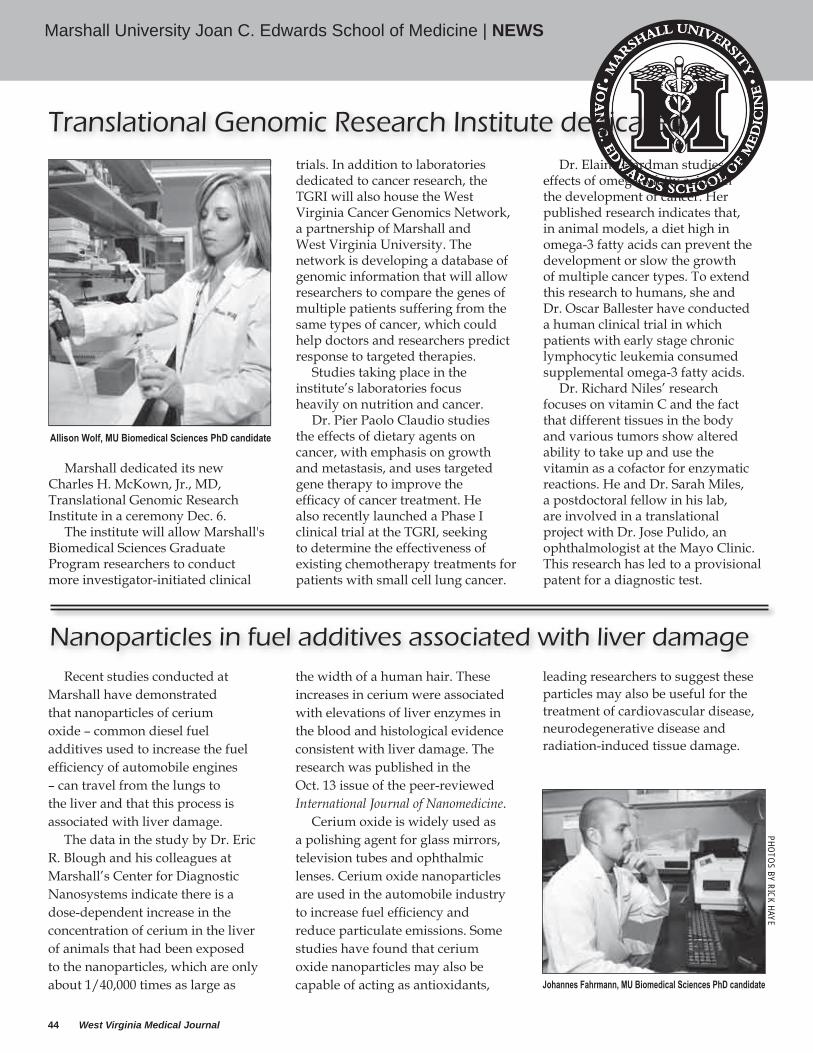
44 West Virginia Medical Journal
Marshall University Joan C. Edwards School of Medicine | NEWS
Marshall dedicated its new Charles H. McKown, Jr., MD, Translational Genomic Research Institute in a ceremony Dec. 6.
The institute will allow Marshall's Biomedical Sciences Graduate Program researchers to conduct more investigator-initiated clinical
trials. In addition to laboratories dedicated to cancer research, the TGRI will also house the West Virginia Cancer Genomics Network, a partnership of Marshall and West Virginia University. The network is developing a database of genomic information that will allow researchers to compare the genes of multiple patients suffering from the same types of cancer, which could help doctors and researchers predict response to targeted therapies.
Studies taking place in the institute’s laboratories focus heavily on nutrition and cancer.
Dr. Pier Paolo Claudio studies the effects of dietary agents on cancer, with emphasis on growth and metastasis, and uses targeted gene therapy to improve the efficacy of cancer treatment. He also recently launched a Phase I clinical trial at the TGRI, seeking to determine the effectiveness of existing chemotherapy treatments for patients with small cell lung cancer.
Dr. Elaine Hardman studies the effects of omega-3 fatty acids on the development of cancer. Her published research indicates that, in animal models, a diet high in omega-3 fatty acids can prevent the development or slow the growth of multiple cancer types. To extend this research to humans, she and Dr. Oscar Ballester have conducted a human clinical trial in which patients with early stage chronic lymphocytic leukemia consumed supplemental omega-3 fatty acids.
Dr. Richard Niles’ research focuses on vitamin C and the fact that different tissues in the body and various tumors show altered ability to take up and use the vitamin as a cofactor for enzymatic reactions. He and Dr. Sarah Miles, a postdoctoral fellow in his lab, are involved in a translational project with Dr. Jose Pulido, an ophthalmologist at the Mayo Clinic. This research has led to a provisional patent for a diagnostic test.
Nanoparticles in fuel additives associated with liver damageRecent studies conducted at
Marshall have demonstrated that nanoparticles of cerium oxide – common diesel fuel additives used to increase the fuel efficiency of automobile engines – can travel from the lungs to the liver and that this process is associated with liver damage.
The data in the study by Dr. Eric R. Blough and his colleagues at Marshall’s Center for Diagnostic Nanosystems indicate there is a dose-dependent increase in the concentration of cerium in the liver of animals that had been exposed to the nanoparticles, which are only about 1/40,000 times as large as
the width of a human hair. These increases in cerium were associated with elevations of liver enzymes in the blood and histological evidence consistent with liver damage. The research was published in the Oct. 13 issue of the peer-reviewed International Journal of Nanomedicine.
Cerium oxide is widely used as a polishing agent for glass mirrors, television tubes and ophthalmic lenses. Cerium oxide nanoparticles are used in the automobile industry to increase fuel efficiency and reduce particulate emissions. Some studies have found that cerium oxide nanoparticles may also be capable of acting as antioxidants,
leading researchers to suggest these particles may also be useful for the treatment of cardiovascular disease, neurodegenerative disease and radiation-induced tissue damage.
Translational Genomic Research Institute dedicated
Allison Wolf, MU Biomedical Sciences phd candidate
Johannes Fahrmann, MU Biomedical Sciences phd candidate
PHOTOS BY RICK HAYE
January/February 2012 | Vol. 108 45
| New Members
Please direct all membership inquiries to: Mona Thevenin, WVSMA Membership Director at 304.925.0342, ext. 16 or [email protected].
Cabell County Medical SocietyGregory Bills, FYMSAlizabeth Blankenship, FYMSJay Bonder, FYMSJames Buchanhan Jr., FYMSRudolf Burcl, FYMSJoseph Cart, FYMSCarl Chotas, FYMSGeorge Clements, FYMSCharles Clements III, FYMSShane Cook, FYMSCarrie Cox, FYMSChristopher Damron, FYMSJohn Davitt, FYMSAaron Dom, FYMSChristopher Fine, FYMSJames Flannery, FYMSHunter Garland, FYMSKelley Groves, FYMSShayne Gue, FYMSWilliam Hall, FYMSJacob Hamm, FYMSMelinda Hodge, FYMSSarah Johnson, FYMS
Anthony Johnson, FYMSRahul Khetan, FYMSMathew Lemberger, FYMSClinton McDaniel, FYMSSteven Nakano, FYMSLouie Olive, FYMSAron Pickering, FYMSRonald Reyes, FYMSJoseph Russo, FYMSB Trent Schambach, FYMSSarah Sexton, FYMSMadhvi Shah, FYMSBrandon Shiflett, FYMSAlexander Slocum Jr, FYMSAmos Turner IV, FYMSAdam VanHorn, FYMSJohn Waddell, FYMSAfton Wickline, FYMS
Harrison County Medical SocietyC. Bradley Franz, MD
Kanawha County Medical SocietyFatima Aziz-Ashraf, MDJack Depriest, MD
Nancy Elwood, MDJennifer Ladd, DOShaen Reinert, DOMark Richards, MD
Monongalia County Medical SocietyMichael Best, FYMSNicholas Corridoni, FYMSSydney Clark, FYMSKarina Geronilla, SYMSJohn Grantham, FYMSJeremy Hustead, SYMSCaitlin Kocher, FYMSAaron Lucas, FYMSBrandon Luck-Wold, FYMSDaniel McFarland, FYMSLindsay Miller, FYMSMark Plumby, FYMSRachel Schillinger, FYMSNicole Shockor, FYMSJason Statler, FYMS
raleigh County Medical SocietyOmar Hasan, MD
ObituariesThe WVsMA remembers our esteemed colleagues…
John Blair Morton IIDr. John Blair Morton II,
47, of Charleston passed away Wednesday, Nov. 30, 2011, at home after a long illness, surrounded by his loving family.
John was a physician, specializing in gastroenterology. He returned to Charleston with his family in 2007 and became a partner in Charleston Gastroenterology Associates.
He served in the U.S. Navy as a commissioned officer from 1987 to 1992.
John was a 1982 graduate of George Washington High School. He graduated with high honors from the University of Virginia in chemical
engineering in 1986. In 1996, he graduated from the West Virginia University School of Medicine.
He completed a gastroenterology fellowship at Oschner Clinic Foundation, New Orleans, La.
He was an avid runner, enjoyed traveling, loved spending time with his children and really enjoyed life.
John was preceded in death by his father, William A. Morton Sr.
He is survived by his wife, Erma Morton of Charleston; son, John "Jack" Morton III, age 6; daughter, Margaret "Maggie" Elizabeth Morton, age 5; mother, Barbara O' Connor Morton of Charleston; sisters, Julia Anne Morton and Mary Leslie
Morton of Charleston; and brother, William A. and his wife, Yvonne Fay Morton, of Whittier, Calif.
John leaves behind his loving family and caring friends.
As his final wish, his body was donated to the West Virginia University School of Medicine Human Gift Registry, Morgantown, to further medical research.
In lieu of flowers, donations may be made to his children's college fund, Harford Smart 529 Plan, c/o Heritage Investment Group, 318 5th Ave., South Charleston, WV 25303, or First Presbyterian Church, 16 Leon Sullivan Way, Charleston, WV 25301.
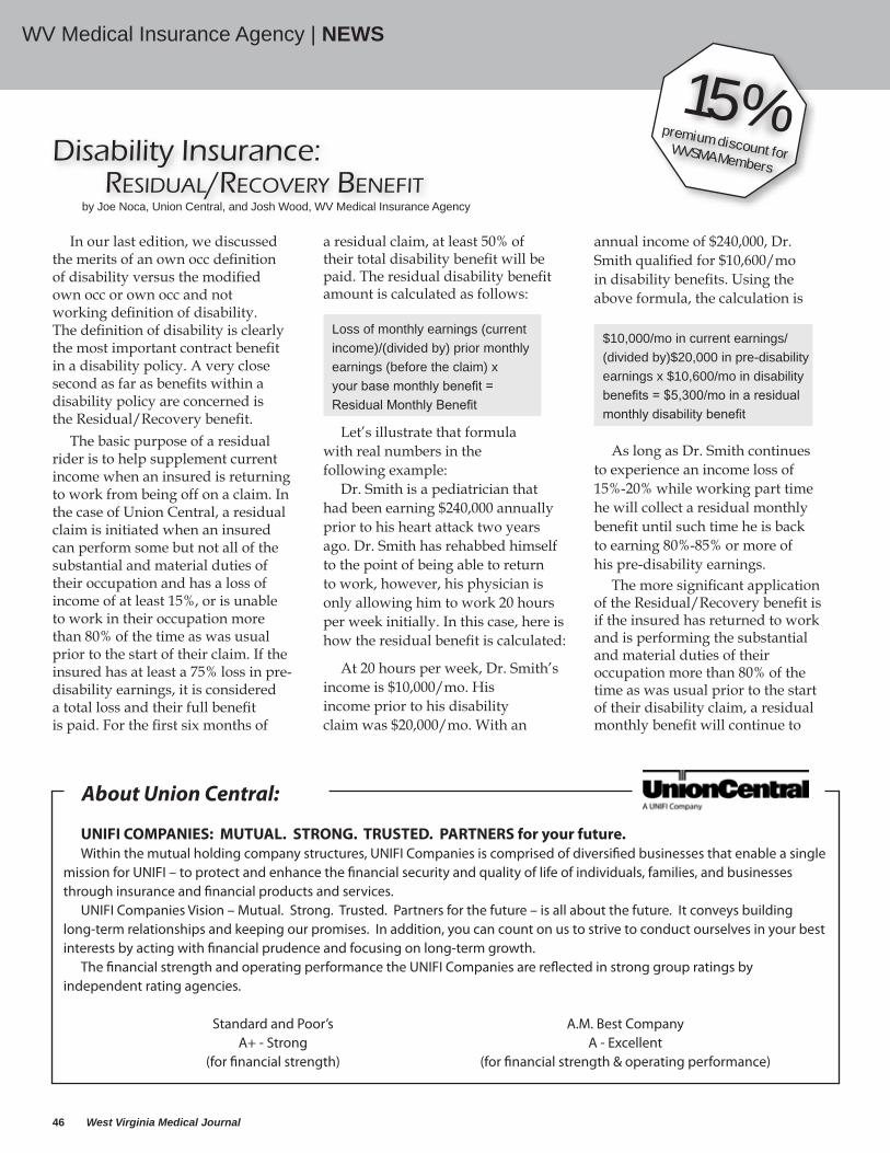
In our last edition, we discussed the merits of an own occ definition of disability versus the modified own occ or own occ and not working definition of disability. The definition of disability is clearly the most important contract benefit in a disability policy. A very close second as far as benefits within a disability policy are concerned is the Residual/Recovery benefit.
The basic purpose of a residual rider is to help supplement current income when an insured is returning to work from being off on a claim. In the case of Union Central, a residual claim is initiated when an insured can perform some but not all of the substantial and material duties of their occupation and has a loss of income of at least 15%, or is unable to work in their occupation more than 80% of the time as was usual prior to the start of their claim. If the insured has at least a 75% loss in pre-disability earnings, it is considered a total loss and their full benefit is paid. For the first six months of
a residual claim, at least 50% of their total disability benefit will be paid. The residual disability benefit amount is calculated as follows:
Loss of monthly earnings (current income)/(divided by) prior monthly earnings (before the claim) x your base monthly benefit = Residual Monthly Benefit
Let’s illustrate that formula with real numbers in the following example:
Dr. Smith is a pediatrician that had been earning $240,000 annually prior to his heart attack two years ago. Dr. Smith has rehabbed himself to the point of being able to return to work, however, his physician is only allowing him to work 20 hours per week initially. In this case, here is how the residual benefit is calculated:
At 20 hours per week, Dr. Smith’s income is $10,000/mo. His income prior to his disability claim was $20,000/mo. With an
annual income of $240,000, Dr. Smith qualified for $10,600/mo in disability benefits. Using the above formula, the calculation is
$10,000/mo in current earnings/(divided by)$20,000 in pre-disability earnings x $10,600/mo in disability benefits = $5,300/mo in a residual monthly disability benefit
As long as Dr. Smith continues to experience an income loss of 15%-20% while working part time he will collect a residual monthly benefit until such time he is back to earning 80%-85% or more of his pre-disability earnings.
The more significant application of the Residual/Recovery benefit is if the insured has returned to work and is performing the substantial and material duties of their occupation more than 80% of the time as was usual prior to the start of their disability claim, a residual monthly benefit will continue to
Disability Insurance:Residual/RecoveRy Benefit
WV Medical Insurance Agency | NEWS
by Joe Noca, Union Central, and Josh Wood, WV Medical Insurance Agency
UNIFI COMPANIES: MUTUAL. STRONG. TRUSTED. PARTNERS for your future. Within the mutual holding company structures, UNIFI Companies is comprised of diversified businesses that enable a single
mission for UNIFI – to protect and enhance the financial security and quality of life of individuals, families, and businesses through insurance and financial products and services.
UNIFI Companies Vision – Mutual. Strong. Trusted. Partners for the future – is all about the future. It conveys building long-term relationships and keeping our promises. In addition, you can count on us to strive to conduct ourselves in your best interests by acting with financial prudence and focusing on long-term growth.
The financial strength and operating performance the UNIFI Companies are reflected in strong group ratings by independent rating agencies.
Standard and Poor’s A.M. Best Company A+ - Strong A - Excellent (for financial strength) (for financial strength & operating performance)
15%premium discount forWVSMA Members
About Union Central:
46 West Virginia Medical Journal
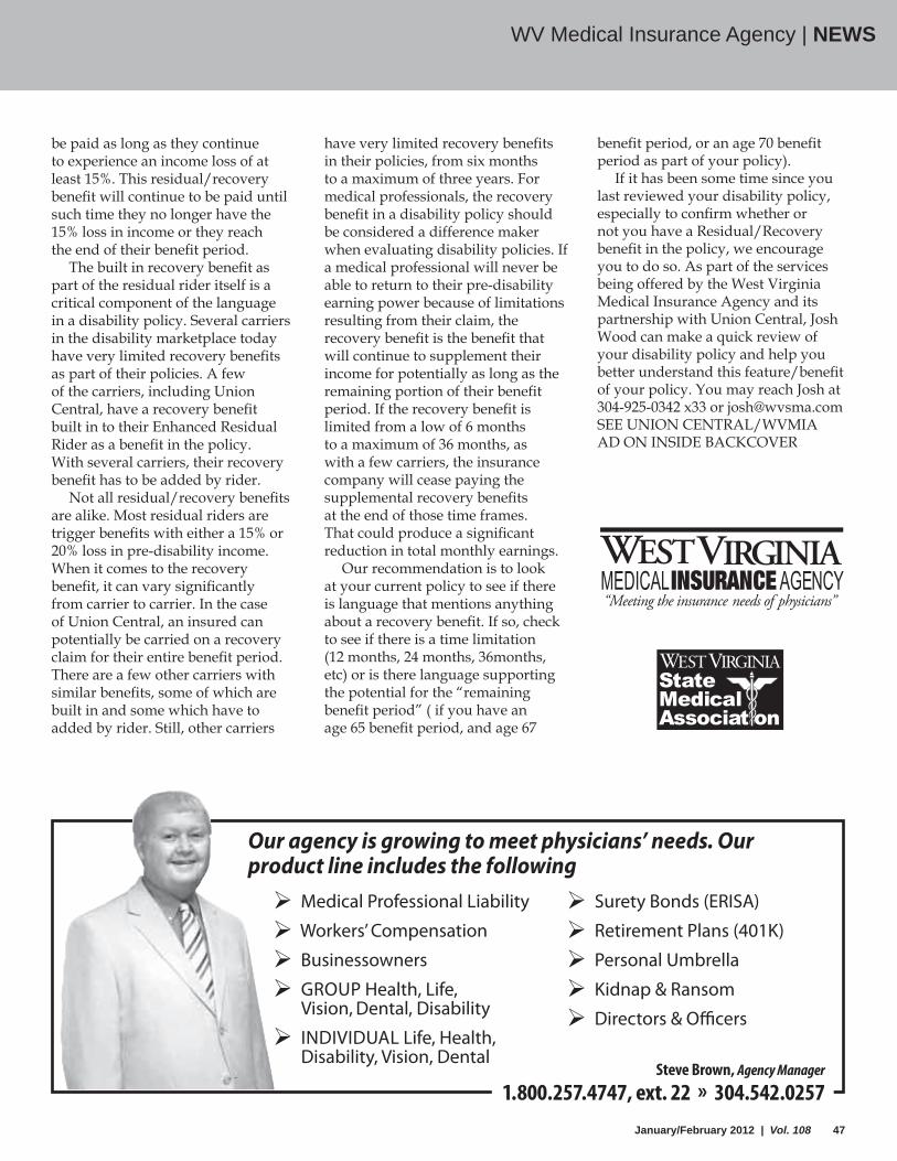
January/February 2012 | Vol. 108 47
WV Medical Insurance Agency | NEWS
be paid as long as they continue to experience an income loss of at least 15%. This residual/recovery benefit will continue to be paid until such time they no longer have the 15% loss in income or they reach the end of their benefit period.
The built in recovery benefit as part of the residual rider itself is a critical component of the language in a disability policy. Several carriers in the disability marketplace today have very limited recovery benefits as part of their policies. A few of the carriers, including Union Central, have a recovery benefit built in to their Enhanced Residual Rider as a benefit in the policy. With several carriers, their recovery benefit has to be added by rider.
Not all residual/recovery benefits are alike. Most residual riders are trigger benefits with either a 15% or 20% loss in pre-disability income. When it comes to the recovery benefit, it can vary significantly from carrier to carrier. In the case of Union Central, an insured can potentially be carried on a recovery claim for their entire benefit period. There are a few other carriers with similar benefits, some of which are built in and some which have to added by rider. Still, other carriers
have very limited recovery benefits in their policies, from six months to a maximum of three years. For medical professionals, the recovery benefit in a disability policy should be considered a difference maker when evaluating disability policies. If a medical professional will never be able to return to their pre-disability earning power because of limitations resulting from their claim, the recovery benefit is the benefit that will continue to supplement their income for potentially as long as the remaining portion of their benefit period. If the recovery benefit is limited from a low of 6 months to a maximum of 36 months, as with a few carriers, the insurance company will cease paying the supplemental recovery benefits at the end of those time frames. That could produce a significant reduction in total monthly earnings.
Our recommendation is to look at your current policy to see if there is language that mentions anything about a recovery benefit. If so, check to see if there is a time limitation (12 months, 24 months, 36months, etc) or is there language supporting the potential for the “remaining benefit period” ( if you have an age 65 benefit period, and age 67
benefit period, or an age 70 benefit period as part of your policy).
If it has been some time since you last reviewed your disability policy, especially to confirm whether or not you have a Residual/Recovery benefit in the policy, we encourage you to do so. As part of the services being offered by the West Virginia Medical Insurance Agency and its partnership with Union Central, Josh Wood can make a quick review of your disability policy and help you better understand this feature/benefit of your policy. You may reach Josh at 304-925-0342 x33 or [email protected] SEE UNION CENTRAL/WVMIA AD ON INSIDE BACKCOVER
Our agency is growing to meet physicians’ needs. Our product line includes the following Medical Professional Liability
Workers’ Compensation
Businessowners
GroUP Health, Life, Vision, Dental, Disability
INDIVIDUAL Life, Health, Disability, Vision, Dental
Surety Bonds (ErISA)
retirement Plans (401K)
Personal Umbrella
Kidnap & ransom
Directors & officers
Steve Brown, Agency Manager
1.800.257.4747, ext. 22 » 304.542.0257
C o M p o u n d i n g p h A r M A C yLOOP PhaRMaCy & hOME MEdICaL
The Region’s only PCAB Accredited Compounding Pharmacy serving the medical community for over
25 years. Hormone Replacement, Pain Management, Sterile Compounding, Pediatrics, Autism, Dermatology, and
much more. Contact us today for more information.
1-800-696-3170Email: [email protected]
Web: www.Looppharmacy.com
i n h o M E C A r ESarahCare Of baRbOURSVILLE
Adult Day Care Center
2 Courtyard LaneBarboursville, WV 25504
304-736-3005www.sarahcare.com/barboursville/
n E u r o Lo g yaLVaRO R. GUTIERREZ, Md
NEUROLOGY
Academic results with private practice convenience.Headache Rescue Services/EMG/Consultations.
Self-referrals accepted.
2199 Cheat Road, Morgantown, WV 26508
304-594-3258304-594-3498 fax
o b S t Et r i C S / g y n E C o Lo g ySCOTT a. NaEGELE, Md, PLLC
OB/GYNDaVinci Robotic Surgery
Advanced Bladder ProceduresIn-OfficeSterilizations&EndoAblations
SCOTT A. NAEGELE, MD, FACOG830 Pennsylvania Avenue Suite 108
Charleston, WV 25302304.344.8368 l 304.342.8938 faX
www.drscottnaegele.com/[email protected]
WOMEN’S hEaLTh CaRE Of MORGaNTOWN“Experienced,professionalcarethatputsyoufirst”
Diplomates of the American Collegeof Obstetrics and Gynecology
William Hamilton, MD Jan Thomas, CNMLouise Van Riper, MD Gail Rock, CNMMurshid Latif, MD Lisa Stout, CNMCraig Herring, MD Rhonda Conley, CNMShane Prettyman, MD Bjarni Thomas, CNM
Complete OB/GYN care:Prenatal care and delivery with our MD’s or Nurse •MidwivesNon-surgical solutions and advanced surgical care•Well woman screenings for all ages•Sneak peek 3D/4D ultrasound•
1249 Suncrest Towne Centre, Morgantown, WV 26505
304-599-6353www.whcofmorgantown.com
pA i n M A n A g E M E n tThE CENTER fOR PaIN RELIEf, INC.
Multidisciplinary Interventional Pain Management
TIMOTHY DEER, MD RICHARD BOWMAN, MD
CHRISTOPHER KIM, MD MATTHEW RANSON, MD
St. Francis Hospital Location400 Court Street, Suite 100, Charleston, WV 25301
304.347.6120
ThE CENTER fOR PaIN RELIEf, INC. Teays Valley Hospital Location Doctors Park, 1400 Hospital Drive
Hurricane, WV 25526304.757.5420
Physical Therapy and Rehabilitation Center, Southridge Location
100 Peyton Way, Charleston WV, 25309304.720.6747
www.centerforpainrelief.com
Professional Directory

Continuing Medical Education Opportunities at CAMC Health Education
and Research InstituteThe CAMC Health Education and Research Institute is dedicated to improving health through research, education and community health development. The Institute’s Education Division offers live conferences, seminars, workshops, teleconferences and on-site programs to health care professionals. The CAMC Institute’s CME program is accredited by the Accreditation Council for Continuing Medical Education to sponsor continuing medical education for physicians. The CAMC Institute designates this educational activity for a maximum of 1 AMA PRA Category 1 Credit(s)™. Physicians should only claim credit commensurate with the extent of their participation in the activity. For more information on these and future programs provided by the Institute, call (304) 388-9960 or fax (304) 388-9966.
©Charleston Area Medical Center Health System, Inc. 2011 23725-K11
SEMINARS
31st Cardiovascular Conference at SnowshoeFriday through Sunday, January 27-29, 2012 Snowshoe, WV
West Virginia Trauma Symposium Wednesday through Saturday, February 7-11, 2012 Stonewall Jackson Resort Roanoke, WV
3rd Annual Junior Fellows OB/Gyn Symposium Saturday, March 3, 2012 Stonewall Jackson Resort Roanoke, WV
LIFE SUPPORT TRAINING Please log-on to our website to view upcoming schedule and register at www.camcinstitute.org under CAMC Life Support Training Center.
CME ONLINE PROGRAMS/ARCHIVED GUEST LECTURE PROGRAMSLog-on to our website at www.camcinstitute.org
System RequirementsEnvironment: Windows 98, SE, NT, 2000 or XP Resolution: 800 x 600Web Browser: Microsoft’s Internet Explorer 5.0 or above or Netscape Navigator 4.7x. (Do not use Netscape 7.1)Video Player: Windows Media Player 6.4 or better.Dial-Up or Broadband Connection. Minimum Speed, 56k (Broadband is Recommended)
OTHER ARCHIVED CME OPPORTUNITIES:Geriatric Series Research Series NET Reach library
50 West Virginia Medical Journal
Sharing in the Joy of Healthcare Education
304.881.4633 www.mccabemedicalcoding.com
• Claim Processing Guidelines
• Coding Concepts and Guidelines
• Medical Billing and Reimbursement
• Quality Assurance and Risk Analysis
• Medical Record or Chart Review
Professional Instruction Personal Consulting
| Classified Ads
RENAL CONSULTANTS inWest Virginia is recruiting
nephrologists for Boone, Putnam
and Kanawha Counties.
Practice consists of busy CKD
clinic, ESRD patients, home
dialysis and post-transplant
care. Applicant must be BC/
BE and prepared to be busy
immediately. Salary awarded
proportionate to productivity.
J1 Visas welcomed.E-mail resume to:
needed to join two oncologists in private practice in northern
West Virginia. BC/BE. Competitive salary and
benefits.
Reply to:
Oncologist-HematologistPO Box 4106
Charleston, WV 25364
Medical oncologist-hematologist
MEDICAL PRACTICE FOR SALE
Includes land, building and equipment.Two Locations
Location #1: 163 Greenbrier Street, Rupert, WV 25984
Approximately 2500 square feet; completely remodeled office building, fully equipped w/ 6 exam rooms, 3 dr. offices, 2 nurses stations,
staff break room, 2 waiting rooms, a lab and plenty of parking.Price: $175,000
Location #2: 219 @ Dunlap Street Union, WV 25984
Approximately 1000 square feet; 2 exam rooms, a dr. office, lab & comes fully equipped.
Price: $135,000 For more information contact
Patricia Long304-645-4043
3000 Washington St. West
OFFICE MANAGERS ASSOCIATIONOF HEALTHCARE PROVIDERS, INC.
www.officemanagersassociation.comWe invite you to join our organization which consists of members
who manage the daily business of healthcare providers.Our objectives are to promote educational opportunities, professional knowledge
and to provide channels of communication to officemanagers in all areas of healthcare. We currently have
eleven chapters in West Virginia.
Visit us on our website for more information or contactDonna Zahn (President) at 740-283-4770 ext. 105 or
Tammy Mitchell (Membership) at 304-324-2703.

Presented through an exclusive partnership between:
Coding and Medicare UpdateThe West Virginia State Medical Association (WVSMA), through our exclusive partnership with the Practice Manage ment Institute
(PMI), is bringing these classes to you.For your convenience, you may choose either the morning or afternoon session. Each session will offer 3 CEU credits for Certified
Medical office Managers (CMoMs) or other certified professionals. The cost of the course is $199.00 per person. All PMI certified professionals, as well as WVSMA members, receive a 10% discount.
CHOOSE YOURLOCATION & DATE:
TUESDAy, JAnUARy 10in Morgantown(Mon General)*
WEDnESDAy, JAnUARy 11in Beckley
(Raleigh General)*
THURSDAy, JAnUARy 12in Huntington
(St. Mary’s)*
CHOOSE YOURSESSION:
9:00 AM - Noonor
1:00 PM - 4:00 PM
*Each location/date has two sessions to choose from.
2 0 1 2Coding update information
critical to your practice!
Participant Informationregistrant 1: ________________________________________________________________
registrant 2: ________________________________________________________________
registrant 3: ________________________________________________________________E-mail: _____________________________________________________________________
Practice Name: ______________________________________________________________
Street Address: ______________________________________________________________
City: ________________________ State: _______ Zip: ______________________________
Phone: ______________________________ Fax: _______________________________
Registration Fee: $199 (10% discount for PMI certified professionals and/or WVSMA members)
Payment Method: q American Express q MasterCard q Visa
q Check Enclosed $______________ AMOUNT PAID
Card No: Exp. Date: V Code:
Name As It Appears on Card: ____________________________________________________
Signature: ___________________________________________________________________
Payable to:West Virginia State Medical Association
Three digit number
Registration Methods:Mail registration form to: Karie Sharp • West Virginia State Medical Association
Po Box 4106, Charleston, WV 25364Fax registration form to: Karie Sharp • (304) 925-0345
Charge by phone: Karie Sharp • (304) 925-0342, ext. 12 • E-mail: [email protected]
Your registration will be confirmed by email. Please provide your address here.
3 Locations ~ 3 Dates ~ 3 CEUs2 Session Times Available at Each Location!
January 10, 11 & 12
Register Today! Seating is Limited. must know information for full, fair physician
reimbursement.

52 West Virginia Medical Journal
The WVSMA reserves the right to deny advertising space to any individual, company, group or association whose products or services interfere with the mission, objectives, endorsement agreement(s) and/or any contractual obligations of the WVSMA. The WVSMA, in its sole discretion, retains the right to decline any submitted advertisement or to discontinue publishing any advertisement previously accepted. The Journal does not accept paid political advertisements.The fact that an advertisement for a product, service, or company appears in the Journal is not a guarantee by the WVSMA of the product, service or company or the claims made for the product in such advertising. The WVSMA reserves the right to enter into endorsements, sponsorship and/or marketing agreements that may limit the placement of advertisements for certain products or services.
Subscription Rates: $60 a year in the United States$100 a year in foreign countries$10 per single copy
POSTMASTER: Send address changes to the West Virginia Medical Journal, P.O. Box 4106, Charleston, WV 25364. Periodical postage paid at Charleston, WV.
USPS 676 740 ISSN 0043 - 3284Claims for back issues should be made within six months after publication. Microfilm editions beginning with the 1972 volume are available from University Microfilms International, 300 N. Zeeb Rd., Ann Arbor, MI 48106. ©2012 , West Virginia State Medical Association
Thanks To Our Advertisers!
Advertising policy
Beckley ARH Hospital................................................... 17, 21
CAMC Health Education & Research Institute ................... 49
Center for Rural Health Development Loan Fund .............. 25
Centers for Medicare & Medicaid Services .......................... 1
Chapman Printing .......................................................... 27,50
EHR Solutions .................................................................... 14
EN&T Associates ................................................................ 31
Eye & Ear Clinic Physicians, Inc......................................... 38
Flaherty Sensabaugh & Bonasso ...................................... 33
Highmark West Virginia ........................................................ 5
McCabe Medical Coding .................................................... 50
Medical Practice FOR SALE .............................................. 50
Oncologists/Hematologists Opportunity ............................. 50
Physician’s Business Office .................................................. 9
Renal Consultants .............................................................. 50
Suttle & Stalnaker ............................................................... 35
TOTAL ACCESS ................................................................... 7
Unicare ............................................................................... 40
Union Central Life Insurance Company......... Inside Back Cover
West Virginia Medical Insurance Agency ..................... 47, 52
West Virginia Mutual Insurance Co. . ................................. 22
WV Medical Professionals Health Program........................ 41
West Virginia REDI ...............................................Back Cover
West Virginia University SOM ...................Inside Front Cover
1.800.257.4747, ext. 22 » 304.542.0257
Your Practice.Your Future.Your Agency.
In case it’s not this obvious...In case it’s not this obvious...
To review the Manuscript
Guidelines, please visit our website at
wvsma.com


West Virginia Medical JournalP.O. Box 4106
Charleston, WV 25364www.wvsma.com
BA
CK
CO
VER
What is WV REDI?
West Virginia Responder Emergency Deployment Information system
• WV REDI is a web-based registration system developed to facilitate health and
medical response through identification of West Virginians willing to serve in public
health emergency and non-emergency situations
Who can register?
• Registration is open to West Virginia’s health and medical professionals, and others
who live or work in West Virginia
How can I help?
• You can help by being willing to assist during a health related emergency or event and
by registering in WV REDI
What if I can’t go when called?
• Please remember that “volunteer” truly means volunteer. You can choose, at any time,
to decline any request that you receive for deployment
How do I register?
• To register go to www.wvredi.org and click on “register now”
Where do I get more information?
• For more information, call 304-558-6900 ext. 2009
Visit the
www.wvredi.org homepage and click on
“register now.”
Register today to be
prepared for tomorrow!
West Virginia Responder Emergency Deployment Information