regulation of immune cell functions by metabolic...
TRANSCRIPT

Review ArticleRegulation of Immune Cell Functions byMetabolic Reprogramming
Jaehong Kim 1,2
1Department of Biochemistry, School of Medicine, Gachon University, Incheon 21999, Republic of Korea2Department of Health Sciences and Technology, Gachon Advanced Institute for Health Science and Technology, Gachon University,Incheon 21999, Republic of Korea
Correspondence should be addressed to Jaehong Kim; [email protected]
Received 19 October 2017; Accepted 14 January 2018; Published 13 February 2018
Academic Editor: Abdallah Elkhal
Copyright © 2018 Jaehong Kim. This is an open access article distributed under the Creative Commons Attribution License, whichpermits unrestricted use, distribution, and reproduction in any medium, provided the original work is properly cited.
Recent findings show that the metabolic status of immune cells can determine immune responses. Metabolic reprogrammingbetween aerobic glycolysis and oxidative phosphorylation, previously speculated as exclusively observable in cancer cells, existsin various types of immune and stromal cells in many different pathological conditions other than cancer. Themicroenvironments of cancer, obese adipose, and wound-repairing tissues share common features of inflammatory reactions. Inaddition, the metabolic changes in macrophages and T cells are now regarded as crucial for the functional plasticity of theimmune cells and responsible for the progression and regression of many pathological processes, notably cancer. It is possiblethat metabolic changes in the microenvironment induced by other cellular components are responsible for the functionalplasticity of immune cells. This review explores the molecular mechanisms responsible for metabolic reprogramming inmacrophages and T cells and also provides a summary of recent updates with regard to the functional modulation of theimmune cells by metabolic changes in the microenvironment, notably the tumor microenvironment.
1. Introduction
Pleiotropic interactions between various cells are responsiblefor the maintenance and disturbance of homeostasis in thetissue microenvironment of physiological and pathologicalconditions. For example, from early carcinogenesis toprogression and metastasis, cancer cells interact with varioustypes of stromal cells, for example, cancer-associated fibro-blasts, endothelial cells, and immune cells in the tumormicroenvironment (TME). The TME is flooded withcytokines and growth factors responsible for “smolderingpersistent inflammation.” This reactive stroma is a well-characterized component of the TME that shows similaritiesto the repair response in injured tissue [1]. Recent findingsrevealed that various immune cell subsets are dominantregulators of the delicate balance between homeostasis anddisturbance in the tissue microenvironment [2–5]. For exam-ple, macrophages can form a major component of immune
cell infiltrate in the TME, constituting as much as half of atumor mass [6, 7]. Immune responses of M1 and M2 macro-phages describe the opposing activities of killing or repairing.The typical M1 macrophages drive inflammation and showhigh antigen presentation, high production of inflammatorycytokines such as IL-12 and IL-23, and high production ofnitric oxide (NO) and reactive oxygen intermediates. In con-trast, M2-type responses are the “resting” phenotype and areobserved in the resolution of inflammation without infec-tions, tissue remodeling, and repair. It has been widelyaccepted that IFNγ alone or with microbial LPS or cytokinessuch as GM-CSF and TNF induces classically activated M1macrophages, and IL-4, IL-6, IL-10, IL-13, IL-21, IL-33,immune complexes, and Notch can induce the M2 form ofmacrophage activation [8, 9]. Notably, truly polarizedmacrophages are rare [10–13] and tumor-associated macro-phages (TAMs) can be also described as M(IL-4), M(Ig),M(IL-10), M(GC: glucocorticoid), M(IFNγ), M(LPS), and
HindawiJournal of Immunology ResearchVolume 2018, Article ID 8605471, 12 pageshttps://doi.org/10.1155/2018/8605471

so forth, according to a recently attempted nomenclaturebased on specific activation standard [12]. Evidence supportsa tumor-promoting role of TAMs, and high frequencies ofTAMs are generally associated with poor prognosis in mosthuman cancers [2, 14, 15]. TAMs infiltrating establishedtumors generally show the properties of an M2-like activatedanti-inflammatory, protumoral properties rather than M1-like activated proinflammatory, antitumoral phagocyticproperties [16–18].
Macrophages can also form a major component ofimmune cell infiltrate in obese adipose tissue (AT), constitut-ing as much as 40% of all AT cells [19]. In the progression ofobesity, a switch from M2-like to M1-like activation of themacrophage population occurs and inflammatory cyto-kines such as tumor necrosis factor (TNF) contribute toinsulin resistance in adipocytes characterized by animpaired insulin response such as hypertriglyceridemiaand elevated fasting glucose [20, 21]. In addition, lym-phoid as well as myeloid cells infiltrates and expands inthe liver tissue and the obese AT and these immune cellsubsets are responsible for the development of obesity-related metabolic dysregulation due to excessive nutrientintake and exacerbation of low-grade inflammatorychanges in the microenvironment. CD8+ T cells also pro-mote inflammation and metabolic disturbance in the AT[22]. In addition to macrophages and T cells, neutrophilsand mast cells can also disturb the homeostasis in thetissue microenvironment.
Many recent findings in the field of immunometabolismnow show that metabolic status in immune cells can deter-mine various types of immune responses. Immune cells haveremarkably diverse functions and cellular activities that areassociated with distinct metabolic demands. The traditionalsimple concept of production of cellular ATP is that glycoly-sis generates two molecules of ATPs from one molecule ofglucose. Glycolysis metabolizes glucose to pyruvate first,and the pyruvate is further metabolized to carbon dioxide,NADH, and FADH2 in the mitochondria. The reducingequivalents (NADH and FADH2) drive oxidative phosphor-ylation (OXPHOS) for more ATP synthesis. In the 1920s, itwas demonstrated that cancer tissues can metabolize, evenin aerobic conditions, about tenfolds more glucose toproduce lactate than normal tissues can and this is knownas aerobic glycolysis or the Warburg effect [23]. Since pyru-vate is metabolized to lactate and secrete, lactate appears tobe wasted in aerobic glycolysis. However, lactate secretionout of cells allows increased continuous glucose influxfrom the generation of NAD+ and resultant accumulationof glycolytic intermediates facilitates biomass synthesisfor rapidly proliferating cells. Since the observation anddramatic revitalization of the Warburg effect, the domi-nant glycolysis and relatively reduced OXPHOS werethought to be confined to cancer cells. However, recentfindings clearly show that the Warburg effect-like meta-bolic reprogramming also exists in rapidly proliferatingcells including various types of immune cells, most notablyin macrophages and T cells, and determines the functionof the immune cell subsets in disease conditions such asthose in inflamed tissue or cancer [24–27].
2. Metabolic Regulation ofMacrophage Phenotypes
The function of macrophages is not limited to the mainte-nance of homeostasis in the tissue microenvironment butalso includes many activities such as cytokine productionand phagocytosis upon their activation. Importantly, macro-phages are famous for their plasticity and adoption of variousactivation states in response to their functional requirementssignaled from their microenvironment. For example, aninnate arm of the immune system can have an importantcapacity to adapt after challenged with pathogens [28].This is known as innate immune memory or trainedimmunity. Trained immunity from epigenetic reprogram-ming of macrophages shows high glucose consumptionand a high ratio of NAD+ to its reduced form NADH,reflecting a shift in metabolism with an increase in glycol-ysis and M1-like activation of macrophages, dependent onthe activation of mTOR through the Akt-HIF-1α pathway[29]. M2-like activated macrophages exploit fatty acidoxidation (FAO) to fuel OXPHOS rather than aerobic gly-colysis for ATP production [30–32].
Of note, HIF1α and NFκB drive the M1 phenotypes[33, 34] and PGC1β, and peroxisome proliferator-activatedreceptors and STAT6 drive the M2 phenotypes (Figure 1)[35–38]. Phosphorylation and activation of a nutritionalsensor, AMPK, regulate mitochondrial biogenesis via deace-tylation of regulating proteins, including SIRT1 withNAD+, and suppress HIF1α and NFκB [38–40]. AMPK andNAD+-SIRT1-PGC1β signaling are key factors for nutri-tional state-dependent M1/M2-like activation of macro-phages in inflammatory conditions [39, 41]. HIF-1α alsoenhances the lactate dehydrogenase- (LDH-) mediated con-version of pyruvate-to-lactate [42] and increases expres-sion of GLUT1, GLUT3, and MCT4 to increase glucoseuptake and expression of pyruvate kinase M2 (PKM2),resulting in an increase in the secretion of lactate anduncoupled glycolysis and oxidative phosphorylation [43, 44](Figure 2). Pyruvate dehydrogenase (PDH) inactivationfrom phosphorylation by pyruvate dehydrogenase kinases(PDKs) prevents pyruvate from entering the mitochondrialKrebs cycle [45]. HIF-1α transcriptionally activates thePDKs [46, 47].
LPS-activated dendritic cells and M1-like activated mac-rophages show enhanced aerobic glycolysis, flux through thepentose phosphate pathway, and fatty acid synthesis but haveincomplete OXPHOS at the level of succinate dehydrogenase(SDH) and isocitrate dehydrogenase, blocking the synthesisof mitochondrial ATP. In these cells, glucose is used for thebiosynthesis of large quantities of cytokines and effectormolecules, and inactivation of OXPHOS directs metabolitesfrom the Krebs cycle for inflammatory reaction [32, 48].Accumulation of succinate and citrate from the truncatedOXPHOS leads to stabilization of HIF1α by limiting prolylhydroxylase activity to maintain a proinflammatory, antitu-moral response [49–51].
Recently, itaconic acid-mediated inhibition of SDHhas also been found as a driver for succinate accumula-tion in LPS-stimulated M1-like activated proinflammatory
2 Journal of Immunology Research
macrophages [52]. Immunoresponsive gene 1 (Irg1) is highlyexpressed in mammalian macrophages during inflammationand Irg1 gene silencing in macrophages results in signifi-cantly decreased intracellular itaconic acid levels as well assignificantly reduced antimicrobial activity during bacterialinfections [53].
High intracellular iron levels in M1-like activated mac-rophages stabilize HIF1α through low levels of ferroportinand high levels of H-ferritin, involved in iron export andstorage, respectively [54, 55]. Hemeoxygenase-1 (HO-1)catabolizes heme to ferrous ion, biliverdin, and carbonmonooxide, and suppression of HO-1 results in M2-likeactivation of TAMs [56].
HIF1α can also be stabilized from nitrosylation withperoxynitrites from increased iNOS [57], favoring aerobicglycolysis in M1 phenotypes. NFκB transcriptionally acti-vates proinflammatory genes including iNOS, which formsNO in the presence of arginine. Peroxynitrite, formed fromNO and superoxide anions in the mitochondria, nitrosylatesiron-sulfur proteins in the mitochondrial electron transportchain, and the resultant nitrosylation can inhibit OXPHOS[58], also favoring aerobic glycolysis in M1 phenotypes.Unlike iNOS-mediated catabolism of arginine to NO inM1-like activated macrophages, M2-like activated macro-phages catalyze arginine to urea and ornithine by arginase 1(ARG1); ARG1 is a representative marker for M2-like activa-tion. As NO production is limited in M2-like activated mac-rophages, the nitrosylation-mediated inhibition of OXPHOSis dampened, now favoring M2 phenotypes [48]. AlthoughHIF1α drives the M1 phenotypes in hypoxic conditions,lactate produced by cancer cells, as a by-product of aerobicglycolysis, has an unexpected critical function in HIF1α-dependent expression of ARG1 and resultant M2-like activa-tion of TAMs in normoxic conditions [59] (Figure 3). These
findings clearly indicate highly interconnected signaling forthe conservation of HIF-1-centered metabolic phenotypes.
As stated, M2-like activated macrophages show loweredglycolysis and enhanced FAO to fuel OXPHOS. Th2 cytokineand IL-4-induced PGC1β increase mitochondrial biogenesisand FAO in a STAT6-dependent manner [38, 41, 60].PGC1β plays a key role in increasing mitochondrial biogen-esis and OXPHOS by upregulating the expression of FAO-involved genes [41]. IL-4-/IL-13-stimulated macrophagesexpress PFKFB1, which produces a low level of a glycolyticactivator, fructose 2,6 bisphosphate [61, 62]. In IL-4-stimulated macrophages, fatty acid sources such as LDLand VLDL are taken up via the scavenger receptor CD36and metabolized in the lysosome. The CD36-mediated lyso-somal lipolysis is essential for the M2-like activation [31].
An orphan nuclear receptor, estrogen-related receptor α(ESRRα), is required for the increased mitochondrial biogen-esis [63]. Importantly, ESRRα-deficient macrophages show adecrease in phagosomal maturation and antimicrobial activ-ity [64]. Another study reported an M1-like phenotype ofincreased glycolysis but impaired mitochondrial respiratoryfunction and biosynthesis as a result of ESRRα deficiency[65]. Interestingly, VLDLR expression is a determinant factorin inflammation and in M1-like activation of macrophages inAT [66].
In spite of our knowledge gained from macrophages ininflammatory disease conditions, our understanding of themetabolic regulations in TAMs is surprisingly limited andthe signals involved in communication between tumorsand macrophages are still poorly defined [67]. However,emerging evidence strongly indicates that the metabolicreprogramming of macrophages is closely related to the pro-tumoral or antitumoral function of macrophages [68, 69] andthat unraveling the TAM phenotype might lead to the
RTK
PTEN P13K
AKT mTOR H1F1�훼
PD-L1 PD-1
TEFF
Aerobicglycolysis
STAT6 PGC1 AMPKSIRT1
NF�휅B
FAOormitochondrialbiogenesis
NO
Inflammation
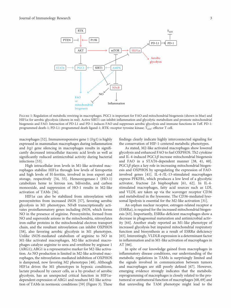
Figure 1: Regulation of metabolic rewiring in macrophages. PGC1 is important for FAO and mitochondrial biogenesis (shown in blue) andHIF1α for aerobic glycolysis (shown in red). Active SIRT1 can inhibit inflammation and glycolytic metabolism and promote mitochondrialbiogenesis and FAO. Interaction of PD-L1 and PD-1 induces FAO and suppresses aerobic glycolysis and immune functions in Teff. PD-1:programmed death-1; PD-L1: programmed death ligand-1; RTK: receptor tyrosine kinase; TEFF: effector T cell.
3Journal of Immunology Research

NAPDH,nucleotidesynthesis
Pentosephosphatepathway
Glucose‑6‑P
Glucose‑6‑PPFK1 Fructose‑
2,6‑BPFructose‑1,6‑BPMalonyl‑Co‑A
Acetyl‑CoA LCFacyl‑CoA�훽‑oxidation
Glucose
Glucose
G3P
3PG
2PG
PEP
Pyruvate
PyruvateMalate Citrate
PDH
PS
TCAT
CPT‑1Lactate
MCT1
(PFKFB3)
ACC‑2
CitrateCholesterol
TAGs
LCFacyl‑CoA
Mevalonatepathway
GLUTs
HK2
PFK2
PKM2LDHMCT4
PDK
Krebs cycle
Acetyl‑CoA
(a)
Aspartate
Pyruvate
Isocitrate
Citrate
�훼‑KGIDH1,2
Arginosuccinate
Arginine
NOS ARG
NOUreaOrnithine
Citruline
Acetyl‑CoA
�훼‑KG,
PyruvateCitrate
Oxaloa
cetat
e
Isocitrate
Malate
FumarateSuccinate
SDH
IDH3
NADH
(b)
Figure 2: Metabolism of glucose and fatty acid at a glance. (a) Stabilization of HIF-1α upregulates GLUTs, HK2, PFK2, PKM2, LDH, PDK,and MCT4 shown in red. ACC: acetyl-CoA carboxylase; ARG: arginase; CPT-1: carnitine palmitoyltransferase 1; FAT: fatty acid translocase;G3P: glyceraldehyde 3-phosphate; GLUT: glucose transporter; HK2: hexokinase 2; IDH: isocitrate dehydrogenase; LCFacyl-CoAs: long-chainfatty acyl-CoAs; MCT: monocarboxylate transporter; 2PG: 2-phosphoglycerate; 3PG: 3-phosphoglycerate; PEP: phosphoenolpyruvate; PDH:pyruvate dehydrogenase; PDK: pyruvate dehydrogenase kinase; PFK: phosphofructokinase; PS: pyruvate symporter; SDH: succinatedehydrogenase; TAG: triacylglyceride; TCAT: tricarboxylic acid transporter. (b) A schematic of the Krebs cycle and metabolites exportedout of the mitochondria. Arginine is metabolized to urea and ornithine in M2-like macrophages that do not express NOS. ARG: arginase;NOS: nitric oxide synthase.
4 Journal of Immunology Research
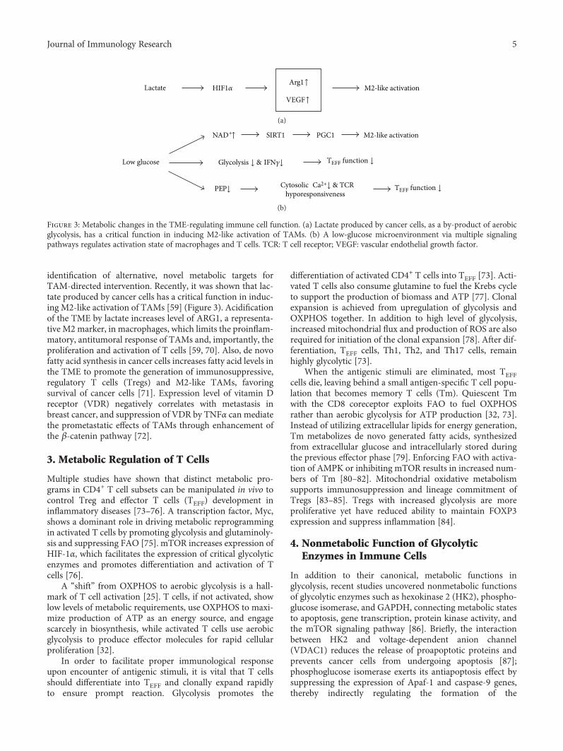
identification of alternative, novel metabolic targets forTAM-directed intervention. Recently, it was shown that lac-tate produced by cancer cells has a critical function in induc-ing M2-like activation of TAMs [59] (Figure 3). Acidificationof the TME by lactate increases level of ARG1, a representa-tive M2 marker, in macrophages, which limits the proinflam-matory, antitumoral response of TAMs and, importantly, theproliferation and activation of T cells [59, 70]. Also, de novofatty acid synthesis in cancer cells increases fatty acid levels inthe TME to promote the generation of immunosuppressive,regulatory T cells (Tregs) and M2-like TAMs, favoringsurvival of cancer cells [71]. Expression level of vitamin Dreceptor (VDR) negatively correlates with metastasis inbreast cancer, and suppression of VDR by TNFα can mediatethe prometastatic effects of TAMs through enhancement ofthe β-catenin pathway [72].
3. Metabolic Regulation of T Cells
Multiple studies have shown that distinct metabolic pro-grams in CD4+ T cell subsets can be manipulated in vivo tocontrol Treg and effector T cells (TEFF) development ininflammatory diseases [73–76]. A transcription factor, Myc,shows a dominant role in driving metabolic reprogrammingin activated T cells by promoting glycolysis and glutaminoly-sis and suppressing FAO [75]. mTOR increases expression ofHIF-1α, which facilitates the expression of critical glycolyticenzymes and promotes differentiation and activation of Tcells [76].
A “shift” from OXPHOS to aerobic glycolysis is a hall-mark of T cell activation [25]. T cells, if not activated, showlow levels of metabolic requirements, use OXPHOS to maxi-mize production of ATP as an energy source, and engagescarcely in biosynthesis, while activated T cells use aerobicglycolysis to produce effector molecules for rapid cellularproliferation [32].
In order to facilitate proper immunological responseupon encounter of antigenic stimuli, it is vital that T cellsshould differentiate into TEFF and clonally expand rapidlyto ensure prompt reaction. Glycolysis promotes the
differentiation of activated CD4+ T cells into TEFF [73]. Acti-vated T cells also consume glutamine to fuel the Krebs cycleto support the production of biomass and ATP [77]. Clonalexpansion is achieved from upregulation of glycolysis andOXPHOS together. In addition to high level of glycolysis,increased mitochondrial flux and production of ROS are alsorequired for initiation of the clonal expansion [78]. After dif-ferentiation, TEFF cells, Th1, Th2, and Th17 cells, remainhighly glycolytic [73].
When the antigenic stimuli are eliminated, most TEFFcells die, leaving behind a small antigen-specific T cell popu-lation that becomes memory T cells (Tm). Quiescent Tmwith the CD8 coreceptor exploits FAO to fuel OXPHOSrather than aerobic glycolysis for ATP production [32, 73].Instead of utilizing extracellular lipids for energy generation,Tm metabolizes de novo generated fatty acids, synthesizedfrom extracellular glucose and intracellularly stored duringthe previous effector phase [79]. Enforcing FAO with activa-tion of AMPK or inhibiting mTOR results in increased num-bers of Tm [80–82]. Mitochondrial oxidative metabolismsupports immunosuppression and lineage commitment ofTregs [83–85]. Tregs with increased glycolysis are moreproliferative yet have reduced ability to maintain FOXP3expression and suppress inflammation [84].
4. Nonmetabolic Function of GlycolyticEnzymes in Immune Cells
In addition to their canonical, metabolic functions inglycolysis, recent studies uncovered nonmetabolic functionsof glycolytic enzymes such as hexokinase 2 (HK2), phospho-glucose isomerase, and GAPDH, connecting metabolic statesto apoptosis, gene transcription, protein kinase activity, andthe mTOR signaling pathway [86]. Briefly, the interactionbetween HK2 and voltage-dependent anion channel(VDAC1) reduces the release of proapoptotic proteins andprevents cancer cells from undergoing apoptosis [87];phosphoglucose isomerase exerts its antiapoptosis effect bysuppressing the expression of Apaf-1 and caspase-9 genes,thereby indirectly regulating the formation of the
Lactate HIF1�훼Arg1↑
VEGF↑M2-like activation
(a)
Low glucose
NAD+↑ SIRT1 PGC1 M2-like activation
Glycolysis ↓ & IFN�훾↓ TEFF function ↓
PEP↓ Cytosolic Ca2+↓ & TCRhyporesponsiveness
TEFF function ↓
(b)
Figure 3: Metabolic changes in the TME-regulating immune cell function. (a) Lactate produced by cancer cells, as a by-product of aerobicglycolysis, has a critical function in inducing M2-like activation of TAMs. (b) A low-glucose microenvironment via multiple signalingpathways regulates activation state of macrophages and T cells. TCR: T cell receptor; VEGF: vascular endothelial growth factor.
5Journal of Immunology Research

apoptosome [88, 89]. GAPDH exerts controversial pro- andantiapoptotic effects through interaction with VDAC1 andinduction of autophagy, respectively [86, 90].
Other studies have shown that GAPDH, HK, and enolaseare also RNA-binding proteins [91–93] and the “REM(RNA–enzyme–metabolite) hypothesis” proposes a regula-tory interaction between gene expression and cellular metab-olism by RNA-binding metabolic enzymes [94, 95]. It isnotable that many glycolytic enzymes, formerly known toexclusively function in glycolytic metabolic events in thecytoplasm or mitochondria, have now been shown to regu-late transcription and translation [86, 91]. Indeed, numerousmetabolic enzymes that also function in glycolysis, fatty acidsynthesis, and the Krebs cycle are also RNA-binding proteins[96], although the significance for immune response is notclearly known except for that of GAPDH [95] and enolase[32, 83]. Recently, a REM connection with GAPDH wasproven in T cell activation [25]. GAPDH is diverted to glycol-ysis and translation of IFNγ and IL-2 is not perturbed inhighly glycolytic T cells. However, when aerobic glycolysisis blocked, GAPDH binds to IFNγ and IL-2 mRNA inCD4+ T cells to suppress their translation [25]. In myeloidcells, GAPDH is a component of the IFNγ-activated inhib-itor of translation (GAIT) complex that controls transla-tion of inflammatory genes [97, 98]. High glycolytic fluxsuppresses the interaction between GAPDH and Rheband thus allows Rheb to activate mTORC1 and stimulate cellgrowth [99]. By modulating expression of Foxp3-splicingvariants with exon 2(Foxp3-E2), enolase-1-mediated glycoly-sis controls induction of human Tregs with a potent immu-nosuppressive function [83]. When glycolysis is inhibited,enolase-1 translocates to the nucleus and represses expres-sion of the Foxp3-E2 splice variant in Tregs and suppressesTreg induction.
The level of the glycolytic intermediate phosphoenolpyr-uvate (PEP) is controlled by a balance between enolase-mediated formation of PEP and pyruvate kinase-mediatedconversion to pyruvate. PKM2 exists either as an inactivedimer or as more active tetramer, and the transition betweenthe two conformations is subject to posttranslational mod-ifications [100]. Dimeric PKM2, previously regarded ascrucial for metabolic reprogramming exclusively in cancercell, is also important in promoting aerobic glycolysis inimmune cells [101, 102]. Enhanced expression of dimericPKM2 reduces the rate of PEP conversion to pyruvateand results in an accumulation of glycolytic products thatcan be otherwise metabolized in biosynthetic pathways[32]. Importantly, PEP enhances antitumor effector func-tions in activated T cells by regulating Ca2+ import intothe endoplasmic reticulum, thus sustaining translocationof nuclear factor of activated T cells (NFAT) into thenucleus and the expression of a set of genes that arerequired for T cell activation [103].
These findings imply that a direct and strong interactionexists between the nonmetabolic function of glycolyticenzymes and the generation of immune responses and alsothat enhanced glycolysis sustains antitumoral and proinflam-matory functions via highly interconnected signaling inimmune cells.
5. Metabolic Changes in the TME InfluencingImmune Cell Functions
The microenvironment determines the metabolism ofimmune cells, which in turn adjust to a broad spectrum ofconfigurations to meet the demands of various cellular activ-ities. For example, changes in the metabolic profiles ofimmune cells by cancer cells can alter the function of theimmune cells [103, 104]. A protracted aerobic glycolysisacidifies and destabilizes the TME and this is consistentwith the view of the tumor as an unhealed wound [105].Similarities of utilizing nutrients and engaging metabolicregulation to sustain cellular proliferation and survivalare shared by cancer and immune cells. Notably, nutri-tional competition between cancer cells and antitumoralimmune cells in the TME shifts the activation and differ-entiation status of T-cells to favoring the survival of cancercells [103, 104, 106, 107].
Recently, it was shown that lactate produced by cancercells, as a by-product of aerobic glycolysis, has a critical func-tion in signaling that induces M2-like activation of TAMs[59] (Figure 3). Interestingly, lactate-induced M2-like activa-tion was fromHIF1α-dependent expression of ARG1. Deple-tion of glucose and a glucose-rich hypoxic ROS environmentfavor M2-like activation and M1-like activation of TAMs,respectively, and depletion of glucose can disarm T cells inthe TME [27, 104]. Low levels of ATP from dietaryrestrictions or energy consumption induces nicotinamidephosphoribosyltransferase that generates NAD+, which isa key factor for SIRT1 activation. SIRT1 acetylates and acti-vates PGC1β to increase OXPHOS [35, 67, 108]. Pyruvateis metabolized by LDH-A, producing lactate and NAD+.NAD+ acts as an electron acceptor in the Krebs cycleand the electron transport system in mitochondria. Itappears feasible from these findings that glucose-depleted,low ATP, and NAD+-rich states (in cachexic patient withadvanced cancer) may drive the M2-like activation ofmacrophages, while the macrophage population stillretains its phagocytic activity in maintaining biosynthesiswith molecules acquired from their microenvironment[32]. For the identification of alternative, novel targetsfor TAM-directed intervention, it would be necessary toshow whether these events can predominantly happen inTAMs of the TME.
A recent study observed that hypoxia-induced upregula-tion of the immunosuppressive programmed death ligand-1(PD-L1) is directly mediated by HIF1α [109]. In the TME,cancer cells, macrophages, and dendritic cells express PD-L1, a notable ligand for immune checkpoint, programmedcell death-1(PD-1) in TEFF. The interaction of PD-1 andPD-L1 directly inhibits glycolysis and promotes lipolysisand FAO in T cells, resulting in failure of the antitumoralfunction of T cells [110] (Figure 1). The lessons that applica-tion of immune checkpoint blockade antibodies against cyto-toxic T lymphocyte antigen-4 (CTLA-4), PD-1, and PD-L1,which are used clinically, restore glucose in the TME, permit-ting T cell glycolysis and IFNγ production clearly show thatnutrient availability in the microenvironment can changethe metabolic status of immune cells. Another study also
6 Journal of Immunology Research
revealed that PD-1 expression by TAMs correlates withprotumoral activity, and blockage of PD-1–PD-L1 in vivoincreases phagocytosis, reduces growth of cancer cells,and increases the survival of mice in mouse models ofcancer in a macrophage-dependent way [111]. In addition,blocking PD-L1 directly on cancer cells decreases glycoly-sis and restores glucose in the TME, resulting in allowanceof antitumoral function of T cells from glycolysis andIFNγ production [104].
In conclusion, these findings indicate that signals such ascytokines, growth factors, hypoxia, and nutrient availabilitythat emanate from the microenvironment can inducemetabolic changes in immune cell subsets, resulting inchanges in immune functions and pathological responses.An interesting perspective is whether immune cells can sup-ply their microenvironment with lactate and antioxidativeresources as stromal cells in the TME are able to (the reverseWarburg effect). Considering their preponderance in theTME, there is an ample possibility that metabolic changesin a large subgroup of macrophages or T cells may also affectthe metabolic state of their microenvironment and functionsof other cellular components.
6. Potential Metabolic Targets for theManipulation of Immune Cell Function
Since the function of immune cells is dependent on a delicatemetabolic balance, results of many clinical trials performedwith inhibitors of metabolic enzymes and oncogenes willprovide valuable insights for the prospect of immunomodu-lation by specific metabolic regulation [112]. Of note, resultsfrom targeting cancer metabolism in vivo have been disap-pointing and less prominent than results from targetingimmune cell metabolism [85]. The PKM2 inhibitor, TLN-232, was tested in a clinical trial for refractory renal cellcarcinoma (NCT00422786). Inactive dimeric PKM2 acti-vates the mTORC1 signaling pathway by phosphorylatingthe mTOR inhibitor, AKT1S1, and leads to an acceleratedoncogenic growth and autophagy inhibition of cancer cells[113]. In line with this, increase in the tetrameric, active formof PKM2, attenuated the LPS-induced proinflammatory M1-like macrophage phenotypes while promoting M2-like mac-rophage phenotypes [114]. Many AMPK activators are nowtested in clinical and preclinical studies for diabetes, cancer,and cardiovascular disease [115]. Importantly, AMPK stimu-lation inhibiting mTORC1 was sufficient to decrease Glut1and increase generation of Tregs in an animal model, imply-ing AMPK activation as a potential manipulable checkpointfor immune response [73]. REDD1, an inhibitor of mTOR,is highly expressed in M2-like TAMs. Inhibition of REDD1stimulates glycolysis in the TAMs and competition of glucosebetween TAMs and endothelial cells prevents vascular hyper-activation and promotes the formation of quiescent vascularjunctions in the TME [69]. Suppression of REDD1 wasattempted in phase 2 clinical trial (NCT00713518) for thetreatment of neovascularization in AMD patients. Nitrosyla-tion of HIF1α prevents its degradation. If denitrosylation ofHIF1α is observed, its modulation may be potentially
applicable for the inhibition of glycolytic enzymes and thealleviation of M1-like phenotypes.
Isoprenylation of ubiquinone is important for OXPHOSand isoprenylation of Ras, Rho, and Rab guanosine tripho-sphatases is involved in immunological synapse formation,migration, proliferation, and cytotoxic effector response ofT cells. The intracellular availability of sterols is crucial forisoprenylation modification of proteins for plasma mem-brane attachment and represents a checkpoint for metabolicreprogramming that modulates T cell responses [116]. Statinand other chemical inhibitors of the mevalonate pathway cansuppress isoprenylation of Rho proteins [117] and have beentested in many clinical trials.
Clinical trials involving agents that inhibit PD-L1 andPD-1 are now being performed. Atezolizumab is the solemember of this class currently approved for the treatmentof bladder cancer, but approvals for avelumab, durvalumab,nivolumab, and pembrolizumab in the treatment of variouscancer are anticipated in the near future [118]. Therefore, itappears possible that the combined use of metabolism-targeting reagents with immune checkpoint inhibitors canalter the activation and differentiation of T cells.
7. Conclusions
Immunity and metabolism advance together. Consideringthe significant contribution of immune cell functions in pro-moting and suppressing various types of disease progression,repolarization of immune cells from the potential targetsstated above shows an ample possibility to become noveltherapeutic approaches. Extension of our knowledge of thefunctional plasticity of macrophages and T cells spanningfrom inflammation biology to cancer immunology and thepersistent reprogramming effect achievable from stable epi-genetic changes in the metabolic pathways of macrophages[29] and potentially T cells by potential modulators mayprovide new information for immune therapeutic strategiesapplicable for different disease conditions. Importantly, can-cer cells and host primary cell constituents such as immunecells and stromal cells can form microanatomical compart-ments within the cancer tissue to regulate metabolic needs,immune surveillance, survival, invasion, and metastasis.Indeed, different signals from particular locations in theTME seem to influence activation of TAMs and T cells andoverall tumor prognosis [119]. TAMs can be diverse withinthe microanatomical compartments, including the accumu-lation of M1-like activated cells with protumoral propertiesin hypoxic areas [120] and differences in inflammatory com-ponents and pathways between tumors originating in distinctanatomical sites [120, 121]. The notion that metaboliccompetition between cancer cells, immune cells, and otherstromal cells can determine function and fate of each cell sub-set proposing that identification of which of specific niches inthe microenvironment can impede immune cells fromproper metabolic engagement will encourage significantcontributions to this research field. Generation of metaboli-cally fit T cells prior to adoptive cell transfer will improve Tcell-based immunotherapy against cancer by surviving theunfavorable, hostile TME. Furthermore, successful therapies
7Journal of Immunology Research
targeting the function of macrophages and T cells will requireidentification of targets that specifically allow metabolicreprogramming of immune cells while, at the same time,not causing an increase in proliferation and survival of cancercells or systemic inflammatory changes or autoimmunity.Our understanding of the metabolic regulations in B cells issurprisingly limited, and the mechanisms about how cellularmetabolism supports and regulates function of B cells are stillpoorly defined. B cell immunometabolism is anticipated tobecome an exciting research field.
Conflicts of Interest
The authors declare that they have no conflicts of interest.
Acknowledgments
This study was supported by a grant of the Korea Instituteof Radiological and Medical Sciences (KIRAMS), fundedby Ministry of Science and ICT (MSIT), Republic of Korea(1711045557, 1711045538, and 1711045554) and by theBasic Science Research Program through the NationalResearch Foundation of Korea (NRF of Korea) fundedby the Ministry of Science, ICT & Future Planning(NRF-2017R1D1A1B03029063).
References
[1] H. F. Dvorak, “Tumors: wounds that do not heal. Similaritiesbetween tumor stroma generation and wound healing,” TheNew England Journal of Medicine, vol. 315, no. 26,pp. 1650–1659, 1986.
[2] L. Bingle, N. J. Brown, and C. E. Lewis, “The role of tumour-associated macrophages in tumour progression: implicationsfor new anticancer therapies,” The Journal of Pathology,vol. 196, no. 3, pp. 254–265, 2002.
[3] J. M. Fernandez-Real and J. C. Pickup, “Innate immunity,insulin resistance and type 2 diabetes,” Trends in Endocrinol-ogy and Metabolism, vol. 19, no. 1, pp. 10–16, 2008.
[4] D. A. Winer, S. Winer, L. Shen et al., “B cells promote insulinresistance through modulation of T cells and production ofpathogenic IgG antibodies,” Nature Medicine, vol. 17, no. 5,pp. 610–617, 2011.
[5] J. Jager, M. Aparicio-Vergara, and M. Aouadi, “Liver innateimmune cells and insulin resistance: the multiple facets ofKupffer cells,” Journal of Internal Medicine, vol. 280, no. 2,pp. 209–220, 2016.
[6] P. M. Kelly, R. S. Davison, E. Bliss, and J. O. McGee, “Macro-phages in human breast disease: a quantitative immunohisto-chemical study,” British Journal of Cancer, vol. 57, no. 2,pp. 174–177, 1988.
[7] E. Van Overmeire, D. Laoui, J. Keirsse, J. A. Van Ginderach-ter, and A. Sarukhan, “Mechanisms driving macrophagediversity and specialization in distinct tumor microenviron-ments and parallelisms with other tissues,” Frontiers inImmunology, vol. 5, p. 127, 2014.
[8] A. Mantovani, A. Sica, S. Sozzani, P. Allavena, A. Vecchi, andM. Locati, “The chemokine system in diverse forms of macro-phage activation and polarization,” Trends in Immunology,vol. 25, no. 12, pp. 677–686, 2004.
[9] Y. C. Wang, F. He, F. Feng et al., “Notch signaling determinesthe M1 versus M2 polarization of macrophages in antitumorimmune responses,” Cancer Research, vol. 70, no. 12,pp. 4840–4849, 2010.
[10] F. Rae, K. Woods, T. Sasmono et al., “Characterisation andtrophic functions of murine embryonic macrophages basedupon the use of a Csf1r-EGFP transgene reporter,” Develop-mental Biology, vol. 308, no. 1, pp. 232–246, 2007.
[11] A. Sica and A. Mantovani, “Macrophage plasticity and polar-ization: in vivo veritas,” The Journal of Clinical Investigation,vol. 122, no. 3, pp. 787–795, 2012.
[12] P. J. Murray, J. E. Allen, S. K. Biswas et al., “Macrophage acti-vation and polarization: nomenclature and experimentalguidelines,” Immunity, vol. 41, no. 1, pp. 14–20, 2014.
[13] J. Van den Bossche, J. Baardman, and M. P. de Winther,“Metabolic characterization of polarized M1 and M2 bonemarrow-derived macrophages using real-time extracellularflux analysis,” Journal of Visualized Experiments, no. 105,article e53424, 2015.
[14] B. Z. Qian and J. W. Pollard, “Macrophage diversity enhancestumor progression and metastasis,” Cell, vol. 141, no. 1,pp. 39–51, 2010.
[15] Q. W. Zhang, L. Liu, C. Y. Gong et al., “Prognostic signifi-cance of tumor-associated macrophages in solid tumor: ameta-analysis of the literature,” PLoS One, vol. 7, no. 12, arti-cle e50946, 2012.
[16] A. Mantovani, P. Allavena, A. Sica, and F. Balkwill, “Can-cer-related inflammation,” Nature, vol. 454, no. 7203,pp. 436–444, 2008.
[17] M. Zhang, Y. He, X. Sun et al., “A high M1/M2 ratio oftumor-associated macrophages is associated with extendedsurvival in ovarian cancer patients,” Journal of OvarianResearch, vol. 7, no. 1, p. 19, 2014.
[18] A. Yuan, Y. J. Hsiao, H. Y. Chen et al., “Opposite effects of M1and M2 macrophage subtypes on lung cancer progression,”Scientific Reports, vol. 5, no. 1, p. 14273, 2015.
[19] S. P. Weisberg, D. McCann, M. Desai, M. Rosenbaum, R. L.Leibel, and A. W. Ferrante Jr, “Obesity is associated withmacrophage accumulation in adipose tissue,” The Journal ofClinical Investigation, vol. 112, no. 12, pp. 1796–1808, 2003.
[20] C. N. Lumeng, J. L. Bodzin, and A. R. Saltiel, “Obesity inducesa phenotypic switch in adipose tissue macrophage polariza-tion,” The Journal of Clinical Investigation, vol. 117, no. 1,pp. 175–184, 2007.
[21] A. Castoldi, C. Naffah de Souza, N. O. Camara, and P. M.Moraes-Vieira, “The macrophage switch in obesity develop-ment,” Frontiers in Immunology, vol. 6, p. 637, 2016.
[22] S. Nishimura, I. Manabe, M. Nagasaki et al., “CD8+ effector Tcells contribute to macrophage recruitment and adipose tis-sue inflammation in obesity,” Nature Medicine, vol. 15,no. 8, pp. 914–920, 2009.
[23] O. Warburg, F. Wind, and E. Negelein, “The metabolism oftumors in the body,” The Journal of General Physiology,vol. 8, no. 6, pp. 519–530, 1927.
[24] M. Chang, J. A. Hamilton, G. M. Scholz, and C. L. Elsegood,“Glycolytic control of adjuvant-induced macrophage sur-vival: role of PI3K, MEK1/2, and Bcl-2,” Journal of LeukocyteBiology, vol. 85, no. 6, pp. 947–956, 2009.
[25] C. H. Chang, J. D. Curtis, L. B. Maggi Jr et al., “Posttranscrip-tional control of T cell effector function by aerobic glycoly-sis,” Cell, vol. 153, no. 6, pp. 1239–1251, 2013.
8 Journal of Immunology Research

[26] S. K. Biswas, “Metabolic reprogramming of immune cells incancer progression,” Immunity, vol. 43, no. 3, pp. 435–449,2015.
[27] M. Corrado, L. Scorrano, and S. Campello, “Changing per-spective on oncometabolites: from metabolic signature ofcancer to tumorigenic and immunosuppressive agents,”Oncotarget, vol. 7, no. 29, pp. 46692–46706, 2016.
[28] M. G. Netea, J. Quintin, and J. W. van der Meer,“Trained immunity: a memory for innate host defense,” CellHost & Microbe, vol. 9, no. 5, pp. 355–361, 2011.
[29] S. C. Cheng, J. Quintin, R. A. Cramer et al., “mTOR- andHIF-1α-mediated aerobic glycolysis as metabolic basis fortrained immunity,” Science, vol. 345, no. 6204, article1250684, 2014.
[30] D. Vats, L. Mukundan, J. I. Odegaard et al., “Oxidative metab-olism and PGC-1β attenuate macrophage-mediated inflam-mation,” Cell Metabolism, vol. 4, no. 1, pp. 13–24, 2006.
[31] S. C. Huang, B. Everts, Y. Ivanova et al., “Cell-intrinsiclysosomal lipolysis is essential for alternative activation ofmacrophages,” Nature Immunology, vol. 15, no. 9, pp. 846–855, 2014.
[32] N. Assmann and D. K. Finlay, “Metabolic regulation ofimmune responses: therapeutic opportunities,” The Journalof Clinical Investigation, vol. 126, no. 6, pp. 2031–2039,2016.
[33] T. Cramer, Y. Yamanishi, B. E. Clausen et al., “HIF-1alpha isessential for myeloid cell-mediated inflammation,” Cell,vol. 112, no. 5, pp. 645–657, 2003.
[34] C. H. Fong, M. Bebien, A. Didierlaurent et al., “An antiin-flammatory role for IKKβ through the inhibition of “classi-cal” macrophage activation,” The Journal of ExperimentalMedicine, vol. 205, no. 6, pp. 1269–1276, 2008.
[35] A. Chawla, “Control of macrophage activation and functionby PPARs,” Circulation Research, vol. 106, no. 10, pp. 1559–1569, 2010.
[36] N. Wang, H. Liang, and K. Zen, “Molecular mechanisms thatinfluence the macrophage m1-m2 polarization balance,”Frontiers in Immunology, vol. 5, p. 614, 2014.
[37] N. Kapoor, J. Niu, Y. Saad et al., “Transcription factorsSTAT6 and KLF4 implement macrophage polarization viathe dual catalytic powers of MCPIP,” Journal of Immunology,vol. 194, no. 12, pp. 6011–6023, 2015.
[38] H. R. Griffiths, D. Gao, and C. Pararasa, “Redox regulation inmetabolic programming and inflammation,” Redox Biology,vol. 12, pp. 50–57, 2017.
[39] C. Canto, Z. Gerhart-Hines, J. N. Feige et al., “AMPKregulates energy expenditure by modulating NAD+ metabo-lism and SIRT1 activity,” Nature, vol. 458, no. 7241,pp. 1056–1060, 2009.
[40] D. B. Shackelford, D. S. Vasquez, J. Corbeil et al., “mTOR andHIF-1α-mediated tumor metabolism in an LKB1 mousemodel of Peutz-Jeghers syndrome,” Proceedings of theNational Academy of Sciences of the United States of America,vol. 106, no. 27, pp. 11137–11142, 2009.
[41] L. A. O'Neill and D. G. Hardie, “Metabolism of inflammationlimited by AMPK and pseudo-starvation,” Nature, vol. 493,no. 7432, pp. 346–355, 2013.
[42] J. G. Sonanez-Organis, M. Rodriguez-Armenta, B. Leal-Rubio, A. B. Peregrino-Uriarte, S. Gomez-Jimenez, andG. Yepiz-Plascencia, “Alternative splicing generates two lac-tate dehydrogenase subunits differentially expressed during
hypoxia via HIF-1 in the shrimp Litopenaeus vannamei,”Biochimie, vol. 94, no. 5, pp. 1250–1260, 2012.
[43] M. S. Ullah, A. J. Davies, and A. P. Halestrap, “The plasmamembrane lactate transporter MCT4, but not MCT1, is up-regulated by hypoxia through a HIF-1α-dependent mecha-nism,” The Journal of Biological Chemistry, vol. 281, no. 14,pp. 9030–9037, 2006.
[44] H. Pelicano, W. Lu, Y. Zhou et al., “Mitochondrial dysfunc-tion and reactive oxygen species imbalance promote breastcancer cell motility through a CXCL14-mediated mecha-nism,” Cancer Research, vol. 69, no. 6, pp. 2375–2383, 2009.
[45] T. Zhao, Y. Zhu, A. Morinibu et al., “HIF-1-mediated meta-bolic reprogramming reduces ROS levels and facilitates themetastatic colonization of cancers in lungs,” ScientificReports, vol. 4, p. 3793, 2014.
[46] J. W. Kim, I. Tchernyshyov, G. L. Semenza, and C. V.Dang, “HIF-1-mediated expression of pyruvate dehydroge-nase kinase: a metabolic switch required for cellularadaptation to hypoxia,” Cell Metabolism, vol. 3, no. 3,pp. 177–185, 2006.
[47] C. W. Lu, S. C. Lin, K. F. Chen, Y. Y. Lai, and S. J. Tsai,“Induction of pyruvate dehydrogenase kinase-3 by hypoxia-inducible factor-1 promotes metabolic switch and drug resis-tance,” The Journal of Biological Chemistry, vol. 283, no. 42,pp. 28106–28114, 2008.
[48] J. Van den Bossche, J. Baardman, N. A. Otto et al., “Mito-chondrial dysfunction prevents repolarization of inflamma-tory macrophages,” Cell Reports, vol. 17, no. 3, pp. 684–696,2016.
[49] G. M. Tannahill, A. M. Curtis, J. Adamik et al., “Succinate isan inflammatory signal that induces IL-1β through HIF-1α,”Nature, vol. 496, no. 7444, pp. 238–242, 2013.
[50] K. Ito and T. Suda, “Metabolic requirements for the mainte-nance of self-renewing stem cells,”Nature Reviews. MolecularCell Biology, vol. 15, no. 4, pp. 243–256, 2014.
[51] P. Koivunen, M. Hirsila, A. M. Remes, I. E. Hassinen, K. I.Kivirikko, and J. Myllyharju, “Inhibition of hypoxia-inducible factor (HIF) hydroxylases by citric acid cycle inter-mediates: possible links between cell metabolism and stabili-zation of HIF,” The Journal of Biological Chemistry, vol. 282,no. 7, pp. 4524–4532, 2007.
[52] V. Lampropoulou, A. Sergushichev, M. Bambouskovaet al., “Itaconate links inhibition of succinate dehydrogenasewith macrophage metabolic remodeling and regulation ofinflammation,” Cell Metabolism, vol. 24, no. 1, pp. 158–166,2016.
[53] A. Michelucci, T. Cordes, J. Ghelfi et al., “Immune-responsivegene 1 protein links metabolism to immunity by catalyzingitaconic acid production,” Proceedings of the National Acad-emy of Sciences of the United States of America, vol. 110,no. 19, pp. 7820–7825, 2013.
[54] G. Cairo, S. Recalcati, A. Mantovani, and M. Locati, “Irontrafficking and metabolism in macrophages: contribution tothe polarized phenotype,” Trends in Immunology, vol. 32,no. 6, pp. 241–247, 2011.
[55] I. Siegert, J. Schodel, M. Nairz et al., “Ferritin-mediated ironsequestration stabilizes hypoxia-inducible factor-1α uponLPS activation in the presence of ample oxygen,” Cell Reports,vol. 13, no. 10, pp. 2048–2055, 2015.
[56] R. Deng, S. M. Wang, T. Yin et al., “Inhibition of tumorgrowth and alteration of associated macrophage cell type
9Journal of Immunology Research
by an HO-1 inhibitor in breast carcinoma-bearing mice,”Oncology Research, vol. 20, no. 10, pp. 473–482, 2013.
[57] F. Li, P. Sonveaux, Z. N. Rabbani et al., “Regulation of HIF-1alpha stability through S-nitrosylation,” Molecular Cell,vol. 26, no. 1, pp. 63–74, 2007.
[58] R. J. Mailloux and W. G. Willmore, “S-glutathionylationreactions in mitochondrial function and disease,” Frontiersin Cell and Development Biology, vol. 2, p. 68, 2014.
[59] O. R. Colegio, N. Q. Chu, A. L. Szabo et al., “Functional polar-ization of tumour-associated macrophages by tumour-derived lactic acid,” Nature, vol. 513, no. 7519, pp. 559–563,2014.
[60] S. J. Bensinger and P. Tontonoz, “Integration of metabolismand inflammation by lipid-activated nuclear receptors,”Nature, vol. 454, no. 7203, pp. 470–477, 2008.
[61] J. C. Rodriguez-Prados, P. G. Traves, J. Cuenca et al., “Sub-strate fate in activated macrophages: a comparison betweeninnate, classic, and alternative activation,” Journal of Immu-nology, vol. 185, no. 1, pp. 605–614, 2010.
[62] X. Geeraerts, E. Bolli, S. M. Fendt, and J. A. Van Ginderach-ter, “Macrophage metabolism as therapeutic target for can-cer, atherosclerosis, and obesity,” Frontiers in Immunology,vol. 8, p. 289, 2017.
[63] S. N. Schreiber, R. Emter, M. B. Hock et al., “Theestrogen-related receptor alpha (ERRα) functions inPPARγ coactivator 1α (PGC-1α)-induced mitochondrialbiogenesis,” Proceedings of the National Academy of Sciencesof the United States of America, vol. 101, no. 17, pp. 6472–6477, 2004.
[64] S. Y. Kim, C. S. Yang, H. M. Lee et al., “ESRRA (estrogen-related receptor α) is a key coordinator of transcriptionaland post-translational activation of autophagy to promoteinnate host defense,” Autophagy, pp. 1–17, 2017.
[65] J. M. Yuk, T. S. Kim, S. Y. Kim et al., “Orphan nuclearreceptor ERRα controls macrophage metabolic signalingand A20 expression to negatively regulate TLR-inducedinflammation,” Immunity, vol. 43, no. 1, pp. 80–91,2015.
[66] A. Nguyen, H. Tao, M. Metrione, and T. Hajri, “Very lowdensity lipoprotein receptor (VLDLR) expression is a deter-minant factor in adipose tissue inflammation and adipocyte-macrophage interaction,” The Journal of Biological Chemistry,vol. 289, no. 3, pp. 1688–1703, 2014.
[67] J. Kim and J. S. Bae, “Metabolic regulation of macrophages intumor microenvironment,” Current Opinion in Hematology,vol. 25, no. 1, pp. 52–59, 2017.
[68] H. L. Penny, J. L. Sieow, G. Adriani et al., “Warburgmetabolism in tumor-conditioned macrophages promotesmetastasis in human pancreatic ductal adenocarcinoma,”OncoImmunology, vol. 5, no. 8, article e1191731, 2016.
[69] M. Wenes, M. Shang, M. Di Matteo et al., “Macrophagemetabolism controls tumor blood vessel morphogenesis andmetastasis,” Cell Metabolism, vol. 24, no. 5, pp. 701–715,2016.
[70] T. Ohashi, T. Akazawa, M. Aoki et al., “Dichloroacetateimproves immune dysfunction caused by tumor-secretedlactic acid and increases antitumor immunoreactivity,” Inter-national Journal of Cancer, vol. 133, no. 5, pp. 1107–1118,2013.
[71] L. Zhu, Q. Zhao, T. Yang, W. Ding, and Y. Zhao, “Cellularmetabolism and macrophage functional polarization,”
International Reviews of Immunology, vol. 34, no. 1, pp. 82–100, 2015.
[72] Y. Zhang, Q. Guo, Z. Zhang et al., “VDR status arbitrates theprometastatic effects of tumor-associated macrophages,”Molecular Cancer Research, vol. 12, no. 8, pp. 1181–1191,2014.
[73] R. D. Michalek, V. A. Gerriets, S. R. Jacobs et al., “Cuttingedge: distinct glycolytic and lipid oxidative metabolicprograms are essential for effector and regulatory CD4+ T cellsubsets,” Journal of Immunology, vol. 186, no. 6, pp. 3299–3303, 2011.
[74] A. N. Macintyre, V. A. Gerriets, A. G. Nichols et al., “The glu-cose transporter Glut1 is selectively essential for CD4 T cellactivation and effector function,” Cell Metabolism, vol. 20,no. 1, pp. 61–72, 2014.
[75] R. Wang, C. P. Dillon, L. Z. Shi et al., “The transcriptionfactor Myc controls metabolic reprogramming upon T lym-phocyte activation,” Immunity, vol. 35, no. 6, pp. 871–882,2011.
[76] E. V. Dang, J. Barbi, H. Y. Yang et al., “Control of TH17/Tregbalance by hypoxia-inducible factor 1,” Cell, vol. 146, no. 5,pp. 772–784, 2011.
[77] E. L. Carr, A. Kelman, G. S. Wu et al., “Glutamine uptake andmetabolism are coordinately regulated by ERK/MAPK dur-ing T lymphocyte activation,” Journal of Immunology,vol. 185, no. 2, pp. 1037–1044, 2010.
[78] L. A. Sena, S. Li, A. Jairaman et al., “Mitochondria arerequired for antigen-specific T cell activation through reac-tive oxygen species signaling,” Immunity, vol. 38, no. 2,pp. 225–236, 2013.
[79] D. O'Sullivan, G. J. van der Windt, S. C. Huang et al., “Mem-ory CD8+ T cells use cell-intrinsic lipolysis to support themetabolic programming necessary for development,” Immu-nity, vol. 41, no. 1, pp. 75–88, 2014.
[80] E. L. Pearce, M. C. Walsh, P. J. Cejas et al., “Enhancing CD8T-cell memory by modulating fatty acid metabolism,”Nature, vol. 460, no. 7251, pp. 103–107, 2009.
[81] K. Araki, A. P. Turner, V. O. Shaffer et al., “mTOR regulatesmemory CD8 T-cell differentiation,” Nature, vol. 460,no. 7251, pp. 108–112, 2009.
[82] C. Herbel, N. Patsoukis, K. Bardhan, P. Seth, J. D. Weaver,and V. A. Boussiotis, “Clinical significance of T cell metabolicreprogramming in cancer,” Clinical and Translational Medi-cine, vol. 5, no. 1, p. 29, 2016.
[83] V. De Rosa, M. Galgani, A. Porcellini et al., “Glycolysis con-trols the induction of human regulatory T cells by modulat-ing the expression of FOXP3 exon 2 splicing variants,”Nature Immunology, vol. 16, no. 11, pp. 1174–1184, 2015.
[84] V. A. Gerriets, R. J. Kishton, M. O. Johnson et al., “Foxp3 andToll-like receptor signaling balance Treg cell anabolic metab-olism for suppression,” Nature Immunology, vol. 17, no. 12,pp. 1459–1466, 2016.
[85] G. Andrejeva and J. C. Rathmell, “Similarities and distinc-tions of cancer and immune metabolism in inflammationand tumors,” Cell Metabolism, vol. 26, no. 1, pp. 49–70,2017.
[86] X. Yu and S. Li, “Non-metabolic functions of glycolyticenzymes in tumorigenesis,” Oncogene, vol. 36, no. 19,pp. 2629–2636, 2017.
[87] J. G. Pastorino and J. B. Hoek, “Hexokinase II: the integra-tion of energy metabolism and control of apoptosis,”
10 Journal of Immunology Research
Current Medicinal Chemistry, vol. 10, no. 16, pp. 1535–1551, 2003.
[88] A. Haga, T. Funasaka, Y. Niinaka, A. Raz, and H. Nagase,“Autocrine motility factor signaling induces tumor apoptoticresistance by regulations Apaf-1 and Caspase-9 apoptosomeexpression,” International Journal of Cancer, vol. 107, no. 5,pp. 707–714, 2003.
[89] T. Yanagawa, T. Funasaka, S. Tsutsumi, H. Watanabe, andA. Raz, “Novel roles of the autocrine motility factor/phos-phoglucose isomerase in tumor malignancy,” Endocrine-Related Cancer, vol. 11, no. 4, pp. 749–759, 2004.
[90] A. Tarze, A. Deniaud, M. Le Bras et al., “GAPDH, a novelregulator of the pro-apoptotic mitochondrial membranepermeabilization,” Oncogene, vol. 26, no. 18, pp. 2606–2620,2007.
[91] J. W. Kim and C. V. Dang, “Multifaceted roles of glycolyticenzymes,” Trends in Biochemical Sciences, vol. 30, no. 3,pp. 142–150, 2005.
[92] F. Grosse, H. P. Nasheuer, S. Scholtissek, and U. Schomburg,“Lactate dehydrogenase and glyceraldehyde-phosphatedehydrogenase are single-stranded DNA-binding proteinsthat affect the DNA-polymerase-alpha-primase complex,”European Journal of Biochemistry, vol. 160, no. 3, pp. 459–467, 1986.
[93] S. Choudhary, B. P. De, and A. K. Banerjee, “Specific phos-phorylated forms of glyceraldehyde 3-phosphate dehydroge-nase associate with human parainfluenza virus type 3 andinhibit viral transcription in vitro,” Journal of Virology,vol. 74, no. 8, pp. 3634–3641, 2000.
[94] M. W. Hentze and T. Preiss, “The REM phase of gene regula-tion,” Trends in Biochemical Sciences, vol. 35, no. 8, pp. 423–426, 2010.
[95] A. Castello, M. W. Hentze, and T. Preiss, “Metabolic enzymesenjoying new partnerships as RNA-binding proteins,” Trendsin Endocrinology and Metabolism, vol. 26, no. 12, pp. 746–757, 2015.
[96] A. Castello, B. Fischer, K. Eichelbaum et al., “Insights intoRNA biology from an atlas of mammalian mRNA-bindingproteins,” Cell, vol. 149, no. 6, pp. 1393–1406, 2012.
[97] R. Mukhopadhyay, J. Jia, A. Arif, P. S. Ray, and P. L. Fox,“The GAIT system: a gatekeeper of inflammatory geneexpression,” Trends in Biochemical Sciences, vol. 34, no. 7,pp. 324–331, 2009.
[98] P. Sampath, B. Mazumder, V. Seshadri, and P. L. Fox, “Tran-script-selective translational silencing by gamma interferon isdirected by a novel structural element in the ceruloplasminmRNA 3′ untranslated region,” Molecular and CellularBiology, vol. 23, no. 5, pp. 1509–1519, 2003.
[99] M. N. Lee, S. H. Ha, J. Kim et al., “Glycolytic flux signals tomTOR through glyceraldehyde-3-phosphate dehydrogenase-mediated regulation of Rheb,”Molecular and CellularBiology, vol. 29, no. 14, pp. 3991–4001, 2009.
[100] D. Anastasiou, Y. Yu, W. J. Israelsen et al., “Pyruvate kinaseM2 activators promote tetramer formation and suppresstumorigenesis,” Nature Chemical Biology, vol. 8, no. 10,pp. 839–847, 2012.
[101] J. C. Alves-Filho and E. M. Palsson-McDermott, “Pyruvatekinase M2: a potential target for regulating inflammation,”Frontiers in Immunology, vol. 7, p. 145, 2016.
[102] T. Shirai, R. R. Nazarewicz, B. B. Wallis et al., “The glycolyticenzyme PKM2 bridges metabolic and inflammatory
dysfunction in coronary artery disease,” The Journal of Exper-imental Medicine, vol. 213, no. 3, pp. 337–354, 2016.
[103] P. C. Ho, J. D. Bihuniak, A. N. Macintyre et al., “Phospho-enolpyruvate is a metabolic checkpoint of anti-tumor T cellresponses,” Cell, vol. 162, no. 6, pp. 1217–1228, 2015.
[104] C. H. Chang, J. Qiu, D. O'Sullivan et al., “Metabolic competi-tion in the tumor microenvironment is a driver of cancerprogression,” Cell, vol. 162, no. 6, pp. 1229–1241, 2015.
[105] H. F. Dvorak, “Tumors: wounds that do not heal-redux,”Cancer Immunology Research, vol. 3, no. 1, pp. 1–11, 2015.
[106] M. Sukumar, R. Roychoudhuri, and N. P. Restifo, “Nutrientcompetition: a new axis of tumor immunosuppression,” Cell,vol. 162, no. 6, pp. 1206–1208, 2015.
[107] P. C. Ho and P. S. Liu, “Metabolic communication intumors: a new layer of immunoregulation for immuneevasion,” Journal for ImmunoTherapy of Cancer, vol. 4,no. 1, p. 4, 2016.
[108] T. T. Vellinga, T. Borovski, V. C. de Boer et al., “SIRT1/PGC1α-dependent increase in oxidative phosphorylationsupports chemotherapy resistance of colon cancer,” ClinicalCancer Research, vol. 21, no. 12, pp. 2870–2879, 2015.
[109] M. Z. Noman, G. Desantis, B. Janji et al., “PD-L1 is anovel direct target of HIF-1α, and its blockade under hyp-oxia enhanced MDSC-mediated T cell activation,” TheJournal of Experimental Medicine, vol. 211, no. 5,pp. 781–790, 2014.
[110] N. Patsoukis, K. Bardhan, P. Chatterjee et al., “PD-1 alters T-cell metabolic reprogramming by inhibiting glycolysis andpromoting lipolysis and fatty acid oxidation,” Nature Com-munications, vol. 6, p. 6692, 2015.
[111] S. R. Gordon, R. L. Maute, B. W. Dulken et al., “PD-1 expres-sion by tumour-associated macrophages inhibits phagocyto-sis and tumour immunity,” Nature, vol. 545, no. 7655,pp. 495–499, 2017.
[112] J. Kim, J. Kim, and J. S. Bae, “ROS homeostasis and metabo-lism: a critical liaison for cancer therapy,” Experimental &Molecular Medicine, vol. 48, no. 11, article e269, 2016.
[113] C. L. He, Y. Y. Bian, Y. Xue et al., “Pyruvate kinase M2 acti-vates mTORC1 by phosphorylating AKT1S1,” ScientificReports, vol. 6, no. 1, p. 21524, 2016.
[114] E. M. Palsson-McDermott, A. M. Curtis, G. Goel et al., “Pyru-vate kinase M2 regulates Hif-1alpha activity and IL-1betainduction and is a critical determinant of the warburg effectin LPS-activated macrophages,” Cell Metabolism, vol. 21,no. 1, pp. 65–80, 2015.
[115] M. Goodman, Z. Liu, P. Zhu, and J. Li, “AMPK activators as adrug for diabetes, cancer and cardiovascular disease,” Phar-maceutical Regulatory Affairs: Open Access, vol. 03, no. 02,2014.
[116] M. Thurnher and G. Gruenbacher, “T lymphocyte regulationby mevalonate metabolism,” Science Signaling, vol. 8, no. 370,p. re4, 2015.
[117] G. Sorrentino, N. Ruggeri, V. Specchia et al., “Metabolic con-trol of YAP and TAZ by the mevalonate pathway,” NatureCell Biology, vol. 16, no. 4, pp. 357–366, 2014.
[118] J. Bellmunt, T. Powles, and N. J. Vogelzang, “A review on theevolution of PD-1/PD-L1 immunotherapy for bladder can-cer: the future is now,” Cancer Treatment Reviews, vol. 54,pp. 58–67, 2017.
[119] A. Casazza, D. Laoui, M.Wenes et al., “Impedingmacrophageentry into hypoxic tumor areas by Sema3A/Nrp1 signaling
11Journal of Immunology Research

blockade inhibits angiogenesis and restores antitumor immu-nity,” Cancer Cell, vol. 24, no. 6, pp. 695–709, 2013.
[120] K. Movahedi, D. Laoui, C. Gysemans et al., “Different tumormicroenvironments contain functionally distinct subsets ofmacrophages derived from Ly6C(high) monocytes,” CancerResearch, vol. 70, no. 14, pp. 5728–5739, 2010.
[121] B. Ruffell, N. I. Affara, and L. M. Coussens, “Differential mac-rophage programming in the tumor microenvironment,”Trends in Immunology, vol. 33, no. 3, pp. 119–126, 2012.
12 Journal of Immunology Research

Stem Cells International
Hindawiwww.hindawi.com Volume 2018
Hindawiwww.hindawi.com Volume 2018
MEDIATORSINFLAMMATION
of
EndocrinologyInternational Journal of
Hindawiwww.hindawi.com Volume 2018
Hindawiwww.hindawi.com Volume 2018
Disease Markers
Hindawiwww.hindawi.com Volume 2018
BioMed Research International
OncologyJournal of
Hindawiwww.hindawi.com Volume 2013
Hindawiwww.hindawi.com Volume 2018
Oxidative Medicine and Cellular Longevity
Hindawiwww.hindawi.com Volume 2018
PPAR Research
Hindawi Publishing Corporation http://www.hindawi.com Volume 2013Hindawiwww.hindawi.com
The Scientific World Journal
Volume 2018
Immunology ResearchHindawiwww.hindawi.com Volume 2018
Journal of
ObesityJournal of
Hindawiwww.hindawi.com Volume 2018
Hindawiwww.hindawi.com Volume 2018
Computational and Mathematical Methods in Medicine
Hindawiwww.hindawi.com Volume 2018
Behavioural Neurology
OphthalmologyJournal of
Hindawiwww.hindawi.com Volume 2018
Diabetes ResearchJournal of
Hindawiwww.hindawi.com Volume 2018
Hindawiwww.hindawi.com Volume 2018
Research and TreatmentAIDS
Hindawiwww.hindawi.com Volume 2018
Gastroenterology Research and Practice
Hindawiwww.hindawi.com Volume 2018
Parkinson’s Disease
Evidence-Based Complementary andAlternative Medicine
Volume 2018Hindawiwww.hindawi.com
Submit your manuscripts atwww.hindawi.com