regulation of vascular endothelial growth factor...
TRANSCRIPT
Retina
Regulation of Vascular Endothelial Growth FactorExpression by Extra Domain B Segment of Fibronectin inEndothelial Cells
Shali Chen,1 Rana Chakrabarti,1 Emily C. Keats,1 Megan Chen,1 Subrata Chakrabarti,*,1,2
and Zia A. Khan*,1,2
PURPOSE. Diabetic retinopathy entails proliferation of vascularendothelial cells (ECs) and unregulated angiogenesis. We havepreviously shown that ECs increase the expression of anembryonic variant of fibronectin (FN), called extra domain-BFN (ED-B FN) in response to high glucose. We also showed thatED-B FN regulates EC tube morphogenesis, possibly throughvascular endothelial growth factor (VEGF). In the presentstudy, we have attempted to decipher the mechanisms bywhich ED-B FN may modulate EC phenotype.
METHODS. We hypothesized that ED-B FN regulates VEGFexpression in ECs through interaction with selected integrinreceptors. To test this hypothesis, we first cultured ECs in highlevels of glucose to investigate for any alteration. We then usedintegrin-specific matrix mimetic peptides, neutralizing antibod-ies, and RNAi to identify the integrin(s) involved in VEGFexpression. Finally, we used an animal model of diabetes to studywhether these in vitro mechanisms also take place in the retina.
RESULTS. Our results show that exposure of ECs to high levels ofglucose increased VEGF expression. ED-B FN mediated thisincrease since knockdown of ED-B FN completely preventedglucose-induced VEGF expression. We then identified b1integrin as the essential receptor involved in high glucose-induced VEGF expression. We also showed that diabetesincreased b1 integrin and VEGF expression in the retina, whichnormalized upon ED-B knockdown.
CONCLUSIONS. These findings showed that high levels of glucosein diabetes increased VEGF expression in ECs through ED-B FNand b1 integrin interaction. These results provide novelmechanistic basis of increased VEGF expression in diabetes.(Invest Ophthalmol Vis Sci. 2012;53:8333–8343) DOI:10.1167/iovs.12-9766
Diabetic retinopathy (DR), a microvascular complication oflongstanding diabetes, is the most common cause of
blindness in the working population. All people with diabetes(types 1 and 2) eventually develop some form of retinopathy.1
Early pathogenic events in DR include altered expression ofvasoactive factors and basement membrane (BM) proteins.Ultimately, endothelial cell (EC) dysfunction ensues. The loss ofvasoregulation, thickening of the BM, increased permeability,and the eventual EC loss converge to create an ischemicretina.2,3 In response, new blood vessels form, however, thisreactive angiogenesis is unregulated and leads to blindness.2–4
The mechanisms of unregulated reactive angiogenesis in DRare becoming known. We know that a number of growth factorsare elaborated, including the EC-specific mitogen, vascularendothelial growth factor (VEGF).2 Numerous VEGF isoformsarise from alternative splicing.5–7 These include isoforms of 121,145, 148, 165, 183, 189, and 206 amino acids.8 VEGF121 is themost soluble form, whereas VEGF189 and VEGF206 are almostexclusively sequestered in the extracellular matrix (ECM). VEGFbinds to VEGF receptors (VEGFR1 and VEGFR2) to alter ECproperties such as proliferation and permeability.9–11 Evidencefor a role of VEGF in DR comes from studies that show elevatedlevels in the vitreous samples of patients with active late-stageDR.12–14 In experimental models, chimeric VEGF receptorproteins (which sequester VEGF) have been shown to suppressneovascularization in almost all animal models studied.15–17
The source of elevated VEGF in DR is not fully elucidated,but may include retinal ECs and pigment epithelial cells.2,18,19
EC-derived VEGF has been recently shown to be an essentialautocrine mechanism for cell survival.20,21 We and others haveshown that culture of ECs in high levels of glucose increasesVEGF mRNA and protein levels.22,23 The signaling pathwayinvolved in high glucose–induced VEGF expression remainsenigmatic. One possible mechanism may be through alteredECM. ECM of cells expressing VEGF can stimulate prolifera-tion.24 This could be because of sequestration of VEGF in theECM or because the ECM composition of VEGF-expressing cellsis different. We have demonstrated that high levels of glucose,which increase VEGF expression, also alter the composition ofEC-derived ECM proteins. Specifically, we reported increasedexpression of an embryonic splice variant of fibronectin (FN)(called extra domain-B FN [ED-B FN]) by ECs challenged withhigh levels of glucose. Targeted inhibition of ED-B FN causeddecreased EC proliferation and tube morphogenesis.22 Thesetwo cellular activities are reminiscent of EC response to VEGF.Therefore, we hypothesized that ED-B FN regulated autogenousVEGF expression in ECs. This ECM protein-mediated VEGFexpression may be due to an interaction between matrix proteinreceptors on EC surface. In the present study, we tested thishypothesis in cultured ECs. Using integrin-binding peptides,neutralizing antibodies, and RNAi, we showed that ED-B caused
From the 1Department of Pathology, University of WesternOntario, London, Ontario, Canada; and the 2Metabolism andDiabetes Research Program, Lawson Health Research Institute,London, Ontario, Canada.
Supported by grants from the Canadian Institutes of HealthResearch (SCha) and the Canadian Diabetes Association (ZAK), and aNew Investigator Award from the Heart and Stroke Foundation ofCanada (ZAK).
Submitted for publication February 27, 2012; revised June 25,August 15, and September 27, 2012; accepted November 5, 2012.
Disclosure: S. Chen, None; R. Chakrabarti, None; E.C. Keats,None; M. Chen, None; S. Chakrabarti, None; Z.A. Khan, None
*Each of the following is a corresponding author: Zia A. Khan,Department of Pathology, University of Western Ontario, 4011Dental Sciences Building, 1151 Richmond Street, London, ON N6A5C1; [email protected].
Subrata Chakrabarti, Department of Pathology, University ofWestern Ontario, 4045 Dental Sciences Building, 1151 RichmondStreet, London, ON N6A 5C1; [email protected].
Investigative Ophthalmology & Visual Science, December 2012, Vol. 53, No. 13
Copyright 2012 The Association for Research in Vision and Ophthalmology, Inc. 8333
Downloaded From: http://iovs.arvojournals.org/pdfaccess.ashx?url=/data/journals/iovs/933249/ on 06/10/2018
increased VEGF expression through interaction with b1integrin.
METHODS
EC Culture
Human umbilical vein endothelial cells (HUVECs) (Cat # PCS-100-010;
American Type Culture Collection, Rockville, MD) were plated at 2500
cells/cm2 in endothelial basal medium (EBM) (CC-3121; Lonza Inc.,
Walkerville, MD) with endothelial growth supplements (EGM Single-
Quots, CC-4133; Lonza Inc.) and 2% fetal bovine serum. Human retinal
microvascular endothelial cells (HRMECs) (Cat # HEC09; Olaf Pharma-
ceuticals, Worcester, MA) were cultured in EBM2 media (CC-3156; Lonza
Inc.) supplemented with 20% fetal bovine serum and endothelial growth
supplements (EGM-2 SingleQuots, CC-4176; Lonza Inc.). Before
performing experiments, cells were washed in PBS and incubated in
basal media containing 1% FBS for 24 hours. To study the effect of high
glucose levels, we cultured ECs in 25 mmol/L D-glucose (with 1% FBS).
L-glucose (25 mmol/L) was used as the osmotic control. All experiments
were carried out after 24 hours of glucose incubation unless otherwise
indicated. At least three different batches of cells, each in triplicate, were
used for each experiment.
Cellular Activity Assays
Tube formation assay was performed as described by us previously.22
Briefly, we plated ECs on growth factor–reduced matrix (Matrigel; BD
Biosciences, Mississauga, Ontario, Canada). After allowing cells to
attach, we washed the plates with PBS and overlaid the cells with a
collagen gel.25 Type 1 collagen (C7661; Sigma-Aldrich, Oakville,
Ontario, Canada) was prepared by mixing seven parts of a 1.4 mg/
mL type I collagen with one part EBM2 basal media and with two parts
of an 11.8 mg/mL sodium bicarbonate. Two investigators assessed EC
branching after 24-hour treatment.
Cell viability and proliferation were determined by 2-(4-iodophen-
yl)-3-(4-nitrophenyl)-5-(2,4-disulfophenyl)-2H-tetrazolium (WST-1;
Roche, Laval, Quebec, Canada) assay. ECs were seeded onto 96-well
plates at a density of 1.0 3 104 cells per well in 100 lL of culture
medium with or without specific reagents for 24 hours. To measure the
number of viable cells, 10 microliters of WST-1 was added per well, and
the cells were incubated for 4 hours at 378C. We then measured
absorbance at 450 nm.
Integrin-Binding Peptides, ED-B FN Peptide, andNeutralizing Antibodies
ED-B FN peptide (sequence as reported in Khan et al.22) was obtained
from Sigma-Aldrich and used at 2 lg/mL concentration. We have
previously shown that 2 lg/mL ED-B peptide increases EC tube
formation and enhances EC proliferation.22 To examine the effect of
VEGF on FN expression, we treated ECs with 10 ng/mL VEGF
(VEGF165) (293-VE-010; R & D Systems, Minneapolis, MN) for 24 hours
(HUVECs and HRMECs) or 48 hours (HRMECs). Integrin-binding
peptides were used at 5 mmol/L concentration. The peptides included
DGEA (a2b1 binding) (H-Asp-Gly-Glu-Ala-OH; Bachem Americas Inc.,
Torrance, CA), KDGE (a2b1 scramble sequence) (H-Lys-Asp-Gly-Glu-
OH; Bachem Americas Inc.), and RGDS (a5b1) (H-Arg-Gly-Asp-Ser-OH;
Bachem Americas Inc.).26 Integrin a2b1, a5b1, b1, and b3 antibodies
were obtained from Millipore (Temecula, CA) and used at 0.1 to 0.5 lg/
mL. ECs were pretreated with the integrin-binding peptides or the
neutralizing antibodies for 30 minutes before exposure to high glucose
or ED-B peptide. For some experiments, we coated the culture plates
with ED-B peptide (1 lg/cm2) before treating the cells with IgG control
antibody or integrin neutralizing antibodies.
siRNA Transfections
We used small interfering RNA (siRNA) to silence the expression of ED-
B FN and integrins in ECs. Four pooled ED-B FN siRNAs (100 nmol/L
final concentration) were used in a single reaction using lipid siRNA
transfection reagent (1 lL reagent per 500 lL transfection volume)
(siPORT; Ambion Inc., Burlington, Ontario, Canada). The sequence of
the siRNAs is the same as shown by us previously.22 Predesigned and
validated integrin b1 (ITGB1) and b3 (ITGB3) siRNA were obtained
from (gene IDs: 3688, 3690, respectively; Dharmacon Inc., Chicago,
IL). Scrambled siRNAs were used as controls. siRNA transfection
efficiency was assessed by measuring mRNA expression by real time
reverse transcriptase-PCR (RT-PCR). We performed all experiments at
optimal conditions with at least 75% knockdown.
Animal Model of Chronic Diabetes
Male B6 mice (Charles River Canada Ltd., Quebec, Canada), weighing
approximately 20 g, were randomly divided into four groups (n¼6 per
group): nondiabetic controls (Control), nondiabetic animals treated
with ED-B siRNA (ED-B siRNA), diabetic animals treated with scrambled
siRNA (Diab), and diabetic animals treated with ED-B siRNA (DiabþED-
B siRNA). Diabetes was induced by three intravenous injections of
streptozotocin (STZ) (65 mg/kg in citrate buffer), while the control
animals received the same volume of citrate buffer. Animals received
150 lg ED-B siRNAs weekly for 4 weeks by intravenous route (tail vein)
using Lipofectamine 2000 transfection reagent. The animals were
sacrificed 1 week after the fourth injection, and the retinal tissues were
collected for gene expression analyses. All animal were treated in
accordance with the ARVO Statement for the Use of Animals in
TABLE. Primer Sequences for RT-PCR
Genes (sp) Sequence (50 � 30)
b1 integrin (hu) CGAGGTCATGGTTCATGTTC
CAGTGTTGTGGGATTTGCAC
b1 integrin (mo) GGTGTCGTGTTTGTGAATGC
CTCCTGTGCACACGTGTCTT
ED-B FN (hu/mo) CCGCCATTAATGAGAGTGAT
AGTTAGTTGCGGCAGGAGAAG
Total FN (hu) GATAAATCAACAGTGGGAGC
CCCAGATCATGGAGTCTTTA
VEGF (hu) ATCTTCAAGCCATCCTGTGTGC
GCTCACCGCCTCGGCTTGT
VEGF (mo) CCATGAACTTTCTGCTCTCTTG
GGTGAGAGGTCTGGTTCCCGAA
VEGF121 (hu) ATCTTCAAGCCATCCTGTGTGC
TGCGCTTGTCACATTTTTCTTG
VEGF145 (hu) ATCTTCAAGCCATCCTGTGTGC
TCGGCTTGTCACATACGCTCC
VEGF148 (hu) ATCTTCAAGCCATCCTGTGTGC
TCGGCTTGTCACATCTTGCAAC
VEGF165 (hu) ATCTTCAAGCCATCCTGTGTGC
CAAGGCCCACAGGGATTTTC
VEGF183 (hu) ATCTTCAAGCCATCCTGTGTGC
GCCCACAGGGACGGGATTT
VEGF189 (hu) ATCTTCAAGCCATCCTGTGTGC
CACAGGGAACGCTCCAGGAC
VEGFR1 (hu) AGGGGAAGAAATCCTCCAGA
CGTGCTGCTTCCTGGTCC
VEGFR2 (hu) GTGACCAACATGGAGTCGTG
TGCTTCACAGAAGACCATGC
b-actin (hu/mo) CCTCTATGCCAACACAGTGC
CATCGTACTCCTGCTTGCTG
18S rRNA (hu/mo) GTAACCCGTTGAACCCCATT
CCATCCAACGGTAGTAGCG
Hu, human; mo, mouse.
8334 Chen et al. IOVS, December 2012, Vol. 53, No. 13
Downloaded From: http://iovs.arvojournals.org/pdfaccess.ashx?url=/data/journals/iovs/933249/ on 06/10/2018
Ophthalmic and Vision Research. All experiments were approved by
the University of Western Ontario Council on Animal Care Committee.
RNA Isolation and RT-PCR
A nucleic acid isolation reagent (TRIzol; Life Technologies, Burlington,
Ontario, Canada) was used to isolate total RNA as described
previously.22 First-strand cDNA synthesis was performed with reverse
transcriptase (Superscript II system; Life Technologies). The resulting
cDNA products were stored at �208C. Real-time quantitative PCR was
performed with fast cycling real time instrument (LightCycler; Roche
Diagnostic Canada, Laval, Quebec, Canada) as described previously.22
The data were analyzed by the standard curve method and normalized
to 18S rRNA or b-actin mRNA. Primer sequences are given in the Table.
Immunofluorescence Staining and WesternBlotting
We performed immunofluorescence staining for ED-B FN, CD31 (EC
cell surface marker), and b1 integrin. Integrin antibody was obtained
from Millipore, CD31 antibody from Santa Cruz Biotechnology (Santa
Cruz, CA), and ED-B antibody was previously generated in our
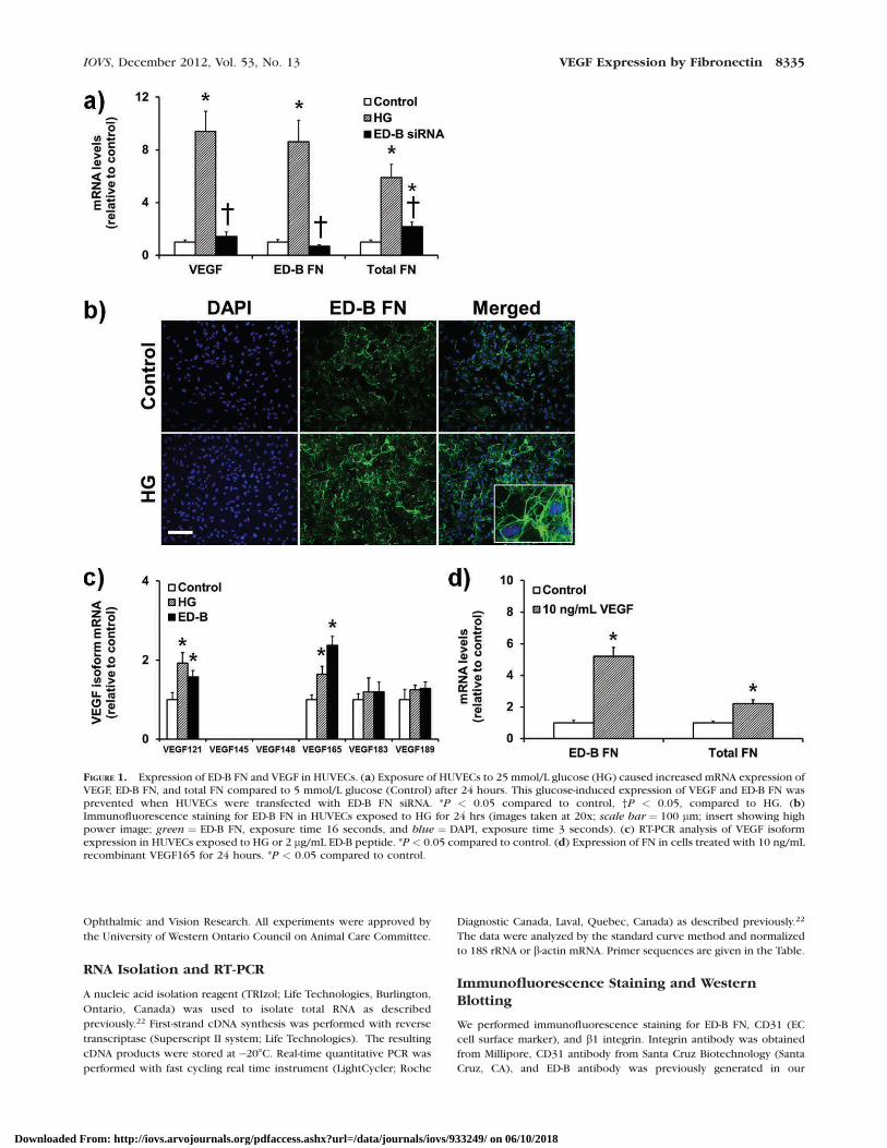
FIGURE 1. Expression of ED-B FN and VEGF in HUVECs. (a) Exposure of HUVECs to 25 mmol/L glucose (HG) caused increased mRNA expression ofVEGF, ED-B FN, and total FN compared to 5 mmol/L glucose (Control) after 24 hours. This glucose-induced expression of VEGF and ED-B FN wasprevented when HUVECs were transfected with ED-B FN siRNA. *P < 0.05 compared to control, †P < 0.05, compared to HG. (b)Immunofluorescence staining for ED-B FN in HUVECs exposed to HG for 24 hrs (images taken at 20x; scale bar ¼ 100 lm; insert showing highpower image; green ¼ ED-B FN, exposure time 16 seconds, and blue ¼ DAPI, exposure time 3 seconds). (c) RT-PCR analysis of VEGF isoformexpression in HUVECs exposed to HG or 2 lg/mL ED-B peptide. *P < 0.05 compared to control. (d) Expression of FN in cells treated with 10 ng/mLrecombinant VEGF165 for 24 hours. *P < 0.05 compared to control.
IOVS, December 2012, Vol. 53, No. 13 VEGF Expression by Fibronectin 8335
Downloaded From: http://iovs.arvojournals.org/pdfaccess.ashx?url=/data/journals/iovs/933249/ on 06/10/2018
laboratory.22 After treating the cells with high glucose levels, we used
methanol to fix the cells. Primary antibodies were used at 1:200
concentrations in PBS (containing 1% bovine serum albumin) and the
cells were incubated for 1 hour at room temperature.
Alexa Fluor-488 and -594 conjugated secondary antibodies (Life
Technologies) were used for detection (1:200 at room temperature)
and DAPI (Vector Laboratories, Burlington, Ontario, Canada) for
counterstaining. Images were taken using a compound fluorescent
microscope (Olympus BX-51; Olympus Canada Inc., Richmond Hill,
Ontario, Canada) equipped with a digital camera (Spot Pursuit; SPOT
Imaging Solutions, Sterling Heights, MI).
For western blotting, we used 20 micrograms of total proteins per
lane, resolved by 10% sodium dodecyl sulfate-polyacrylamide gel
electrophoresis, and analyzed using VEGF and b-actin antibodies (Santa
Cruz Biotechnology). The signals were detected with horseradish
peroxidase-conjugated secondary antibody (Santa Cruz Biotechnology)
and developed with the chemiluminescent substrate (Amersham
Pharmacia Biotechnology, Amersham, UK).
Statistical Analysis
The data were expressed as means 6 SEM. Differences were
determined using ANOVA with post hoc Bonferroni’s correction. P
values < 0.05 were considered statistically significant.
RESULTS
High Glucose-Induced VEGF Expression IsMediated by ED-B FN
We first investigated whether high levels of glucose alter theexpression of VEGF in ECs and whether any change in VEGF
was mediated by ED-B FN. A previous study has reported 40%induction in VEGF protein after 72 hours treatment with 30mmol/L glucose.23 We have shown that glucose increases FNmRNA and protein in as early as 24 hours in HUVECs.22,27–29
Therefore, we exposed the cells to media containing 25 mmol/L glucose for 24 hours and assayed for VEGF transcript levels.Our results showed that HG increased VEGF mRNA levels ascompared to cells exposed to 5 mmol/L glucose (Fig. 1a). Anincrease was also observed in ED-B containing FN mRNAlevels. Twenty-five mmol/L L-glucose, used as osmotic control,did not produce any changes in VEGF mRNA levels (data notshown). These results showed that the newly expressed FN(induced by HG) predominantly contains ED-B. We hypothe-sized that the VEGF induction by high glucose levels ismediated by ED-B FN. To test this, we silenced ED-B FN in ECsand measured VEGF expression. If the hypothesis is true, weshould see a reduction in glucose-induced VEGF expressionupon ED-B FN knockdown. Our results showed that, indeed,HG-induced VEGF was completely normalized with ED-BsiRNA transfection (Fig. 1a).
We next determined whether HG increases ED-B proteinlevels or localization by immunofluorescence staining. Ourresults showed no significant changes in ED-B FN fibrilformation upon glucose challenge (Fig. 1b). However, theED-B FN immunoreactivity was clearly higher in cells exposedto HG (images taken at the same exposure time).
Selective Upregulation of VEGF121 and VEGF165by HG and ED-B Peptide
Alternative splicing of VEGF produces a number of isoformsthat differ in their heparin– and heparin–sulfate-bindingcapacities.30 We wanted to determine whether HG or ED-Bpeptide selectively upregulated certain VEGF isoforms. There-fore, we performed qPCR analysis (primer sequences present-ed in the Table; VEGF isoform sequences from Zygalaki et al.31)of major VEGF isoforms in ECs exposed to HG or ED-B peptidefor 24 hours. Our results showed that both HG and ED-Bpeptide upregulated VEGF121 and VEGF165 isoforms (Fig. 1c).Other isoforms including VEGF145, 148, 183, and 189 showedno significant changes. It is possible that HG- or ED-B FN-induced VEGF may create a positive feedback loop to increaseFN. In support of this notion are reports of VEGF increasingECM proteins in ECs.32 We treated ECs with recombinantVEGF165 and assayed for FN expression. Our results show apositive feedback loop where exogenous VEGF increasedexpression of total FN and ED-B containing FN (Fig. 1d).
Human Retinal Microvascular Endothelial CellsShow the Same Alterations as HUVECs
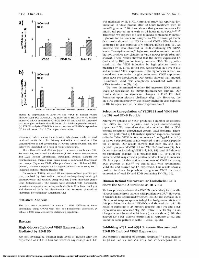
We have previously shown that ED-B FN is selectively increased invitreous samples from patients with proliferative DR.33 However,it remains to be determined whether HRMECs also increase ED-BFN expression upon exposure to high levels of glucose. We testedthis possibility in cultured HRMECs and showed that with 48hours of exposure to 25 mmol/L glucose, ED-B FN and VEGFexpression was increased (Fig. 2a). Unlike HUVECs (Fig. 1), nochanges were observed at 24 hours (data not shown). We alsoassayed for VEGF isoform expression in response to HG andfound the same pattern as with HUVECs (Fig. 2b).
Inhibiting a2b1 and a5b1 Prevents Glucose- andED-B FN Induced VEGF Expression
ECs express a number of integrin receptors.34 These include4x b1 (a1, a2, a3, and a5), avb3, and avb5 integrins. FN is
FIGURE 2. Expression of ED-B FN and VEGF in human retinalmicrovascular ECs (HRMECs). (a) Exposure of HRMECs to HG causedincreased mRNA expression of VEGF, ED-B FN, and total FN comparedto control glucose levels after 48 hours. *P < 0.05 compared to control.(b) RT-PCR analysis of VEGF isoform expression in HRMECs exposed toHG for 48 hours. *P < 0.05 compared to control.
8336 Chen et al. IOVS, December 2012, Vol. 53, No. 13
Downloaded From: http://iovs.arvojournals.org/pdfaccess.ashx?url=/data/journals/iovs/933249/ on 06/10/2018
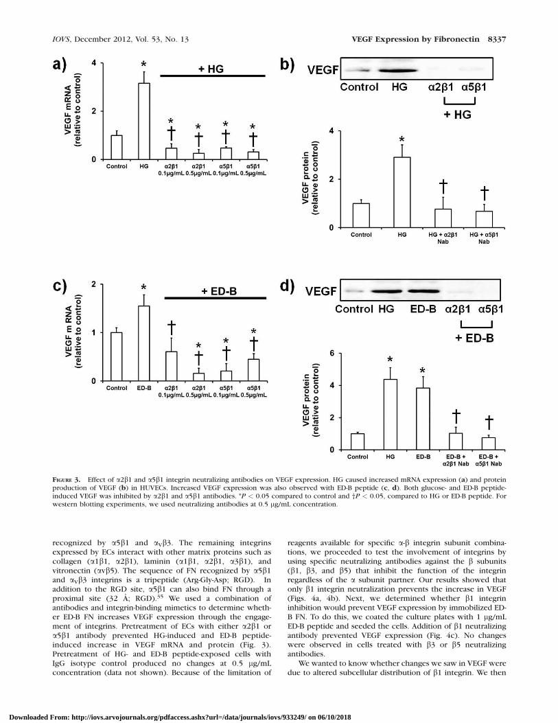
recognized by a5b1 and aVb3. The remaining integrinsexpressed by ECs interact with other matrix proteins such ascollagen (a1b1, a2b1), laminin (a1b1, a2b1, a3b1), andvitronectin (avb5). The sequence of FN recognized by a5b1and aVb3 integrins is a tripeptide (Arg-Gly-Asp; RGD). Inaddition to the RGD site, a5b1 can also bind FN through aproximal site (32 A; RGD).35 We used a combination ofantibodies and integrin-binding mimetics to determine wheth-er ED-B FN increases VEGF expression through the engage-ment of integrins. Pretreatment of ECs with either a2b1 ora5b1 antibody prevented HG-induced and ED-B peptide-induced increase in VEGF mRNA and protein (Fig. 3).Pretreatment of HG- and ED-B peptide-exposed cells withIgG isotype control produced no changes at 0.5 lg/mLconcentration (data not shown). Because of the limitation of
reagents available for specific a-b integrin subunit combina-tions, we proceeded to test the involvement of integrins byusing specific neutralizing antibodies against the b subunits(b1, b3, and b5) that inhibit the function of the integrinregardless of the a subunit partner. Our results showed thatonly b1 integrin neutralization prevents the increase in VEGF(Figs. 4a, 4b). Next, we determined whether b1 integrininhibition would prevent VEGF expression by immobilized ED-B FN. To do this, we coated the culture plates with 1 lg/mLED-B peptide and seeded the cells. Addition of b1 neutralizingantibody prevented VEGF expression (Fig. 4c). No changeswere observed in cells treated with b3 or b5 neutralizingantibodies.
We wanted to know whether changes we saw in VEGF weredue to altered subcellular distribution of b1 integrin. We then
FIGURE 3. Effect of a2b1 and a5b1 integrin neutralizing antibodies on VEGF expression. HG caused increased mRNA expression (a) and proteinproduction of VEGF (b) in HUVECs. Increased VEGF expression was also observed with ED-B peptide (c, d). Both glucose- and ED-B peptide-induced VEGF was inhibited by a2b1 and a5b1 antibodies. *P < 0.05 compared to control and †P < 0.05, compared to HG or ED-B peptide. Forwestern blotting experiments, we used neutralizing antibodies at 0.5 lg/mL concentration.
IOVS, December 2012, Vol. 53, No. 13 VEGF Expression by Fibronectin 8337
Downloaded From: http://iovs.arvojournals.org/pdfaccess.ashx?url=/data/journals/iovs/933249/ on 06/10/2018

performed immunofluorescence staining for b1 integrin in ECsexposed to HG. Our results showed no significant changes inthe localization of b1 integrin upon HG exposure (Fig. 4d).Finally, we knocked down the expression of b1 integrin in ECsand observed similar results as with the neutralizing antibodies(Figs. 5a, 5b). Both HG- and ED-B peptide-mediated VEGFexpression were completely normalized when b1 integrin wassilenced.
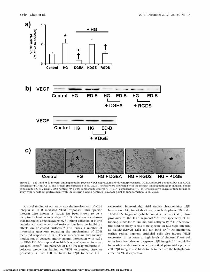
So far, our results showed that HG increases ED-B FN, whichthen leads to VEGF expression through the b1 integrin. To
show that these changes were mediated by direct binding ofED-B FN to integrin, we used integrin-binding peptides tocompetitively block the interaction between b1 integrin andED-B FN. DGEA peptide is recognized by the a2b1 integrin, andthe RGDS peptide is recognized by a5b1. Pretreatment withthese peptides prevented HG-induced increase in VEGF mRNAand protein (Figs. 6a, 6b). In contrast, KDGE (scrambledpeptide; negative control for DGEA) showed no effect on VEGFexpression at the mRNA or protein level (Figs. 6a, 6b). As afunctional readout of these effects, we assayed for tube
FIGURE 4. Involvement of b1 integrin in HG and ED-B FN mediated VEGF expression. VEGF expression by HG (a) or ED-B peptide (b) was inhibitedby a specific neutralizing antibody against integrin b1. b3 neutralizing antibody had no significant effect. (c) Effect of the neutralizing antibodies oncontrol (5 mmol/L glucose) cells plated on ED-B FN peptide coated plates (1 lg/cm2). All neutralizing antibodies were applied at a concentration of0.5 lg/mL. For (a, b), *P < 0.05 compared to control, †P < 0.05, compared to HG or ED-B peptide. For (c), *P < 0.05 compared to IgG control. (d)Immunofluorescence staining of b1 integrin in HUVECs exposed to HG (images taken at 20x; scale bar ¼ 100 lm; inserts showing high powerimage; green ¼ b1 integrin, red ¼ CD31, and blue ¼ DAPI).
8338 Chen et al. IOVS, December 2012, Vol. 53, No. 13
Downloaded From: http://iovs.arvojournals.org/pdfaccess.ashx?url=/data/journals/iovs/933249/ on 06/10/2018
formation by ECs. Exposure of ECs to HG increases tubemorphogenesis (branching), which was prevented by DGEAand RGDS peptide, but not KDGE peptide (Fig. 6c). Theseresults confirmed that the interaction between ED-B FN anda2b1/ a5b1 was necessary for the increased VEGF expression.
High Glucose Leads to Altered VEGFR2 Expressionthrough ED-B Containing FN
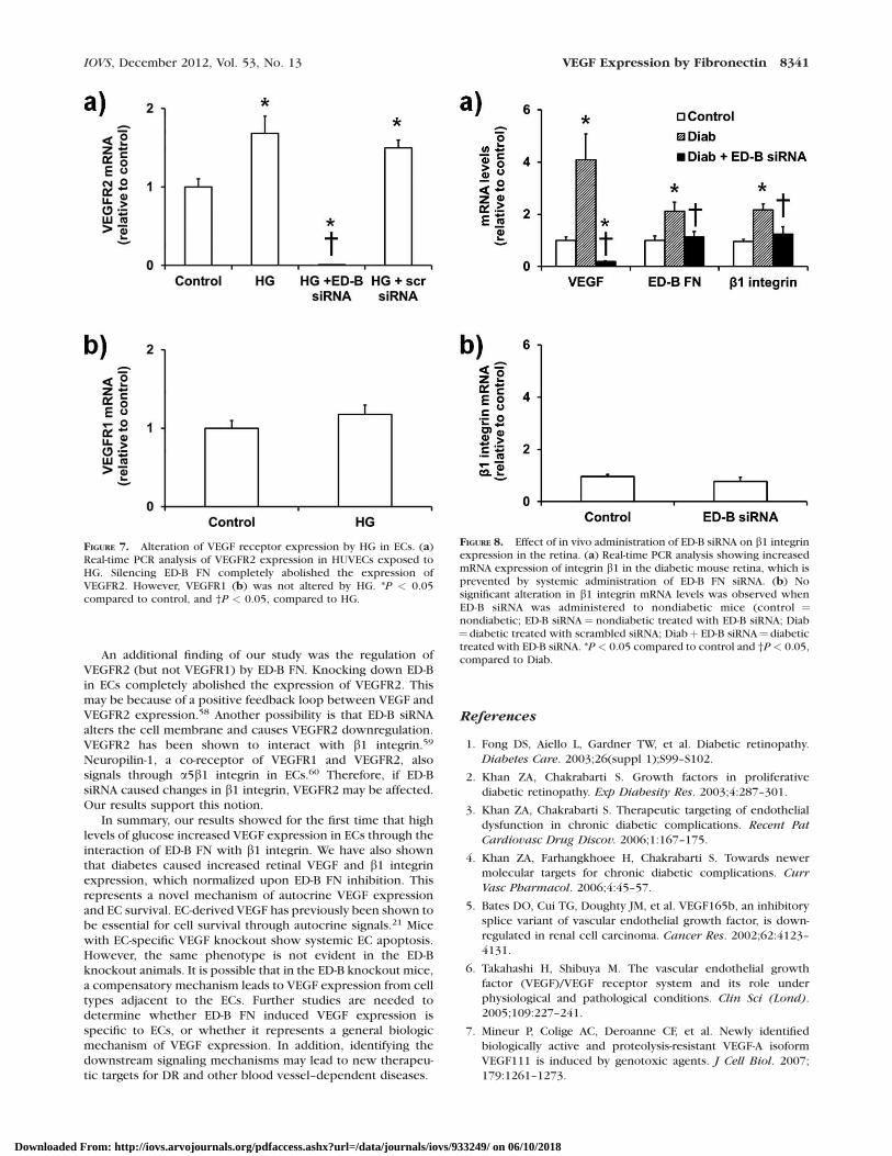
We next determined whether HG altered the expression ofVEGF receptors in ECs, and whether ED-B FN mediated theseeffects. Culture of ECs in HG significantly increased VEGFR2expression (Fig. 7a), but not VEGFR1 (Fig. 7b). Since VEGFR1has been shown to regulate the binding of VEGF to VEGFR2(and thus the proliferative actions of VEGF),36 these findingsillustrate that HG increases VEGF-VEGFR2 signaling in ECs.Interestingly, we found that the HG-induced expression ofVEGFR2 was mediated by ED-FN, as knockdown completelyabolished VEGFR2 mRNA. VEGFR2 has been shown to clusteralongside integrins on the cell surface. It is possible thatsilencing ED-B FN modulates VEGFR2–integrin coupling andreduced VEGF expression.
Inhibiting ED-B FN in the Retina Prevents Diabetes-Induced b1 Integrin Expression and VEGFProduction
In order to study whether the changes we observed in invitro studies translate to the in vivo setting, we used an
established animal model of chronic diabetes. We havepreviously shown that this STZ-diabetic model exhibits allmolecular alterations reminiscent of human diabetes.37–39
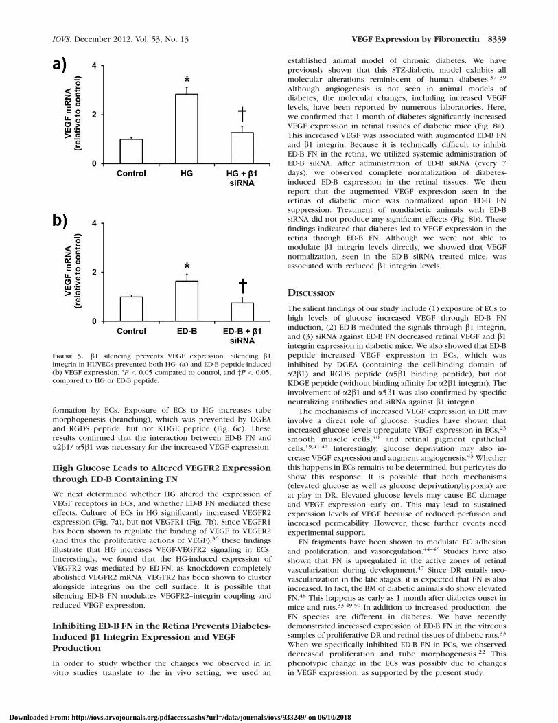
Although angiogenesis is not seen in animal models ofdiabetes, the molecular changes, including increased VEGFlevels, have been reported by numerous laboratories. Here,we confirmed that 1 month of diabetes significantly increasedVEGF expression in retinal tissues of diabetic mice (Fig. 8a).This increased VEGF was associated with augmented ED-B FNand b1 integrin. Because it is technically difficult to inhibitED-B FN in the retina, we utilized systemic administration ofED-B siRNA. After administration of ED-B siRNA (every 7days), we observed complete normalization of diabetes-induced ED-B expression in the retinal tissues. We thenreport that the augmented VEGF expression seen in theretinas of diabetic mice was normalized upon ED-B FNsuppression. Treatment of nondiabetic animals with ED-BsiRNA did not produce any significant effects (Fig. 8b). Thesefindings indicated that diabetes led to VEGF expression in theretina through ED-B FN. Although we were not able tomodulate b1 integrin levels directly, we showed that VEGFnormalization, seen in the ED-B siRNA treated mice, wasassociated with reduced b1 integrin levels.
DISCUSSION
The salient findings of our study include (1) exposure of ECs tohigh levels of glucose increased VEGF through ED-B FNinduction, (2) ED-B mediated the signals through b1 integrin,and (3) siRNA against ED-B FN decreased retinal VEGF and b1integrin expression in diabetic mice. We also showed that ED-Bpeptide increased VEGF expression in ECs, which wasinhibited by DGEA (containing the cell-binding domain ofa2b1) and RGDS peptide (a5b1 binding peptide), but notKDGE peptide (without binding affinity for a2b1 integrin). Theinvolvement of a2b1 and a5b1 was also confirmed by specificneutralizing antibodies and siRNA against b1 integrin.
The mechanisms of increased VEGF expression in DR mayinvolve a direct role of glucose. Studies have shown thatincreased glucose levels upregulate VEGF expression in ECs,23
smooth muscle cells,40 and retinal pigment epithelialcells.19,41,42 Interestingly, glucose deprivation may also in-crease VEGF expression and augment angiogenesis.43 Whetherthis happens in ECs remains to be determined, but pericytes doshow this response. It is possible that both mechanisms(elevated glucose as well as glucose deprivation/hypoxia) areat play in DR. Elevated glucose levels may cause EC damageand VEGF expression early on. This may lead to sustainedexpression levels of VEGF because of reduced perfusion andincreased permeability. However, these further events needexperimental support.
FN fragments have been shown to modulate EC adhesionand proliferation, and vasoregulation.44–46 Studies have alsoshown that FN is upregulated in the active zones of retinalvascularization during development.47 Since DR entails neo-vascularization in the late stages, it is expected that FN is alsoincreased. In fact, the BM of diabetic animals do show elevatedFN.48 This happens as early as 1 month after diabetes onset inmice and rats.33,49,50 In addition to increased production, theFN species are different in diabetes. We have recentlydemonstrated increased expression of ED-B FN in the vitreoussamples of proliferative DR and retinal tissues of diabetic rats.33
When we specifically inhibited ED-B FN in ECs, we observeddecreased proliferation and tube morphogenesis.22 Thisphenotypic change in the ECs was possibly due to changesin VEGF expression, as supported by the present study.
FIGURE 5. b1 silencing prevents VEGF expression. Silencing b1integrin in HUVECs prevented both HG- (a) and ED-B peptide-induced(b) VEGF expression. *P < 0.05 compared to control, and †P < 0.05,compared to HG or ED-B peptide.
IOVS, December 2012, Vol. 53, No. 13 VEGF Expression by Fibronectin 8339
Downloaded From: http://iovs.arvojournals.org/pdfaccess.ashx?url=/data/journals/iovs/933249/ on 06/10/2018
A novel finding of our study was the involvement of a2b1integrin in ED-B mediated VEGF responses. This specificintegrin (also known as VLA-2) has been shown to be areceptor for laminin and collagen.51,52 Studies have also shownthat antibodies directed against a2b1 inhibit adhesion of ECs tolaminin- and collagen-coated surfaces, but have no inhibitoryeffects on FN-coated surfaces.53 This raises a number ofinteresting questions regarding the mechanisms of ED-Bmediated responses in ECs. These mechanisms may includemodulation of collagen and/or laminin interaction with a2b1by ED-B FN. ECs exposed to high levels of glucose increasecollagen levels.54 The presence of ED-B FN may modulate EC-collagen interaction leading to VEGF expression. Anotherpossibility is that ED-B FN binds to a2b1 to cause VEGF
expression. Interestingly, initial studies characterizing a2b1
have shown binding of this integrin to both plasma FN and a
110-Kd FN fragment (which contains the RGD site; close
proximity to the ED-B segment).55,56 The specificity of FN
binding is similar to laminin and collagen IV.55 Furthermore,
this binding ability seems to be specific for ECs a2b1 integrin,
as platelet-derived a2b1 did not bind FN.56 As mentioned
earlier, retinal pigment epithelial cells also induce VEGF
expression in response to high levels of glucose. These cell
types have been shown to express a2b1 integrin.57 It would be
interesting to determine whether retinal pigmental epithelial
cell a2b1 integrin also binds to FN to mediate the high-glucose
effect on VEGF expression.
FIGURE 6. a2b1 and a5b1 integrin-binding peptides prevent VEGF expression and tube morphogenesis. DGEA and RGDS peptides, but not KDGE,prevented VEGF mRNA (a) and protein (b) expression in HUVECs. The cells were pretreated with the integrin-binding peptides (5 mmol/L) beforeexposure to HG or 2 lg/mL ED-B peptide. *P < 0.05 compared to control, †P < 0.05, compared to HG. (c) Representative images of tube formationassay with or without pretreatment with the integrin-binding peptides (asterisks point to tube formation in HUVECs).
8340 Chen et al. IOVS, December 2012, Vol. 53, No. 13
Downloaded From: http://iovs.arvojournals.org/pdfaccess.ashx?url=/data/journals/iovs/933249/ on 06/10/2018
An additional finding of our study was the regulation ofVEGFR2 (but not VEGFR1) by ED-B FN. Knocking down ED-Bin ECs completely abolished the expression of VEGFR2. Thismay be because of a positive feedback loop between VEGF andVEGFR2 expression.58 Another possibility is that ED-B siRNAalters the cell membrane and causes VEGFR2 downregulation.VEGFR2 has been shown to interact with b1 integrin.59
Neuropilin-1, a co-receptor of VEGFR1 and VEGFR2, alsosignals through a5b1 integrin in ECs.60 Therefore, if ED-BsiRNA caused changes in b1 integrin, VEGFR2 may be affected.Our results support this notion.
In summary, our results showed for the first time that highlevels of glucose increased VEGF expression in ECs through theinteraction of ED-B FN with b1 integrin. We have also shownthat diabetes caused increased retinal VEGF and b1 integrinexpression, which normalized upon ED-B FN inhibition. Thisrepresents a novel mechanism of autocrine VEGF expressionand EC survival. EC-derived VEGF has previously been shown tobe essential for cell survival through autocrine signals.21 Micewith EC-specific VEGF knockout show systemic EC apoptosis.However, the same phenotype is not evident in the ED-Bknockout animals. It is possible that in the ED-B knockout mice,a compensatory mechanism leads to VEGF expression from celltypes adjacent to the ECs. Further studies are needed todetermine whether ED-B FN induced VEGF expression isspecific to ECs, or whether it represents a general biologicmechanism of VEGF expression. In addition, identifying thedownstream signaling mechanisms may lead to new therapeu-tic targets for DR and other blood vessel–dependent diseases.
References
1. Fong DS, Aiello L, Gardner TW, et al. Diabetic retinopathy.
Diabetes Care. 2003;26(suppl 1);S99–S102.
2. Khan ZA, Chakrabarti S. Growth factors in proliferative
diabetic retinopathy. Exp Diabesity Res. 2003;4:287–301.
3. Khan ZA, Chakrabarti S. Therapeutic targeting of endothelial
dysfunction in chronic diabetic complications. Recent Pat
Cardiovasc Drug Discov. 2006;1:167–175.
4. Khan ZA, Farhangkhoee H, Chakrabarti S. Towards newer
molecular targets for chronic diabetic complications. Curr
Vasc Pharmacol. 2006;4:45–57.
5. Bates DO, Cui TG, Doughty JM, et al. VEGF165b, an inhibitory
splice variant of vascular endothelial growth factor, is down-
regulated in renal cell carcinoma. Cancer Res. 2002;62:4123–
4131.
6. Takahashi H, Shibuya M. The vascular endothelial growth
factor (VEGF)/VEGF receptor system and its role under
physiological and pathological conditions. Clin Sci (Lond).
2005;109:227–241.
7. Mineur P, Colige AC, Deroanne CF, et al. Newly identified
biologically active and proteolysis-resistant VEGF-A isoform
VEGF111 is induced by genotoxic agents. J Cell Biol. 2007;
179:1261–1273.
FIGURE 7. Alteration of VEGF receptor expression by HG in ECs. (a)Real-time PCR analysis of VEGFR2 expression in HUVECs exposed toHG. Silencing ED-B FN completely abolished the expression ofVEGFR2. However, VEGFR1 (b) was not altered by HG. *P < 0.05compared to control, and †P < 0.05, compared to HG.
FIGURE 8. Effect of in vivo administration of ED-B siRNA on b1 integrinexpression in the retina. (a) Real-time PCR analysis showing increasedmRNA expression of integrin b1 in the diabetic mouse retina, which isprevented by systemic administration of ED-B FN siRNA. (b) Nosignificant alteration in b1 integrin mRNA levels was observed whenED-B siRNA was administered to nondiabetic mice (control ¼nondiabetic; ED-B siRNA¼ nondiabetic treated with ED-B siRNA; Diab¼ diabetic treated with scrambled siRNA; Diabþ ED-B siRNA¼ diabetictreated with ED-B siRNA. *P < 0.05 compared to control and †P< 0.05,compared to Diab.
IOVS, December 2012, Vol. 53, No. 13 VEGF Expression by Fibronectin 8341
Downloaded From: http://iovs.arvojournals.org/pdfaccess.ashx?url=/data/journals/iovs/933249/ on 06/10/2018
8. Biselli-Chicote PM, Oliveira AR, Pavarino EC, Goloni-BertolloEM. VEGF gene alternative splicing: pro- and anti-angiogenicisoforms in cancer. J Cancer Res Clin Oncol. 2012;138:363–370.
9. Mustonen T, Alitalo K. Endothelial receptor tyrosine kinasesinvolved in angiogenesis. J Cell Biol. 1995;129:895–898.
10. Waltenberger J, Claesson-Welsh L, Siegbahn A, Shibuya M,Heldin CH. Different signal transduction properties of KDRand Flt1, two receptors for vascular endothelial growth factor.J Biol Chem. 1994;269:26988–26995.
11. Zachary I. Signal transduction in angiogenesis. EXS. 2005;267–300.
12. Aiello LP, Avery RL, Arrigg PG, et al. Vascular endothelialgrowth factor in ocular fluid of patients with diabeticretinopathy and other retinal disorders. N Engl J Med. 1994;331:1480–1487.
13. Endo H, Naito T, Asahara T, Kajima M, Shiota H. [Cytokines inthe vitreous fluid of patients with proliferative diabeticretinopathy–vascular endothelial growth factor and platelet-derived growth factor are elevated in proliferative diabeticretinopathy]. Nihon Ganka Gakkai Zasshi. 2000;104:711–716.
14. Endo M, Yanagisawa K, Tsuchida K, et al. Increased levels ofvascular endothelial growth factor and advanced glycation endproducts in aqueous humor of patients with diabeticretinopathy. Horm Metab Res. 2001;33:317–322.
15. Aiello LP, Pierce EA, Foley ED, et al. Suppression of retinalneovascularization in vivo by inhibition of vascular endothelialgrowth factor (VEGF) using soluble VEGF-receptor chimericproteins. Proc Natl Acad Sci U S A. 1995;92:10457–10461.
16. Adamis AP, Shima DT, Tolentino MJ, et al. Inhibition of vascularendothelial growth factor prevents retinal ischemia-associatediris neovascularization in a nonhuman primate. Arch Oph-
thalmol. 1996;114:66–71.
17. Ozaki H, Seo MS, Ozaki K, et al. Blockade of vascularendothelial cell growth factor receptor signaling is sufficientto completely prevent retinal neovascularization. Am J Pathol.2000;156:697–707.
18. Young TA, Wang H, Munk S, et al. Vascular endothelial growthfactor expression and secretion by retinal pigment epithelialcells in high glucose and hypoxia is protein kinase C-dependent. Exp Eye Res. 2005;80:651–662.
19. Sone H, Kawakami Y, Okuda Y, et al. Vascular endothelialgrowth factor is induced by long-term high glucose concen-tration and up-regulated by acute glucose deprivation incultured bovine retinal pigmented epithelial cells. Biochem
Biophys Res Commun. 1996;221:193–198.
20. Vega-Diaz B, Herron GS, Michel S. An autocrine loop mediatesexpression of vascular endothelial growth factor in humandermal microvascular endothelial cells. J Invest Dermatol.2001;116:525–530.
21. Lee S, Chen TT, Barber CL, et al. Autocrine VEGF signaling isrequired for vascular homeostasis. Cell. 2007;130:691–703.
22. Khan ZA, Chan BM, Uniyal S, et al. EDB fibronectin andangiogenesis–a novel mechanistic pathway. Angiogenesis.2005;8:183–196.
23. Hsu CC, Yin MC, Tian R. Ascorbic acid and uric acid suppressglucose-induced fibronectin and vascular endothelial growthfactor production in human endothelial cells. J Diabetes
Complications. 2005;19:96–100.
24. Park JE, Keller GA, Ferrara N. The vascular endothelial growthfactor (VEGF) isoforms: differential deposition into thesubepithelial extracellular matrix and bioactivity of extracel-lular matrix-bound VEGF. Mol Biol Cell. 1993;4:1317–1326.
25. Sweeney SM, DiLullo G, Slater SJ, et al. Angiogenesis incollagen I requires alpha2beta1 ligation of a GFP*GERsequence and possibly p38 MAPK activation and focaladhesion disassembly. J Biol Chem. 2003;278:30516–30524.
26. Xiao G, Wang D, Benson MD, Karsenty G, Franceschi RT. Roleof the alpha2-integrin in osteoblast-specific gene expressionand activation of the Osf2 transcription factor. J Biol Chem.1998;273:32988–32994.
27. Khan ZA, Barbin YP, Farhangkhoee H, Beier N, Scholz W,Chakrabarti S. Glucose-induced serum- and glucocorticoid-regulated kinase activation in oncofetal fibronectin expres-sion. Biochem Biophys Res Commun. 2005;329:275–280.
28. Xin X, Khan ZA, Chen S, Chakrabarti S. Glucose-induced Akt1activation mediates fibronectin synthesis in endothelial cells.Diabetologia. 2005;48:2428–2436.
29. Xin X, Khan ZA, Chen S, Chakrabarti S. Extracellular signal-regulated kinase (ERK) in glucose-induced and endothelin-mediated fibronectin synthesis. Lab Invest. 2004;84:1451–1459.
30. Neufeld G, Cohen T, Gengrinovitch S, Poltorak Z. Vascularendothelial growth factor (VEGF) and its receptors. FASEB J.1999;13:9–22.
31. Zygalaki E, Stathopoulou A, Kroupis C, et al. Real-time reversetranscription-PCR quantification of vascular endothelialgrowth factor splice variants. Clin Chem. 2005;51:1518–1520.
32. Kuiper EJ, Hughes JM, Van Geest RJ, et al. Effect of VEGF-A onexpression of profibrotic growth factor and extracellularmatrix genes in the retina. Invest Ophthalmol Vis Sci. 2007;48:4267–4276.
33. Khan ZA, Cukiernik M, Gonder JR, Chakrabarti S. Oncofetalfibronectin in diabetic retinopathy. Invest Ophthalmol Vis Sci.2004;45:287–295.
34. Rupp PA, Little CD. Integrins in vascular development. Circ
Res. 2001;89:566–572.
35. Johansson S, Svineng G, Wennerberg K, Armulik A, LohikangasL. Fibronectin-integrin interactions. Front Biosci. 1997;2:d126–146.
36. Zeng H, Dvorak HF, Mukhopadhyay D. Vascular permeabilityfactor (VPF)/vascular endothelial growth factor (VEGF)peceptor-1 down-modulates VPF/VEGF receptor-2-mediatedendothelial cell proliferation, but not migration, throughphosphatidylinositol 3-kinase-dependent pathways. J Biol
Chem. 2001;276:26969–26979.
37. Chiu J, Farhangkhoee H, Xu BY, Chen S, George B, ChakrabartiS. PARP mediates structural alterations in diabetic cardiomy-opathy. J Mol Cell Cardiol. 2008;45:385–393.
38. Chiu J, Xu BY, Chen S, Feng B, Chakrabarti S. Oxidative stress-induced, poly(ADP-ribose) polymerase-dependent upregula-tion of ET-1 expression in chronic diabetic complications. Can
J Physiol Pharmacol. 2008;86:365–372.
39. Xin X, Chen S, Khan ZA, Chakrabarti S. Akt activation andaugmented fibronectin production in hyperhexosemia. Am J
Physiol Endocrinol Metab. 2007;293:E1036–1044.
40. Williams B, Gallacher B, Patel H, Orme C. Glucose-inducedprotein kinase C activation regulates vascular permeabilityfactor mRNA expression and peptide production by humanvascular smooth muscle cells in vitro. Diabetes. 1997;46:1497–1503.
41. Ahmado A, Carr AJ, Vugler AA, et al. Induction of differenti-ation by pyruvate and DMEM in the human retinal pigmentepithelium cell line ARPE-19. Invest Ophthalmol Vis Sci. 2011;52:7148–7159.
42. Wang W, Matsukura M, Fujii I, et al. Inhibition of high glucose-induced VEGF and ICAM-1 expression in human retinalpigment epithelium cells by targeting ILK with smallinterference RNA. Mol Biol Rep. 2012;39:613–620.
43. Kim BS, Chen J, Weinstein T, Noiri E, Goligorsky MS. VEGFexpression in hypoxia and hyperglycemia: reciprocal effect onbranching angiogenesis in epithelial-endothelial co-cultures. J
Am Soc Nephrol. 2002;13:2027–2036.
44. Wilson SH, Ljubimov AV, Morla AO, et al. Fibronectinfragments promote human retinal endothelial cell adhesion
8342 Chen et al. IOVS, December 2012, Vol. 53, No. 13
Downloaded From: http://iovs.arvojournals.org/pdfaccess.ashx?url=/data/journals/iovs/933249/ on 06/10/2018
and proliferation and ERK activation through alpha5beta1integrin and PI 3-kinase. Invest Ophthalmol Vis Sci. 2003;44:1704–1715.
45. Laplante C, St-Pierre S, Beaulieu AD, Marceau F. Smallfibronectin fragments induce endothelium-dependent vascularrelaxations. Can J Physiol Pharmacol. 1988;66:745–748.
46. Grant MB, Caballero S, Bush DM, Spoerri PE. Fibronectinfragments modulate human retinal capillary cell proliferationand migration. Diabetes. 1998;47:1335–1340.
47. Jiang B, Liou GI, Behzadian MA, Caldwell RB. Astrocytesmodulate retinal vasculogenesis: effects on fibronectin expres-sion. J Cell Sci. 1994;107;2499–2508.
48. Nishikawa T, Giardino I, Edelstein D, Brownlee M. Changes indiabetic retinal matrix protein mRNA levels in a commontransgenic mouse strain. Curr Eye Res. 2000;21:581–587.
49. Evans T, Deng DX, Chen S, Chakrabarti S. Endothelin receptorblockade prevents augmented extracellular matrix componentmRNA expression and capillary basement membrane thicken-ing in the retina of diabetic and galactose-fed rats. Diabetes.2000;49:662–666.
50. Chen S, Khan ZA, Cukiernik M, Chakrabarti S. Differentialactivation of NF-kappa B and AP-1 in increased fibronectinsynthesis in target organs of diabetic complications. Am J
Physiol Endocrinol Metab. 2003;284:E1089–1097.
51. van der Flier A, Sonnenberg A. Function and interactions ofintegrins. Cell Tissue Res. 2001;305:285–298.
52. Zutter MM, Edelson BT. The alpha2beta1 integrin: a novelcollectin/C1q receptor. Immunobiology. 2007;212:343–353.
53. O’Connell PJ, Faull R, Russ GR, D’Apice AJ. VLA-2 is a collagenreceptor on endothelial cells. Immunol Cell Biol. 1991;69;103–110.
54. Moritani S, Negishi K, Watanabe T, et al. Glucose-inducedproduction of type IV collagen and laminin P1 from culturedhuman umbilical vein endothelial cells. J Diabet Complica-
tions. 1991;5:201–203.
55. Languino LR, Gehlsen KR, Wayner E, Carter WG, Engvall E,Ruoslahti E. Endothelial cells use alpha 2 beta 1 integrin as alaminin receptor. J Cell Biol. 1989;109:2455–2462.
56. Kirchhofer D, Languino LR, Ruoslahti E, Pierschbacher MD.Alpha 2 beta 1 integrins from different cell types showdifferent binding specificities. J Biol Chem. 1990;265:615–618.
57. Bando H, Ikuno Y, Hori Y, Sayanagi K, Tano Y. Mitogen-activated protein kinase (MAPK) and phosphatidylinositol-3kinase (PI3K) pathways differently regulate retinal pigmentepithelial cell-mediated collagen gel contraction. Exp Eye Res.2006;82:529–537.
58. Cao Y, EG, Bhattacharya S, Dutta S, Wang E, Mukhopadhyay D.Endogenous Vascular Endothelial Growth Factor-A (VEGF-A)Maintains Endothelial Cell Homeostasis by Regulating VEGFReceptor-2 Transcription. J Biol Chem. 2012;287:3029–3041.
59. Chen TT, Luque A, Lee S, Anderson SM, Segura T, Iruela-ArispeML. Anchorage of VEGF to the extracellular matrix conveysdifferential signaling responses to endothelial cells. J Cell Biol.2010;188:595–609.
60. Valdembri D, Caswell PT, Anderson KI, et al. Neuropilin-1/GIPC1 signaling regulates alpha5beta1 integrin traffic andfunction in endothelial cells. PLoS Biol. 2009;7:e25.
IOVS, December 2012, Vol. 53, No. 13 VEGF Expression by Fibronectin 8343
Downloaded From: http://iovs.arvojournals.org/pdfaccess.ashx?url=/data/journals/iovs/933249/ on 06/10/2018