reininger, l., garcia, m., tomlins, a., muller, s., and
TRANSCRIPT

Enlighten – Research publications by members of the University of Glasgow http://eprints.gla.ac.uk
Reininger, L., Garcia, M., Tomlins, A., Muller, S., and Doerig, C. (2012) The Plasmodium falciparum, Nima-related kinase Pfnek-4: a marker for asexual parasites committed to sexual differentiation. Malaria Journal, 11 (1). p. 250. ISSN 1475-2875 http://eprints.gla.ac.uk/69545/ Deposited on: 12 September 2012

This Provisional PDF corresponds to the article as it appeared upon acceptance. Fully formattedPDF and full text (HTML) versions will be made available soon.
The Plasmodium falciparum, Nima-related kinase Pfnek-4: a marker for asexualparasites committed to sexual differentiation
Malaria Journal 2012, 11:250 doi:10.1186/1475-2875-11-250
Luc Reininger ([email protected])Miguel Garcia ([email protected])
Andrew Tomlins ([email protected])Sylke Müller ([email protected])
Christian Doerig ([email protected])
ISSN 1475-2875
Article type Research
Submission date 30 April 2012
Acceptance date 13 July 2012
Publication date 31 July 2012
Article URL http://www.malariajournal.com/content/11/1/250
This peer-reviewed article was published immediately upon acceptance. It can be downloaded,printed and distributed freely for any purposes (see copyright notice below).
Articles in Malaria Journal are listed in PubMed and archived at PubMed Central.
For information about publishing your research in Malaria Journal or any BioMed Central journal, goto
http://www.malariajournal.com/authors/instructions/
For information about other BioMed Central publications go to
http://www.biomedcentral.com/
Malaria Journal
© 2012 Reininger et al. ; licensee BioMed Central Ltd.This is an open access article distributed under the terms of the Creative Commons Attribution License (http://creativecommons.org/licenses/by/2.0),
which permits unrestricted use, distribution, and reproduction in any medium, provided the original work is properly cited.
The Plasmodium falciparum, Nima-related kinase
Pfnek-4: a marker for asexual parasites committed
to sexual differentiation
Luc Reininger1,2,3,*
Email: [email protected]
Miguel Garcia4
Email: [email protected]
Andrew Tomlins2
Email: [email protected]
Sylke Müller2
Email: [email protected]
Christian Doerig2,3,5
Email: [email protected]
1 CNRS USR3151, Station Biologique, Place Georges Teissier, 29680 Roscoff,
France
2 Wellcome Trust Centre for Molecular Parasitology, Institute of Infection,
Immunity & Inflammation, College of Medical, Veterinary and Life Sciences,
University of Glasgow, 120 University Place, Glasgow G12 8TA, UK
3 INSERM U609, INSERM-EPFL Joint Laboratory, GHI-SV-EPFL, Station 19,
Lausanne CH-1015, Switzerland
4 FCCF-SV-EPFL, Station 15, Lausanne CH-1015, Switzerland
5 Department of Microbiology, Monash University, Wellington Road, Clayton,
VIC 3800, Australia
* Corresponding author. INSERM U609, INSERM-EPFL Joint Laboratory, GHI-
SV-EPFL, Station 19, Lausanne CH-1015, Switzerland
Abstract
Background
Malaria parasites undergo, in the vertebrate host, a developmental switch from asexual
replication to sexual differentiation leading to the formation of gametocytes, the only form
able to survive in the mosquito vector. Regulation of the onset of the sexual phase remains
largely unknown and represents an important gap in the understanding of the parasite’s
complex biology.
Methods
The expression and function of the Nima-related kinase Pfnek-4 during the early sexual
development of the human malaria parasite Plasmodium falciparum were investigated, using
three types of transgenic Plasmodium falciparum 3D7 lines: (i) episomally expressing a
Pfnek-4-GFP fusion protein under the control of its cognate pfnek-4 promoter; (ii) episomally
expressing negative or positive selectable markers, yeast cytosine deaminase-uridyl
phosphoribosyl transferase, or human dihydrofolate reductase, under the control of the
pfnek-4 promoter; and (iii) lacking a functional pfnek-4 gene. Parasite transfectants were
analysed by fluorescence microscopy and flow cytometry. In vitro growth rate and
gametocyte formation were determined by Giemsa-stained blood smears.
Results
The Pfnek-4-GFP protein was found to be expressed in stage II to V gametocytes and,
unexpectedly, in a subset of asexual-stage parasites undergoing schizogony. Culture
conditions stimulating gametocyte formation resulted in significant increase of this schizont
subpopulation. Moreover, sorted asexual parasites expressing the Pfnek-4-GFP protein
displayed elevated gametocyte formation when returned to in vitro culture in presence of
fresh red blood cells, when compared to GFP- parasites from the same initial population.
Negative selection of asexual parasites expressing pfnek-4 showed a marginal reduction in
growth rate, whereas positive selection caused a marked reduction in parasitaemia, but was
not sufficient to completely abolish proliferation. Pfnek-4- clones are not affected in their
asexual growth and produced normal numbers of stage V gametocytes.
Conclusions
The results indicate that Pfnek-4 is not strictly gametocyte-specific, and is expressed in a
small subset of asexual parasites displaying high rate conversion to sexual development.
Pfnek-4 is not required for erythrocytic schizogony and gametocytogenesis. This is the first
study to report the use of a molecular marker for the sorting of sexually-committed schizont
stage P. falciparum parasites, which opens the way to molecular characterization of this pre-
differentiated subpopulation.
Keywords
Gametocytes, NIMA, Plasmodium falciparum, Ser/Thr protein kinase
Background
The life cycle of Plasmodium falciparum parasites includes multiple rounds of asexual
replication in human host erythrocytes. A small subset of parasites, upon invasion of new red
blood cells, do not enter schizogony, but develop into cell cycle-arrested gametocytes [1].
Gametocytes are the only forms able to survive in the mosquito vector, and their formation is
therefore essential for malaria transmission. How malaria parasites regulate the switch from
erythrocytic schizogony to gametocytogenesis is still not understood. Bruce et al. [2] showed
that merozoites released from a single schizont become either all asexual parasites or all
gametocytes, indicating that the switch to sexual differentiation is likely to occur during the

preceding asexual red blood cell cycle. Sex determination occurs at the same time or soon
after the switch to sexual development, as the entire progeny of a given gametocyte-
producing schizont comprises exclusively either male or female cells [3]. The mechanism of
parasite commitment to sexual differentiation appears to be constitutive but may be
modulated by the environment, as reviewed in [4].
The NIMA-related protein kinases (Neks) constitute a family of eukaryotic serine/threonine
kinases implicated in cell cycle control, and whose main role is to regulate centrosome and
cilia function [5,6]. The P. falciparum kinome includes four Nek kinases, two of which,
Pfnek-2 and Pfnek-4, were shown to be predominantly or exclusively expressed in sexual
stages, suggesting a possible role in the sexual development of the parasite. Consistent with
this hypothesis, it has been previously shown that the rodent malaria parasite Plasmodium
berghei NIMA-related kinases Pbnek-2 and Pbnek-4, displaying female gametocyte-specific
expression, are essential for pre-meiotic genome replication in the zygote and ookinete
formation in the mosquito host [7-9]. Here, the expression and function of the P. falciparum
Nima-related kinase Pfnek-4 were investigated at the onset and during progression of
gametocytogenesis. A subpopulation of asexual stage parasites undergoing schizogony and
expressing the Pfnek-4 protein was identified. Further analyses indicate that the progeny of
these asexual parasites is more likely to differentiate into gametocytes in the subsequent red
blood cell cycle. Since not all asexual parasites expressing Pfnek-4 appear to be committed to
sexual development, altogether the data suggest that Pfnek-4 identifies a population of
committed and reversibly pre-committed parasites.
Methods
Molecular cloning and plasmid constructs
Genomic and cDNA sequence data were accessed via PlasmoDB [10]. The pfnek-4 gene
PlasmoDB identifier is PF3D7_0719200, MAL7P1.100 in earlier versions. The Pfnek-4-GFP
plasmid (pCHD-Pfnek-4) was generated by using the pHGB and pCHD-1/2 transfection
vectors based on GatewayTM
recombinational cloning and described in Tonkin et al. [11].
The 997-bp 5’-flanking region of Pfnek-4 was amplified from 3D7 genomic DNA using the
forward OL-306 (GGGTCGACGAACTCATCATTCATA) and reverse OL-308
(CCAGATCTTGAATGGTTATAAGATATAC) oligonucleotides containing SalI and BglII
sites, respectively. The 933-bp Pfnek-4 open reading frame was amplified from a gametocyte
cDNA library using the oligonucleotides forward OL-598
(CCCAGATCTATGAATAAATATGAAAAGATTAGAG) and reverse OL-599
(CCCCCTAGGAGTATCAACAACATCCAG) containing BglII and AvrII sites, respectively.
The digested products, ~1-kb 5’-flanking region and open reading frame of Pfnek-4, were
sequentially ligated into the plasmid pHGB to produce the pHGB-Pfnek-4-GFP entry clone.
This plasmid was used in a recombination reaction with the pCHD-1/2 destination vector
containing the cassette responsible for expression of hDHFR conferring resistance to
WR99210 treatment, to produce the final transfection vector pCHD-Pfnek-4-GFP.
The pCC1 and pCC4 vectors constructed for negative selection of single crossover
recombinants in the generation of knock-out parasites have been described [12] and formed
the basis for the generation of plasmids pScCDUPPfnek-4 and phDHFRPfnek-4 described here.
The pScCDUPPfnek-4 plasmid was constructed by replacing the 857-bp SacII-XhoI hsp86 5’
sequence of pCC4 by the 997-bp 5’-flanking region of pfnek-4 amplified from genomic DNA
with forward OL-1082 (GGCCGCGGGAACTCATCATTCATA) and reverse OL-1083
(GGCTCGAGTGAATGGTTATAAGATATAC) containing SacII and XhoI sites,
respectively, generating an expression vector in which the yeast cytosine deaminase gene is
placed under the control of the pfnek-4 promoter. The phDHFRPfnek-4 plasmid was constructed
by replacing the ~1.1–kb XhoI-XmaI yeast cytosine deaminase coding sequence from the
pScCDUPPfnek-4 plasmid by ~0.6–kb of the hDHFR sequence amplified from plasmid pCC1
with forward OL-1084 (GGCTCGAGATGCATGGTTCGCTAAACTGC) and reverse OL-
1085 (GGCCCGGGTTAATCATTCTTCTCATATAC) containing XhoI and XmaI sites,
respectively, generating an expression vector in which the hDHFR gene is placed under the
control of the pfnek-4 promoter.
A pfnek-4 gene disruption plasmid was produced in the plasmid pCAM-BSD that contains
the gene conferring resistance to blasticidin. The oligonucleotide pair forward OL-46
(GGGGGGATCCAATTATGGAAATACAATACT) and reverse OL-
47(GGGGCGCCGGCGTGGACTTAAATAATAAGG) containing BamHI and NotI sites
was used to amplify a 1,017 bp fragment for insertion to pCAM-BSD. Ring-stage parasites
were electroporated with 50–100 μg plasmid DNA, as previously described. Blasticidin
(Calbiochem) was added to a final concentration of 2.5 μg/ml 48 hours after transfection to
select for transformed parasites. Resistant parasites appeared after three to four weeks and
were maintained under selection. After verification by PCR that pfnek-4- parasites were
present, the population was cloned by limiting dilution in 96-well plates (0.25/0.5/1.0 parasite
per well). Genotypic analysis enabled selection of independent pfnek4- clones for further
phenotypic analysis. All constructs were sent to the Dundee Sequencing Service, University
of Dundee, UK, for sequence verification before being used.
Parasite culture, transfection and determination of the growth rate
The 3D7 clone of P. falciparum, its F12 subclone and the 3D7 and F12 transfectants were
grown in human erythrocytes using 0.5% Albumax II (Invitrogen) and synchronized using
sorbitol as described previously [13]. For transfections, synchronized ring-stage parasites
(3D7 and F12) were electroporated with 50–100 μg of plasmid DNA using standard
procedures. Transformed parasites were selected in presence of 5 nM WR22910 (Jacobus
Pharmaceutical Co Inc, Princeton, NJ, USA) or 2.5 μg ml-1
blasticidin (Calbiochem).
Parasitaemia was monitored by Giemsa-stained thin blood smears. Induction of
gametocytogenesis was performed according to the protocol of Carter et al. [14], culturing
asexual blood stage parasites for four to five days in 6%-haematocrit blood cultures to high 8-
10% parasitaemia.
Nested RT-PCR and diagnostic PCR
Total RNA samples were extracted from parasite pellets using TRIzol lysis solution
(Invitrogen). DNase treatment of RNA samples prior to RT-PCR was performed by
incubation at 37°C for 30 min using the RQ1 RNase-free DNase I purchased from Promega.
The DNase was inactivated by incubation at 65°C for 10 min. RT-PCRs were performed with
500 ng of total RNA/reaction using the ImPromII reverse transcription system purchased
from Promega. The RT reactions were incubated at 42°C for 1 h. For the first round of PCR
(30 cycles at 94°C for 45 sec, 55°C for 45 sec, and 68°C for 2 min), Pfnek-4-specific primers
were forward OL-587 (GAGAGGGATCCATGAATAAATATGAAAAGA) and reverse OL-
586 (GAGAGGTCGACTTAAGTATCAACAACATCC). For the second round of PCR (25
cycles at 94°C for 45 sec, 55°C for 45 sec, and 68°C for 2 min), 1 μl (1/25) of each PCR

product was reamplified using the Pfnek-4-specific primers forward OL-46
(GGGGGGATCCAATTATGGAAATACAATACT) and reverse OL-47
(GGGGCGCCGGCGTGGACTTAAATAATAAGG).
Disruption of the pfnek-4 gene was analysed by diagnostic PCR using three primer pairs. The
primer pair forward OL-761(CACGACATTACATAATAAAAGC) and reverse OL-763
(ATCCCTTTTATGAATTTACTG) produced a 1288-bp fragment corresponding to the
undisrupted Pfnek-4 locus from wild-type 3D7. Primer pairs forward OL-761 and reverse
OL-168 (CAATTAACCCTCACTAAAG), and forward OL-167
(TATTCCTAATCATGTAAATCTTAAA) and reverse OL-764
(TCGAAGAGGTCATTATATATC) amplified across the 5’ and 3’ ends of the integration
site, giving rise to 1,179- and 1,277-bp products only in the disrupted locus, respectively.
Western blot analysis
Western blot analysis was performed on cell-free extracts prepared by resuspending parasite
pellets in M-PER Mammalian Protein Extraction Reagent (Pierce) supplemented with 1 mM
phenylmethylsulphonyl fluoride and ComplexTM
mixture protease inhibitor tablet from Roche
Applied Science. Immunoblotting was performed as described [13], using monoclonal anti-
GFP antibodies (Roche) and horseradish peroxidase-conjugated sheep anti-mouse IgG
antiserum (Sigma). Lambda protein phosphatase (New England BioLabs) was used to
dephosphorylate protein extracts (20 μg) 30 min at 30°C following the supplier
recommendations prior to western blot analysis.
Fluorescence microscopy
Live imaging of parasite transfectants was performed on a Delta vision deconvolution
fluorescence microscope (100x/1.4 oil immersion objective Olympus IX-70). Images were
processed using IMARIS version 7.0. For live imaging, parasite nuclei were stained by
incubation 10 min at 37°C in complete medium containing 1 μg ml-1
Hoechst 3342
(Invitrogen).
Flow cytometry and cell sorting
Flow cytometer analysis of parasite subpopulations was performed on a FACScan flow
cytometer (Becton Dickinson Biosciences). Cells were either fixed in 0.025% glutaraldehyde
and propidium iodide (PI)-stained or directly stained with 1 μg ml-1
Hoechst 33258
(Invitrogen) prior to analysis. The threshold was set to a value eliminating uninfected red
cells. For sorting Pfnek-4-GFP-expressing transfectants, late trophozoite-stage parasites were
first enriched by using the VarioMACS separator and CS MACS columns (MiltenyiBiotec),
then returned to culture conditions for three to four hours prior to cell sorting using a FACS
ARIA II SORP Instrument (Becton Dickinson Biosciences). In two independent experiments,
Pfnek-4-GFP- (4 10
6 and 12 10
6) and Pfnek-4-GFP
+ (0.8 10
6 and 1.6 10
6) parasites,
respectively, were collected over a four-hour cell sorting period and returned to culture
conditions in flat-bottom 96-well plates with fresh red blood cells to 2% -haematocrit and 1%
-parasitaemia. Gating of GFP- and GFP
+ parasite populations has been set in a restrictive way
such that the sorted cells exhibited high levels of purity in both experiments (purity of GFP-
sorted cells, 100%; GFP+ sorted cells, 92.8 and 97.2%) when re-analysed by FACS analysis.

Results
Transgenic expression of a Pfnek-4-GFP fusion protein in gametocytes and a
subset of asexual blood stage parasites
P. falciparum 3D7 parasites were transfected with a plasmid containing the Pfnek-4 coding
sequence fused C-terminally to GFP under the control of its cognate (Pfnek-4) promoter,
using ~1 kb of genomic sequence upstream of the Pfnek-4 translation initiation codon.
Episomal propagation of the construct was maintained in the presence of antifolate drug
pressure. Stage II transgenic gametocytes displayed a single punctuate Pfnek-4-GFP
fluorescent structure, which coincides with or is located nearby one tip of the parasite and
which is often closely associated with the nucleus (Figure 1A-B). The Pfnek-4-GFP protein
appeared to accumulate in the cytosol of stage III gametocytes (Figure 1C) and remained
present at high levels throughout the formation of mature stage V gametocytes (Additional
file 1, panel A). Interestingly, while the bulk of examined schizonts did not show any
detectable signal, as expected for a protein thought to be gametocyte-specific, fluorescence
microscopy of asexual stage cultures consistently identified a small subpopulation (a few
percents, see below) of schizonts with dots (single or doublets) of concentrated Pfnek-4-GFP
protein associated with nuclei (Figure 1D). Fluorescence was never detected in ring and
trophozoite stages. Noteworthy, all nuclei within a single schizont appeared to be associated
with punctuate Pfnek-4-GFP fluorescence from early developing schizont to multinucleated
schizonts (Additional file 1, panel B).
Figure 1 Episomal expression of GFP-tagged Pfnek-4 protein in gametocytes and a
subset of asexual stage parasites. Live images of Hoechst-stained stage II (A-B), stage III
gametocytes (C), and multinucleated-schizont stage (D), from transgenic 3D7 parasites
expressing GFP-tagged Pfnek-4 controlled by cognate (Pfnek-4) promoter. Overlay between
the green and blue channels, and corresponding DIC images are shown as well. Scale bars,
2.0 μm. E, Immunoblots showing ~62-kDa protein band (arrow) recognized by mouse anti-
GFP mAbs (Roche) using parasite extracts (10 μg) from wild type (Wt) 3D7 and Pfnek-4-
GFP transgenic (Tg) stage II-V gametocytes and synchronized schizonts of 3D7 and the non-
gametocyte-producer F12 clones at day 4 of induction of gametocytogenesis, and from
Pfnek-4-GFP transgenic 3D7 synchronized schizonts before (−) and after (+) 30 min-
incubation with λ-protein phosphatase. High MW Pfnek-4-GFP species susceptible to λ-
protein phosphatase treatment are marked with an asterisk. Note the much lower expression
level in asexual parasite extracts as compared to gametocyte extract
Western blot analysis of parasite extracts from gametocyte and asexual blood stage parasite
cultures, using anti-GFP antibodies, detected a specific protein of ~62 kDa, consistent with
the predicted mass of the Pfnek-4-GFP fusion protein (Figure 1E). This is in contrast to the
previously reported lack of detectable Pfnek-4 protein in cultures of asexual 3D7 parasites,
using chicken anti-Pfnek-4 IgY antibodies. Possible explanations for this discrepancy
include: (i) the use of synchronized schizont stage parasites grown under conditions inducing
gametocyte formation in the present series of experiments (in contrast to mixed asexual not
under gametocytogenesis-inducing conditions in the earlier study [8]), and (ii) higher
sensitivity of the anti-GFP monoclonal antibodies versus the anti-Pfnek-4 IgY. Pfnek-4-GFP
transfectants produced in the F12 clone background, a 3D7 subclone having lost the ability to
produce gametocytes, allow to exclude that the Pfnek-4 signal from 3D7 asexual cultures is
solely caused by contaminating gametocytes (Figure 1E). Anti-GFP reactive protein species

of slightly higher molecular mass likely corresponds to Pfnek-4 phosphorylated forms as
indicated by their susceptibility to λ protein phosphatase treatment (Figure 1E).
Nested RT-PCR using RNA isolated from asexual stages of the 3D7 parental clone and the
gametocyte-less F12 clone yielded amplicons of the expected size for Pfnek-4 cDNA (Figure
2), the identity of which was verified by cloning and sequencing, further demonstrating that
Pfnek-4 is not strictly gametocyte-specific, and that low mRNA levels are also present in
untransformed asexual parasites.
Figure 2 Expression of Pfnek-4 in untransformed asexual parasites. Nested RT-PCR
detection of Pfnek-4 transcripts in untransformed gametocyte-less Plasmodium falciparum
F12 substrain. Two rounds of amplification performed on RNA samples untreated or treated
with Dnase I and reverse transcriptase from both 3D7 and F12 clones yielded amplified
fragments of the expected size for Pfnek-4 genomic DNA (0.99 kb) and cDNA (0.48 kb). C,
water control
Asexual stage parasites expressing Pfnek-4 display high rate commitment to
sexual differentiation
Pfnek-4-GFP transgenic 3D7 and F12 parasites, either maintained under standard culture
conditions or grown under conditions inducing commitment to sexual differentiation were
subjected to FACS analysis. Figure 3 shows dot plots of samples of glutaraldehyde-fixed,
propidium iodide (PI)-stained 3D7 and F12 transfectants grown under standard culture
conditions, the GFP-positive (GFP+) schizonts consisting of 13.7% and 2.1% of the total
schizont population, respectively; the high DNA content as measured by PI fluorescence
allow to exclude that these are gametocytes. Under culture conditions stimulating sexual
differentiation, 3D7 and F12 GFP+ schizonts significantly increased to 26.5% and 9.6% of the
total schizont population, respectively. FACS analysis of live, 3D7 Hoechst-stained Pfnek-4-
GFP transfectants confirmed the ~2-fold increase (paired t-test; p<0.0001) in the asexual
multinucleated GFP+ parasite population in culture conditions inducing sexual differentiation
(Table 1). It should be stressed that the GFP+ asexual parasites generated at high parasitaemia
display a punctuate pattern and intensity of Pfnek-4-GFP fluorescence similar to the GFP+
asexual parasites observed in standard culture conditions, arguing against any major
deregulation of the episomal promoter due to stress conditions (data not shown).
Figure 3 Culture conditions inducing sexual differentiation result in increased number
of multinucleated-schizont stage parasites expressing Pfnek-4. Dot plots representations
of GFP and propidium iodide (PI) fluorescence intensity of Pfnek-4-GFP 3D7 (upper panels)
and F12 (lower panels) transfectants grown for four days either in standard culture conditions
(low parasitaemia and every second-day dilution) or in conditions known to induce
commitment to gametocytogenesis (high haematocrit blood cultures and high parasitaemia).
Cells were fixed with 0.025% glutaraldehyde, stained for DNA with the fluorescent dye
propidium iodide (PI) and subsequently analysed by a FACScan flow cytometer (Becton
Dickinson). Uninfected red blood cells with low FL-2 (PI) fluorescence were excluded from
the selection. Parasite numbers are given related to total number of infected red cells
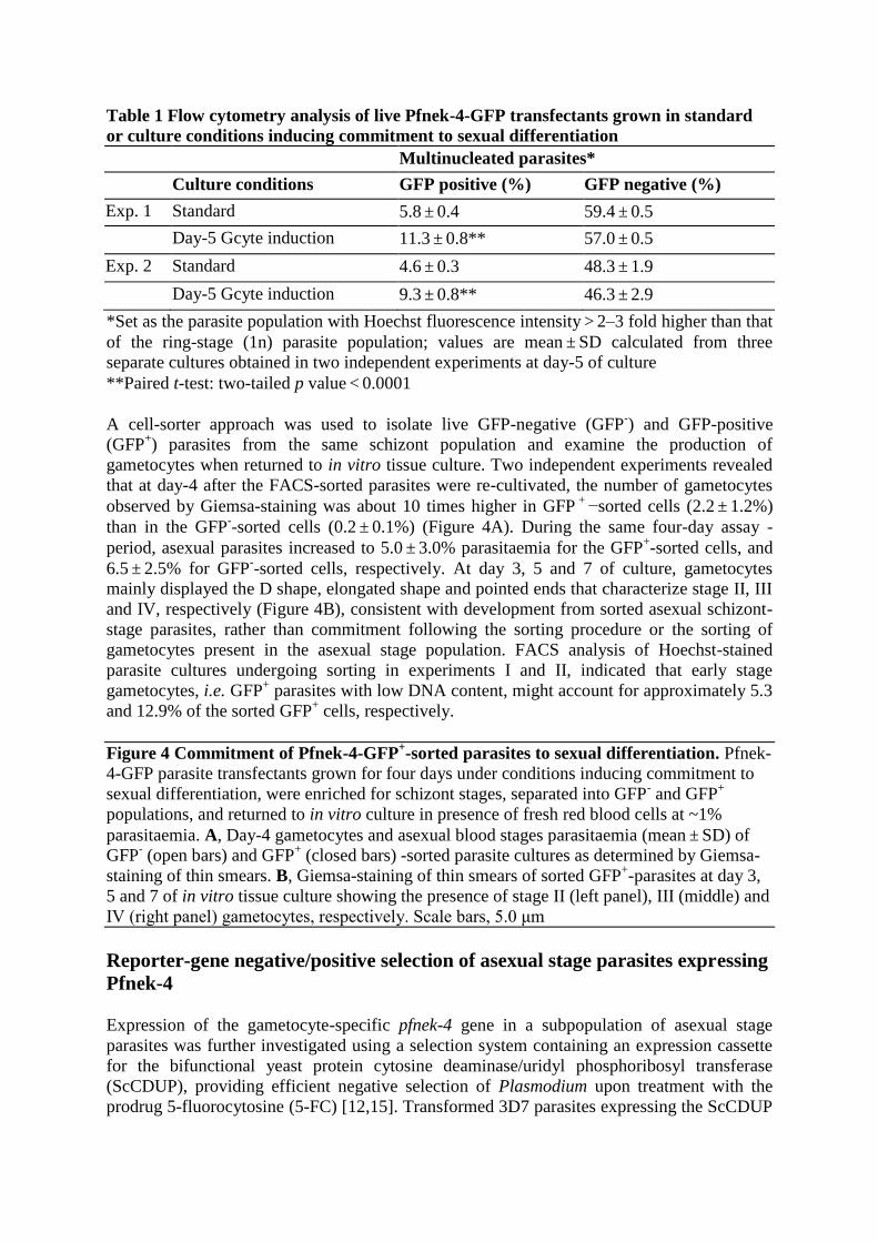
Table 1 Flow cytometry analysis of live Pfnek-4-GFP transfectants grown in standard
or culture conditions inducing commitment to sexual differentiation
Multinucleated parasites*
Culture conditions GFP positive (%) GFP negative (%)
Exp. 1 Standard 5.8 ± 0.4 59.4 ± 0.5
Day-5 Gcyte induction 11.3 ± 0.8** 57.0 ± 0.5
Exp. 2 Standard 4.6 ± 0.3 48.3 ± 1.9
Day-5 Gcyte induction 9.3 ± 0.8** 46.3 ± 2.9
*Set as the parasite population with Hoechst fluorescence intensity > 2–3 fold higher than that
of the ring-stage (1n) parasite population; values are mean ± SD calculated from three
separate cultures obtained in two independent experiments at day-5 of culture
**Paired t-test: two-tailed p value < 0.0001
A cell-sorter approach was used to isolate live GFP-negative (GFP-) and GFP-positive
(GFP+) parasites from the same schizont population and examine the production of
gametocytes when returned to in vitro tissue culture. Two independent experiments revealed
that at day-4 after the FACS-sorted parasites were re-cultivated, the number of gametocytes
observed by Giemsa-staining was about 10 times higher in GFP + −sorted cells (2.2 ± 1.2%)
than in the GFP--sorted cells (0.2 ± 0.1%) (Figure 4A). During the same four-day assay -
period, asexual parasites increased to 5.0 ± 3.0% parasitaemia for the GFP+-sorted cells, and
6.5 ± 2.5% for GFP--sorted cells, respectively. At day 3, 5 and 7 of culture, gametocytes
mainly displayed the D shape, elongated shape and pointed ends that characterize stage II, III
and IV, respectively (Figure 4B), consistent with development from sorted asexual schizont-
stage parasites, rather than commitment following the sorting procedure or the sorting of
gametocytes present in the asexual stage population. FACS analysis of Hoechst-stained
parasite cultures undergoing sorting in experiments I and II, indicated that early stage
gametocytes, i.e. GFP+ parasites with low DNA content, might account for approximately 5.3
and 12.9% of the sorted GFP+ cells, respectively.
Figure 4 Commitment of Pfnek-4-GFP+-sorted parasites to sexual differentiation. Pfnek-
4-GFP parasite transfectants grown for four days under conditions inducing commitment to
sexual differentiation, were enriched for schizont stages, separated into GFP- and GFP
+
populations, and returned to in vitro culture in presence of fresh red blood cells at ~1%
parasitaemia. A, Day-4 gametocytes and asexual blood stages parasitaemia (mean ± SD) of
GFP- (open bars) and GFP
+ (closed bars) -sorted parasite cultures as determined by Giemsa-
staining of thin smears. B, Giemsa-staining of thin smears of sorted GFP+-parasites at day 3,
5 and 7 of in vitro tissue culture showing the presence of stage II (left panel), III (middle) and
IV (right panel) gametocytes, respectively. Scale bars, 5.0 μm
Reporter-gene negative/positive selection of asexual stage parasites expressing
Pfnek-4
Expression of the gametocyte-specific pfnek-4 gene in a subpopulation of asexual stage
parasites was further investigated using a selection system containing an expression cassette
for the bifunctional yeast protein cytosine deaminase/uridyl phosphoribosyl transferase
(ScCDUP), providing efficient negative selection of Plasmodium upon treatment with the
prodrug 5-fluorocytosine (5-FC) [12,15]. Transformed 3D7 parasites expressing the ScCDUP

gene under control of the constitutive Hsp86 promoter (pScCDUPHsp86) were rapidly lost in
the presence of 5-FC, as expected (Figure 5A). In contrast, transformed 3D7 parasites
expressing ScCDUP driven by ~1-kb Pfnek-4 5’ flanking region (pScCDUPPfnek-4) showed a
marginal reduction in growth rate in presence of 5-FC, consistent with the Pfnek-4 being
active in only a small sub-population of asexual parasites. In parallel, to achieve a positive
selection of parasites expressing Pfnek-4, the expression cassette was modified such that the
human dihydrofolate reductase (hDHFR) was placed under control of ~1-kb 5’ flanking
region from Pfnek-4 (phDHFRPfnek-4). Figure 5B shows that positive selection of transformed
3D7 parasites expressing hDHFR driven by the Pfnek-4 promoter with the antifolate drug
WR99210 caused a marked reduction in parasitaemia growth rate, but was not sufficient to
completely abolish proliferation. In contrast parental 3D7 parasites were fully susceptible, as
expected.
Figure 5 Selection of parasite transfectants expressing negative or positive selectable
markers directed by Pfnek-4 promoter. A, In vitro growth rate of 3D7 parasite
transfectants expressing the Saccharomyces cerevisiae cytosine deaminase-
uridylphosphoribosyltransferase fusion gene (ScCDUP) driven by Hsp86 (pScCDUPHsp86) or
Pfnek-4 (pScCDUPPfnek-4) 5’ flanking regions, in presence () or absence () of prodrug 5-FC
(1 μM). B, In vitro growth rate of untransformed 3D7 (3D7 control) and 3D7 parasite
transfectants expressing the human dihydrofolate reductase (hDHFR) gene driven by Pfnek-4
5’ flanking region (phDHFRPfnek-4), in presence () or absence () of WR22910 (5 nM).
Parasitaemia (mean of duplicate cultures) were determined daily by Giemsa-stained blood
smears and parasite cultures >3% parasitaemia were diluted to 1%-parasitaemia
Generation of Plasmodium falciparum parasites with a disrupted Pfnek-4 gene
The function of the nek-4 gene has been previously investigated in the rodent malaria parasite
Plasmodium berghei; it was shown that disruption of Pbnek-4 does not affect asexual
replication, gametocytogenesis and gametogenesis, but impairs differentiation of zygotes into
ookinetes [8]. To investigate a possible phenotype caused by the absence of a functional Nek-
4 enzyme in the commitment of P. falciparum parasite to sexual differentiation, the pfnek-4
gene of the 3D7 parasite line was disrupted by single-crossover homologous recombination
(Figure 6A). Clonal lines derived from two independent transfection experiments were
established by limiting dilution, and their genotypes were analysed by PCR. The amplicon
corresponding to the wild-type locus was not detected in clones cl3 and cl4, but was observed
in untransfected 3D7 parasites. In contrast, amplicons diagnostic for the 5’ and 3’ boundaries
of plasmid integration were detectable in the transgenic parasites (Figure 6B). The generation
of pfnek-4- mutant parasite clones demonstrates that the gene is not essential for replication in
erythrocytes. Moreover, no effect on parasite growth rate of the pfnek-4- mutant parasites was
observed (data not shown). At day 12 to 14 of culture the number of gametocytes (~0.5-
0.7%) appears undistinguishable from those of 3D7 wild-type parasite cultures and the
majority of gametocytes were at stage IV and V as assessed by Giemsa-stained smears
(Figure 6C). Most stage V gametocytes displayed the morphological characteristics of mature
female gametocytes [16], indicating that pfnek-4 disruption does not affect the formation of
mature female gametocytes.
Figure 6 Disruption of the Pfnek-4 gene. A, Strategy for gene disruption. The schematic
representation shows the Pfnek-4 locus, the gene-targeting construct used for gene disruption
by homologous recombination, and the pseudo-diploid locus resulting from integration of the
KO construct. The primers used for the diagnostic PCRs are indicated. BSD, blasticidin-

resistance cassette. B, PCR analysis indicating disruption of the Pfnek-4 gene in parasite
clones cl3 and cl4 obtained from two independent transfections. Genomic DNA isolated from
3D7 wild-type parasite and Pfnek-4- clones cl3 and cl4, were subjected to PCR using the
indicated primers OL-761 and OL-763 (diagnostic for the wild-type locus), OL-761 and OL-
168 (diagnostic for 5’ integration), and OL-167 and OL-763 (diagnostic for 3’ integration).
PCR products corresponding to expected sizes for wild-type (1288-bp), 5’ integration (1179-
bp) and 3’ integration (1277-bp) are indicated on the right. C, Giemsa-staining of thin smears
of pfnek-4- gametocytes at days 12–14 of in vitro tissue culture stimulating gametocyte
production, showing the presence of stage IV (left panel) and stage V (right panel)
gametocytes. Scale bars, 5.0 μm
Discussion
In P. falciparum, the commitment to gametocytogenesis occurs during the preceding asexual
RBC cycle [2]. The present study identifies a subset of schizont-stage parasites expressing
Pfnek-4, a protein kinase previously thought to be gametocyte-specific. Upon sorting, Pfnek-
4-GFP positive schizonts produced elevated levels of gametocytes in the subsequent red
blood cell cycle. The experimental approach is based on episomal expression of Pfnek-4-GFP
fusion protein driven by 1 kb-genomic DNA upstream of the Pfnek-4 coding sequence. It
cannot be formally excluded that the proper expression pattern is not maintained in this
conditions. However, a study by Khan et al. [7] reported the ability of a 678 bp-Pbnek-4
promoter to drive the expression of a reporter GFP protein in female gametocytes, but not in
male gametocytes or asexual blood stages. In this latter study, nine out of ten promoters (the
only exception being most likely due to the short 5’ region available from the genome
database), were found to drive the expression of the GFP reporter protein in agreement with
the prediction from proteomic analyses, suggesting that sex-specific expression is mainly
controlled by the 5’ UTR/promoter. In another instance, the episomal expression of the HA-
epitope-tagged P. falciparum protease, PfROM1, placed under control of its own promoter
element, revealed an expression at mature stages and localization to the mononeme, a newly
described apical organelle of P. falciparum merozoites [17]. The nucleus-associated
punctuate pattern observed for Pfnek-4-GFP distribution, which is reminiscent of what is
observed with the P. falciparum Aurora-related kinase Pfark-1, is consistent with the location
of Nima- and Aurora-related kinases, many members of which associate with centrosomal
structures (see below). Thus, it is proposed that there is a high likelihood that the localization
observed with the episomally-encoded Pfnek-4-GFP fusion protein might reflect that of the
endogenous enzyme.
It should be stressed that: (i) the sorted Pfnek-4-GFP positive schizont population not only
generated gametocytes but also parasites undergoing asexual RBC cycles; and (ii) a
proportion of the parasites positively selected for expression of the hDHFR resistance marker
driven by the pfnek-4 gene was still able to undergo asexual cycles. Altogether these findings
support and expand previous studies indicating that malaria parasites have quantitative
sensitivity to gametocyte induction and that multiple stimuli can induce gametocytogenesis,
reflecting the highly flexible mechanism underlying sexual differentiation [4]. Noteworthy,
high rate conversion to sexual forms of asexual parasites grown at high densities was shown
to be reversible by dilution within a time window smaller than the time required for one
asexual cycle (48 hours) [2]. This feature might explain the relatively lower gametocyte
conversion rate observed in the GFP+ sorted parasites (2.2%), as compared to normal 3D7
parasites grown under gametocyte-inducing culture conditions (~4-5%). Altogether, the data
suggest that Pfnek-4 identifies a population of committed and reversibly pre-committed
parasites. Since sex determination appears to occur simultaneously to commitment to sexual
differentiation, the asexual subpopulation expressing Nek-4, a protein shown to be restricted
to female gametocytes in the rodent malaria parasite P. berghei [7,8], is likely to represent the
progenitor of female rather than male gametocytes, although this question remains to be
further investigated.
The expression of a gametocyte-specific gene product in sexually-committed asexual
parasites is consistent with a developmental change in gene expression during sexual
differentiation [18,19], and extends previous studies reporting the expression of gametocyte-
specific genes, such as Pfs16, in sub-populations of asexual-stage parasites [9,20,21]. Since
some of the asexual parasite population expressing Pfnek-4 did not appear to be fully
committed to sexual differentiation (being still able to undergo RBC cycles, see above), a
switch to gametocyte-specific gene expression may occur before the “no-return”decision to
commit to gametocytogenesis is made. Analysing genes expressed in the sexually-committed
population would be of great interest to explore gene regulation in the context of commitment
to gametocytogenesis, and would help to identify signatures of early sexual development of
malaria parasites. Identification of Pfnek-4 as a molecular marker of sexually-committed
schizonts provides a useful tool, making this parasite population amenable to purification
followed by transcriptome and proteome analyses. Transcriptional regulator candidates
controlling expression of subsets of genes are the ApiAP2 family of proteins. De Silva et al.
[22], reported highly coherent expression patterns of predicted downstream targets of P.
falciparum AP2 transcription factors, suggesting essential roles in parasite development.
Translational regulation also plays a critical role during commitment to gametocytogenesis
[18,19]. Targeted disruption of PfPuf2, a member of the Puf family of translational repressors
was shown to promote the formation of gametocytes and the differentiation of male
gametocytes [23].
In P. berghei, the Nek-4 kinase does not appear to be required for gametocytogenesis but is
essential for pre-meiotic DNA replication in the zygote, consistent with cell-cycle related
functions [7,8]. That pfnek-4- P. falciparum parasites are able to undergo gametocytogenesis
and produce mature stage V gametocytes, indicates that Pfnek-4 is not required for the early
stages of the sexual cycle in both P. berghei and P. falciparum. This conclusion is also
supported by the finding that pfnek-4- clones produce female gametocytes. It is intriguing that
the timing of recruitment of the Pfnek-4 protein to schizont nuclear bodies appears to
coincide with the occurrence of nuclear divisions. Noteworthy, all nuclei within a single
schizont appear to be associated with punctuate Pfnek-4-GFP fluorescence from early
developing to multinucleated schizont, in contrast to Pfark-1, a mitotic kinase that marks only
a subset of nuclei in a given schizont as a result of transient recruitment at the spindle pole
bodies, a consequence of asynchronous nuclear division in a single schizont [13]. In contrast
to Pfark-1, the Pfnek-4-GFP protein appears to associate to all nuclei, irrespective of their
nuclear division status. Furthermore, cell cycle-arrested stage II gametocytes were found to
express the Pfnek-4-GFP protein with a punctuate fluorescence similar to parasites
undergoing schizogony. Preliminary results showing a close association of doublets of Pfnek-
4-GFP fluorescence with short mitotic spindle microtubules in schizont-stage parasites (data
not shown), are consistent with a recruitment of Pfnek-4 at nuclear spindle pole bodies, the
centrosome equivalent of Plasmodium parasites, an observation consistent with the known
centrosomal functions of Nima-related kinases. However, the sub-cellular structure to which
Pfnek-4 associates remains to be better defined. Whether the Pfnek-4 enzyme has cell-cycle-
related functions in RBC-stages still remains to be elucidated.

Conclusions
Taken together, the data presented in this study indicate that the Nima-related kinase Pfnek-4
identifies a small subset of schizont-stage P. falciparum parasites displaying high rate
conversion to sexual differentiation, and thus represents a molecular marker of sexually-
committed and -pre-committed schizonts. Pfnek-4 does not appear to control the switching of
asexual stages into gametocytes.
Competing interests
The authors declare that they have no competing interests.
Authors’ contributions
LR carried out molecular cloning, RT-PCR, western blotting, parasite genetic manipulations,
analyses by fluorescence microscopy and flow cytometry, and participated in conception of
the study and writing of the manuscript. MG performed the cell sorting. AT performed and
analysed parasite growth. SM performed and analysed parasite growth and participated in
writing of the manuscript. CD participated in conception of the study and writing of the
manuscript. All authors read and approved the final manuscript.
Acknowledgements
We thank Richard Carter (University of Edinburgh, Edinburgh,UK) for providing the F12
clone, Geoffrey I. McFadden (University of Melbourne, Parkville, Australia) for providing
pHGB and pCHD-1/2 plasmids, Alan F. Cowman (The Walter and Eliza Hall Institute of
Medical Research, Parkville, Australia) for providing pCC1 and pCC4 plasmids. We also
thank Gonzalo Tapia, Flow Cytometry Core Facility (EPFL, Lausanne, Switzerland), and
members of the Bioimaging and Optics Platform (EPFL, Lausanne, Switzerland) for passing
on their expertise in image analysis software IMARIS.
References
1. Sinden RE: Sexual development of malarial parasites. Adv Parasitol 1983, 22:153–216.
2. Bruce MC, Alano P, Duthie S, Carter R: Commitment of the malaria parasite
Plasmodium falciparum to sexual and asexual development. Parasitology 1990, 100(Pt
2):191–200.
3. Silvestrini F, Alano P, Williams JL: Commitment to the production of male and female
gametocytes in the human malaria parasite Plasmodium falciparum. Parasitology 2000,
121(Pt 5):465–471.
4. Dyer M, Day KP: Commitment to gametocytogenesis in Plasmodium falciparum.
Parasitol Today 2000, 16:102–107.
5. O’Regan L, Blot J, Fry AM: Mitotic regulation by NIMA-related kinases. Cell Div
2007, 2:25.
6. Quarmby LM, Mahjoub MR: Caught Nek-ing: cilia and centrioles. J Cell Sci 2005,
118:5161–5169.
7. Khan SM, Franke-Fayard B, Mair GR, Lasonder E, Janse CJ, Mann M, Waters AP:
Proteome analysis of separated male and female gametocytes reveals novel sex-specific
Plasmodium biology. Cell 2005, 121:675–687.
8. Reininger L, Billker O, Tewari R, Mukhopadhyay A, Fennell C, Dorin-Semblat D, Doerig
C, Goldring D, Harmse L, Ranford-Cartwright L, Packer J: A NIMA-related protein kinase
is essential for completion of the sexual cycle of malaria parasites. J Biol Chem 2005,
280:31957–31964.
9. Reininger L, Tewari R, Fennell C, Holland Z, Goldring D, Ranford-Cartwright L, Billker
O, Doerig C: An essential role for the Plasmodium Nek-2 Nima-related protein kinase in
the sexual development of malaria parasites. J Biol Chem 2009, 284:20858–20868.
10. Aurrecoechea C, Brestelli J, Brunk BP, Dommer J, Fischer S, Gajria B, Gao X, Gingle A,
Grant G, Harb OS, Heiges M, Innamorato F, Iodice J, Kissinger JC, Kraemer E, Li W, Miller
JA, Nayak V, Pennington C, Pinney DF, Roos DS, Ross C, Stoeckert CJ Jr, Treatman C,
Wang H: PlasmoDB: a functional genomic database for malaria parasites. Nucleic Acids
Res 2009, 37:D539–D543.
11. Tonkin CJ, van Dooren GG, Spurck TP, Struck NS, Good RT, Handman E, Cowman AF,
McFadden GI: Localization of organellar proteins in Plasmodium falciparum using a
novel set of transfection vectors and a new immunofluorescence fixation method. Mol
Biochem Parasitol 2004, 137:13–21.
12. Maier AG, Rug M, O’Neill MT, Brown M, Chakravorty S, Szestak T, Chesson J, Wu Y,
Hughes K, Coppel RL, Newbold C, Beeson JG, Craig A, Crabb BS, Cowman AF: Exported
proteins required for virulence and rigidity of Plasmodium falciparum-infected human
erythrocytes. Cell 2008, 134:48–61.
13. Reininger L, Wilkes JM, Bourgade H, Miranda-Saavedra D, Doerig C: An essential
Aurora-related kinase transiently associates with spindle pole bodies during Plasmodium falciparum erythrocytic schizogony. Mol Microbiol 2011, 79:205–221.
14. Carter R, Ranford-Cartwright L, Alano P: The culture and preparation of gametocytes
of Plasmodium falciparum for immunochemical, molecular, and mosquito infectivity
studies. Methods Mol Biol 1993, 21:67–88.
15. Braks JA, Franke-Fayard B, Kroeze H, Janse CJ, Waters AP: Development and
application of a positive–negative selectable marker system for use in reverse genetics in
Plasmodium. Nucleic Acids Res 2006, 34:e39.
16. Talman AM, Domarle O, McKenzie FE, Ariey F, Robert V: Gametocytogenesis: the
puberty of Plasmodium falciparum. Malar J 2004, 3:24.
17. Singh S, Plassmeyer M, Gaur D, Miller LH: Mononeme: a new secretory organelle in
Plasmodium falciparum merozoites identified by localization of rhomboid-1 protease. Proc Natl Acad Sci U S A 2007, 104:20043–20048.
18. Hall N, Karras M, Raine JD, Carlton JM, Kooij TW, Berriman M, Florens L, Janssen CS,
Pain A, Christophides GK, James K, Rutherford K, Harris B, Harris D, Churcher C, Quail
MA, Ormond D, Doggett J, Trueman HE, Mendoza J, Bidwell SL, Rajandream MA, Carucci
DJ, Yates JR 3rd, Kafatos FC, Janse CJ, Barrell B, Turner CM, Waters AP, Sinden RE: A
comprehensive survey of the Plasmodium life cycle by genomic, transcriptomic, and
proteomic analyses. Science 2005, 307:82–86.
19. Le Roch KG, Johnson JR, Florens L, Zhou Y, Santrosyan A, Grainger M, Yan SF,
Williamson KC, Holder AA, Carucci DJ, Yates JR 3rd, Winzeler EA: Global analysis of
transcript and protein levels across the Plasmodium falciparum life cycle. Genome Res
2004, 14:2308–2318.
20. Eksi S, Suri A, Williamson KC: Sex- and stage-specific reporter gene expression in
Plasmodium falciparum. Mol Biochem Parasitol 2008, 160:148–151.
21. Alano P: Plasmodium falciparum gametocytes: still many secrets of a hidden life. Mol
Microbiol 2007, 66:291–302.
22. De Silva EK, Gehrke AR, Olszewski K, Leon I, Chahal JS, Bulyk ML, Llinas M:
Specific DNA-binding by apicomplexan AP2 transcription factors. Proc Natl Acad Sci U
S A 2008, 105:8393–8398.
23. Miao J, Li J, Fan Q, Li X, Cui L: The Puf-family RNA-binding protein PfPuf2
regulates sexual development and sex differentiation in the malaria parasite Plasmodium falciparum. J Cell Sci 2010, 123:1039–1049.
Additional files
Additional_file_1 as TIFF
Additional file 1. Expression of the Pfnek-4-GFP protein in gametocytes, early and
multinucleated schizont-stage 3D7 transfectants. Live cell images of stage III, IV and V
gametocytes (A), early developing (2–3 nuclei stage) (higher panel) and multinucleated
(lower panel) schizonts (B) from 3D7 transfectants stained with Hoechst 33258. The Pfnek-4-
GFP protein strongly accumulates in the cytosol of stage III to V gametocytes. Images of
early developing schizont and multinucleated schizonts show that each of Hoechst-stained
nuclear bodies is associated with a dot of Pfnek-4-GFP fluorescence (green). Overlay of all
channels (A, B), and corresponding DIC images are shown as well (B). All images were
acquired using a Deltavision RT wide-field epifluorescence microscope imaging system and
a 100x/1.4 objective and processed using SoftWorx software. Image analysis software was
IMARIS version 5.0. Scale bars, 2.0 μm.

Figure 1

Figure 2

Figure 3
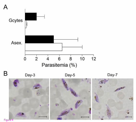
Figure 4

B
A
Figure 5
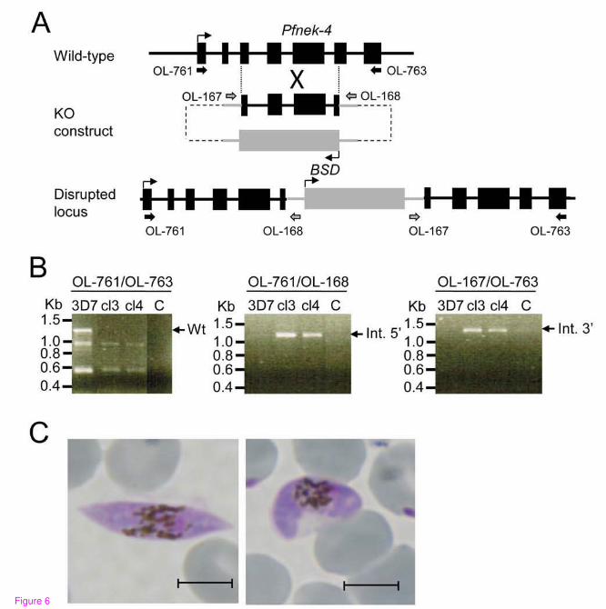
Figure 6

Additional files provided with this submission:
Additional file 1: 1768818127227089sup1.png, 169Khttp://www.malariajournal.com/imedia/1539821016771218/supp1.png