relationship between selenium, selenomethionine and selenocysteine ... · acta chromatographica,...
TRANSCRIPT

ACTA CHROMATOGRAPHICA, NO. 18, 2007
RELATIONSHIP BETWEEN THE SELENIUM, SELENOMETHIONINE, AND SELENOCYSTEINE
CONTENT OF SUBMERGED CULTIVATED MYCELIUM OF Lentinula edodes (Berk.)
J. Turło1,*, B. Gutkowska1, and E. Malinowska2 1Department of Drug Technology, Medical University of Warsaw, 1, Banacha Street, 02-097 Warsaw, Poland
2Faculty of Pharmacy, Medical University of Warsaw, Poland SUMMARY To obtain extracts rich in the organic forms of selenium with puta-tive cancer-preventive properties more effective than those of selenized-yeast, Lentinula edodes mycelia were cultivated in media enriched with selenium at concentrations from 0 to 20 µg mL−1, by addition of sodium selenite either before inoculation or after three days of mycelial growth. The total selenium content of submerged cultivated mycelial biomass was determined by atomic absorption spectroscopy (AAS) and concentrations of selenomethionine and selenocysteine were determined by RP-HPLC. The concentration of selenium in the mycelia increased from 23 µg g−1 dry weight (d.w.) for mycelia cultivated in media not enriched in selenium to 1800 µg g−1 (d.w.) for mycelia cultivated in medium enriched with 20 µg mL−1 selenium. The amount of selenomethionine in the cultivated bio-mass increased in proportion to the selenium content of the mycelium (con-centration range 23–289 µg g−1 d.w.) but the percentage of selenium accu-mulated as the selenoamino acid decreased. For selenium concentrations greater than 1 µg mL−1 the selenocysteine content of the mycelial biomass does not depend directly on the concentration of selenium in the medium. INTRODUCTION Selenium is a trace element of fundamental importance to human health, part of the antioxidant enzymes that protect cells against the effects –––––––––––––––––––––––––––––––––––––––––––––––––––––––––––––––––––––––––––––––––––––––––
Presented at the 5th International Symposium on Chromatography of Natural Products ISCNP 2006, Lublin (Poland), June 19th-22nd, 2006
- 36 -

of free radicals [1,2]. One important health effect of selenium not exclusi-vely linked to its enzymatic functions is connected with functioning of the immune system. Selenium seems to be a key nutrient in cancer prevention and in inhibiting progression of HIV to AIDS [3–5]. It must be kept in mind that the biological activity of selenium is determined by its chemical form and the dose ingested. Selenium supplements in the form of inorga-nic compounds may not be as bioavailable as organically bound Se [6,7]. Clark et al. reported a putative role of selenized yeast in cancer prevention [8]. Supplementation of people’s diet with selenized yeast (Se-yeast) is capable of reducing overall cancer morbidity by almost 50%. Analytical speciation studies have shown that the bulk of selenium (approx. 85%) in Se-yeast was in form of selenomethionine [9]. For higher mushrooms selenium accumulation is comparable with that of yeast [10]. Similarly to yeast, selenomethionine was the main sele-nium compound detected in aqueous extracts of the fruit bodies of Agari-cus bisporus and Lentinula edodes grown on medium (compost) supple-mented with selenium [11,12]. Cell cultures of higher mushrooms, culti-vated in selenium-enriched media, are potentially as good a source of the highly bioactive and safe organic species of selenium as selenized yeast or the fruit bodies of higher mushrooms. Our previous work has shown that the submerged cultivated mycelium of Lentinula edodes very effectively accumulates selenium from cultivation medium. The total concentration of selenium in submerged cultivated mycelial biomass, as determined by atomic absorption spectroscopy (AAS), expressed in mg per 100 g myce-lial dry weight (d.w.), rose from 0.1 mg (mycelium cultivated in unmodi-fied medium) to 5000 mg (mycelium cultivated in medium enriched with 100 µg mL−1 selenium) [13]. Speciation investigations of selenium at these high concentrations accumulated in selenated mycelial biomass, and deter-mination of the relationship between the total selenium content of the cul-tivated mycelium and the concentration of individual Se compounds seem to be interesting problems. Several different techniques have been used for speciation of sele-nium compounds [14,15]. Because our objective was to determine the re-lationship between the concentrations of three amino acids (selenomethio-nine, methionine, and selenocysteine) in mycelia containing different total amounts of selenium, we adapted an RP-HPLC method with o-phthaldi-aldehyde derivatization (OPA method) widely used for analysis of amino acids. The main advantages of this method are good selectivity and effici-ency and low detection limit [16–18].
- 37 -

Lentinula edodes (Shii-take mushroom), selected for our experi-ments, is a medicinal mushroom from which the extracted, highly puri-fied, polysaccharide fraction (lentinan) is an approved drug used in cancer treatment and in AIDS research [19–23]. Hot water extracts of L. edodes mycelium (LEM) and culture media (LAP) have strong antitumor activity [24,25]. The objective of this study was, therefore, to optimize submerged culture conditions for good mycelium growth of L. edodes and high sele-nium content of the mycelial biomass, possibly in the form of the highly bioavailable selenoamino acids. We hoped that high concentrations of the organic forms of selenium in submerged cultivated mycelial biomass would enhance the anticancer activity and inhibition of HIV progression activity of mushroom extracts. EXPERIMENTAL Equipment
A GBC Avanta Ultra Z automatic, single-beam atomic-absorption spectrometer with autosampler, longitudinal Zeeman-effect background-correction system, and transversely heated graphite atomizer was used for determination of the total selenium content of cultivated mycelia. A coded 1.5-inch selenium hollow-cathode lamp (CPI Int.), operated at 10 mA, was used for determination of selenium at 196 nm; the slit width was 2.0 nm. Microwave digestion was performed by use of a Plazmotronika BM-1s closed-vessel microwave system. HPLC analysis of selenoamino acids was performed with a Shi-madzu (USA) instrument including gradient system (SCL-10AVP control-ler, two LC-10AT vp pumps, CTO-10AC vp oven) and fluorescence de-tector (LaChrom L-7480; Merck–Hitachi, Darmstadt, Germany). Compounds were separated on a 250 mm × 4 mm Phenomenex Luna 2 C18 column with appropriate guard column. Mushroom cultures were cultivated in a rotary shaker (New Bruns-wick Scientific, Edison, NY, USA). Materials
L-Selenomethionine and an L-amino acid standard kit for use as external standards were obtained from Sigma (St Louis, MO, USA). A stan-dard solution of carboxymethylselenocysteine was prepared from seleno-
- 38 -
L-cystine (Sigma) by using the same procedure for reductive carboxyme-thylation as for carboxymethylation of mycelial proteins. HNO3 solution (65%) and Suprapur quality water (Merck) were used for preparation of samples. Selenium atomic absorption standard so-lution (1020 µg mL−1 in 1% HNO3; Aldrich) was used for preparation of selenium standards. Nickel(II) nitrate hexahydrate p.a. (Fluka) was used as a chemical modifier for selenium. Sodium phosphate monobasic monohydrate, sodium hydroxide, boric acid, hydrochloric acid, sodium selenite, mercaptoethanol, (GR for analysis), acetonitrile (LC grade), and methanol (LC grade) were obtained from Merck. Microorganism and Cultivation Media
The strain of Lentinula edodes used in this study was taken from the medicinal mushroom strain collection of the Department of Drug Tech-nology of the Medical University of Warsaw (Poland). It originated in Korea. In nature it grew in the Pobwang Peak area, near Mt Myohyang. The seed culture was grown in 500-mL flasks containing 150 mL basal medium (glucose 5% (w/v), yeast extract 1%, casein hydrolysate 1%, KH2PO4 0.1%), at 26°C, on a rotary shaker at 110 rev min−1 for seven days. The fermentation medium selected for experiments on accumula-tion of selenium and for biosynthesis of selenoamino acids by Lentinula edodes submerged cultured mycelial biomass was partially composed of waste products from the food industry (beet molasses 10%, corn steep liquor 0.15%, grain wort 5%) and also contained 0.3% (w/v) KH2PO4. Procedures Fermentation
Culture medium was enriched in selenium by addition of sodium selenite (Na2SeO3). Concentrations of selenium were in the range 0 to 20 µg mL−1 (0, 0.5, 1, 2, 3, 4, 5, 6, 7, 8, 9, 10, and 20 µg mL−1). The sodium selenite was added to the medium either before inoculation or after 3 days of mycelial growth. The initial pH of the medium was 6.5. Mycelia were grown in shake-flask culture in 500-mL flasks con-taining 200 mL medium. The fermentation medium was inoculated with 5% (v/v) seed culture. Cultures were incubated at 26°C in a rotary shaker at 120 rev min−1 for 14 days. Mycelia were harvested by filtration, washed three times with distilled water, and dried at 60°C. All experiments were conducted at least three times to determine reproducibility. Total selenium
- 39 -
content was determined for each sample of mycelium by use of the atomic-absorption spectrometry (AAS). Methionine and selenomethionine content were determined by high-performance liquid chromatography (HPLC). Determination of Selenium in Mycelium and Culture Media by Atomic-Ab-sorption Spectrometry Digestion Procedure
A sample (0.02 g) of mycelium dried at 100°C was placed in a Tef-lon crucible and digested with 3 mL 65% HNO3 in microwave mineralizer according to the program. When cool the solution was diluted to 25 mL with 1% HNO3 in deionized water. If necessary, the solutions were diluted several times in a few steps with 5 mg mL−1 Ni(NO3)2 in 1% HNO3. A blank digest was performed in the same way. Selenium standard solutions (40, 100, and 200 ng mL−1 Se) were prepared under the same conditions. Analytical Procedure
Selenium standard or sample solution (10 µL) was wet-injected into the graphite furnace. The time and temperature program is shown in Tab-le I. Slow solution uptake and slow solution injection conditions were se-lected. Three standard additions (four replicates of each) and peak-height measurements were used for quantification [25]. Table I
Furnace program for determination of selenium in Lentinula edodes mycelium using Ni(II) as chemical modifier
Stage Temperature (°C) Ramp (° s−1) Hold time (s) Air flow (L min−1)
Drying 110 130
10.0 1.0
10.0 10.0
3.0 3.0
Ashing 800 800
10.0 1.0
10.0 1.0
3.0 0.0
Atomizing 2100 0.5 0.5 0.0 Cleaning 2300 0.1 0.9 3.0
Determination of Methionine and Selenomethionine in Mycelial Biomass Sample Preparation
Because methionine partially decomposes during acidic hydrolysis of mycelial proteins in the presence of carbohydrates and heavy metal ca-
- 40 -

tions, alkaline hydrolysis in 5 M NaOH was preferred. NaOH solution (5 M, 10 mL) was added to the pulverized mycelium (0.1 g) in an ampoule and the mixture was purged with nitrogen for 30 min. The ampoule was sealed, full of nitrogen, then hydrolysis was performed at 110°C for 20 h. The my-celial hydrolysate was transferred to a 100-mL graduated flask and diluted to volume with LC-grade water. Derivatization was performed as described in the Merck application note [14]. RP-HPLC of Mycelial Hydrolysates
Selenomethionine and methionine in mycelial hydrolysates were determined by high-performance liquid chromatography of the o-phthal-dialdehyde derivatives (OPA method) [16,17,26] by using retention time standards and standard addition to the samples. The mobile phase was a gradient prepared from 100 mM sodium acetate buffer, pH 7.2, containing 0.1% acetonitrile (component A) and methanol (component B). The gradient breakpoints (min/%B) were: (0/45), (6/45), (12/50), (20/63), (38/63), (49/100), and (55/100). The temperature was 32°C, the injection volume 20 µL, and the flow rate 1.2 mL min−1. The excitation and emission wavelengths were 340 and 435 nm. The re-tention times of methionine and selenomethionine were 34.7 and 40.2 min (Fig. 1).
5 10 15 20 25 30 35 40 45 50 Minutes
Fig. 1
Chromatograms obtained from selenomethionine standard solution (0.002 µmol mL−1) and from a mixture of a mycelial hydrolysate and selenomethionine standard solution
- 41 -

Determination of Selenocysteine (SeCys) in Mycelium Sample Preparation
Selenocysteine was determined in mycelium hydrolysate after de-rivatization with o-phthaldialdehyde [17,18]. Selenocysteine, similarly to cysteine, is very easily oxidized and decomposes during hydrolysis of my-celial proteins. The method normally used for cysteine – formation of cy-steic acid by treatment with performic acid – results in complete decom-position of selenocysteine [17]. After carboxymethylation, however, sele-nocysteine is stable for several weeks. For carboxymethylation of mycelial proteins before acidic hydrolysis an adaptation of the method described by Jones [18] was used. Mycelial biomass homogenate (0.05 g), KBH4 solu-tion (2 mg mL−1, 0.5 mL), mercaptoethanol (50 µL), and iodoacetic acid solution (200 mM, 2 mL) were stirred for 24 h under nitrogen in the dark. The mixture was then evaporated, HCl solution (6 M, 10 mL) was added to the dry residue, and the mixture was purged with nitrogen in an ampoule for 30 min. The ampoule was filled with nitrogen and sealed, and hydroly-sis was performed at 110°C for 20 h. RP-HPLC of Mycelial Hydrolysates
Carboxymethylselenocysteine in mycelial hydrolysates was deter-mined in the same manner as selenomethionine, by reversed-phase HPLC of the o-phthaldialdehyde derivative (OPA method) [16–18,26], using re-tention time standards and standard additions to the sample. A standard solution of carboxymethylselenocysteine was prepared from seleno-L-cystine (Sigma) using the same procedure as for reductive carboxymethylation [17,18]. The mobile phase was a gradient prepared from 100 mM sodium acetate buffer, pH 7, containing 0.1% acetonitrile (component A) and me-thanol (component B). The gradient breakpoints were (min/%B): (0/22), (15/22), (20/100), (22/100). The flow rate was 1.2 mL min−1, the tempe-rature 32°C, and the injection volume 20 µL. Under these conditions the retention time of carboxymethylselenocysteine was 8.7 min (Fig. 2.). RESULTS The efficacy of cultivation of mushroom mycelium in the medium selected was good. The biomass accumulation recorded after cultivation for 14 days (in medium not enriched with selenium) was 7–8 g d.w. L−1 cultu-
- 42 -

1 2 3 4 5 6 7 8 9 10 11 12 13 14 15 16 17 Minutes
Fig. 2
Chromatograms obtained from carboxymethylselenocysteine standard solution (0.003 µmol mL−1), a mixture of carboxymethylselenocysteine and protein amino acid standard solution, and a Lentinula edodes mycelium sample hydrolysed after reductive carboxy-methylation
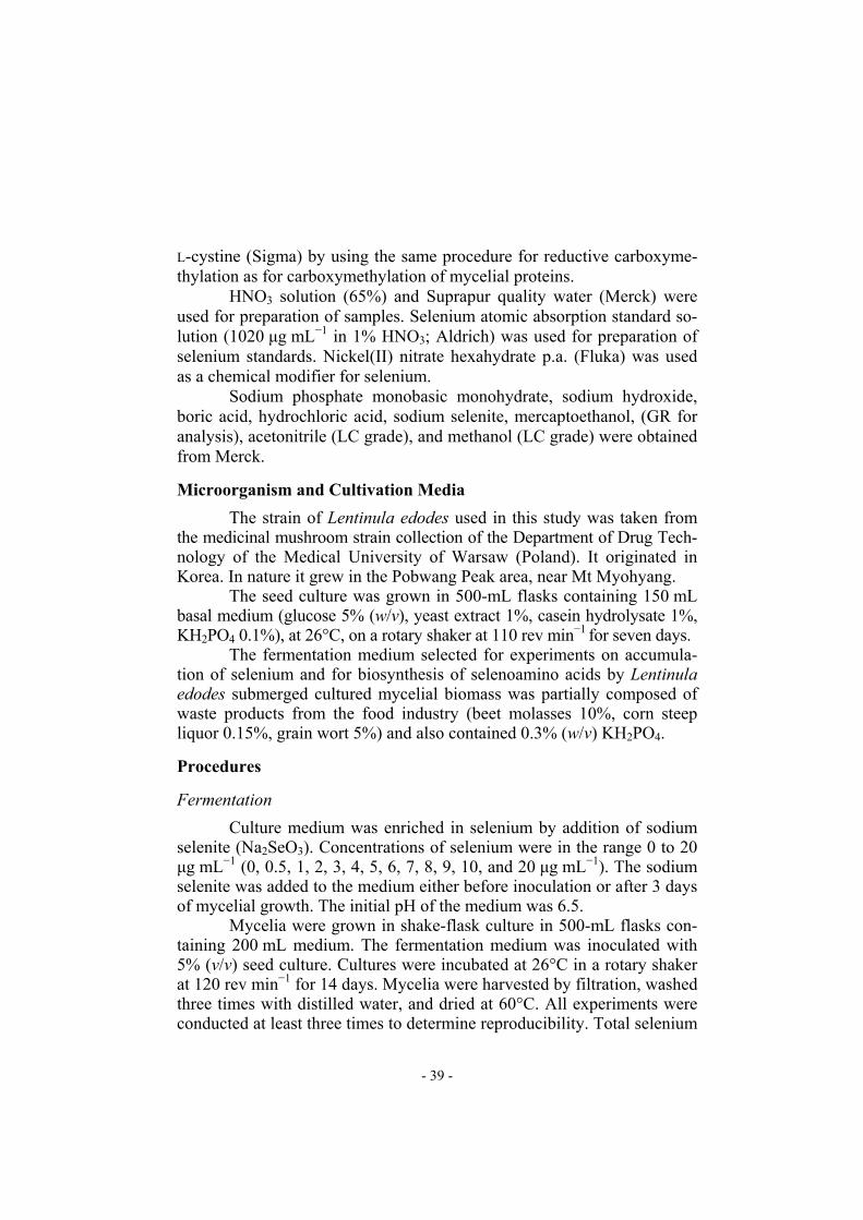
re medium. Mycelial growth was only weakly affected by increasing the concentration of selenium in the medium. Reduction of mycelial growth by selenium-containing compounds was observed only for cultivation in media enriched with selenium before inoculation (Tables II and III). Uptake of selenium by the submerged cultivated mycelium of Len-tinula edodes was very effective. According to our previous observations the effect on the selenium uptake of the time of addition of the selenium-containing compound to the cultivation medium (i.e. before inoculation or after mycelial growth for 3 days) is significant only for concentrations of selenium in the medium greater than 30 µg mL−1 [13]. Thus when the con-centration of selenium in the medium was relatively low (0.5 to 20 µg mL−1), significant differences between concentrations of selenium in mycelia har-vested from media enriched in selenium at different times was not observed (Tables II and III). Addition of 20 µg mL−1 selenium to the medium yielded 1815 µg selenium g−1 (d.w.) of mycelial growth (Fig. 3, Tables II and III). The selenomethionine content of the cultivated mycelia increased as the amount of selenium added was increased (Fig. 3). For low concen-trations of selenium in the mycelium (20 µg g−1) almost 70% of the sele-nium content was accumulated as selenomethionine. For concentrations of selenium in the mycelium greater than 400 µg g−1, however, the propor-
- 43 -

Table II
Relationship between the concentration of selenium in the cultivation medium and my-celial growth, and selenomethionine (SeMet) and methionine (Met) content of mycelia cultivated in media enriched in selenium before inoculation
Concn of Se in medium (µg mL−1)
Mycelial growth (g L−1) (RSD)
Concn of Se in mycelia
(µg g−1 d.w.) (RSD)
Concn of SeMet in mycelia
(µg g−1 d.w.) (RSD)
Concn of Met in mycelia
(µg g−1 d.w.) (RSD)
Amount of methionine
transformed into selenomethionine*
(%) 0 7.54 (0.31) 23.2 (0.13) 2.96 (0.35) 5933.0 (0.15) 0.35 0.5 5.16 (0.08) 60.6 (0.08) 54.9 (0.83) 5411.0 (0.11) 0.77 1 5.42 (0.19) 90.0 (0.28) 74.4 (0.14) 5256.2 (0.09) 1.08 3 5.42 (0.01) 416.7 (0.26) 88.9 (0.07) 5498.3 (0.02) 1.23 5 5.46 (0.27) 571.1 (0.14) 52.2 (0.35) 5192.9 (0.21) 0.76 7 8.63 (0.15) 637.7 (0.34) 101.4 (0.09) 5132.2 (0.04) 1.50 8 5.89 (0.16) 933.2 (0.25) 124.0 (0.29) 5310.2 (0.08) 1.78 9 6.22 (0.33) 1009.1 (0.08) 151.6 (0.16) 4278.5 (0.15) 2.69
10 5.21 (0.12) 1180.8 (0.26) 205.2 (0.29) 4533.7 (0.03) 3.44 20 5.74 (0.19) 1669.9 (0.37) 289.0 (0.21) 6135.0 (0.07) 3.58
Results are average values from three experiments
* Calculated on the basis of the number of moles of SeMet and Met in a sample
Table III
Relationship between the concentration of selenium in the cultivation medium and my-celial growth, and selenomethionine (SeMet) and methionine (Met) content of mycelia cultivated in media enriched in selenium after three days of cultivation
Concn of Se in medium (µg mL−1)
Mycelial growth (g L−1) (RSD)
Concn of Se in mycelia
(µg g−1 d.w.) (RSD)
Concn of SeMet in mycelia
(µg g−1 d.w.) (RSD)
Concn of Met in mycelia
(µg g−1 d.w.) (RSD)
Amount of methionine
transformed into selenomethionine*
(%) 0 7.87 (0.22) 27.0 (0.13) 2.30 (0.30) 6005.3 (0.05) 0.29 0.5 6.12 (0.40) 61.6 (0.09) 81.2 (0.15) 4490.7 (0.19) 1.37 1 8.01 (0.09) 90.4 (0.08) 72.5 (0.47) 6974.3 (0.09) 0.79 3 7.81 (0.17) 325.5 (0.21) 131.0 (0.16) 8096.4 (0.22) 1.23 5 10.86 (0.05) 415.1 (0.08) 164.9 (0.10) 6410.0 (0.03) 1.96 7 8.92 (0.27) 651.9 (0.22) 154.9 (0.38) 7855.9 (0.14) 1.50 8 9.98 (0.10) 644.2 (0.13) 203.6 (0.07) 8447.4 (0.28) 1.83 9 11.52 (0.17) 1173.5 (0.46) 203.5 (0.09) 6409.1 (0.15) 2.41
10 7.34 (0.53) 1289.0 (0.38) 198.8 (0.13) 6791.0 (0.03) 2.23 20 6.72 (0.30) 1815.0 (0.30) 265.4 (0.14) 5876.2 (0.18) 3.43
Results are average values from three experiments
* Calculated on the basis of the number of moles of SeMet and Met in a sample
- 44 -
0,00
50,00
100,00
150,00
200,00
250,00
0 2 4 6 8 10 12 14 16 18 20
Selenium added to the cultivation medium (µg/ ml)
Con
cent
ratio
n of
Se
or S
eMet
in m
ycel
ium
( mg/
100g
of d
ry w
eigh
t)
SeMet series "4"SeMet series "0"Se series "4"Se series "0"
Fig. 3
Relationship between selenium and selenomethionine concentrations in mycelia from me-dia enriched in selenium both before inoculation (series “0”) and after cultivation for three days (series “4”)
0%
20%
40%
60%
80%
100%
% o
f par
ticip
atio
n di
ffere
nt s
elen
ium
spe
cies
in m
ycel
ia
0 0,5 1 3 5 7 8 9 10 20Concentration of selenium in medium enriched before inoculation (µg/ml)
Remaining SeSe accumulated in SeMet
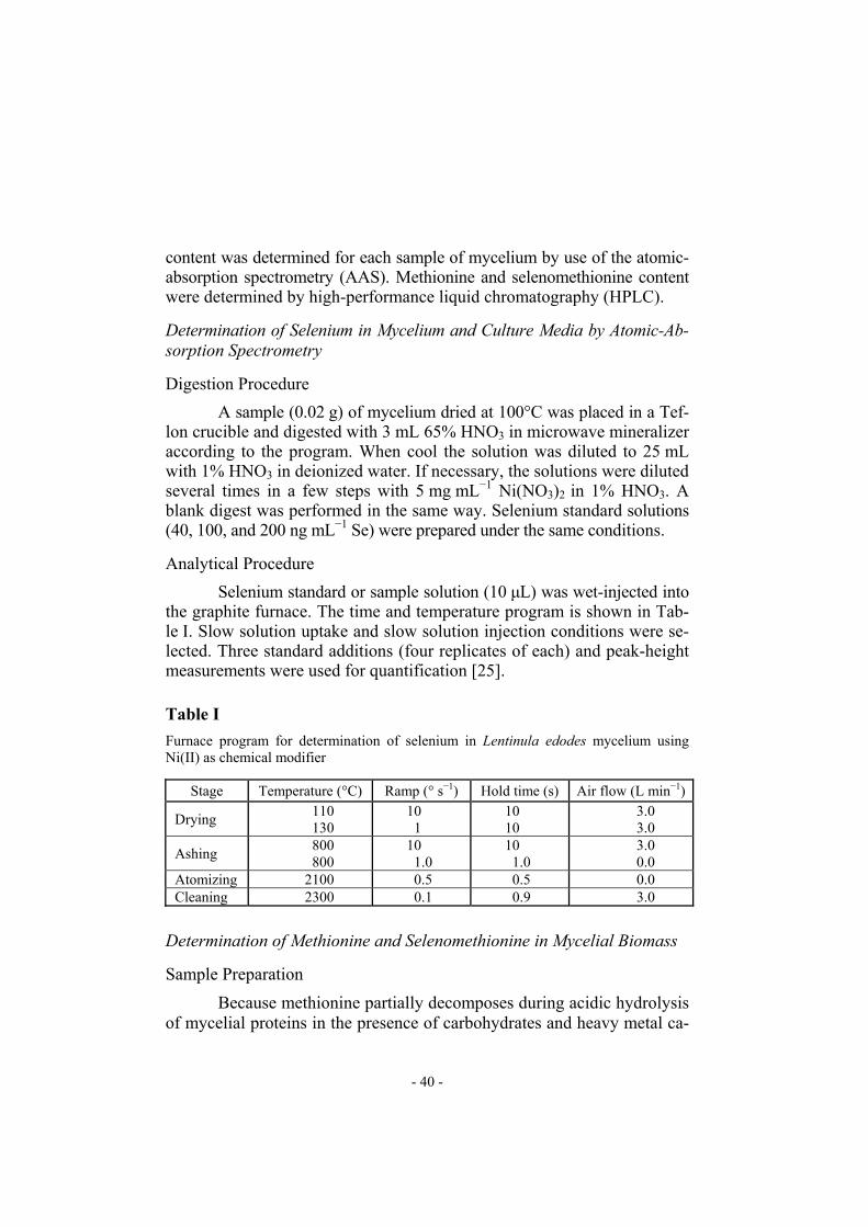
Fig. 4
Selenium accumulated in mycelium as selenomethionine as a percentage of total selenium content (series “0”)
- 45 -
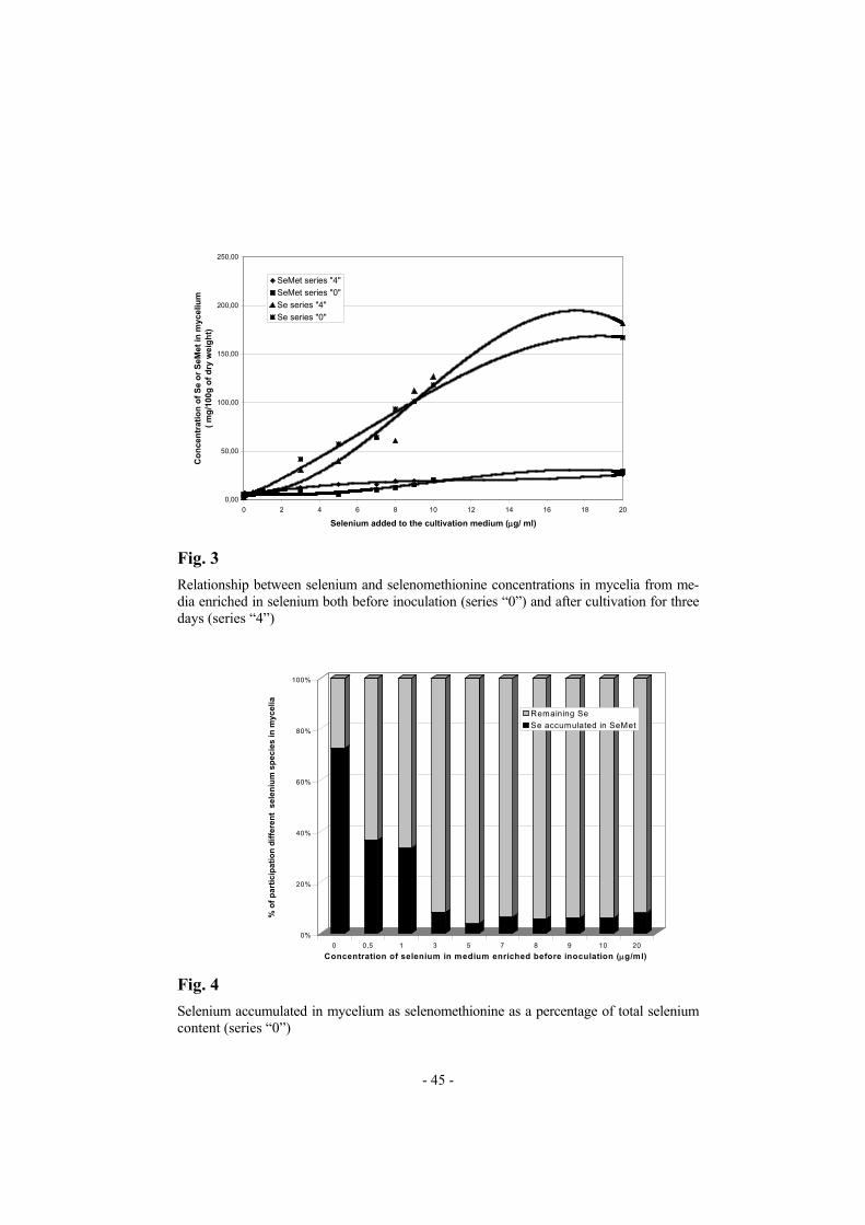
tion of selenomethionine was only almost 6% (Figs 4 and 5). The percen-tage of methionine transformed into selenomethionine rose in proportion to the total selenium content of L. edodes mycelial dry weight (Tables II and III) and reached almost 3%.
0%
10%
20%
30%
40%
50%
60%
70%
80%
90%
100%
% o
f par
ticip
atio
n di
ffere
nt s
elen
ium
spe
cies
in m
ycel
ia
0 0,5 1 3 5 7 8 9 10 20
Concentration of selenium in medium enriched after 3 days (µg/ml)
remaining Seselenium in SeMet
Fig. 5
Selenium accumulated in mycelium as selenomethionine as a percentage of total selenium content (series “4”)
0
5
10
15
20
25
30
0 5 10 15 20 25
Concentration of selenium in medium (µg/ml)
Con
cent
ratio
n of
sel
enoa
min
o ac
ids
in m
ycel
ium
(mg/
g)
SeMetSeCys
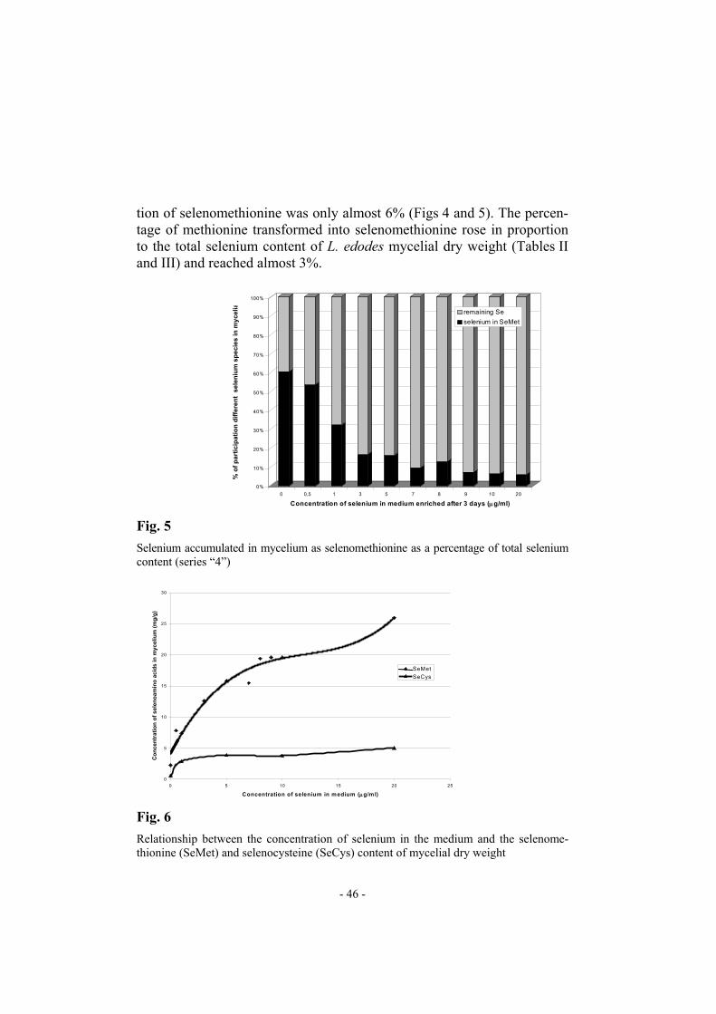
Fig. 6
Relationship between the concentration of selenium in the medium and the selenome-thionine (SeMet) and selenocysteine (SeCys) content of mycelial dry weight
- 46 -

The selenocysteine content of mycelial dry weight seemed to be al-most stable for mycelia cultivated in media enriched in selenium at con-centrations higher than 1 µg mL−1. The amount of selenocysteine in myce-lium cultivated in medium containing 20 µg mL−1 selenium reached almost 50 µg g−1 d.w. (Fig. 6). CONCLUSIONS Amino acid analysis by RP-HPLC with o-phthaldialdehyde deriva-tization is a method with good selectivity and efficiency. The detection limit for most OPA amino acid derivatives is in range 25–50 fmol. The method can be successfully used for determination of selenomethionine and selenocysteine in hydrolysates of proteins. The problem of the oxida-tive decomposition of SeCys during protein hydrolysis can be avoided by carboxymethylation of proteins before hydrolysis. The standard HPLC equipment and relatively cheap C18 columns used are major advantages of this method. Selenomethionine and selenocysteine standards are commer-cially available. This method was successfully used for determination of the relationship between the selenium, selenomethionine, and selenocysteine content of submerged cultivated mycelium of the medicinal mushroom Lentinula edodes. The results indicate that in medium not fortified with selenium over 80% of the selenium in the mycelium is accumulated as se-lenoamino acids – over 70% as selenomethionine and approximately 10% as selenocysteine. Similar data were obtained for selenized yeast. For my-celia rich in selenium the proportion of selenium accumulated as selenoa-mino acids decreases. Speciation of these selenium compounds is the next important objective of this research because of their putative role in cancer prevention. REFERENCES
[1] M.P. Rayman, Lancet, 356, 233 (2000) [2] G.F. Combs Jr. and W.P. Gray, Pharmacol. Ther., 79, 179 (1988) [3] WHO, Food and Agriculture Organisation, International Atomic
Energy Agency expert group. Trace elements in human nutrition and health, Geneva, WHO, 1996
[4] H. Tapiero, D.M. Townsend, and K.D. Tew, Biomed. Pharmacother., 57, 134 (2003)
- 47 -

[5] R. McKenzie, T.S. Rafferty, and G.J. Beckett, Trends, 19, 342 (1998) [6] D.C. Mahan and Y.Y. Kim, J. Anim. Sci., 74, 2711 (1996) [7] C. Wang and R.T. Lovell, Aquaculture, 152, 223 (1997) [8] L.C. Clark, G.F. Combs, B.W. Turnbull, E.H. Slate, D.K. Chalker,
and J. Chow, J. Am. Med. Assoc., 276, 1957 (1996) [9] C. Ip, M. Birringer, E. Block, et al., J. Agric. Food. Chem., 51, 4191
(2003) [10] J. Eltren, Chemosphere, 36, 1787 (1998) [11] V. Gerely, K.M. Kubashka, S. Mounicou, P. Fodor, and J.A. Caruso,
J. Chromatogr. A, 1101, 94 (2006) [12] Y. Ogra, K. Ishwita, J.R. Encinar, R. Łobiński, and K.T. Suzuki,
Anal. Bioanal. Chem., 379, 861 (2004) [13] J. Turło, B. Gutkowska, and P. Suchocki, paper in preparation [14] X. Dauchy, A. Potin-Gauthier, A. Astrus, and M. Astruc, Fresenius
J. Anal. Chem., 348, 792 (1994) [15] I. Arnault and J. Auger, J. Chromatogr. A, 1112, 23 (2006) [16] M.P. Bartolomeo and F. Maisano, J. Biomol. Tech., 17, 131 (2006) [17] C. Mammel, A. Kyriakopoulos, U. Rosick, and D. Behne, Analyst,
122, 1359 (1997) [18] J.E. Shively, Methods of Protein Microcharacterization, Humana
Press, Clifton, New York, 1986 [19] Ajinomoto Company Information Publication, 1984, Lentinan: a new
type of anticancer drug. Ajinomoto Co. Inc. 15-1 Kyobashi, Chuo-ku Tokyo, Japan
[20] P. Mattila, K. Suonpaa, and V. Piironen, Nutrition, 16, 694 (2000) [21] G. Chichara, Y.Y. Maeda, T. Taguchi, and J. Hamuro,
Int. J. Immunol., 5, 145 (1989) [22] G. Chichara, Int. J. Orient. Med., 17, 57 (1992) [23] S.P. Wasser and A.L. Weis, Int. J. Med. Mush., 1, 31 (1999) [24] H. Iizuka, Food Rev. Int., 13, 343 (1997) [25] H. Suzuki, K. Iiyama, O. Yoshida, S. Yamazaki, N. Yamamoto,
and S. Toda, Agric. Biol. Chem., 54, 479 (1990) [26] Merck Application Note, HPLC analysis of amino acids
by automatic precolumn derivatization with OPA. 1996, Merck KGaA, Darmstadt, Germany
- 48 -