relevance of mitochondrial genetics and metabolism in...
TRANSCRIPT

Relevance of Mitochondrial Genetics andMetabolism in Cancer Development
Giuseppe Gasparre1, Anna Maria Porcelli2, Giorgio Lenaz3, and Giovanni Romeo1
1Department of Medical and Surgical Sciences, Unit of Medical Genetics, University of BolognaMedical School, 40138 Bologna, Italy
2Department of Pharmacy and Biotechnologies, University of Bologna, 40126 Bologna, Italy3Department of Biochemistry “Giovanni Moruzzi,” University of Bologna Medical School, 40126Bologna, Italy
Correspondence: [email protected]
Cancer cells are characterized in general by a decrease of mitochondrial respiration andoxidative phosphorylation, together with a strong enhancement of glycolysis, the so-calledWarburg effect. The decrease of mitochondrial activity in cancer cells may have multiplereasons, related either to the input of reducing equivalents to the electron transfer chain or todirect alterations of the mitochondrial respiratory complexes. In some cases, the depressionof respiratory activity is clearly the consequence of disruptive mitochondrial DNA (mtDNA)mutations and leads as a consequence to enhanced generation of reactive oxygen species(ROS). By acting both as mutagens and cellular mitogens, ROS may contribute directly tocancer progression. On the basis of our experimental evidence, we suggest a deep implica-tion of the supercomplex organization of the respiratory chain as a missing link betweenoxidative stress, energy failure, and tumorigenesis. We speculate that under conditions ofoxidative stress, a dissociation of mitochondrial supercomplexes occurs, with destabilizationof complex I and secondary enhanced generation of ROS, thus leading to a vicious circleamplifying mitochondrial dysfunction. An excellent model to dissect the role of pathogenic,disassembling mtDNA mutations in tumor progression and their contribution to the meta-bolic reprogramming of cancer cells (glycolysis vs. respiration) is provided by an oftenunderdiagnosed subset of tumors, namely, the oncocytomas, characterized by disruptivemutations of mtDNA, especially of complex I subunits. Such mutations almost completelyabolish complex I activity, which slows down the Krebs cycle, favoring a high ratio of a-ketoglutarate/succinate and consequent destabilization of hypoxia inducible factor 1a(HIF1a). On the other hand, if complex I is partially defective, the levels of NADþ may besufficient to implement the Krebs cycle with higher levels of intermediates that stabilizeHIF1a, thus favoring tumor malignancy. The threshold model we propose, based on thepopulation-like dynamics of mitochondrial genetics (heteroplasmy vs. homoplasmy),implies that below threshold complex I is present and functioning correctly, thus favoringtumor growth, whereas above threshold, when complex I is not assembled, tumor growth isarrested. We have therefore termed “oncojanus” the mtDNA genes whose disruptive muta-tions have such a double-edged effect.
Editors: Douglas C. Wallace and Richard J. Youle
Additional Perspectives on Mitochondria available at www.cshperspectives.org
Copyright # 2013 Cold Spring Harbor Laboratory Press; all rights reserved; doi: 10.1101/cshperspect.a011411
Cite this article as Cold Spring Harb Perspect Biol 2013;5:a011411
1
on February 16, 2019 - Published by Cold Spring Harbor Laboratory Press http://cshperspectives.cshlp.org/Downloaded from

Cancer cells are characterized by uncon-trolled proliferation due to gain-of-function
of oncogenes and loss of function of tumor-sup-pressor genes. Among the hallmarks of the can-cer phenotype, there is a metabolic reprogram-ming (DeBerardinis et al. 2008). Cancer cells arecharacterized in general by a decrease of mito-chondrial respiration and oxidative phosphor-ylation, together with a strong enhancement ofglycolysis, the so-called Warburg effect, first re-ported by Otto Warburg (Warburg 1956). Ow-ing to space limitations, for this particular topicwe refer you to recent excellent reviews (More-no-Sanchez et al. 2007; Mayevsky 2009; Mathu-pala et al. 2010; Koppenol et al. 2011). Never-theless, it is not clear which of the two processes(i.e., decreased respiration and enhanced glycol-ysis) is the primary event, or even if they areconcomitant consequences of a common causalevent.
Although the decrease of mitochondrial ox-idative phosphorylation is considered by manyinvestigators as a universal feature of neoplasticcells, there are numerous publications in whichcancer cells have been reported to show normalor even high respiratory activity (Zu and Guppy2004; Weinberg and Chandel 2009). This appar-ent paradox can be explained by the hypothesisadvanced by Smolkova et al (2010) that cancerprogression is characterized by waves of geneexpression, where initial waves suppressing mi-tochondrial function and stimulating glycolysisare followed by a resumption of mitochondrialoxidative phosphorylation when glutamine be-comes the leading substrate for energy produc-tion and the major carbon source for anabolicprocesses. Each malignant tumor is character-ized by a distinct metabolic phenotype reflect-ing the history of the different waves during thesequence of carcinogenic mutations.
The enhanced aerobic glycolysis character-izing cancer cells (and, in general, highly prolif-erating cells) at least in some periods of theirprogression toward malignancy may be vital toprovide, besides energy, also the starting mate-rials for active biosynthetic processes (Cuezvaet al. 2009). Indeed, it was recognized earlythat incomplete activity of the Krebs cycle allowsthe use of citrate as a starting intermediate for
the synthesis of fatty acids and cholesterol (Parloand Coleman 1986; Kuhajda et al. 1994), where-as glycolysis may provide the majority of energyrequired for the anabolic processes. The supplyof citrate for lipid biosynthetic processes may beprovided by reductive carboxylation of a-keto-glutarate by reversal of the isocitrate dehydroge-nase reaction when the major carbon source isglutamine (Holleran et al. 1995; Yoo et al. 2008)and pyruvate dehydrogenase activity is de-pressed by the activation of pyruvate dehydro-genase kinase (Kim et al. 2006).
During the cell cycle, the genes for glycolyticenzymes are overexpressed at the time of activeDNA synthesis (Klevecz et al. 2004). Likewise,several oncogenes (such as c-myc, HIF1a, Akt)or tumor suppressors (p53) are involved in thetransactivation of glycolytic enzymes (Cuezvaet al. 2009). The activation of these genes mayinvolve mitochondrial dysregulation, for exam-ple, through unbalance of Krebs cycle interme-diates (see below).
DECREASED MITOCHONDRIALRESPIRATION IN CANCER CELLS
The decrease of mitochondrial activity in cancercells may have multiple reasons:
1. Early in the 1920s, it was observed that en-hanced glycolysis, as in cancer cells, inhibitsrespiration (the so-called Crabtree effect); itwas suggested that respiratory inhibition iscaused by competition of glycolysis and ox-idative phosphorylation for Pi and ADP (Ib-sen 1961).
2. Decrease of oxidative phosphorylation(OXPHOS) may also derive from hypoxiabecause O2 may fall below the Km for cyto-chrome oxidase (Brahimi-Horn and Pouys-segur 2007) and thus induce the Pasteur ef-fect (stimulation of phosphofructokinase byAMP when mitochondrial ATP synthesis isdeficient).
3. On the other hand, decreased OXPHOSbecomes operative during hypoxia becauseof the stabilization of hypoxia inducible fac-tor 1a (HIF1a) (Semenza 2003). This factor
G. Gasparre et al.
2 Cite this article as Cold Spring Harb Perspect Biol 2013;5:a011411
on February 16, 2019 - Published by Cold Spring Harbor Laboratory Press http://cshperspectives.cshlp.org/Downloaded from
at normal oxygen tension undergoes hydrox-ylation of two key proline residues by HIF-prolyl hydroxylases (PHDs), which allow therecognition of the factor by pVHL, the prod-uct of the von Hippel–Lindau gene, thataddresses the protein to ubiquitin-mediateddegradation. The factor becomes stabilizedduring hypoxia by inactivation of PHD andbinds to hypoxia-responsive elements inthe DNA, stimulating a large array of genes(Semenza 2007; Kaluz et al. 2008). Besidesglycolytic enzymes, HIF1a enhances the ex-pression of pyruvate dehydrogenase kinase(PDK), thus inhibiting pyruvate dehydroge-nase (PDH) complex and decreasing the in-put of reducing equivalents to the respiratorychain (Kim et al. 2006). The decreased inputof carbon into the Krebs cycle must necessar-ily be limited, otherwise the lack of citratewould impair the synthesis of lipids andcholesterol that are required for the forma-tion of new cellular membranes during tu-mor growth. This is the reason that glutaminebecomes a preferred substrate for replenish-ing the Krebs cycle for both energetic andbiosynthetic purposes.
It is of interest that HIF1a is also stabi-lized by several nonhypoxic stimuli, for ex-ample, by succinate (that inhibits prolyl hy-droxylase) (King et al. 2006), but also bypyruvate and oxaloacetate (Lu et al. 2005;McFate et al. 2008) and, interestingly, by re-active oxygen species (ROS) (Patten et al.2010), although the activating effect of ROSon HIF is still a controversial issue (Bell et al.2008). ROS could act directly but also bystimulating PDK, thus leading to accumula-tion of pyruvate (Sun et al. 2009; Patten et al.2010). Thus, derangement of the Krebs cyclemay lead to changes in steady-state concen-trations of important metabolites that areable to control the activity of HIF1a.
4. Direct alterations of respiratory activity incancer cells have not been frequently investi-gated. In general, a depression of respiratoryactivity is observed in cancer cells (Putignaniet al. 2008; Park et al. 2009; Jahnke et al. 2010;Ma et al. 2010). In the case of Ras-trans-
formed fibroblasts, we have observed a directalteration of the respiratory chain, in partic-ular, a 50% decrease of complex I contentand activity, and consequently of NAD-linked respiration and ATP synthesis (Ba-racca et al. 2010). This seems to be due inpart to decreased expression as seen by tran-scriptome analysis. However, we do notknow yet if there are disruptive mitochon-drial DNA (mtDNA) mutations as seen inoncocytic tumors (see below). The effect ofthe adenylate cyclase activator forskolin inreversing the changes accompanying trans-formation suggests that the mitochondrialchanges may be caused by impairment ofcAMP-dependent pathways (L Alberghina,pers. comm.).
5. In some cases, the depression of respiratoryactivity is clearly the consequence of disrup-tive mtDNA mutations (see below).
Tumorigenic mtDNA mutations affect therespiratory chain complexes and enhance theproduction of ROS (Brandon et al. 2006); how-ever, the pathological relevance of mtDNA mu-tations in cancer cells is controversial (Frezzaand Gottlieb 2009). Nonetheless, a clear-cutcorrelation between occurrence of pathogenicmtDNA mutations and mitochondrial energet-ic impairment is a well-demonstrated feature ofoncocytic tumors (Bonora et al. 2006a; Porcelliet al. 2010), which is dealt with in detail below.
Accumulation of NADH due to either hy-poxia or alterations of the electron transferchain inhibits isocitrate dehydrogenase andthe Krebs cycle when implemented by acetylCoA may help the exit of citrate to support lipidsynthesis (fatty acids and cholesterol). On theother hand, the entry of reducing equivalentsvia glutamine may not be inhibited, thus sus-taining a truncated Krebs cycle via glutamate,a-ketoglutarate, and the intermediates of theKrebs cycle, thus reaching malate that is con-verted to pyruvate by the malic enzyme afterrelease in the cytosol. Pyruvate would contrib-ute to stabilization of HIF1a and may also beconverted to lactate by lactate dehydrogenase.The extent of this pathway of glutamine oxida-tion is determined by the residual activity of the
Mitrochondrial Genetics and Metabolism in Cancer
Cite this article as Cold Spring Harb Perspect Biol 2013;5:a011411 3
on February 16, 2019 - Published by Cold Spring Harbor Laboratory Press http://cshperspectives.cshlp.org/Downloaded from

respiratory chain, because both glutamate de-hydrogenase and a-ketoglutarate dehydroge-nase require oxidized NADþ, and both theseenzymes together with succinate dehydrogenaserequire an intact respiratory chain for theirturnover.
THE ROLE OF ROS
An overwhelming body of evidence accumu-lated in the last decades has shown that mito-chondria have a central role in the etiology andpathogenesis of most majorchronic diseases andin aging (Wallace 2005; Lenaz et al. 2006; Lenazand Genova 2010). The involvement of mito-chondria in disease, which has generated theterm “mitochondrial medicine” (DiMauro etal. 2006), has been largely ascribed to their cen-tral role in production of ROS and to the dam-aging effect of the latter on these organelles. Inparticular, mtDNA mutations would induce al-terations of the encoded polypeptides in therespiratory complexes, with consequent de-crease of electron transfer activity, leading to fur-ther production of ROS, and thus establishing avicious circle of oxidative stress and energeticdecline (Ozawa 1997). It must be underlinedthat mtDNA mutations leading to increasedROS production are those allowing at least a par-tial assembly of the respiratory complexes. Onthe contrary, for instance, oncocytic tumors dis-play homoplasmic disassembling complex I mu-tations, which may not be ROS-generating mu-tations, if one considers that the main ROSproduction site might be lacking as a whole(Koopman et al. 2007; Moran et al. 2010; Porcelliet al. 2010; Tuppen et al. 2010). Moreover, themutant load ought to be considered when ana-lyzing functional effects of mtDNA mutations.The threshold model we previously proposedimplies that, below threshold, complex I is pre-sent and functioning correctly, in contrast to thecase of a missense mutation leading to the co-existence of nonmutant along with mutant ROSproducing complex I (Park et al. 2009).
Decreased mitochondrial activity is consid-ered to be tumorigenic, mainly because of theenhanced ROS production. H2O2 exported tothe nucleus enhances the transcription of select-
ed genes that favor tumor progression (Wallace2005). ROS act both as mutagens and cellularmitogens (Klaunig and Kamendulis 2004). Ithas been suggested that oncogenesis and neuro-degeneration may share common pathogenicfeatures as differential responses to different in-sults during different phases of the cell cycle,leading either to cell death or to cell prolifera-tion (Morris et al. 2010).
Any lowering of the electron transfer chainactivity, either caused by mtDNA mutationsand hence structural lesions of the respiratorycomplexes or depending directly or indirectlyon hypoxia, would enhance generation ofROS; it is known that ROS generation is en-hanced in State 4 (controlled state in absenceof ADP), when complex I is inhibited, and para-doxically under hypoxic conditions.
ROS may contribute to further mitochon-drial dysfunction by several mechanisms: (1)direct alterations of respiratory complexes, forexample, complex I, particularly on FeS clusters;(2) peroxidation of mitochondrial phospholip-ids, in particular, cardiolipin (Paradies et al.2010), that are required for proper assemblyand activity of mitochondrial complexes (I,III, IV); (3) (further) mtDNA mutations affect-ing respiratory complexes; and (4) stabilizationof HIF1a (Patten et al. 2010).
In this scenario, it is easy to foresee a deepimplication of supercomplex organization asthe missing link between oxidative stress andenergy failure (Lenaz and Genova 2007). It istempting to speculate that under conditions ofoxidative stress a dissociation of complex I–IIIaggregates occurs, with loss of facilitated elec-tron channeling and resumption of a less effi-cient random diffusional behavior with electrontransfer depending on the collisional encoun-ters of the free ubiquinone molecules with thepartner complexes.
As predicted by Lenaz and Genova (2007),dissociation of supercomplexes might have fur-ther deleterious consequences, such as disas-sembly of complex I and III subunits and lossof electron transfer and/or proton transloca-tion; the consequent alteration of electrontransfer may elicit further induction of ROSgeneration. Following this line of thought, the
G. Gasparre et al.
4 Cite this article as Cold Spring Harb Perspect Biol 2013;5:a011411
on February 16, 2019 - Published by Cold Spring Harbor Laboratory Press http://cshperspectives.cshlp.org/Downloaded from
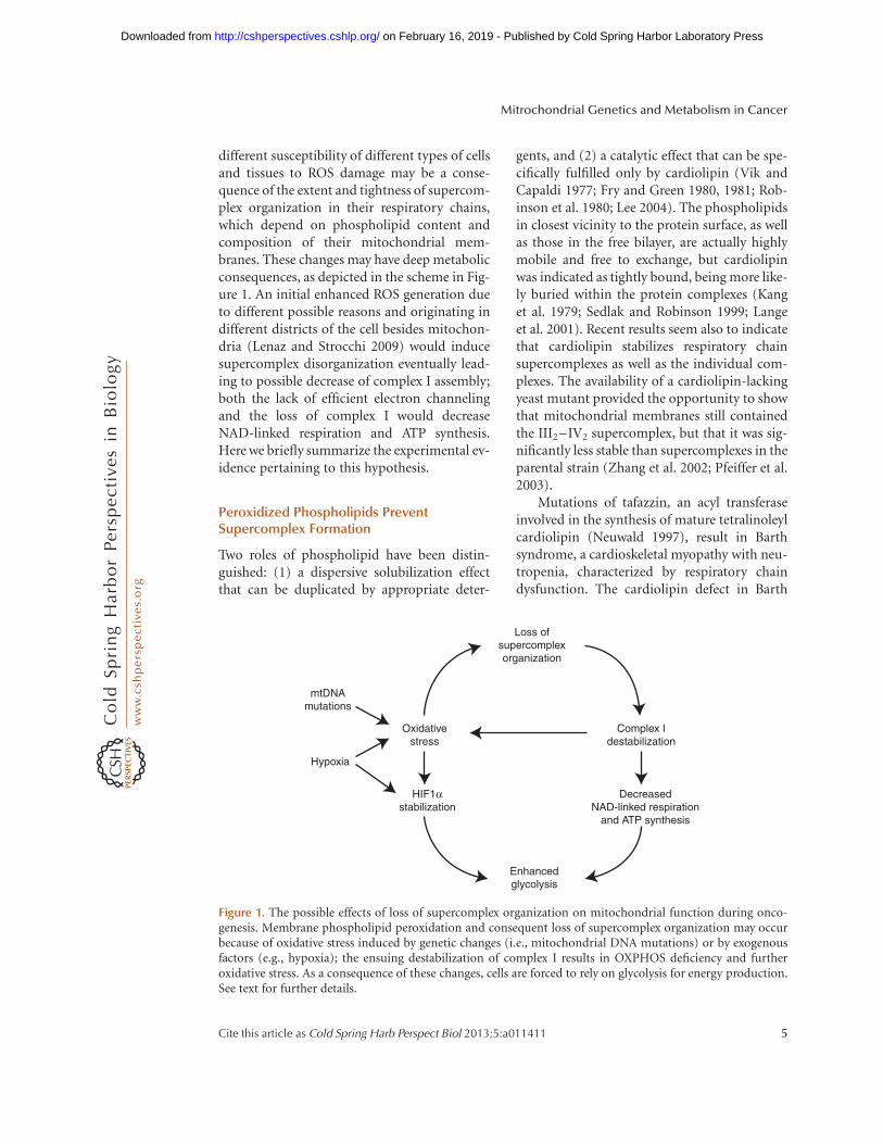
different susceptibility of different types of cellsand tissues to ROS damage may be a conse-quence of the extent and tightness of supercom-plex organization in their respiratory chains,which depend on phospholipid content andcomposition of their mitochondrial mem-branes. These changes may have deep metabolicconsequences, as depicted in the scheme in Fig-ure 1. An initial enhanced ROS generation dueto different possible reasons and originating indifferent districts of the cell besides mitochon-dria (Lenaz and Strocchi 2009) would inducesupercomplex disorganization eventually lead-ing to possible decrease of complex I assembly;both the lack of efficient electron channelingand the loss of complex I would decreaseNAD-linked respiration and ATP synthesis.Here we briefly summarize the experimental ev-idence pertaining to this hypothesis.
Peroxidized Phospholipids PreventSupercomplex Formation
Two roles of phospholipid have been distin-guished: (1) a dispersive solubilization effectthat can be duplicated by appropriate deter-
gents, and (2) a catalytic effect that can be spe-cifically fulfilled only by cardiolipin (Vik andCapaldi 1977; Fry and Green 1980, 1981; Rob-inson et al. 1980; Lee 2004). The phospholipidsin closest vicinity to the protein surface, as wellas those in the free bilayer, are actually highlymobile and free to exchange, but cardiolipinwas indicated as tightly bound, being more like-ly buried within the protein complexes (Kanget al. 1979; Sedlak and Robinson 1999; Langeet al. 2001). Recent results seem also to indicatethat cardiolipin stabilizes respiratory chainsupercomplexes as well as the individual com-plexes. The availability of a cardiolipin-lackingyeast mutant provided the opportunity to showthat mitochondrial membranes still containedthe III2–IV2 supercomplex, but that it was sig-nificantly less stable than supercomplexes in theparental strain (Zhang et al. 2002; Pfeiffer et al.2003).
Mutations of tafazzin, an acyl transferaseinvolved in the synthesis of mature tetralinoleylcardiolipin (Neuwald 1997), result in Barthsyndrome, a cardioskeletal myopathy with neu-tropenia, characterized by respiratory chaindysfunction. The cardiolipin defect in Barth
Enhancedglycolysis
DecreasedNAD-linked respiration
and ATP synthesis
HIF1αstabilization
Complex Idestabilization
Oxidativestress
Loss ofsupercomplexorganization
mtDNAmutations
Hypoxia
Figure 1. The possible effects of loss of supercomplex organization on mitochondrial function during onco-genesis. Membrane phospholipid peroxidation and consequent loss of supercomplex organization may occurbecause of oxidative stress induced by genetic changes (i.e., mitochondrial DNA mutations) or by exogenousfactors (e.g., hypoxia); the ensuing destabilization of complex I results in OXPHOS deficiency and furtheroxidative stress. As a consequence of these changes, cells are forced to rely on glycolysis for energy production.See text for further details.
Mitrochondrial Genetics and Metabolism in Cancer
Cite this article as Cold Spring Harb Perspect Biol 2013;5:a011411 5
on February 16, 2019 - Published by Cold Spring Harbor Laboratory Press http://cshperspectives.cshlp.org/Downloaded from

syndrome results in destabilization of the su-percomplexes by weakening the interactions be-tween respiratory complexes (McKenzie et al.2006).
It is well documented that exposure of mi-tochondria to ROS can affect the respiratoryactivity via oxidative damage of cardiolipin,which is required for the optimal functioningof the enzyme complexes (Paradies et al. 2000,2002, 2010; Petrosillo et al. 2003, 2009). We haveshown that the maintenance of a I–III super-complex after reconstitution into phospholipidvesicles is abolished if lipid peroxidation is in-duced before reconstitution (Bianchi et al. 2003;Genova et al. 2008).
Evidently, the distortion of the lipid bilayerinduced by peroxidation and the alteration ofthe tightly bound phospholipids determine dis-sociation of the supercomplex originally presentin the lipid-poor preparation.
Supercomplex Disassembly AbolishesCoQ Channeling and Resumes LessEfficient Pool Behavior
Studies of respiration in pathological condi-tions (Rosca et al. 2008; van Raam et al. 2008)showed that electron transfer in absence ofsupercomplex organization is lost even if activ-ity of the individual complexes is normal. Inreconstitution studies of complexes I and IIIin phospholipid vesicles (Genova et al. 2008),electron transfer between complex I and com-plex III (NADH cytochrome c reductase) showsrates higher than those predicted by randombehavior in proteoliposomes enriched in com-plexes I and III at a protein-to-phospholipidratio of 1:1 (when kinetic testing according toflux control analysis indicates the presence of asupercomplex I–III). However, at a ratio of 1:30(when the supercomplex is dissociated), therate of NADH cytochrome c reductase is exactlythat predicted by the pool equation on the basisof the individual activities of complex I andcomplex III. This is a demonstration that su-percomplex formation, indeed, enhances therate of electron transfer above that occurringvia a ubiquinone pool in the membrane (Bian-chi et al. 2003; Genova et al. 2008), and loss of
supercomplex organization resumes less effi-cient pool activity of the quinone.
Loss of Supercomplexes Prevents Stabilityand Assembly of Individual Complexes
Analysis of the state of supercomplexes in pa-tients with an isolated deficiency of single com-plexes (Schagger et al. 2004) and in cultured cellmodels harboring cytochrome b mutations(Acin-Perez et al. 2004; Blakely et al. 2005;D’Aurelio et al. 2006; Diaz et al. 2006; McKenzieet al. 2006) provided evidence that the forma-tion of respirasomes is essential for the assem-bly/stability of complex I. Genetic alterationsleading to a loss of complex III prevented respi-rasome formation and led to secondary loss ofcomplex I, and therefore primary complex IIIassembly deficiencies presented as complex III/I defects. Conversely, complex III stability wasnot influenced by the absence of complex I.
From these findings, supercomplex assem-bly emerged as a necessary step for respiration,its defect setting the threshold for respiratoryimpairment in mtDNA mutant cells.
Disassembly of Supercomplex OrganizationEnhances Generation of ROS
Indirect circumstantial evidence suggests thatsupercomplex assembly may limit the extentof superoxide generation by the respiratorychain (Lenaz and Genova 2010). A direct studyon a mitochondrial fraction containing com-plexes I and III and reconstituted with differentamounts of phospholipids shows that at pro-tein:phospholipids 1:30, when the supercom-plex is not formed, the production of superox-ide is 20-fold higher than at a 1:1 ratio, whenmost units are assembled in a supercomplex(ML Genova, G Barbero, E Maranzana, et al.,unpubl.).
Although the molecular structure of the in-dividual complexes does not allow us to envisiona close apposition of the matrix arm of complexI, where FMN is localized, with either complexIII or IV, the actual shape of the I1III2IV1 super-complexes from bovine heart (Schafer et al.2007) suggests a slightly different conformation
G. Gasparre et al.
6 Cite this article as Cold Spring Harb Perspect Biol 2013;5:a011411
on February 16, 2019 - Published by Cold Spring Harbor Laboratory Press http://cshperspectives.cshlp.org/Downloaded from

of complex I in the supercomplexes with a small-er angle of the matrix arm with the membranearm showing a higher bending toward the mem-brane (and presumably complex III), in line withthe notion that complex I may undergo impor-tant conformational changes (Radermacher et al.2006).
Do Cancer Cells Lose SupercomplexAssociation?
The validation of the above working hypothesisrequires direct studies on cancer tissues andneoplastic cells. A general decrease of respirato-ry chain activity is a common feature of severalcancers. Nevertheless, very few studies have ad-dressed the respiratory chain supramolecularorganization.
Baracca et al. (2010) reported that down-regulation of the respiratory chain in Ras-trans-formed fibroblasts is operated through strongdecrease of complex I activity and content, prob-ably because of lack of correct assembly of thesubunits as a consequence of altered supercom-plex organization, as shown by loss of the high-est-molecular-weight I1III2IV1-2 supercomplex.Significantly, ROS generation was strongly en-hanced in the Ras cells. In the XTC-UC1 onco-cytic cell line (see below), we have also found(Genova et al. 2008) by blue-native electropho-resis of mitochondrial proteins, a complete ab-sence of high-molecular-weight aggregates con-taining either complex I or complex IV, that areinstead present in a control cell line from a non-oncocytic thyroid tumor. If the absence ofsupercomplexes comprising complex I is a con-sequence of its disassembly due to lack of ND1(see below), the absence of aggregated complexIV is in line with the working hypothesis.
The loss of supercomplex assembly seems toaccompany a decrease/loss of complex I activi-ty; if this decrease is found to be common tomost tumors, the consequences would dependon the extent of such a decrease. DisruptivemtDNA mutations in the ND subunits of com-plex I (as in oncocytomas; see below) almostcompletely abolish complex I activity, whichpresumably inactivates the Krebs cycle, that re-quires NADþ, favoring a high-ratio a-ketoglu-
tarate/succinate and consequent destabilizationof HIF1a. On the other hand, if complex I ispartially defective (as in RAS-mutated fibro-blasts), the levels of NADþ may be sufficientto implement the Krebs cycle with higher levelsof intermediates that stabilize HIF1a, thus fa-voring tumor malignancy.
mtDNA MUTATIONS IN CANCER: THEPECULIAR CASE OF ONCOCYTIC TUMORS
An excellent model to dissect out the role ofpathogenic, disassembling mtDNA mutationsin tumor progression and their contributionto the metabolic reprogramming of cancer cells(glycolysis vs. respiration) is provided by an of-ten underdiagnosed subset of tumors, namely,the oncocytomas. These neoplasms contain ahigh percentage or they are completely madeof cells characterized by an aberrant mitochon-drial hyperplasia. They originate from epithelialtissues and occur mainly in endocrine and exo-crine organs, although the oncocytic phenotypehas been described in many anatomical districts(for review, see Gasparre et al. 2011a). The ma-jority of oncocytic tumors are considered asbenign, displaying low invasiveness and show-ing a usually favorable prognosis. In the thyroid,nonetheless, the best indicator for prognosis re-mains the degree of differentiation of neoplasticcells, which defines adenoma or carcinoma, re-gardless of the occurrence of the oncocytic phe-notype.
The most striking feature of oncocytic tu-mors is appreciated through histochemical andultrastructural analyses, which highlight cellspacked with enlarged globular or ovate mito-chondria with a stack of lamelliform, tubular,or flat cristae and occupying up to 60% of thecytoplasm (Tallini 1998; Ambu et al. 2000; Rivaand Tandler 2000). Such heterogeneity in mito-chondrial morphology has long been an indica-tor of a functional along with a structural alter-ation of the organelles in oncocytic cells.
The cause of the mitochondrial hyperplasiain oncocytic tumors has been suspected to con-sist in an aberrant mitochondrial biogenesistriggered bya mitochondrial dysfunction, whichmay attempt to compensate for an impaired
Mitrochondrial Genetics and Metabolism in Cancer
Cite this article as Cold Spring Harb Perspect Biol 2013;5:a011411 7
on February 16, 2019 - Published by Cold Spring Harbor Laboratory Press http://cshperspectives.cshlp.org/Downloaded from

energy production. Evidence that a compensa-tory mechanism is responsible for oncocyticphenotype occurrence has accumulated in re-cent years. It is now clear that the mitochondrialrespiration deficiency originally hypothesized(Muller-Hocker et al. 1998a) is, indeed, an eventoccurring in oncocytic tumors and mainly con-cerns respiratory complex I (Zielke et al. 1998;Simonnet et al. 2003). NADH-dehydrogenaseactivity is, in fact, reduced concomitantly withan increase in other respiratory complex abun-dance and, overall, in mitochondrial mass asshown by measurements of citrate synthase ac-tivity and of mtDNA copy number (Zielke et al.1998; Simonnet et al. 2003; Gasparre et al. 2009;Porcelli et al. 2010).
In vitro studies with transmitochondrial cellhybrids (cybrids) have shown that the defectivemitochondrial respiration of thyroid oncocyticcells is transferred to normally respiring os-teosarcoma cells when mitochondria, not nu-clei, are imported (Bonora et al. 2006a). Fromthis observation, as well as from studies inwhich the mitochondrial genome was screenedfor oncocytic cancer-specific genetic lesionssuch as small and large deletions, depletion, orpoint mutations (Tallini et al. 1994; Maximoet al. 1998, 2002; Maximo and Sobrinho-Si-moes 2000; Costa-Guda et al. 2007; Gasparreet al. 2007, 2008, 2009; Mayr et al. 2008; Zim-mermann et al. 2009; Porcelli et al. 2010; Barto-letti-Stella et al. 2011), it is now generallyaccepted that the genetic hallmark of the onco-cytic phenotype is the occurrence of disrup-tive mtDNA mutations, particularly in complexI subunits. This holds true for virtually all typesof oncocytic tumors analyzed to date (Gasparreet al. 2007, 2008, 2009; Porcelli et al. 2010;Bartoletti-Stella et al. 2011), and, inversely,association between the occurrence of disrup-tive mutations and that of oncocytic foci hasbeen shown, for instance, in endometrial car-cinoma (Guerra et al. 2011). Within the pleth-ora of disruptive mutations, both frameshiftand pathogenic missense changes are included,which ultimately lead to complex I impairmentin terms of function and/or assembly. Hence,the direct consequence of these mutations im-pinges on respiration, whose impairment may
subsequently trigger a retrograde signaling fromthe organelles to the nucleus, leading in a vi-cious circle to mitochondrial hyperplasia. Ananalysis of the entire mitochondrial genomessequenced in oncocytic tumor samples has al-lowed identification of MTND1 as a mutationalhotspot (Gasparre et al. 2007). Less importantlythan MTND1, at least two of the remaining sixmitochondria-coded complex I genes display ahigh rate of pathogenic mutations accumu-lation, namely, MTND4 and MTND5, whichare also among the longest mitochondrial genesand richest in homopolymeric stretches, wheremany frameshift mutations occur. Complex IV(cytochrome c oxidase; COX) mitochondrialsubunits appear to be the least involved in con-tributing to the mitochondrial damage leadingto oncocytic transformation, although somecases have been reported of oncocytomas har-boring mutations in COX genes (Maximo et al.2002; Costa-Guda et al. 2007) or being COX-negative upon immunohistochemical analysis(Porcelli et al. 2010). These findings suggestthat complex I disruption may affect, in certaincases, the organization of supercomplexes.
Although for most types of oncocytic tu-mors such as kidney and pituitary oncocytoma(Gasparre et al. 2008; Porcelli et al. 2010), up to70%–100% of cases have been shown to harborpathogenic mtDNA mutations, in heteroge-neous neoplasms such as those of the thyroid, alower percentage display the genetic features de-scribed. Although studies on microdissectedtumors are currently lacking, several oncocyticcancers do not necessarily harbor mtDNA mu-tations, which suggests that nuclear genetic le-sions may create a phenocopy. It is plausible tothink that mutations in nuclear-encoded respi-ratory complexes subunits (38 belonging toNADH-dehydrogenase), or in chaperones, maywell trigger the same bioenergetic damage and,consequently, induce the same phenotype asmtDNA mutations. Nuclear GRIM-19 (aliasNDUFA13) has been one of the candidate genes,in which somatic mutations in sporadic formsof thyroid oncocytic cancer were reported.However, no GRIM-19 mutations were foundto segregate in familial thyroid oncocytoma(TCO) (Maximo et al. 2005). The mitochondrial
G. Gasparre et al.
8 Cite this article as Cold Spring Harb Perspect Biol 2013;5:a011411
on February 16, 2019 - Published by Cold Spring Harbor Laboratory Press http://cshperspectives.cshlp.org/Downloaded from

protein import system subunit TIMM44 hasbeen reported to harbor functional mutationsin familial TCO (Bonora et al. 2006b). However,preliminary investigation of about 40 oncocyticthyroid tumors has not shown a high incidenceof potentially pathogenic variants in nuclear-encoded complex I subunits (E Bonora, G Gas-parre, G Romeo et al., unpubl.). Further inves-tigation of possible nuclear-coded complex Imutations in microdissected homogeneous on-cocytic tissues are still warranted before the in-volvement of nuclear subunits can be ruled out.
ENERGETIC COMPETENCE IN ONCOCYTICTUMORS
Mitochondrial Hyperplasia and theCompensatory Effect Hypothesis
Retrograde signaling in oncocytic tumors fromthe mitochondrion to the nucleus has been hy-pothesized to affect several pathways (Butowand Avadhani 2004). A metabolic stress in tu-mor cells may induce a nuclear response leadingto the activation of mitochondrial biogenesispathways to restore a defective respiration.
Increased mitochondrial biogenesis hasbeen evaluated in oncocytic tumors as well asin the only existing model of oncocytic cancer,the XTC.UC1 cell line. In both models, a de-fective mitochondrial ATP synthesis and anoverexpression of uncoupling proteins and ofthe biogenesis regulator PGC-1-related coacti-vator (PRC) have been reported (Savagner et al.2001a,b, 2003; Higgins et al. 2003; Baris et al.2004; Finley et al. 2004; Jacques et al. 2005).From these data, it was initially concluded thatthe defective ATP synthesis may explain the mi-tochondrial hyperplasia and hence justify thecompensatory effect, caused by an oxidativephosphorylation coupling defect of unknowncause (Savagner et al. 2001b). Moreover, theup-regulation of mitochondrial proteins hasbeen described both in thyroid and kidney on-cocytomas (Higgins et al. 2003; Baris et al. 2004;Finley et al. 2004). In kidney oncocytomas, theincrease in mitochondrial proteins and in theactivity of respiratory complexes II, III, IV, andV as well as of citrate synthase seems to be a
common feature, in correlation with their be-nign behavior (Simonnet et al. 2003). In appar-ent contrast, in oncocytes of both normal andhyperfunctional parathyroids, a deficiency ofcytochrome c oxidase has been observed (Mul-ler-Hocker 1992; Muller-Hocker et al. 1998b).In oncocytic cells of the same tissues, a highermtDNA content was reported without differ-ence in expression of mitochondrial transcrip-tion factor TFAM or mtDNA g polymerase(POLG), suggesting a compensatory effect dueto a complex IV deficiency (Muller-Hocker et al.1998a). Although the compensatory effect hasbeen described as a predominant feature in on-cocytic tumors, the trigger for such a mecha-nism has not been identified. Godinot’s groupreported that the presence of complex I defi-ciency might increase mitochondrial biogenesisto compensate a respiratory dysfunction (Si-monnet et al. 2003), and our group describeda mitochondrial energetic impairment in theXTC.UC1 cell line originally characterized byZielke et al. (1998). In this cell line we reporteda decrease in both complex I and III activity anda strong reduction of ATP synthesis, driven bycomplex I substrates, and mitochondrial mem-brane potential (Bonora et al. 2006a; Porcelliet al. 2009). We also showed that the energeticdysfunction can be transferred along with themtDNA in a transmitochondrial cybrid modeland hence concluded that the complex I defectmust be due to an mtDNA mutation. Thesefunctional results were followed by an increasedlevel of most respiratory chain complexes sub-units, whereas complex I NDUFA9 and ND6were strongly reduced. Moreover, we observedthat complex I mitochondria-coded ND1 sub-unit was absent (Bonora et al. 2006a).
The feature of a mitochondrial energeticimpairment and decrease in complex I subunitswas also reported in a rare case of nasopharynxoncocytoma (Gasparre et al. 2009) and in a pe-culiar case of Warthin tumor (Porcelli et al.2010). Altogether these data point to a dysfunc-tion of at least respiratory complex I as the maintrigger for oncocytic transformation with acompensatory increase in other mitochondrialproteins leading to the high mitochondrial pro-liferation in oncocytic cells.
Mitrochondrial Genetics and Metabolism in Cancer
Cite this article as Cold Spring Harb Perspect Biol 2013;5:a011411 9
on February 16, 2019 - Published by Cold Spring Harbor Laboratory Press http://cshperspectives.cshlp.org/Downloaded from
Structural and Functional Consequencesof mtDNA Mutations
The common occurrence of mtDNA mutationsin complex I genes should result in incompleteor partial assembly and/or function of this en-zyme depending on the mtDNA mutation type.Our group showed that the presence of ho-moplasmic truncating ND mutations mainlycause a loss of complex I subunits in oncocyticneoplasia (Gasparre et al. 2009). Moreover, wetook advantage of XTC.UC1 cells bearing them.3571insC ND1 mutation to clarify the im-pact of the homoplasmic truncating mtDNAmutations on the respiratory complex con-tent/assembly and energetic function. In thesecells, the ND1 subunit was ablated, and the lev-els of other complex I subunits (i.e., NDUFA9and ND6) were significantly reduced (Bonoraet al. 2006a). Similar results were extended to asurvey of a large panel of pituitary, head-and-neck, and thyroid oncocytic tumors for thepresence of homoplasmic mtDNA truncatingmutations (Porcelli et al. 2010).
The presence of an incomplete or partiallyassembled complex I in oncocytic tumors raisesthe question of whether this may contribute toROS generation. Lack of data on the role of ROSin oncocytic tumors is mainly because ROSmeasurements in tumor biopsies are not feasi-ble, and cell models for in vitro studies are veryscarce. In XTC.UC1 cells, different degrees ofheteroplasmy of the 3571insC ND1 mutationdid not influence ROS amounts, probably be-cause of a differential expression of ROS detox-ifying enzymes (i.e., manganese superoxide dis-mutase and catalase) in the presence/absence ofcomplex (Porcelli et al. 2010). Kofler’s groupspeculated that a lack of complex I in oncocyticthyroid may increase ROS production inhibit-ing pro-apoptotic pathways (Zimmermannet al. 2009). Further studies are warranted tounderstand whether ROS may influence theproliferative potential and the accumulation ofmutations in oncocytic tumors, where most ofthe mutations reported are indeed homoplas-mic and complex I appears to be disassembled.
Because elevated ROS have been proposedto induce apoptosis, additional studies are re-
quired to determine the role of apoptosis inregulating the survival and proliferation of on-cocytic cells. It has been reported that mtDNAmutations impairing mitochondrial energeticfunction may protect cells from apoptosis, pro-viding a growth advantage (Shidara et al. 2005;Park et al. 2009), but whether mtDNA muta-tions prevent apoptosis in oncocytic tumors isstill poorly defined.
Alteration of the oxidative metabolism re-sulting from mtDNA mutations may triggerthe Warburg effect permitting a shift towardglycolysis, hence favoring tumor progression(Chen et al. 2007; Smolkova et al. 2010). Severalobservations have linked respiratory complexdysfunctions to alteration of upstream mito-chondrial metabolism, that is, Krebs cycle, incancer. Mutations in enzymes such as succinatedehydrogenase (SDH) and fumarate hydratase(FH) are actively involved in tumorigenesisthrough imbalance of the Krebs cycle and sta-bilization of HIF1a (Pollard et al. 2005; Kinget al. 2006). Mutations in the B, C, and D sub-units of SDH have been shown to lead to theaccumulation of the Krebs metabolite succinateat the expense of a-ketoglutarate. Mutations inFH lead to increase of fumarate, slowing downthe Krebs cycle (Pollard et al. 2005; Pasini andStratakis 2009). The accumulation of succinateand fumarate are thought to inhibit PHD, keyactivators of HIF1a degradation (Boulahbel etal. 2009).
The complex I dysfunction described in on-cocytic tumors raises the question of whethera parallel may exist between these neoplasmsand SDH/FH mutated cancers, and what isthe role played by complex I in regulating cancermetabolism. We recently reported that in theXTC.UC1 cells bearing the homoplasmic trun-cating ND1 mutation, the ratio between succi-nate and a-ketoglutarate is opposite to that re-ported in SDH and FH mutated tumors, withincrease of a-ketoglutarate at the expense ofsuccinate (Porcelli et al. 2010). It is likely thatthe absence of a functional complex I in thesecells leads to accumulation of NADH, whichmay, in turn, inhibit a-ketoglutarate dehy-drogenase and prevent succinate productionwith a consequent a-ketoglutarate increase.
G. Gasparre et al.
10 Cite this article as Cold Spring Harb Perspect Biol 2013;5:a011411
on February 16, 2019 - Published by Cold Spring Harbor Laboratory Press http://cshperspectives.cshlp.org/Downloaded from
Because a-ketoglutarate is the main reagentfeeding PHD, HIF1a should undergo a chron-ic destabilization, even in the hypoxic tumorenvironment. We have, indeed, shown this tobe the case in oncocytic tumors harboring ho-moplasmic disruptive mtDNA mutations caus-ing disassembly of complex I (Porcelli et al.2010).
CONCLUDING REMARKS: GENERALIMPLICATIONS OF THE ONCOCYTICTUMOR MODEL FOR CANCERRESEARCH
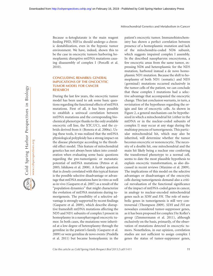
During the last few years, the oncocytic tumormodel has been used to ask some basic ques-tions regarding the functional effects of mtDNAmutations. First of all, it has been possibleto establish a univocal correlation betweenmtDNA mutations and the corresponding bio-chemical phenotype thanks to the only availableoncocytic cell line, the XTC.UC1, and the cy-brids derived from it (Bonora et al. 2006a). Us-ing these tools, it was realized that the mtDNAphysiological polyploidy has a strong impact onthe disease phenotype according to the thresh-old effect model. This feature of mitochondrialgenetics has not always been taken into consid-eration when evaluating some basic questionsregarding the pro-tumorigenic or metastaticpotential of mtDNA mutations (Petros et al.2005; Ishikawa et al. 2008). A further questionthat is closely correlated with this typical featureis the possible selective disadvantage or advan-tage that mtDNA mutations have in vitro as wellas in vivo (Gasparre et al. 2007) as a result of the“population dynamics” that might characterizethe evolution of mtDNA mutations during tu-morigenesis. The possibility of a selective ad-vantage is strongly supported by recent findings(Gasparre et al. 2009), which describe disrup-tive frameshift mtDNA mutations affecting theND5 and ND1 subunits of complex I present inhomoplasmy in a nasopharyngeal oncocytic tu-mor. In both cases, the mutations were inherit-ed at a low degree of heteroplasmy through thegermline in the patient’s family (Gasparre et al.2009) or were germline de novo events (Pradellaet al. 2011) but became homoplasmic in the
patient’s oncocytic tumor. Immunohistochem-istry has shown a perfect correlation betweenpresence of a homoplasmic mutation and lackof the mitochondria-coded ND6 subunit,which suggests impaired complex I assembly.In the described nasopharynx oncocytoma, afew oncocytic areas from the same tumor, ex-pressing ND6 and heteroplasmic for the ND5mutation, harbored instead a de novo homo-plasmic ND1 mutation. Because the shift to ho-moplasmy of both ND1 (somatic) and ND5(germinal) mutations occurred exclusively inthe tumor cells of the patient, we can concludethat these complex I mutations had a selec-tive advantage that accompanied the oncocyticchange. This last conclusion warrants, in turn, arevisitation of the hypotheses regarding the or-igin and fate of oncocytic cells. As shown inFigure 2, a general mechanism can be hypothe-sized in which a mitochondrial hit (either in themtDNA or in the nuclear-coded subunits ofcomplex I) may occur at any stage during themultistep process of tumorigenesis. This partic-ular mitochondrial hit, which may also beinherited, will determine whether the tumorbecomes oncocytic or nononcocytic. The neces-sity of a double hit, one mitochondrial and themain hit likely being a nuclear one conferringthe transformed phenotype to a normal cell,seems to date the most plausible hypothesis toexplain oncocytic transformation, as also dis-cussed in recent reviews (Maximo et al. 2009).The implications of this model on the selectiveadvantages or disadvantages of the oncocyticcells during tumorigenesis demand also a criti-cal reevaluation of the functional significanceof the impact of mtDNA-coded genes in cancer,in analogy to nuclear-encoded mitochondrialgenes such as SDH and FH. The role of meta-bolic genes in tumorigenesis is still very con-troversial (Thompson 2009). SDH and FH arenowadays considered tumor-suppressor genes,as it has been proposed for complex I by Kofler’sgroup (Zimmermann et al. 2011), althoughexclusively on the basis, primarily, of the obser-vation of mutations detected in oncocytic tu-mors. Nonetheless, in our opinion, correlationstudies are not sufficient to assign complex Igenes the status of tumor-suppressor genes,
Mitrochondrial Genetics and Metabolism in Cancer
Cite this article as Cold Spring Harb Perspect Biol 2013;5:a011411 11
on February 16, 2019 - Published by Cold Spring Harbor Laboratory Press http://cshperspectives.cshlp.org/Downloaded from

LDHAVEGF
GLUT1HIF1α
OH
4
5
PHD
O2
Fe2+
α-KG
S-CoA-s
NADH
NADHAcetyl CoA
Succinyl CoASuccinate
Citrate
Isocitrate
α-KG
NAD+
KG-D
IDH
AC
CS
Oncocytic neoplasia/biogenesis
3
(Pre)neoplasia
2
1
Mitochondrial hit
Nuclear hit
Nononcocytic neoplasia
Figure 2. The origin and fate of the oncocytic cell. Nuclear hits (light blue lightning arrow) presiding to cellulartransformation contribute to the initiation stage of neoplastic or pre-neoplastic development. 1. No mtDNAmutations occur, or such mutations are detrimental to cell replication and mutated mitochondria are selectedagainst, preventing the development of an oncocytic phenotype. 2. During cell replication a mutation (redlightning arrow) may occur in mtDNA (mitochondrial hit) either conferring a metabolic advantage by con-tributing to cell adaptation through the Warburg effect or remaining neutral until a mutation load threshold isreached. 3. Positive selection or random drift may set in through mitochondrial biogenesis, generating theoncocytic phenotype.The pink dotted lines lead to an enlarged dynamic view of an “oncocytic” mitochondrionschematically divided into two parts: a white area showing wild-type mtDNA molecules (gray circles) andcorrectly assembled complex I, and a shaded pink area with mutated mtDNA and partially or fully disassembledcomplex I. An mtDNA mutation disassembling complex I, such as those typical of oncocytic tumors, may notallow NADH consumption, shifting the NADH/NADþ ratio toward NADH and leading to an increase of the a-KG/SA ratio. Such Krebs cycle alteration may foster activity of PHD with subsequent HIF1a destabilization/degradation even in low oxygen tension conditions (pseudonormoxia). 4. Inactivation of this transcriptionfactor may functionally lead, on one hand, to the inhibition of expression of pro-tumorigenic and glycolyticgenes (i.e., LDHA, GLUT1, and VEGF) and, on the other hand, to the down-regulation of glycolysis needed tocompensate for the defective mitochondrial respiration (light blue arrow). This scenario does not allow themetabolic adaptation of tumor cells (Warburg effect), hampering the tumorigenic potential of cells, explainingthe low-proliferative and low-aggressive features of oncocytic tumors. 5. Furthermore, HIF1a destabilizationmay not prevent the inhibition of mitochondrial biogenesis that should occur upon hypoxia sensing, hencefeeding a vicious loop ultimately leading to the pathological mitochondrial hyperplasia of oncocytic cells (redarrow). CS, citrate synthase; AC, aconitase; IDH: isocitrate dehydrogenase; a-KG, a-ketoglutarate; KG-D, a-ketoglutarate dehydrogenase; S-CoA-s, succinyl-CoA synthase; PHD, prolyl hydroxylase; HIF1a, hypoxia-in-ducible factor 1a; GLUT1, glucose trasporter 1; VEGF, vascular endothelial growth factor; LDHA, lactatedehydrogenase A.
G. Gasparre et al.
12 Cite this article as Cold Spring Harb Perspect Biol 2013;5:a011411
on February 16, 2019 - Published by Cold Spring Harbor Laboratory Press http://cshperspectives.cshlp.org/Downloaded from

especially because such definition overlooks thefundamental differences between mitochondri-al genetics (whose dynamics are analogous tothose of population genetics) and Mendeliangenetics. The typical homoplasmic complex Imutations found in oncocytic tumors andshown to generate the phenotype in vivo ham-per the tumorigenic potential of cells that har-bor them (Park et al. 2009; Gasparre et al.2011b), accounting for the low-proliferativeand low-aggressive behavior of most oncocytictumors. This effect has been shown by ourgroup to be independent from apoptosis andlikely due to the lack of HIF1a stabilization de-scribed above and pictured in Figure 2. Suchpseudonormoxic status may also explain whyonly a complete lack of complex I may deter-mine the oncocytic phenotype. HIF1a is a piv-otal intracellular oxygen sensor, whose role,when stabilized during hypoxia/anoxia, is en-visioned to block mitochondrial biogenesiswhen O2 is limiting. The release of a HIF1ablock may reinforce the induction of the com-pensatory mitochondrial biogenesis in complexI-lacking cells, finally leading to mitochondrialhyperplasia.
Last, we have defined the threshold muta-tion load needed to induce an anti-tumorigenicphenotype, which suggests that these genes aremore likely to be lethality associated than ca-nonical tumor suppressors. Mutated mtDNAgenes that cause complex I disassembly mayhence be anti-tumorigenic, in close dependenceon their mutant load, which needs to be care-fully measured (Kurelac et al. 2011). Such fea-ture does not generally apply to nuclear genes.We have therefore proposed the denominationof “oncojanus,” reminiscent of the double-fac-eted Roman god Janus, to indicate the thresh-old-dependent double-edged properties of CImtDNA-encoded genes (Gasparre et al. 2011b).The understanding of the different properties ofmutations affecting complex I genes and thedissection of the mechanisms leading to pseu-donormoxia following complex I disassemblyshould provide fundamental insights linkingcancer metabolism to hypoxia adaptation andwill likely lead to the discovery of novel ap-proaches in cancer therapy.
ACKNOWLEDGMENTS
This work is supported by the Italian Ministryof University and Research (MIUR) grant FIRB“Futuro in Ricerca” and by Fondazione Umber-to Veronesi grant “Disco trip” to G.G., by MIURgrant PRIN 2008 to A.M.P., and by the Associa-zione Italiana Ricerca sul Cancro (AIRC) toG.R.
REFERENCES
Acin-Perez R, Bayona-Bafaluy MP, Fernandez-Silva P, Mo-reno-Loshuertos R, Perez-Martos A, Bruno C, Mo-raes CT, Enriquez JA. 2004. Respiratory complex III isrequired to maintain complex I in mammalian mito-chondria. Mol Cell 13: 805–815.
Ambu R, Riva A, Lai ML, Loffredo F, Riva FT, Tandler B.2000. Scanning electron microscopy of the interior ofcells in Hurthle cell tumors. Ultrastruct Pathol 24:211–219.
Baracca A, Chiaradonna F, Sgarbi G, Solaini G, Alber-ghina L, Lenaz G. 2010. Mitochondrial complex I de-crease is responsible for bioenergetic dysfunction inK-ras transformed cells. Biochim Biophys Acta 1797:314–323.
Baris O, Savagner F, Nasser V, Loriod B, Granjeaud S,Guyetant S, Franc B, Rodien P, Rohmer V, Bertucci F, etal. 2004. Transcriptional profiling reveals coordinatedup-regulation of oxidative metabolism genes in thyroidoncocytic tumors. J Clin Endocrinol Metab 89: 994–1005.
Bartoletti-Stella A, Salfi NC, Ceccarelli C, Attimonelli M,Romeo G, Gasparre G. 2011. Mitochondrial DNA muta-tions in oncocytic adnexal lacrimal glands of the con-junctiva. Arch Ophthalmol 129: 664–666.
Bell EL, Klimova T, Chandel NS. 2008. Targeting the mito-chondria for cancer therapy: Regulation of hypoxia-in-ducible factor by mitochondria. Antioxid Redox Signal10: 635–640.
Bianchi C, Fato R, Genova ML, Parenti Castelli G, Lenaz G.2003. Structural and functional organization of complexI in the mitochondrial respiratory chain. Biofactors 18:3–9.
Blakely EL, Mitchell AL, Fisher N, Meunier B, Nijtmans LG,Schaefer AM, Jackson MJ, Turnbull DM, Taylor RW.2005. A mitochondrial cytochrome b mutation causingsevere respiratory chain enzyme deficiency in humansand yeast. FEBS J 272: 3583–3592.
Bonora E, Porcelli AM, Gasparre G, Biondi A, Ghelli A,Carelli V, Baracca A, Tallini G, Martinuzzi A, Lenaz G,et al. 2006a. Defective oxidative phosphorylation in thy-roid oncocytic carcinoma is associated with pathogenicmitochondrial DNA mutations affecting complexes I andIII. Cancer Res 66: 6087–6096.
Bonora E, Evangelisti C, Bonichon F, Tallini G, Romeo G.2006b. Novel germline variants identified in the innermitochondrial membrane transporter TIMM44 andtheir role in predisposition to oncocytic thyroid carcino-mas. Br J Cancer 4: 1529–1536.
Mitrochondrial Genetics and Metabolism in Cancer
Cite this article as Cold Spring Harb Perspect Biol 2013;5:a011411 13
on February 16, 2019 - Published by Cold Spring Harbor Laboratory Press http://cshperspectives.cshlp.org/Downloaded from
Boulahbel H, Duran RV, Gottlieb E. 2009. Prolyl hydroxy-lases as regulators of cell metabolism. Biochem Soc Trans37: 291–294.
Brahimi-Horn MC, Pouyssegur J. 2007. Oxygen, a source oflife and stress. FEBS Lett 581: 3582–3591.
Brandon M, Baldi P, Wallace DC. 2006. Mitochondrial mu-tations in cancer. Oncogene 25: 4647–4662.
Butow RA, Avadhani NG. 2004. Mitochondrial signaling:The retrograde response. Mol Cell 14: 1–15.
Chen Z, Lu W, Garcia-Prieto C, Huang P. 2007. The Warburgeffect and its cancer therapeutic implications. J BioenergBiomembr 39: 267–274.
Costa-Guda J, Tokura T, Roth SI, Arnold A. 2007. Mito-chondrial DNA mutations in oxyphilic and chief cellparathyroid adenomas. BMC Endocr Disord 7: 8.
Cuezva JM, Ortega AD, Willers I, Sanchez-Cenizo L,Aldea M, Sanchez-Arago M. 2009. The tumor suppressorfunction of mitochondria: Translation into the clinics.Biochim Biophys Acta 1792: 1145–1158.
D’Aurelio M, Gajewski CD, Lenaz G, Manfredi G. 2006.Respiratory chain supercomplexes set the threshold forrespiration defects in human mtDNA mutant cybrids.Hum Mol Genet 15: 2157–2169.
DeBerardinis RJ, Lum JJ, Hatzivassiliou G, Thompson CB.2008. The biology of cancer: Metabolic reprogrammingfuels cell growth and proliferation. Cell Metab 7: 11–20.
Diaz F, Fukui H, Garcia S, Moraes CT. 2006. Cytochrome coxidase is required for the assembly/stability of respira-tory complex I in mouse fibroblasts. Mol Cell Biol 26:4872–4881.
DiMauro S, Hirano M, Schon EA. 2006. Mitochondrial med-icine. Informa Healthcare, London.
Finley DJ, Zhu B, Fahey TJ 3rd. 2004. Molecular analysis ofHurthle cell neoplasms by gene profiling. Surgery 136:1160–1168.
Frezza C, Gottlieb E. 2009. Mitochondria in cancer: Not justinnocent bystanders. Semin Cancer Biol 19: 4–11.
Fry M, Green DE. 1980. Cardiolipin requirement by cyto-chrome oxidase and the catalytic role of phospholipid.Biochem Biophys Res Commun 93: 1238–1246.
Fry M, Green DE. 1981. Cardiolipin requirement for elec-tron transfer in complex I and III of the mitochondrialrespiratory chain. J Biol Chem 256: 1874–1880.
Gasparre G, Porcelli AM, Bonora E, Pennisi LF, Toller M,Iommarini L, Ghelli A, Moretti M, Betts CM, Mar-tinelli GN, et al. 2007. Disruptive mitochondrial DNAmutations in complex I subunits are markers of oncocyticphenotype in thyroid tumors. Proc Natl Acad Sci 104:9001–9006.
Gasparre G, Hervouet E, de Laplanche E, Demont J,Pennisi LF, Colombel M, Mege-Lechevallier F, Scoazec JY,Bonora E, Smeets R, et al. 2008. Clonal expansion ofmutated mitochondrial DNA is associated with tumorformation and complex I deficiency in the benign renaloncocytoma. Hum Mol Genet 17: 986–995.
Gasparre G, Iommarini L, Porcelli AM, Lang M, Ferri GG,Kurelac I, Zuntini R, Mariani E, Pennisi LF, Pasquini E, etal. 2009. An inherited mitochondrial DNA disruptivemutation shifts to homoplasmy in oncocytic tumor cells.Hum Mutat 30: 391–396.
Gasparre G, Romeo G, Rugolo M, Porcelli AM. 2011a.Learning from oncocytic tumors: Why choose inefficientmitochondria? Biochim Biophys Acta 1807: 633–642.
Gasparre G, Kurelac I, Capristo M, Iommarini L, Ghelli A,Ceccarelli C, Nicoletti G, Nanni P, De Giovanni C,Scotlandi K, et al. 2011b. A mutation threshold distin-guishes the anti-tumorigenic effects of the mitochondrialgene MTND1, an oncojanus function. Cancer Res 71:6220–6229.
Genova ML, Baracca A, Biondi A, Casalena G, Faccioli M,Falasca AI, Formiggini G, Sgarbi G, Solaini G, Lenaz G.2008. Is supercomplex organization of the respiratorychain required for optimal electron transfer activity? Bio-chim Biophys Acta 1777: 740–746.
Guerra F, Kurelac I, Cormio A, Zuntini R, Amato LB,Ceccarelli C, Santini D, Cormio G, Fracasso F, Selvaggi L,et al. 2011. Placing mitochondrial DNA mutations with-in the progression model of type I endometrial carcino-ma. Hum Mol Genet 20: 2394–2405.
Higgins JP, Shinghal R, Gill H, Reese JH, Terris M, Cohen RJ,Fero M, Pollack JR, van de Rijn M, Brooks JD. 2003. Geneexpression patterns in renal cell carcinoma assessed bycomplementary DNA microarray. Am J Pathol 162: 925–932.
Holleran AL, Briscoe DA, Fiskum G, Kelleher JK. 1995. Glu-tamine metabolism in AS-30D hepatoma cells. Evidencefor its conversion into lipids via reductive carboxylation.Mol Cell Biochem 152: 95–101.
Ibsen KH. 1961. The Crabtree effect: A review. Cancer Res21: 829–841.
Ishikawa K, Takenaga K, Akimoto M, Koshikawa N,Yamaguchi A, Imanishi H, Nakada K, Honma Y,Hayashi J. 2008. ROS-generating mitochondrial DNAmutations can regulate tumor cell metastasis. Science320: 661–664.
Jacques C, Baris O, Prunier-Mirebeau D, Savagner F,Rodien P, Rohmer V, Franc B, Guyetant S, Malthiery Y,Reynier P. 2005. Two-step differential expression analysisreveals a new set of genes involved in thyroid oncocytictumors. J Clin Endocrinol Metab 90: 2314–2320.
Jahnke VE, Sabido O, Defour A, Castells J, Lefai E,Roussel D, Freyssenet D. 2010. Evidence for mitochon-drial respiratory deficiency in rat rhabdomyosarcomacells. PLoS ONE 5: e8637.
Kaluz S, Kaluzova M, Stanbridge EJ. 2008. Rational design ofminimal hypoxia-inducible enhancers. Biochem BiophysRes Commun 370: 613–618.
Kang SY, Gutowsky HS, Hsung JC, Jacobs R, King TE,Rice D, Oldfield E. 1979. Nuclear magnetic resonanceinvestigation of the cytochrome oxidase–phospholipidinteraction: A new model for boundary lipid. Biochemis-try 18: 3257–3267.
Kim JW, Tchernyshyov I, Semenza GL, Dang CV. 2006. HIF-1-mediated expression of pyruvate dehydrogenase ki-nase: A metabolic switch required for cellular adaptationto hypoxia. Cell Metab 3: 177–185.
King A, Selak MA, Gottlieb E. 2006. Succinate dehydroge-nase and fumarate hydratase: Linking mitochondrial dys-function and cancer. Oncogene 25: 4675–4682.
Klaunig JE, Kamendulis LM. 2004. The role of oxidativestress in carcinogenesis. Annu Rev Pharmacol Toxicol44: 239–267.
G. Gasparre et al.
14 Cite this article as Cold Spring Harb Perspect Biol 2013;5:a011411
on February 16, 2019 - Published by Cold Spring Harbor Laboratory Press http://cshperspectives.cshlp.org/Downloaded from

Klevecz RR, Bolen J, Forrest G, Murray DB. 2004. A genome-wide oscillation in transcription gates DNA replicationand cell cycle. Proc Natl Acad Sci 101: 1200–1205.
Koopman WJ, Verkaart S, Visch HJ, van Emst-de Vries S,Nijtmans LG, Smeitink JA, Willems PH. 2007. HumanNADH:ubiquinone oxidoreductase deficiency: Radicalchanges in mitochondrial morphology? Am J PhysiolCell Physiol 293: C22–C29.
Koppenol WH, Bounds PL, Dang CV. 2011. Otto Warburg’scontributions to current concepts of cancer metabolism.Nat Rev Cancer 11: 325–337.
Kuhajda FP, Jenner K, Wood FD, Hennigar RA, Jacobs LB,Dick JD, Pasternack GR. 1994. Fatty acid synthesis: Apotential selective target for antineoplastic therapy. ProcNatl Acad Sci 91: 6379–6383.
Kurelac I, Lang M, Zuntini R, Calabrese C, Simone D,Vicario S, Santamaria M, Attimonelli M, Romeo G,Gasparre G. 2011. Searching for a needle in the haystack:Comparing six methods to evaluate heteroplasmy in dif-ficult sequencecontext. Biotechnol Adv 30: 363–371.
Lange C, Nett JH, Trumpower BL, Hunte C. 2001. Specificroles of protein–phospholipid interactions in the yeastcytochrome bc1 complex structure. EMBO J 20: 6591–6600.
Lee AG. 2004. How lipids affect the activities of integralmembrane proteins. Biochim Biophys Acta 1666: 62–87.
Lenaz G, Genova ML. 2007. Kinetics of integrated electrontransfer in the mitochondrial respiratory chain: Randomcollisions vs. solid state electron channeling. Am J PhysiolCell Physiol 292: C1221–C1239.
Lenaz G, Genova ML. 2010. Structure and organization ofmitochondrial respiratory complexes: A new under-standing of an old subject. Antioxid Redox Signal 12:961–1008.
Lenaz G, Strocchi P. 2009. Reactive oxygen species in theinduction of toxicity. In General and applied toxicologyreview (ed. Ballantyne B, et al.), Vol. 2, pp. 367–410.Wiley, Chichester, UK.
Lenaz G, Baracca A, Fato R, Genova ML, Solaini G. 2006.New insights into structure and function of mitochon-dria and their role in aging and disease. Antioxid RedoxSignal 8: 417–437.
Lu H, Dalgard CL, Mohyeldin A, McFate T, Tait AS,Verma A. 2005. Reversible inactivation of HIF-1 prolylhydroxylases allows cell metabolism to control basal HIF-1. J Biol Chem 280: 41928–41939.
Ma Y, Bai RK, Trieu R, Wong LJ. 2010. Mitochondrial dys-function in human breast cancer cells and their trans-mitochondrial cybrids. Biochim Biophys Acta 1797:29–37.
Mathupala SP, Ko YH, Pedersen PL. 2010. The pivotal rolesof mitochondria in cancer: Warburg and beyond andencouraging prospects for effective therapies. BiochimBiophys Acta 1797: 1225–1230.
Maximo V, Sobrinho-Simoes M. 2000. Mitochondrial DNA“common” deletion in Hurthle cell lesions of the thyroid.J Pathol 192: 561–562.
Maximo V, Sores P, Rocha AS, Sobrinho-Simoes M. 1998.The common deletion of mitochondrial DNA is found ingoiters and thyroid tumors with and without oxyphil cellchange. Ultrastruct Pathol 22: 271–273.
Maximo V, Soares P, Lima J, Cameselle-Teijeiro J, Sobrinho-Simoes M. 2002. Mitochondrial DNA somatic mutations(point mutations and large deletions) and mitochondrialDNAvariants in human thyroid pathology: A study withemphasis on Hurthle cell tumors. Am J Pathol 160:1857–1865.
Maximo V, Botelho T, Capela J, Soares P, Lima J, Taveira A,Amaro T, Barbosa AP, Preto A, Harach HR, et al. 2005.Somatic and germline mutation in GRIM-19, a dualfunction gene involved in mitochondrial metabolismand cell death, is linked to mitochondrion-rich (Hurthlecell) tumours of the thyroid. Br J Cancer 23: 1892–1898.
Maximo V, Lima J, Soares P, Sobrinho-Simoes M. 2009.Mitochondria and cancer. Virchows Arch 454: 481–495.
Mayevsky A. 2009. Mitochondrial function and energy me-tabolism in cancer cells: Past overview and future per-spectives. Mitochondrion 9: 165–179.
Mayr JA, Meierhofer D, Zimmermann F, Feichtinger R,Kogler C, Ratschek M, Schmeller N, Sperl W, Kofler B.2008. Loss of complex I due to mitochondrial DNA mu-tations in renal oncocytoma. Clin Cancer Res 14: 2270–2275.
McFate T, Mohyeldin A, Lu H, Thakar J, Henriques J,Halim ND, Wu H, Schell MJ, Tsang TM, Teahan O, etal. 2008. Pyruvate dehydrogenase complex activity con-trols metabolic and malignant phenotype in cancer cells.J Biol Chem 283: 22700–22708.
McKenzie M, Lazarou M, Thorburn DR, Ryan MT. 2006.Mitochondrial respiratory chain supercomplexes are de-stabilized in Barth syndrome patients. J Mol Biol 361:462–469.
Moran M, Rivera H, Sanchez-Arago M, Blazquez A,Merinero B, Ugalde C, Arenas J, Cuezva JM, Martin MA.2010. Mitochondrial bioenergetics and dynamics inter-play in complex I–deficient fibroblasts. Biochim BiophysActa 1802: 443–453.
Moreno-Sanchez R, Rodriguez-Enriquez S, Marin-Her-nandez A, Saavedra E. 2007. Energy metabolism in tumorcells. FEBS J 274: 1393–1418.
Morris LG, Veeriah S, Chan TA. 2010. Genetic determinantsat the interface of cancer and neurodegenerative disease.Oncogene 29: 3453–3464.
Muller-Hocker J. 1992. Random cytochrome-c-oxidase de-ficiency of oxyphil cell nodules in the parathyroid gland.A mitochondrial cytopathy related to cell ageing? PatholRes Pract 188: 701–706.
Muller-Hocker J, Jacob U, Seibel P. 1998a. Hashimoto thy-roiditis is associated with defects of cytochrome-c oxi-dase in oxyphil Askanazy cells and with the commondeletion (4,977) of mitochondrial DNA. UltrastructPathol 22: 91–100.
Muller-Hocker J, Schafer S, Copeland WC, Wiesner R,Seibel P. 1998b. Immunohistochemical detection of hu-man mtDNA polymerase g and of human mitochondrialtranscription factor A in cytochrome-c-oxidase-deficientoxyphil cells of hyperfunctional parathyroids. VirchowsArch 433: 529–536.
Neuwald AF. 1997. Barth syndrome may be due to an acyl-transferase deficiency. Curr Biol 7: R465–R466.
Ozawa T. 1997. Genetic and functional changes in mito-chondria associated with aging. Physiol Rev 77: 425–464.
Mitrochondrial Genetics and Metabolism in Cancer
Cite this article as Cold Spring Harb Perspect Biol 2013;5:a011411 15
on February 16, 2019 - Published by Cold Spring Harbor Laboratory Press http://cshperspectives.cshlp.org/Downloaded from
Paradies G, Petrosillo G, Pistolese M, Ruggiero FM. 2000.The effect of reactive oxygen species generated from themitochondrial electron transport chain on the cyto-chrome c oxidase activity and on the cardiolipin contentin bovine heart submitochondrial particles. FEBS Lett466: 323–326.
Paradies G, Petrosillo G, Pistolese M, Ruggiero FM. 2002.Reactive oxygen species affect mitochondrial electrontransport complex I activity through oxidative cardioli-pin damage. Gene 286: 135–141.
Paradies G, Petrosillo G, Paradies V, Ruggiero FM. 2010.Oxidative stress, mitochondrial bioenergetics, and cardi-olipin in aging. Free Radic Biol Med 48: 1286–1295.
Park JS, Sharma LK, Li H, Xiang R, Holstein D, Wu J,Lechleiter J, Naylor SL, Deng JJ, Lu J, et al. 2009. A het-eroplasmic, not homoplasmic, mitochondrial DNA mu-tation promotes tumorigenesis via alteration in reactiveoxygen species generation and apoptosis. Hum Mol Genet18: 1578–1589.
Parlo RA, Coleman PS. 1986. Continuous pyruvate carbonflux to newly synthesized cholesterol and the suppressedevolution of pyruvate-generated CO2 in tumors: Furtherevidence for a persistent truncated Krebs cycle in hepa-tomas. Biochim Biophys Acta 886: 169–176.
Pasini B, Stratakis CA. 2009. SDH mutations in tumorigen-esis and inherited endocrine tumours: Lesson from thephaeochromocytoma-paraganglioma syndromes. J In-tern Med 266: 19–42.
Patten DA, Lafleur VN, Robitaille GA, Chan DA, Giaccia AJ,Richard DE. 2010. Hypoxia-inducible factor-1 activationin nonhypoxic conditions: The essential role of mito-chondrial-derived reactive oxygen species. Mol Biol Cell21: 3247–3257.
Petros JA, Baumann AK, Ruiz-Pesini E, Amin MB, Sun CQ,Hall J, Lim S, Issa MM, Flanders WD, Hosseini SH, et al.2005. mtDNA mutations increase tumorigenicity inprostate cancer. Proc Natl Acad Sci 102: 719–724.
Petrosillo G, Ruggiero FM, Di Venosa N, Paradies G. 2003.Decreased complex III activity in mitochondria isolatedfrom rat heart subjected to ischemia and reperfusion:Role of reactive oxygen species and cardiolipin. FASEB J17: 714–716.
Petrosillo G, Matera M, Moro N, Ruggiero FM, Paradies G.2009. Mitochondrial complex I dysfunction in rat heartwith aging: Critical role of reactive oxygen species andcardiolipin. Free Radic Biol Med 46: 88–94.
Pfeiffer K, Gohil V, Stuart RA, Hunte C, Brandt U,Greenberg ML, Schagger H. 2003. Cardiolipin stabilizesrespiratory chain supercomplexes. J Biol Chem 278:52873–52880.
Pollard PJ, Briere JJ, Alam NA, Barwell J, Barclay E,Wortham NC, Hunt T, Olpin S, Moat SJ, Hargreaves IP,et al. 2005. Accumulation of Krebs cycle intermediatesand over-expression of HIF1a in tumours which resultfrom germline FH and SDH mutations. Hum Mol Genet14: 2231–2239.
Porcelli AM, Angelin A, Ghelli A, Mariani E, Martinuzzi A,Carelli V, Petronilli V, Bernardi P, Rugolo M. 2009. Respi-ratory complex I dysfunction due to mitochondrial DNAmutations shifts the voltage threshold for opening of thepermeability transition pore toward resting levels. J BiolChem 284: 2045–2052.
Porcelli A, Ghelli A, Ceccarelli C, Lang M, Cenacchi G,Capristo M, Pennisi LF, Ciccarelli E, Melcarne A, Barto-letti-Stella A, et al. 2010. The genetic and metabolic sig-nature of oncocytic transformation implicates HIF1adestabilization. Hum Mol Genet 19: 1019–1032.
Pradella LM, Zuntini R, Magini P, Ceccarelli C, Neri I,Cerasoli S, Graziano C, Gasparre G, Turchetti D. 2011.Two distinct thyroid tumours in a patient with Cowdensyndrome carrying both a 10q23 and a mitochondrialDNA germline deletion. J Med Genet 48: 779–782.
Putignani L, Raffa S, Pescosolido R, Aimati L, Signore F,Torrisi MR, Grammatico P. 2008. Alteration of expres-sion levels of the oxidative phosphorylation system(OXPHOS) in breast cancer cell mitochondria. BreastCancer Res Treat 110: 439–452.
Radermacher M, Ruiz T, Clason T, Benjamin S, Brandt U,Zickermann V. 2006. The three-dimensional structure ofcomplex I from Yarrowia lipolytica: A highly dynamicenzyme. J Struct Biol 154: 269–279.
Riva A, Tandler B. 2000. Three-dimensional structure ofoncocyte mitochondria in human salivary glands: Ascanning electron microscope study. Ultrastruct Pathol24: 145–150.
Robinson NC, Strey F, Talbert L. 1980. Investigation of theessential boundary layer phospholipids of cytochrome coxidase using Triton X-100 delipidation. Biochemistry 19:3656–3661.
Rosca MG, Vazquez EJ, Kerner J, Parland W, Chandler MP,Stanley W, Sabbah HN, Hoppel CL. 2008. Cardiac mito-chondria in heart failure: Decrease in respirasomes andoxidative phosphorylation. Cardiovasc Res 80: 30–39.
Savagner F, Chevrollier A, Loiseau D, Morgan C, Reynier P,Clark O, Stepien G, Malthiery Y. 2001a. Mitochondrialactivity in XTC.UC1 cells derived from thyroid oncocy-toma. Thyroid 11: 327–333.
Savagner F, Franc B, Guyetant S, Rodien P, Reynier P,Malthiery Y. 2001b. Defective mitochondrial ATP synthe-sis in oxyphilic thyroid tumors. J Clin Endocrinol Metab86: 4920–4925.
Savagner F, Mirebeau D, Jacques C, Guyetant S, Morgan C,Franc B, Reynier P, Malthiery Y. 2003. PGC-1-related co-activator and targets are upregulated in thyroid oncocy-toma. Biochem Biophys Res Commun 310: 779–784.
Schafer E, Dencher NA, Vonck J, Parcej DN. 2007. Three-dimensional structure of the respiratory chain supercom-plex I1III2IV1 from bovine heart mitochondria. Bio-chemistry 46: 12579–12585.
Schagger H, de Coo R, Bauer MF, Hofmann S, Godinot C,Brandt U. 2004. Significance of respirasomes for the as-sembly/stability of human respiratory chain complex I. JBiol Chem 279: 36349–36353.
Sedlak E, Robinson NC. 1999. Phospholipase A(2) digestionof cardiolipin bound to bovine cytochrome c oxidasealters both activity and quaternary structure. Biochemis-try 38: 14966–14972.
Semenza GL. 2003. Targeting HIF-1 for cancer therapy. NatRev Cancer 3: 721–732.
Semenza GL. 2007. Oxygen-dependent regulation of mito-chondrial respiration by hypoxia-inducible factor 1. Bio-chem J 405: 1–9.
G. Gasparre et al.
16 Cite this article as Cold Spring Harb Perspect Biol 2013;5:a011411
on February 16, 2019 - Published by Cold Spring Harbor Laboratory Press http://cshperspectives.cshlp.org/Downloaded from

Shidara Y, Yamagata K, Kanamori T, Nakano K, Kwong JQ,Manfredi G, Oda H, Ohta S. 2005. Positive contributionof pathogenic mutations in the mitochondrial genome tothe promotion of cancer by prevention from apoptosis.Cancer Res 65: 1655–1663.
Simonnet H, Demont J, Pfeiffer K, Guenaneche L,Bouvier R, Brandt U, Schagger H, Godinot C. 2003. Mi-tochondrial complex I is deficient in renal oncocytomas.Carcinogenesis 24: 1461–1466.
Smolkova K, Plecita-Hlavata L, Bellance N, Benard G,Rossignol R, Jezek P. 2010. Waves of gene regulation sup-press and then restore oxidative phosphorylation in can-cer cells. Int J Biochem Cell Biol 43: 950–968.
Sun W, Zhou S, Chang SS, McFate T, Verma A, Califano JA.2009. Mitochondrial mutations contribute to HIF1a ac-cumulation via increased reactive oxygen species and up-regulated pyruvate dehydrogenease kinase 2 in head andneck squamous cell carcinoma. Clin Cancer Res 15:476–484.
Tallini G. 1998. Oncocytic tumours. Virchows Arch 433:5–12.
Tallini G, Ladanyi M, Rosai J, Jhanwar SC. 1994. Analysis ofnuclear and mitochondrial DNA alterations in thyroidand renal oncocytic tumors. Cytogenet Cell Genet 66:253–259.
Thompson CB. 2009. Metabolic enzymes as oncogenes ortumor suppressors. N Engl J Med 360: 813–815.
Tuppen HA, Blakely EL, Worgan L, Al-Dosary M,Saretzki G, Alston CL, Morris AA, Clarke M, Jones S,Devlin AM, et al. 2010. The p.M292T NDUFS2 mutationcauses complex I–deficient Leigh syndrome in multiplefamilies. Brain 133: 2952–2963.
van Raam BJ, Sluiter W, de Wit E, Roos D, Verhoeven AJ,Kuijpers TW. 2008. Mitochondrial membrane potentialin human neutrophils is maintained by complex III ac-
tivity in the absence of supercomplex organisation. PLoSONE 3: e2013.
Vik SB, Capaldi RA. 1977. Lipid requirements for cyto-chrome c oxidase activity. Biochemistry 16: 5755–5759.
Wallace DC. 2005. A mitochondrial paradigm of metabolicand degenerative diseases, aging, and cancer: A dawn forevolutionary medicine. Annu Rev Genet 39: 359–407.
Warburg O. 1956. On the origin of cancer cells. Science 123:309–314.
Weinberg F, Chandel NS. 2009. Mitochondrial metabolismand cancer. Ann NY Acad Sci 1177: 66–73.
Yoo H, Antoniewicz MR, Stephanopoulos G, Kelleher JK.2008. Quantifying reductive carboxylation flux of gluta-mine to lipid in a brown adipocyte cell line. J Biol Chem283: 20621–20627.
Zhang M, Mileykovskaya E, Dowhan W. 2002. Gluing therespiratory chain together. Cardiolipin is required forsupercomplex formation in the inner mitochondrialmembrane. J Biol Chem 277: 43553–43556.
Zielke A, Tezelman S, Jossart GH, Wong M, Siperstein AE,Duh QY, Clark OH. 1998. Establishment of a highly dif-ferentiated thyroid cancer cell line of Hurthle cell origin.Thyroid 8: 475–483.
Zimmermann FA, Mayr JA, Neureiter D, Feichtinger R,Alinger B, Jones ND, Eder W, Sperl W, Kofler B. 2009.Lack of complex I is associated with oncocytic thyroidtumours. Br J Cancer 100: 1434–1437.
Zimmermann FA, Mayr JA, Feichtinger R, Neureiter D,Lechner R, Koegler C, Ratschek M, Rusmir H,Sargsyan K, Sperl W, et al. 2011. Respiratory chain com-plex I is a mitochondrial tumor suppressor of oncocytictumors. Front Biosci 1: 315–325.
Zu XL, Guppy M. 2004. Cancer metabolism: Facts, fantasy,and fiction. Biochem Biophys Res Commun 313: 459–465.
Mitrochondrial Genetics and Metabolism in Cancer
Cite this article as Cold Spring Harb Perspect Biol 2013;5:a011411 17
on February 16, 2019 - Published by Cold Spring Harbor Laboratory Press http://cshperspectives.cshlp.org/Downloaded from

2013; doi: 10.1101/cshperspect.a011411Cold Spring Harb Perspect Biol Giuseppe Gasparre, Anna Maria Porcelli, Giorgio Lenaz and Giovanni Romeo DevelopmentRelevance of Mitochondrial Genetics and Metabolism in Cancer
Subject Collection Mitochondria
EncephalopathyS) Metabolism in Ethylmalonic2Altered Sulfide (H
Valeria Tiranti and Massimo ZevianiMitochondrial Membrane during Apoptosis
Permeabilization of the Outer−−Where Killers Meet
Tom Bender and Jean-Claude Martinou
DiseaseHeteroplasmy Conundrum in Evolution and Mitochondrial DNA Genetics and the
Douglas C. Wallace and Dimitra Chalkia
Nuclear Signaling ProteinsMitochondrial Biogenesis through Activation of
John E. Dominy and Pere Puigserver
Processes, and DiseasesProtein Biogenesis: Mechanisms, Connected
Sulfur−The Role of Mitochondria in Cellular Iron
Oliver Stehling and Roland Lill
Mitochondrial Trafficking in NeuronsThomas L. Schwarz
Mechanisms of Mitochondrial Fission and Fusion
Sumihiro KawajiriAlexander M. van der Bliek, Qinfang Shen and
Muscular DystrophiesAutophagy in the Pathogenesis of Collagen VI Mitochondrial Dysfunction and Defective
Paolo Bernardi and Paolo Bonaldo
Mitochondrial DNA into Cellular HomeostasisThe Mitochondrial Nucleoid: Integrating
al.Robert Gilkerson, Liliana Bravo, Iraselia Garcia, et
Mitochondrial Disease-RelatedPOLGClinical and Molecular Features of
CopelandJeffrey D. Stumpf, Russell P. Saneto and William C.
Metabolism in Cancer DevelopmentRelevance of Mitochondrial Genetics and
Lenaz, et al.Giuseppe Gasparre, Anna Maria Porcelli, Giorgio
Mitochondrial Metabolism, Sirtuins, and AgingMichael N. Sack and Toren Finkel
and Parkin: Links to ParkinsonismMitochondrial Quality Control Mediated by PINK1
Derek Narendra, John E. Walker and Richard Youle
Mechanisms of Protein Sorting in Mitochondria
Pfanner, et al.Diana Stojanovski, Maria Bohnert, Nikolaus
http://cshperspectives.cshlp.org/cgi/collection/ For additional articles in this collection, see
Copyright © 2013 Cold Spring Harbor Laboratory Press; all rights reserved
on February 16, 2019 - Published by Cold Spring Harbor Laboratory Press http://cshperspectives.cshlp.org/Downloaded from
Mitochondrial EvolutionMichael W. Gray
http://cshperspectives.cshlp.org/cgi/collection/ For additional articles in this collection, see
Copyright © 2013 Cold Spring Harbor Laboratory Press; all rights reserved
on February 16, 2019 - Published by Cold Spring Harbor Laboratory Press http://cshperspectives.cshlp.org/Downloaded from