remodeling of membrane properties and dendritic architecture accompanies the postembryonic
TRANSCRIPT

Remodeling of Membrane Properties and Dendritic ArchitectureAccompanies the Postembryonic Conversion of a Slow into aFast Motoneuron
Carsten Duch and R. B. Levine
Division of Neurobiology, University of Arizona, Tucson, Arizona 85721
The postembryonic acquisition of behavior requires alterations inneuronal circuitry, which ultimately must be understood as spe-cific changes in neuronal structure, membrane properties, andsynaptic connectivity. This study addresses this goal by describ-ing the postembryonic remodeling of the excitability and den-dritic morphology of an identified motoneuron, MN5, which dur-ing the metamorphosis of Manduca sexta (L.) changes from aslow motoneuron that is involved in larval-crawling behavior intoa fast adult flight motoneuron. A fivefold lower input resistance, ahigher firing threshold, and an increase in voltage-activated K1
current contribute to a lower excitability of the adult MN5, whichis a prerequisite for its newly acquired behavioral role. In addition,the adult MN5 displays larger Ca21 currents. The dendrites ofMN5 undergo extensive remodeling. Drastic regression of larvaldendrites during early pupal stages is followed by rapid growth of
new dendrites. Critical changes in excitability take place duringthe onset of adult dendrite formation. Larval Ca21 currents areabsent when dendritic remodeling is most dramatic but increasemarkedly during later development. Changes in Ca21 and K1
currents follow different time courses, allowing the transient oc-currence of Ca21 spikes during pupal stages when new dendriticbranching ceases. The adult MN5 can produce prolonged Ca21
spikes after K1 currents are reduced. We suggest that alterationsin Ca21 and K1 currents are necessary for the participation ofMN5 in flight behavior and that the transient production of Ca21
spikes may influence postembryonic dendritic remodeling.
Key words: postembryonic development; calcium current; po-tassium current; neuronal differentiation; insect; Manduca sexta;CNS; flight behavior
Changes in the excitability and dendritic architecture of centralneurons accompany the development and plasticity of behavior(McCobb et al., 1989, 1990; Baines and Bate, 1998; Gao andZiskind-Conhaim, 1998; Reynolds et al., 1998; Sun and Dale, 1998;Martin-Caraballo and Greer, 1999). As neuronal structure, bio-physical properties, and synaptic interactions differentiate or aremodified postembryonically, the activity patterns of the mature CNSemerge (Casanovas and Meyrand, 1995). These emergent activitypatterns, in turn, influence neuronal differentiation (Spitzer, 1991;Ribiera and Spitzer, 1992; Muller et al., 1998). Activity-dependentCa21 influx controls neuronal growth (Cohan et al., 1987; Mattsonet al., 1988; Fields et al., 1990; Haydon and Zoran, 1994), andtransient patterns of electrical activity regulate synaptic input (Shatz,1990). A satisfactory understanding of these interdependent pro-cesses requires a detailed description of the temporal relationshipbetween the structural and functional remodeling of identified neu-rons in a behavioral context.
Although behavior changes postembryonically in all organisms,the particularly extreme example of metamorphosis offers a uniquemodel system for such studies. In holometabolous insects the par-ticipation of central neurons in larval behavior ceases as novelbehavior is acquired during postembryonic life (Weeks and Le-vine, 1990). For example, in Manduca sexta (L.), five motoneuronsthat innervate slowly contracting muscles of the larval dorsal bodywall persist throughout metamorphosis to innervate the new, fast-contracting, dorsal longitudinal flight muscle (DLM) of the adult(Casaday and Camhi, 1976; Rheuben and Kammer, 1980; Duch etal., 2000). Identified flight motoneurons, such as MN5, mediateslow tonic contractions of larval target muscles during crawling but
fast twitch contractions of newly generated adult target musclesduring flight.
Many motoneurons that survive metamorphosis to participate inadult-specific behavior undergo dendritic remodeling (Levine andTruman, 1982, 1985; Truman and Reiss, 1988; Weeks and Ernst-Utzschneider, 1989; Kent and Levine, 1993). Little is known,however, about electrophysiological changes that may accompanystructural remodeling and the development of new behavior. Theisolated somata of cultured Manduca leg motoneurons displaydifferences in ionic currents among different developmental stages(Hayashi and Levine, 1992), some of which are mediated by thesteroid hormone 20-hydroxyecdysone (Grunewald and Levine,1998). The functional consequences of such changes have yet to bedetermined, however, and it is not clear how accurately the char-acteristics of isolated motoneuron cell bodies reflect the propertiesof cells undergoing the normal remodeling of dendrites and syn-aptic interactions in vivo.
This study describes both the dendritic remodeling of MN5 andchanges in its membrane properties that are appropriate for thepostembryonic conversion of a slow into a fast motoneuron. More-over, specific changes in excitability and voltage-dependent ioniccurrents are correlated temporally with critical phases of dendriticremodeling.
MATERIALS AND METHODSAnimals. Manduca sexta obtained from a laboratory culture were reared onan artificial diet (Bell and Joachim, 1976) under a long-day photoperiodregimen (17:7 hr light /dark cycle) at 26°C and ;60% humidity. Bothchronological and morphological criteria were used for the staging ofanimals (Nijhout and Williams, 1974; Bell and Joachim, 1976; Tolbert etal., 1983; Consoulas et al., 1996). In summary, L5 represents the fifth larvalinstar, W0 signifies the first day of wandering, and W1 to W4 represent theremaining 4 d of wandering. P0 indicates the day of the pupal molt, and P1to P18 are the following 18 d of adult development.
Dissection for intracellular recordings. The animals were anesthetized bychilling on ice for 15 min. Animals were dissected along the dorsal midlineand superfused with saline. The thoracic and the first two abdominalganglia were removed, transferred into a Sylgard-coated Petri dish, andpinned down at the cut ends of their lateral nerves in saline. The ganglionicsheath was removed mechanically with a fine pair of forceps. Nerve 1 of the
Received April 19, 2000; accepted June 28, 2000.We gratefully acknowledge the support by National Institutes of Health Grant NS
28495 and by the Deutsche Forschungsgemeinschaft Fellowship DU 331/2-1 (C.D.).We thank Dr. C. Consoulas for many helpful comments on this manuscript.
Correspondence should be addressed to Dr. Carsten Duch, University of Arizona,Division of Neurobiology, 611 Gould-Simpson Building, Tucson, AZ 85721. E-mail:[email protected] © 2000 Society for Neuroscience 0270-6474/00/206950-12$15.00/0
The Journal of Neuroscience, September 15, 2000, 20(18):6950–6961
mesothoracic ganglion was left intact toward its specific peripheral branchthat contains only the axons of the five DLM motoneurons (Duch et al.,2000). MN5 is the only motoneuron in the mesothoracic ganglion thatcarries an axon in this particular nerve branch (Duch et al., 2000). Anti-dromic stimulation from this nerve branch was used to identify MN5during intracellular recordings.
Intracellular staining techniques. Rhodamine Dextran 3000 (MolecularProbes, Eugene, OR) was used for all intracellular stainings. The tips ofthin-walled borosilicate electrodes (resistances, 20–25 MV) were filledwith 5% dextran in 1 M potassium acetate. The electrode shafts were filledwith 1 M potassium acetate, and an air bubble was left between the tip andthe shaft to prevent dye dilution. After intracellular penetration andantidromic identification of MN5, dye was injected iontophoretically byapplying depolarizing current pulses of 3–4 nA amplitude and 200 msecduration with a frequency of 2.5 Hz for 30–60 min. After dye injection,ganglia were left in saline for an additional 30 min to allow dye diffusion.Subsequently, ganglia were fixed in 4% paraformaldehyde and washedthree times in 10 mM PBS, pH 7.4, for 15 min each. Then preparations were
dehydrated in ethanol, cleared in methyl salicylate, and mounted in Per-mount (Fisher Scientific, Fair Lawn, NJ).
Confocal microscopy. Digital images were captured on a Nikon PCM2000 laser-scanning confocal microscope using Simple PCI (Compix, Tu-alatin, OR) image acquisition software. Images were further processedusing Corel Draw 8 software (Corel, Ottawa, Ontario, Canada). Prepara-tions were scanned with a helium–neon laser line with an excitationmaximum at 546 nm using a long-pass filter at 565 nm. All preparationswere scanned with a 403 lens at optical planes of 1 mm. All images shownare the projections of all optical planes of a given stack into one focalplane, to show the entire dendritic field in one two-dimensional image.
Electrophysiology. An Axoclamp 2B amplifier (Axon Instruments, FosterCity, CA) was used for all recordings. Synaptic potentials and spike shapeswere recorded in bridge mode with the same electrode shapes used forintracellular dye injections (see above), but electrode tips were coated withsilicon oil. Input resistance was determined in discontinuous current-clampmode (DCC) to avoid the problem of an inadequate bridge balance, whichcan occur because of changes in electrode resistance while penetrating the
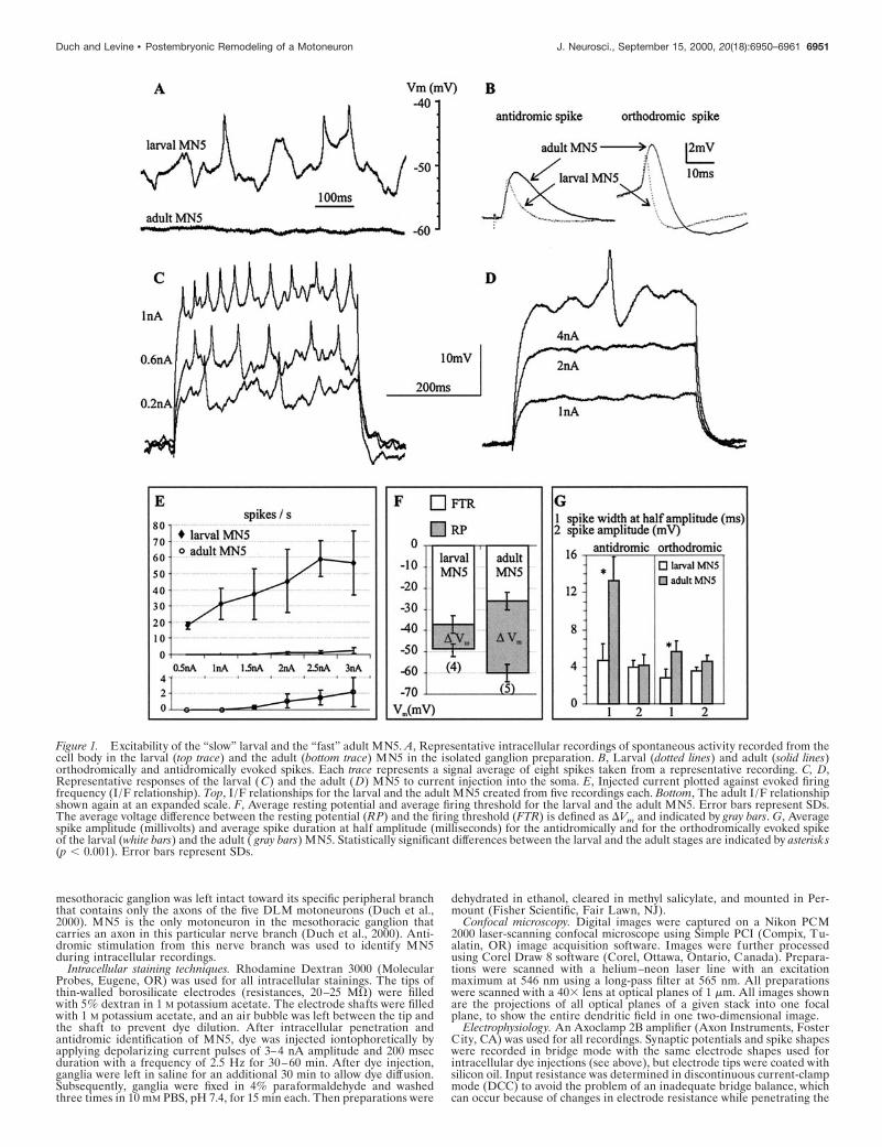
Figure 1. Excitability of the “slow” larval and the “fast” adult MN5. A, Representative intracellular recordings of spontaneous activity recorded from thecell body in the larval (top trace) and the adult (bottom trace) MN5 in the isolated ganglion preparation. B, Larval (dotted lines) and adult (solid lines)orthodromically and antidromically evoked spikes. Each trace represents a signal average of eight spikes taken from a representative recording. C, D,Representative responses of the larval (C) and the adult (D) MN5 to current injection into the soma. E, Injected current plotted against evoked firingfrequency (I/F relationship). Top, I /F relationships for the larval and the adult MN5 created from five recordings each. Bottom, The adult I /F relationshipshown again at an expanded scale. F, Average resting potential and average firing threshold for the larval and the adult MN5. Error bars represent SDs.The average voltage difference between the resting potential (RP) and the firing threshold (FTR) is defined as DVm and indicated by gray bars. G, Averagespike amplitude (millivolts) and average spike duration at half amplitude (milliseconds) for the antidromically and for the orthodromically evoked spikeof the larval (white bars) and the adult ( gray bars) MN5. Statistically significant differences between the larval and the adult stages are indicated by asterisks(p , 0.001). Error bars represent SDs.
Duch and Levine • Postembryonic Remodeling of a Motoneuron J. Neurosci., September 15, 2000, 20(18):6950–6961 6951

Figure 2. A, Schematic drawing of the arrangement of the five motoneurons that innervate the mesothoracic DLM. MN5 is the only DLM motoneuronthat is located in the mesothoracic ganglion and that can, therefore, be individually identified by antidromic stimulation throughout all developmentalstages. B, Confocal images of the adult and the larval MN5. Inset, High-order branches of the adult MN5 depicted in a selective enlargement (regionenclosed in the dotted line in the adult image; see asterisk).
Figure 3. The larval dendrites of MN5 are retracted during late larval and earlypupal life. Confocal images of the dendritic region of MN5 taken from repre-sentative preparations of sequential stages between the fifth larval instar and thethird day of pupal life (P2). All images are projections of all optical planes ofeach preparation (see Materials and Methods). The cell body is located to theright, and the axon is leaving the field of view to the lef t.
6952 J. Neurosci., September 15, 2000, 20(18):6950–6961 Duch and Levine • Postembryonic Remodeling of a Motoneuron

ganglionic tissue. Electrodes with resistances between 20 and 25 MV werefilled either with 1 M potassium acetate or with 5% rhodamine dextran in1 M potassium acetate. No differences between these methods were ob-served. Sampling rates between 3.5 and 5 kHz were reached without anysignal cutoff. The slope input resistance was calculated from the linearportion of the voltage/current relationship using 0.2 nA current stepsbetween 22 and 10.2 nA. The firing threshold was measured as theminimum depolarization from the resting potential necessary to trigger anaction potential.
Voltage-activated Ca 21 and K 1 currents were measured in discon-tinuous single-electrode voltage-clamp mode (dSEVC). Electrodes of12–15 MV resistance were filled with 2 M potassium acetate (for mea-suring K 1 current) or with 1.5 M cesium chloride (for measuring Ca 21
currents). In dSEVC mode, sampling rates of 3.5–5 kHz could be used
without any signal cutoff. The amplifier was controlled using pClamp8software (Axon Instruments) running on an IBM-compatible personalcomputer. A linearly scaled leak current obtained from hyperpolarizingsteps from 260 to 290 mV was subtracted from each current tracebefore further analysis with Clampfit8 (Axon Instruments). All tracesshown are averages of at least four trials. All recordings were made atroom temperature (22–25°C).
Solutions. External saline for dissection and recording consisted of (inmM): 140 NaCl, 5 KCl, 4 CaCl2, 28 D-glucose, and 5 HEPES; pH wasadjusted to 7.4 with 1 M NaOH. K 1 conductance saline contained (in mM):140 NaCl, 5 KCl, 4 MgCl2, 28 D-glucose, 5 HEPES, and 0.5 CdCl2. Ca 21
conductance saline contained (in mM): 110 NaCl, 5 KCl, 4 MgCl2, 28D-glucose, 5 HEPES, and 30 tetraethylammonium chloride (TEA; Sigma,St. Louis, MO). After antidromic identification of MN5, a small volume of
Figure 4. Sprouting and subsequent growth ofnew adult dendrites during pupal life. Confocalimages of the dendritic region of MN5 takenfrom representative preparations of sequentialstages between the third day of pupal life (P2)and adulthood. All images are projections of alloptical planes of each preparation (see Materialsand Methods). Inset (bottom lef t), The regionenclosed in the dotted line in the stage P3 earlypreparation. Note the growth cone-like structureat the tip of the growing dendrite. Filopodia-likeprocesses are marked by arrows.
Duch and Levine • Postembryonic Remodeling of a Motoneuron J. Neurosci., September 15, 2000, 20(18):6950–6961 6953

concentrated tetrodotoxin (TTX; 10 22 M in H2O; Sigma) was added toachieve a final concentration of 10 28 M in the bath.
RESULTSDifferences in excitability between the larval and theadult MN5The excitability of MN5 changed during postembryonic life. In theisolated ganglion preparation, the larval MN5 received many syn-aptic inputs and fired spontaneously (n 5 8), whereas the adultMN5 received fewer synaptic inputs, and no spontaneous actionpotentials were observed in any of 12 preparations (Fig. 1A). Inaddition the resting membrane potential of the adult MN5 wasmore hyperpolarized than was that of the larval MN5 (Fig. 1A,F).The larval MN5 displayed tonic firing in response to currentinjection into the soma (Fig. 1C). In contrast, the adult MN5required current injection of higher amplitudes and responded onlyoccasionally with a single spike (Fig. 1D). The larval MN5 had alinear current/firing frequency relationship between 0.5 and 2.5 nAcurrent injection, before reaching a maximal firing frequency of
;60 Hz (Fig. 1E). In contrast, in the adult MN5 only two spikesper second occurred on average at 3 nA of current injection (Fig.1E). The lower excitability of the adult MN5 was caused in part byits lower resting membrane potential and its higher firing threshold.The membrane voltage difference between the resting potentialand firing threshold was 33 mV for the adult but only 11 mV for thelarval MN5 (Fig. 1F).
Spike shape differed significantly between the larval and theadult stage (Fig. 1B,G). Antidromically evoked spikes displayed athreefold longer duration at half amplitude in the adult. Spikesevoked by current injection into the soma had a twofold longerduration in the adult. Spike amplitude did not differ significantlybetween stages. Neither the orthodromic nor the antidromic spikeswere Ca21 dependent, because replacing Ca21 with Mg 21 had noeffect on spike shape in either developmental stage. TTX blockedthe spikes at both stages.
Remodeling of the dendrites of MN5The adult MN5 was significantly larger than the larval MN5 (Fig.2B). In both stages the cell body was located on the contralateralside of the mesothoracic ganglion in relation to the target muscle.The elaborate dendritic field was located ipsilateral to the targetmuscle. All compartments, including dendrites, axon, and cellbody, of the adult MN5 were larger than those of the larval MN5[quantitative morphometric analysis (F. Libersat and C. Duch,unpublished results)]. This was correlated with an increase inganglionic size between the larval and the adult stage. To under-stand whether the changes in the excitability of MN5 were causedby the increase in cell size during metamorphosis or by changes inmembrane properties, we studied the active and passive membraneproperties and the morphology of MN5 throughout postembryonicdevelopment.
Although the larval and the adult MN5 exhibited similarities intheir central projections, the dendritic field underwent drasticremodeling during metamorphosis. The end of larval life wasaccompanied by a regression of most larval dendrites (Fig. 3). Nochanges in the dendritic architecture were observed until stage W2(Fig. 3). Dendritic regression started on the third day of thewandering phase (Fig. 3, W3). All secondary and higher orderdendrites became thicker and more compact as compared withearlier larval stages (Fig. 3, larva, W2). Two days later in develop-ment, on the first day of pupal life (Fig. 3, P0), most secondary andhigher order larval dendrites were lost, although the length of theprimary dendrite remained relatively constant. During the follow-ing 2 d of pupal life (Fig. 3, P1, P2) dendritic regression continued,and the primary dendrite became shorter. The third day of pupallife (Fig. 3, P2) was the stage of maximal dendritic regression. Thefollowing 6 d of pupal life were accompanied by dramatic dendriticbranching and growth (Fig. 4). At early stage P3 the dendritesformed growth cone-like structures at their tips and began to grow.These growth cone-like structures (see Fig. 4, inset) were observedbetween pupal stages P3 and P5 (Fig. 4), although dendritic growthand the sprouting of higher order dendrites continued at least untilpupal stage P8 (Fig. 4). Dendritic branching ceased between pupalstages P8 and P10 (Fig. 4). Therefore, most adult dendrites wereformed by 40–50% of pupal life, although the dendrites, the neu-rite, and the cell body grew in overall size until pupal stage P16.
Changes in electrical properties of MN5The adult MN5 had a fourfold to fivefold lower input resistance ascompared with the larval MN5 (Fig. 5A), perhaps contributing toits lower excitability (Fig. 1C–E). To test whether this was simply aconsequence of the larger size of the adult motoneuron, the inputresistance of MN5 was followed throughout development. Inputresistance did not decrease gradually while MN5 was growing butinstead decreased dramatically between pupal stages P2 and P3(Fig. 5C), although the cell body size increased gradually through-out all stages of metamorphosis (Fig. 5B) and the dendrites grewbetween pupal stages P2 and P16 (Fig. 4). Therefore, the abruptdecrease in input resistance within 1 d of pupal life was not caused
Figure 5. Changes in input resistance during postembryonic development.The input resistance of MN5 was recorded in DCC mode at differentdevelopmental stages. A, Representative voltage responses of MN5 at thelarval stage, pupal stage P3, and the adult stage to 12 steps of currentinjection between 22 and 0.2 nA. B, Confocal images of the cell bodies ofMN5 at different developmental stages. C, Average input resistances fordifferent developmental stages. The number of recordings for each group ispresented above each bar. Error bars represent SDs.
6954 J. Neurosci., September 15, 2000, 20(18):6950–6961 Duch and Levine • Postembryonic Remodeling of a Motoneuron

simply by a sudden increase in cell size. By comparison, variationsof input resistance that occurred during other developmentalstages were minor (Fig. 5C).
The active properties of MN5 were also modified during postem-bryonic life. Spike shape differed not only between the larval andthe adult stage (Fig. 1) but changed more drastically during specificpupal stages (Fig. 6). Before pupal stage P2, the spike shape, thetonic firing response of MN5, and the spontaneous patterns ofpostsynaptic potentials (PSPs) remained larval-like (Fig. 6A,B). Infact, MN5 received many PSPs at all stages of dendritic regression(Fig. 6B, P1). At pupal stage P3, which also marked the onset ofdendritic growth, both the amplitude and duration of the spikesincreased (Fig. 6C). Furthermore, MN5 responded phasicallyrather than tonically to current injection (Fig. 6C,D). This changein responsiveness to current injection was correlated in time withthe large decrease in input resistance (Fig. 5).
A dramatic change in spike shape occurred during pupal stagesP7 to P9, when dendritic sprouting ceased. At pupal stage P8,action potential amplitude was eightfold larger than that at pupalstage P4, measured as the distance from firing threshold to peak(see Fig. 6D,E, vertical bars). This prominent action potential,which was always followed by an afterhyperpolarization, occurredonly during pupal stages P7 to P9 and was not affected by bathapplication of TTX. In contrast, at all other stages the actionpotentials could be blocked by bath application of TTX (1028 M).In 3 out of 12 recordings of MN5 at pupal stages P7, P8, and P9,fast events were clearly visible on top of the prominent TTX-independent action potential (see Fig. 6E, arrows). These fastevents could be blocked by bath application of TTX. From pupalstage P10 onward, the action potential was of the adult shape andamplitude (Fig. 6F). Furthermore, the low excitability of the adultMN5 (Fig. 1) was established by pupal stages P10 to P11.
Unlike the small Na1-dependent action potentials that ordi-narily invade the somata of insect motoneurons passively, theprominent TTX-independent action potentials that occurred be-tween pupal stages P7 and P9 were dependent on external Ca21
(Fig. 7). Replacing Ca21 with Mg21 in the bath led to a reduction
in spike amplitude (Fig. 7A). Figure 7B shows selective time pointsof the continuous recording shown in Figure 7A. In regular saline,four large-amplitude action potentials were evoked by somaticcurrent injection. Thirty seconds after Ca21 was replaced withMg21 only the first two of the four spikes were of full amplitude,and by 90 sec none of the four spikes was of full amplitude (Fig.7B). The small action potentials that remained could be blockedfully by bath application of TTX (1028 M; data not shown). Re-placing Mg21 with Ca 21 in the bath led to a full recovery of theaction potential amplitude (Fig. 7A,B). Addition of the nonspecificCa21 current blocker Cd21 [0.5 mM (Hayashi and Levine, 1992)]to the bath solution reduced the spike amplitude irreversibly (Fig.7C). Between pupal stage 10 and the adult, the action potentialswere not dependent on external Ca21, and Cd 21 did not affecttheir shape, but they could be blocked by TTX. Therefore, we usethe term “Ca21 spike” for the Ca21-dependent action potentialthat occurs only at the pupal stages P7, P8, and P9, and the term“Na1 spike” for the TTX-sensitive action potentials.
The transient Ca21 spike could not only be evoked by currentinjection into the soma of MN5 but also occurred spontaneouslyand could be elicited by stimulation of sensory nerves (Fig. 7D).Between pupal stages P7 and P9, the action potential threshold was241 6 4 mV (n 5 8), and the average resting potential was 54 63 mV (n 5 12).
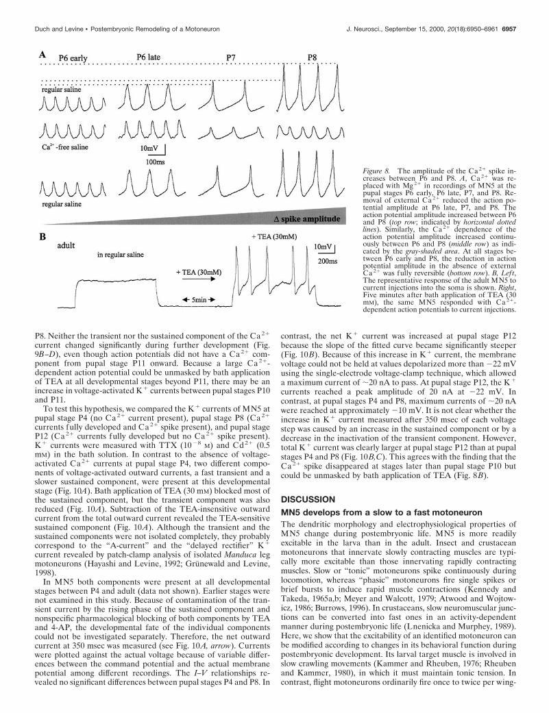
The size of the Ca21 spike increased continually from late pupalstage P6 to pupal stage P8 (Fig. 8A). Replacing Ca21 with Mg21
in the bath did not affect the spike shape at early pupal stage P6(Fig. 8A). Approximately 12 hr later in development ;30% of theaction potential amplitude was Ca21 dependent (Fig. 8A). Atpupal stage P7, 60% and, at pupal stage P8, 80% of the spikeamplitude was Ca21 dependent. The Ca21 component was absentin the small action potentials recorded from MN5 later than pupalstage P10. However, in these later pupal stages and in the adult, itcould be unmasked by bath application of TEA (Fig. 8B). Thissuggests that an increase in K1 current at approximately pupalstage 10 ordinarily masked the Ca21 spike. In vivo voltage-clampexperiments were performed to reveal the ionic currents underly-
Figure 6. Developmental changes infiring responses of MN5. Representa-tive intracellular recordings of firing re-sponses of MN5 to somatic current in-jections at different developmentalstages (A, larval stage; B, P1; C, P3;D, P4; E, P8; F, P16). In all preparations,MN5 was stimulated by square-pulsecurrent injections into the soma at ;0.5nA above the firing threshold, as indi-cated below each voltage trace. The hor-izontal dotted lines indicate the restingmembrane potentials. The vertical barsin D and E indicate where the actionpotential amplitude was measured. Atpupal stage P3 (C), action potential am-plitude and width increase, and MN5responds phasically rather than tonicallyas compared with earlier stages (A, B).A longer current pulse was injected in Cand D to illustrate this fact. At pupalstage P8 ( E), large-amplitude action po-tentials were evoked (arrows). At pupalstage 16 (F), MN5 fired only occasionallyafter current injections into the soma.
Duch and Levine • Postembryonic Remodeling of a Motoneuron J. Neurosci., September 15, 2000, 20(18):6950–6961 6955

ing the transient Ca21 spike during postembryonic dendritic re-modeling of MN5.
Changes in calcium and potassium currents duringadult developmentFor the analysis of Ca21 currents, TTX (1028 M) was added to thebath to block fast Na1 currents, electrodes were filled with 1.5 M
CsCl, and TEA was applied to the bath (30 mM). Nevertheless,outward currents were not blocked completely (Fig. 9A). Afterobtaining these records, therefore, Ca21 currents were blockedwith Cd21 (0.5 mM), and the residual outward current was sub-tracted (Fig. 9A) to allow calculation of the total Ca21 current.Because there are some limitations to current analysis using single-electrode voltage-clamp recordings in situ, the actual membranevoltage was monitored (Fig. 9A, top traces) to detect deviationsfrom the command potential. The actual membrane voltages forcommands between 210 and 10 mV revealed slight depolarizingdeflections during the maximum Ca21 currents, but these werealways ,5 mV (Fig. 9A, top traces, arrow). Therefore, the voltage-clamp control was adequate for comparing large differences inCa21 currents among developmental stages. The differences be-tween the command and actual holding potential were consistent
within a developmental stage, but the actual voltages reached fordepolarizing commands above 220 mV differed among differentdevelopmental stages. Therefore, the instantaneous actual mem-brane voltage was used for the analysis of current/voltage relation-ships (see Fig. 9C).
The larval MN5 displayed a sustained Ca21 current that wasactivated at approximately 235 mV and peaked at ;5 mV (Fig.9B,C). This corresponds to the description of Ca21 currents incultured leg motoneurons of Manduca sexta larvae, as measuredfrom isolated somata with patch pipettes (Hayashi and Levine,1992; Grunewald and Levine, 1998). No Ca21 current was detectedat pupal stages P3 and P4 (Fig. 9B–D). Two days later, at late pupalstage P6, Ca21 currents were again expressed (Fig. 9B). Theactivation and the peak voltage were similar to the larval Ca21
current, but the peak amplitude was twice as large (Fig. 9B).Furthermore, a transient component appeared in addition to thesustained Ca21 current (Fig. 9B, arrows), although the componentshave not been separated pharmacologically or electrically. Theamplitude of the transient Ca21 current increased until pupal stageP8 (Fig. 9B–D). The occurrence of the maximum peak Ca21
current correlated with the prominent Ca21 spike at pupal stage
Figure 7. MN5 exhibits Ca 21 action potentials at pupalstages P7–P9. A, Representative continuous intracellularrecording of MN5 at pupal stage P8. Somatic current injec-tion (bottom trace) of 400 msec duration at 0.6 Hz led tobursts of four large action potentials (top trace). Replacingexternal Ca 21 with Mg 21 (see lef t arrow) reduced theaction potential amplitude significantly. This effect was re-versed when Mg 2 was replaced with Ca 21 (see right arrow).B, Single bursts taken at selected time points from thecontinuous recording shown in A. C, Cd 21 (0.5 mM) shownblocking the Ca 21-dependent component of the action po-tential in a recording of MN5 at pupal stage P7. D, Aspontaneous action potential from a representative record-ing of MN5 at pupal stage P8 (lef t) and responses toelectrical stimulation of a sensory nerve (right). Both sub-threshold postsynaptic responses and Ca 21-dependent ac-tion potentials occurred in response to sensory stimulation.
6956 J. Neurosci., September 15, 2000, 20(18):6950–6961 Duch and Levine • Postembryonic Remodeling of a Motoneuron
P8. Neither the transient nor the sustained component of the Ca21
current changed significantly during further development (Fig.9B–D), even though action potentials did not have a Ca21 com-ponent from pupal stage P11 onward. Because a large Ca21-dependent action potential could be unmasked by bath applicationof TEA at all developmental stages beyond P11, there may be anincrease in voltage-activated K1 currents between pupal stages P10and P11.
To test this hypothesis, we compared the K1 currents of MN5 atpupal stage P4 (no Ca21 current present), pupal stage P8 (Ca21
currents fully developed and Ca21 spike present), and pupal stageP12 (Ca21 currents fully developed but no Ca21 spike present).K1 currents were measured with TTX (1028 M) and Cd21 (0.5mM) in the bath solution. In contrast to the absence of voltage-activated Ca21 currents at pupal stage P4, two different compo-nents of voltage-activated outward currents, a fast transient and aslower sustained component, were present at this developmentalstage (Fig. 10A). Bath application of TEA (30 mM) blocked most ofthe sustained component, but the transient component was alsoreduced (Fig. 10A). Subtraction of the TEA-insensitive outwardcurrent from the total outward current revealed the TEA-sensitivesustained component (Fig. 10A). Although the transient and thesustained components were not isolated completely, they probablycorrespond to the “A-current” and the “delayed rectifier” K1
current revealed by patch-clamp analysis of isolated Manduca legmotoneurons (Hayashi and Levine, 1992; Grunewald and Levine,1998).
In MN5 both components were present at all developmentalstages between P4 and adult (data not shown). Earlier stages werenot examined in this study. Because of contamination of the tran-sient current by the rising phase of the sustained component andnonspecific pharmacological blocking of both components by TEAand 4-AP, the developmental fate of the individual componentscould not be investigated separately. Therefore, the net outwardcurrent at 350 msec was measured (see Fig. 10A, arrow). Currentswere plotted against the actual voltage because of variable differ-ences between the command potential and the actual membranepotential among different recordings. The I–V relationships re-vealed no significant differences between pupal stages P4 and P8. In
contrast, the net K1 current was increased at pupal stage P12because the slope of the fitted curve became significantly steeper(Fig. 10B). Because of this increase in K1 current, the membranevoltage could not be held at values depolarized more than 222 mVusing the single-electrode voltage-clamp technique, which alloweda maximum current of ;20 nA to pass. At pupal stage P12, the K1
currents reached a peak amplitude of 20 nA at 222 mV. Incontrast, at pupal stages P4 and P8, maximum currents of ;20 nAwere reached at approximately 210 mV. It is not clear whether theincrease in K1 current measured after 350 msec of each voltagestep was caused by an increase in the sustained component or by adecrease in the inactivation of the transient component. However,total K1 current was clearly larger at pupal stage P12 than at pupalstages P4 and P8 (Fig. 10B,C). This agrees with the finding that theCa 21 spike disappeared at stages later than pupal stage P10 butcould be unmasked by bath application of TEA (Fig. 8B).
DISCUSSION
MN5 develops from a slow to a fast motoneuronThe dendritic morphology and electrophysiological properties ofMN5 change during postembryonic life. MN5 is more readilyexcitable in the larva than in the adult. Insect and crustaceanmotoneurons that innervate slowly contracting muscles are typi-cally more excitable than those innervating rapidly contractingmuscles. Slow or “tonic” motoneurons spike continuously duringlocomotion, whereas “phasic” motoneurons fire single spikes orbrief bursts to induce rapid muscle contractions (Kennedy andTakeda, 1965a,b; Meyer and Walcott, 1979; Atwood and Wojtow-icz, 1986; Burrows, 1996). In crustaceans, slow neuromuscular junc-tions can be converted into fast ones in an activity-dependentmanner during postembryonic life (Lnenicka and Murphey, 1989).Here, we show that the excitability of an identified motoneuron canbe modified according to changes in its behavioral function duringpostembryonic development. Its larval target muscle is involved inslow crawling movements (Kammer and Rheuben, 1976; Rheubenand Kammer, 1980), in which it must maintain tonic tension. Incontrast, flight motoneurons ordinarily fire once to twice per wing-
Figure 8. The amplitude of the Ca 21 spike in-creases between P6 and P8. A, Ca 21 was re-placed with Mg 21 in recordings of MN5 at thepupal stages P6 early, P6 late, P7, and P8. Re-moval of external Ca 21 reduced the action po-tential amplitude at P6 late, P7, and P8. Theaction potential amplitude increased between P6and P8 (top row; indicated by horizontal dottedlines). Similarly, the Ca 21 dependence of theaction potential amplitude increased continu-ously between P6 and P8 (middle row) as indi-cated by the gray-shaded area. At all stages be-tween P6 early and P8, the reduction in actionpotential amplitude in the absence of externalCa 21 was fully reversible (bottom row). B, Left,The representative response of the adult MN5 tocurrent injections into the soma is shown. Right,Five minutes after bath application of TEA (30mM), the same MN5 responded with Ca 21-dependent action potentials to current injections.
Duch and Levine • Postembryonic Remodeling of a Motoneuron J. Neurosci., September 15, 2000, 20(18):6950–6961 6957

beat cycle to mediate fast twitch contractions but no muscle tonus(Zarnack and Mohl, 1977; Burrows, 1996).
The lower excitability of the adult MN5 is caused by a morehyperpolarized resting potential, a more depolarized firing thresh-old, and a fivefold lower input resistance. Major decreases in inputresistance and tonic firing occur during the onset of dendriticgrowth, but input resistance remains relatively constant throughoutall later stages despite significant cell growth. Therefore, changes inexcitability are not simply reflective of changes in cell size. Theexcitability of MN5 is adult-like at pupal stage P11. This correlateswith an increase in the net K1 current between pupal stages P8 andP12. Similarly, in Xenopus embryonic development, modificationsin K1 currents in spinal neurons are correlated with the matura-tion of motor patterns (Sun and Dale, 1998). For MN5, the lowexcitability during late pupal stages is important because contrac-tions of the pupal DLM muscle, which is striated and functionallyinnervated by pupal stage P8 (Duch et al., 2000), would tear the softnewly formed adult cuticle.
Ca21 conductances that are independent of the spike-initiatingzone play a major role in the generation of plateau potentials(Hartline and Russel, 1984; Schwindt and Crill, 1984; Hancox and
Pitman, 1991, 1993; Kiehn, 1991). Although the adult MN5 dis-played no plateau potentials in the isolated ganglion preparation,prolonged Ca21 action potentials could be evoked after applyingTEA. In many systems, plateau potentials can be induced byneuromodulators or any intervention that sufficiently reduces op-posing outward currents (Ramirez and Pearson, 1991; Hultbornand Kiehn, 1992; Kiehn and Harris-Warrick, 1992; Bal et al.,1994). Flight is strongly influenced by neuromodulators, such asoctopamine, which evokes plateau potentials in locust flight inter-neurons (Orchard et al., 1993). Moths display a prolonged warm-upphase during which neuromodulators may prepare the CNS for flightbehavior (Claassen and Kammer, 1986). Thus, the Ca21currentdetected in the adult MN5 might be important in shaping its motoroutput, particularly if neuromodulators reduce K1 currents duringflight.
In contrast to the adult, Ca21 spikes are readily produced duringpupal stages P7 to P9. At later stages the Ca21 spike is masked bythe increased K1 currents. The large amplitude of the Ca21 spikein somatic recordings suggests that these spikes are generated inthe cell body, although it is unclear where they are initiated. Incontrast, the small amplitude of the Na1 spike as recorded from
Figure 9. Developmental modifications of calcium currents in MN5. A, Representative membrane current (Im) recordings in dSEVC mode of MN5at pupal stage P7 to demonstrate the method used for isolating Ca 21 currents (see Results). The actual membrane potential was monitored inall experiments. Slight voltage fluctuations were sometimes observed at the membrane potentials at which the maximum Ca 21 currents occurred, but thesewere always ,5 mV (see arrow). B, Representative traces of inward currents for different developmental stages after subtraction (see A). The time pointswhere peak and sustained Ca 21 currents were measured are indicated with arrows. C, I–V relationships for the peak Ca 21 current created from fourrecordings each in the larval stage, pupal stages P4 and P8, and the adult. D, Average peak and sustained Ca 21 currents for different developmental stages.The number of recordings for each group is presented below each set of bars. Error bars represent SDs.
6958 J. Neurosci., September 15, 2000, 20(18):6950–6961 Duch and Levine • Postembryonic Remodeling of a Motoneuron

the soma at most stages, and evident at stages P7 to P9 afterremoval of external Ca21, indicates that these spikes are notactively generated in the cell body, similar to the usual case forinsect motoneurons (Gwilliam and Burrows, 1980).
MN5 undergoes sequential changes in ionic currentsduring postembryonic lifeEmbryonic and postnatal maturation of central neurons is accom-panied by a sequential expression of different voltage-activatedionic currents (for review, see Spitzer, 1991). We report sequential
modifications in Ca21 and K1 currents during the postembryonicacquisition of a new behavior.
The validity of the in situ single-electrode voltage-clamp mea-surements is supported by findings obtained from a population ofcultured motoneurons recorded in the whole-cell patch configura-tion. Leg motoneuron somata that were isolated acutely from earlypupal Manduca had smaller Ca21 current densities than did thoseisolated from larvae or adults but retained some K1 currents(Hayashi and Levine, 1992). Although these measurements fromisolated somata avoided space-clamp problems, the extent to whichthey represented normal physiology and their functional conse-quences were unclear. Thus, the in situ measurements reportedhere were indispensable for defining the normal sequence of ioniccurrent expression during development. The absence of Ca21
currents in MN5 at early pupal stages in vivo was not an artifactattributable to space-clamp problems, because MN5 was muchsmaller at these stages than at other stages in which Ca21 currentswere detected. By contrast, the net K1 current did not changebetween P4 and P8 but increased after stage P12. These findingsallow a correlation of physiological changes that are evident duringnormal development with the time course of dendritic remodeling.
Putative signals for changes in ionic currents duringpostembryonic developmentThe steroid hormone 20-hydroxyecdysone (20E) may regulate themodification of Ca21 currents. Manduca leg motoneurons culturedduring early pupal stages show a 20E-dependent increase in Ca21
current (Grunewald and Levine, 1998). Systemic ecdysteroid levelsrise continuously between stages P4 and P8 (Bollenbacher et al.,1981), while Ca21 currents in MN5 increase. Ecdysteroids regulatethe excitability of neurosecretory neurons in Manduca (Hewes andTruman, 1994), perhaps by altering ionic currents, and steroidhormones are potent modulators of ionic currents in the vertebrateCNS (Kerr et al., 1992; Rendt et al., 1992; Dunlap et al., 1997).Alternatively, activity-dependent mechanisms could be involved,because electrical activity influences the postnatal shaping of neu-ronal connections (Shatz, 1990; Fields and Nelson, 1992; Gu andSpitzer, 1995) and the expression of ion currents (Garcia et al.,1994; Lnenicka et al., 1998). Finally, interactions with the degen-erating larval target muscle during late larval and early pupal stages(Duch et al., 2000) might play a role. The trigger for the increasein net K1 current between pupal stages P8 and P12 also remains tobe investigated. Systemic ecdysteroid levels decline during thesestages (Bollenbacher et al., 1981), but 20E had no effect on the K1
currents of cultured pupal leg motoneurons (Grunewald and Le-vine, 1998). Alternatively, the large Ca21 current at stage P8 mightplay a role, similar to the role of a Ca21 influx-dependent increasein K1 channel expression during the postnatal maturation of Pur-kinje cells (Muller et al., 1998).
Possible role of transient calcium signals indendritic differentiationSimilar to other central neurons in Manduca (Levine and Truman,1985; Weeks and Ernst-Utzschneider, 1989; Kent and Levine,1993), MN5 undergoes dramatic dendritic remodeling duringmetamorphosis. Some aspects of the dendritic plasticity are con-trolled directly by 20E (for review, see Weeks and Levine, 1990,1995), but interactions with the periphery are also involved (Kentand Levine, 1993). This is the first study in which dendritic remod-eling can be correlated temporally with changes in active andpassive membrane properties during normal postembryonic devel-opment. This correlation is consistent with a role for Ca21 influxin dendritic remodeling. Prominent growth cone-like structuresform at the tips of the growing adult dendrites between pupalstages P2 and P6. This corresponds in time with a low level of Ca21
currents. In culture systems, low levels of Ca21 influx promotegrowth cone extension and branching, but high levels of influx andprolonged internal elevation inhibit these processes (for review, seeKater et al., 1988; Kater and Mills, 1991). The cessation of the
Figure 10. Developmental modifications of potassium currents in MN5.A, Panel 1, Total potassium current was recorded in dSEVC mode fromMN5 at pupal stage P4 with TTX (10 28 M) and cadmium (0.5 M) in thebath. Panel 2, Bath application of TEA (30 mM) affected the sustainedcomponent of the outward current significantly, although the transientcomponent was also reduced. Panel 3, Subtraction of the residual tran-sient component from the total outward current revealed the sustainedcomponent. B, I–V relationships for the total potassium current (seetraces in A) at pupal stages P4, P8, and P12 are shown. Total current wasmeasured at 350 msec of each voltage step (see arrow in A) and plottedagainst the actual instantaneous membrane potential (see Results). Fiverecordings were plotted for each stage, and curves were fitted using thesimplex method. The I–V relationships for pupal stages P4 and P8showed no significant difference, but the fitted curve for pupal stage P12was significantly steeper. C, Typical current responses of MN5 at thestages P4, P8, and P12 are shown after bath application of TTX andcadmium to voltage steps from a holding potential of 250 mV to anactual membrane potential of 225 mV. The bottom traces show thevoltages recorded at the three different stages, and the top traces showthe current responses.
Duch and Levine • Postembryonic Remodeling of a Motoneuron J. Neurosci., September 15, 2000, 20(18):6950–6961 6959
formation of high-order branches of MN5 is correlated with thetransient occurrence of Ca21 spikes during pupal stages P7 to P9,because of the differential timing of Ca21 versus K1 currentmodifications. Because these spikes occurred spontaneously andcould be evoked by sensory stimulation in the isolated ganglionpreparation, it is likely that they also occur during normal devel-opment. During embryonic spinal cord development, the frequencyof growth cone Ca 21 transients is inversely proportional to the rateof axon outgrowth, with large elevations in internal Ca21 at paus-ing sites (Gomez and Spitzer, 1999), causing an increase in theactivity of calcineurin (Lautermilch and Spitzer, 2000). Further-more, the Ca21-sensitive enzyme calmodulin kinase II is requiredto limit the elaboration of neuronal arbors in the developingXenopus optical tectum (Zou and Cline, 1999). The effects ofpostembryonic Ca21 current modifications on cytosolic Ca21 lev-els of MN5 remain to be investigated. However, the ability tocorrelate structural and functional events in their normal context inthis system provides distinct advantages for examining whetherCa21 signals mediate the initiation and cessation of dendriticgrowth during postembryonic life.
REFERENCESAtwood HL, Wojtowicz JM (1986) Short-term and long-term plasticity
and physiological differentiation of crustacean motor synapses. Int RevNeurobiol 28:275–362.
Baines RA, Bate M (1998) Electrophysiological development of centralneurons in the Drosophila embryo. J Neurosci 18:4673–4683.
Bal T, Nagy F, Moulin M (1994) Muscarinic modulation of a pattern-generating network: control of neuronal properties. J Neurosci14:3019–3035.
Bell RA, Joachim FA (1976) Techniques for rearing laboratory coloniesof tobacco hornworms and pink ballworms. Ann Entomol Soc Am69:365–373.
Bollenbacher WE, Smith SL, Goodman W, Gilbert LI (1981) Ecdysteroidtiter during the larval-pupal-adult development of the tobacco hornworm,Manduca sexta. Gen Comp Endocrinol 44:302–306.
Burrows M (1996) Components of the nervous system: motoneurons. In:The neurobiology of an insect brain. New York: Oxford UP.
Casaday GB, Camhi JM (1976) Metamorphosis of flight motoneurons inthe moth, Manduca sexta. J Comp Physiol [A]112:143–158.
Casanovas B, Meyrand P (1995) Functional differentiation of adult neuralcircuits from a single embryonic network. J Neurosci 15:5703–5718.
Claassen DE, Kammer AE (1986) Effects of octopamine, dopamine, andserotonin on production of flight motor output by thoracic ganglia ofManduca sexta. J Neurobiol 17:1–14.
Cohan CS, Connor JA, Kater SB (1987) Electrically and chemically me-diated increases in intracellular calcium in neuronal growth cones. J Neu-rosci 7:3588–3599.
Consoulas C, Kent KS, Levine RB (1996) Remodeling of the peripheralprocesses and presynaptic terminals of leg motoneurons during meta-morphosis of the hawkmoth, Manduca sexta. J Comp Neurol372:415–434.
Duch C, Bayline RJ, Levine RB (2000) Postembryonic development of thedorsal longitudinal flight muscle and its innervation in Manduca sexta.J Comp Neurol, in press.
Dunlap KD, McAnelly ML, Zakon HH (1997) Estrogen modifies an elec-trocommunication signal by altering the electrocyte sodium current in anelectric fish, Sternopygus. J Neurosci 17:2869–2875.
Fields RD, Nelson PG (1992) Activity-dependent development of the ver-tebrate nervous system. Int Rev Neurobiol 25:281–293.
Fields RD, Neale EA, Nelson PG (1990) Effects of patterned electricalactivity on neurite outgrowth from mouse sensory neurons. J Neurosci10:2950–2964.
Gao BX, Ziskind-Conhaim L (1998) Development of ionic currents un-derlying changes in action potential waveforms in rat spinal motoneu-rons. J Neurophysiol 80:3047–3061.
Garcia DE, Cavalie A, Lux HD (1994) Enhancement of voltage-gatedcalcium currents induced by daily stimulation of hippocampal neuronswith glutamate. J Neurosci 14:545–553.
Gomez MG, Spitzer NC (1999) In vivo regulation of axon extension andpathfinding by growth-cone calcium transients. Nature 397:350–355.
Grunewald B, Levine RB (1998) Ecdysteroid control of ionic currentdevelopment in Manduca sexta motoneurons. J Neurobiol 37:211–223.
Gu X, Spitzer NC (1995) Distinct aspects of neuronal differentiation en-coded by frequency of spontaneous calcium transients. Nature375:784–787.
Gwilliam GF, Burrows M (1980) Electrical characteristics of an identifiedinsect motoneuron. J Exp Biol 86:49–60.
Hancox JC, Pitman RC (1991) Plateau potentials drive axonal impulsebursts in insect motoneurons. Proc R Soc Lond B Biol Sci 244:33–38.
Hancox JC, Pitman RC (1993) Plateau potentials in insect motoneuronscan be driven by synaptic input. J Exp Biol 176:307–310.
Hartline DK, Russel DF (1984) Endogenous burst capability in a neuronof the gastric mill pattern generator of the spiny lobster Panulirus inter-ruptus. J Neurobiol 15:345–364.
Hayashi JH, Levine RB (1992) Calcium and potassium currents in legmotoneurons during postembryonic development in the hawkmothManduca sexta. J Exp Biol 171:15–42.
Haydon PG, Zoran MJ (1994) Retrograde regulation of presynaptic de-velopment during synaptogenesis. J Neurobiol 25:694–706.
Hewes RS, Truman JW (1994) Steroid regulation of excitability in identi-fied insect neurosecretory cells. J Neurosci 14:1812–1819.
Hultborn H, Kiehn O (1992) Neuromodulation of vertebrate motoneuronmembrane properties. Curr Opin Neurobiol 2:770–775.
Kammer AE, Rheuben MB (1976) Adult motor patterns produced by themoth pupae during development. J Exp Biol 65:65–84.
Kater SB, Mills LR (1991) Regulation of growth cone behavior by cal-cium. J Neurosci 11:891–899.
Kater SB, Mattson MP, Cohan C, Connor J (1988) Calcium regulation ofneuronal growth cone. Trends Neurosci 11:315–321.
Kennedy D, Takeda K (1965a) Reflex control of abdominal flexor musclesin the crayfish. I. The twitch system. J Exp Biol 43:211–227.
Kennedy D, Takeda K (1965b) Reflex control of abdominal flexor musclesin crayfish. II. The tonic system. J Exp Biol 43:229–246.
Kent KS, Levine RB (1993) Dendritic reorganization of an identifiedneuron during metamorphosis of the moth, Manduca sexta—the influ-ence of interactions with the periphery. J Neurobiol 24:1–22.
Kerr DS, Campbell LW, Thibault O, Landfield PW (1992) Hippocampalglucocorticoid receptor activation enhances voltage-dependent calciumconductances: relevance for brain aging. Proc Natl Acad Sci USA89:8527–8531.
Kiehn O (1991) Plateau potentials and active integration in the “finalcommon pathway” for motor behavior. Trends Neurosci 14:68–73.
Kiehn O, Harris-Warrick RM (1992) Seretonergic stretch receptors in-duce plateau properties in a crustacean motoneuron by a dual-conductance mechanism. J Neurophysiol 68:485–495.
Lautermilch NJ, Spitzer NC (2000) Regulation of calcineurin by growthcone calcium waves controls neurite extension. J Neurosci 20:315–325.
Levine RB, Truman JW (1982) Metamorphosis of the insect nervoussystem: changes in the morphology and synaptic interactions of identifiedcells. Nature 299:250–252.
Levine RB, Truman JW (1985) Dendritic reorganization of abdominalmotoneurons during metamorphosis of the moth Manduca sexta. J Neu-rosci 5:2424–2431.
Lnenicka GA, Murphey RK (1989) The refinement of invertebrate syn-apses during development. J Neurobiol 20:339–355.
Lnenicka GA, Arcaro KF, Calabro JM (1998) Activity-dependent devel-opment of calcium regulation in growing motor axons. J Neurosci18:4966–4972.
Martin-Caraballo M, Greer JJ (1999) Electrophysiological properties ofrat phrenic motoneurons during perinatal development. J Neurophysiol81:1365–1378.
Mattson MP, Dou P, Kater SB (1988) Outgrowth-regulating actions ofglutamate in isolated hippocampal pyramidal neurons. J Neurosci8:2087–2100.
McCobb DP, Best PM, Beam KG (1989) Development alters the expres-sion of calcium currents in chick limb motoneurons. Neuron 2:1633–1643.
McCobb DP, Best PM, Beam KG (1990) The differentiation of excitabilityon embryonic chick limb motoneurons. J Neurosci 10:2974–2984.
Meyer D, Walcott B (1979) Differences in the responsiveness of identifiedmotoneurons in the cockroach: role for the motor program for stepping.Brain Res 178:600–605.
Muller YL, Reitstetter R, Yool AJ (1998) Regulation of Ca 21-dependentK 1 channel expression in rat cerebellum during postnatal development.J Neurosci 18:16–25.
Nijhout HF, Williams CM (1974) Control of molting and metamorphosisin the tobacco hornworm, Manduca sexta (L): growth of the last instarlarva and the decision to pupate. J Exp Biol 61:481–491.
Orchard I, Ramirez JM, Lange AB (1993) A multifunctional role foroctopamine in locust flight. Annu Rev Entomol 38:227–249.
Ramirez JM, Pearson KG (1991) Octopamine induces bursting and pla-teau potentials in insect neurons. Brain Res 549:332–337.
Rendt JM, Toro L, Stefani E, Eruklar SD (1992) Progesterone increasescalcium currents in myometrial cells from immature and nonpregnantadult rats. Am J Physiol 262:C293–C301.
Reynolds SA, French KA, Baader A, Kristan Jr WB (1998) Developmentof spontaneous behaviors in the medical leech. J Comp Neurol14:168–180.
Rheuben MB, Kammer AE (1980) Comparison of slow larval and fastadult muscle innervated by the same motoneuron. J Exp Biol 84:103–112.
Ribiera AB, Spitzer NC (1992) Developmental regulation of potassiumchannels and the impact on neuronal differentiation. Ion Channels3:1–38.
Schwindt PC, Crill WE (1984) Membrane properties of cat spinal mo-toneurons. In: Handbook of the spinal cord (Davidoff RA, ed), pp199–242. New York: Dekker.
6960 J. Neurosci., September 15, 2000, 20(18):6950–6961 Duch and Levine • Postembryonic Remodeling of a Motoneuron

Shatz CJ (1990) Impulse activity and the patterning of connections duringCNS development. Neuron 5:745–756.
Spitzer NC (1991) A developmental handshake: neuronal control of ioniccurrents and their control of neuronal differentiation. J Neurobiol22:659–673.
Sun QQ, Dale N (1998) Developmental changes in expression of ioniccurrents accompany maturation of locomotor pattern in frog tadpoles.J Physiol (Lond) 507:257–264.
Tolbert LP, Matsumotu SG, Hildebrand JG (1983) Development of syn-apses in the antennal lobes of the moth Manduca sexta during metamor-phosis. J Neurosci 3:1158–1175.
Truman JW, Reiss SE (1988) Hormonal regulation of the shape of iden-tified motoneurons in Manduca sexta. J Neurosci 8:765–775.
Weeks JC, Ernst-Utzschneider K (1989) Respecification of larval proleg
motoneurons during metamorphosis of the tobacco hornworm, Manducasexta. Segmental dependence and hormonal regulation. J Neurobiol20:569–592.
Weeks JC, Levine RB (1990) Postembryonic neuronal plasticity and itshormonal control during insect metamorphosis. Annu Rev Neurosci13:183–194.
Weeks JC, Levine RB (1995) Steroid hormone effects on neurons subserv-ing behavior. Curr Opin Neurobiol 5:809–815.
Zarnack W, Mohl B (1977) Activity of the downstroke flight muscles ofLocusta migratoria (L.) during steering behavior in flight. J Comp Physiol[A] 118A:215–233.
Zou DJ, Cline HT (1999) Postsynaptic calcium/calmodulin-dependent ki-nase II is required to limit elaboration of presynaptic and postsynapticneuronal arbors. J Neurosci 19:8909–8918.
Duch and Levine • Postembryonic Remodeling of a Motoneuron J. Neurosci., September 15, 2000, 20(18):6950–6961 6961