remote anesthesia hany el-zahaby, md ain shams university, 2009
TRANSCRIPT

Remote Anesthesia
Hany El-Zahaby, MD
Ain Shams University, 2009
Remote anesthesia
Anesthesiologists are increasingly being asked to provide anesthetic care in locations outside of the OR.
These locations include: radiology suites, cardiac labs, psychiatric units, GI endoscopy suites, CT, MRI, and PACU.
It is the responsibility of the anesthesiologist to ensure that the location meets the ASA guidelines for safety.
Objectives
Understanding that the standards of anesthesia care and patient monitoring are the same regardless of location.
Remember that the key to efficient and safe remote anesthetic relies on open communication between the anesthesiologist and non-operating room personnel.
Realize that remote locations have different safety concerns, such as radiation and powerful magnetic fields.
Common Problems
Patients coming from wards with staff unfamiliar with preoperative preparation
Anesthetic assistance and maintenance of anesthetic equipment may be less than ideal e.g. anesthesia machine the oldest in the hospital, anesthetic machine disconnected and moved when not in use, empty gas cylinders
Common Problems
Unavailability of tools Communication between non-operating room
staff and the anesthesiologist may be poor Recovery facilities are often non-existent
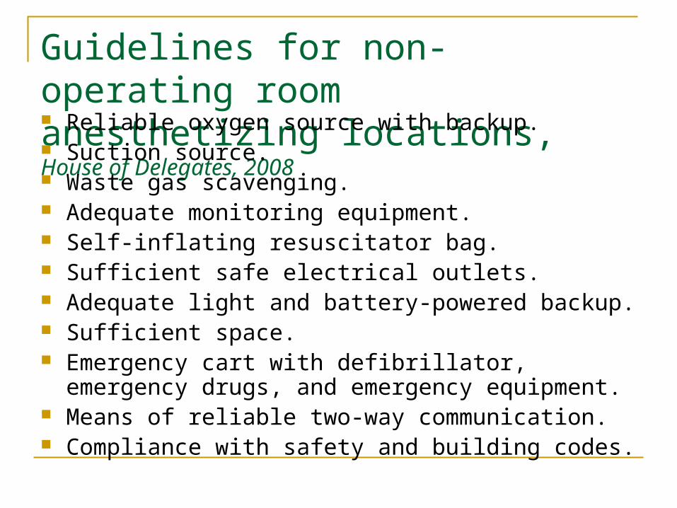
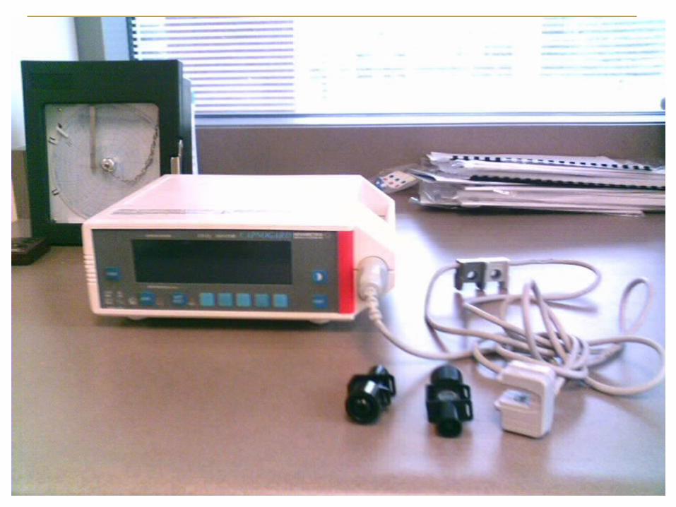
Guidelines for non-operating room anesthetizing locations, House of Delegates, 2008 Reliable oxygen source with backup. Suction source. Waste gas scavenging. Adequate monitoring equipment. Self-inflating resuscitator bag. Sufficient safe electrical outlets. Adequate light and battery-powered backup. Sufficient space. Emergency cart with defibrillator, emergency drugs, and
emergency equipment. Means of reliable two-way communication. Compliance with safety and building codes.
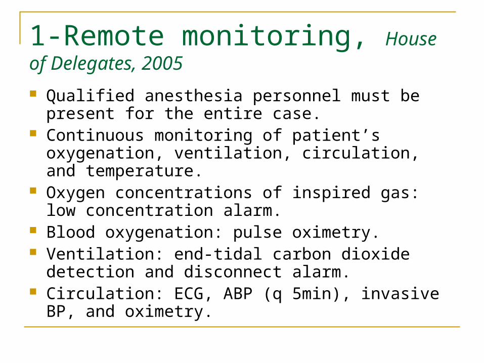
1-Remote monitoring, House of Delegates, 2005
Qualified anesthesia personnel must be present for the entire case.
Continuous monitoring of patient’s oxygenation, ventilation, circulation, and temperature.
Oxygen concentrations of inspired gas: low concentration alarm.
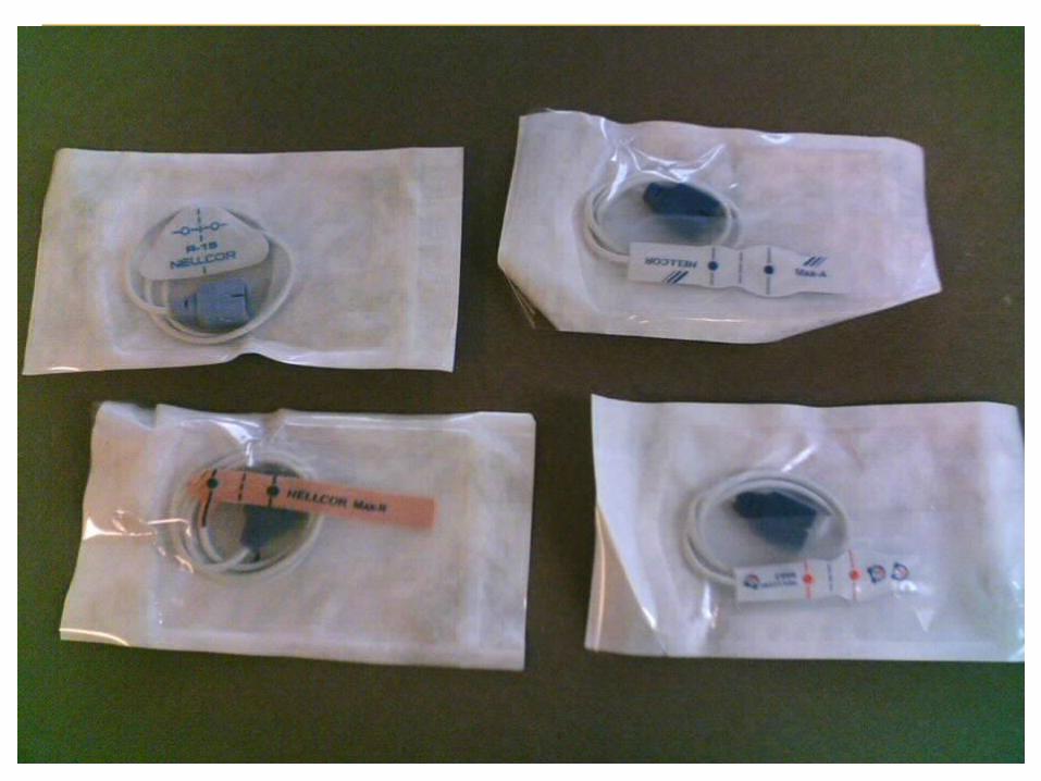
Blood oxygenation: pulse oximetry. Ventilation: end-tidal carbon dioxide detection and
disconnect alarm. Circulation: ECG, ABP (q 5min), invasive BP, and
oximetry.
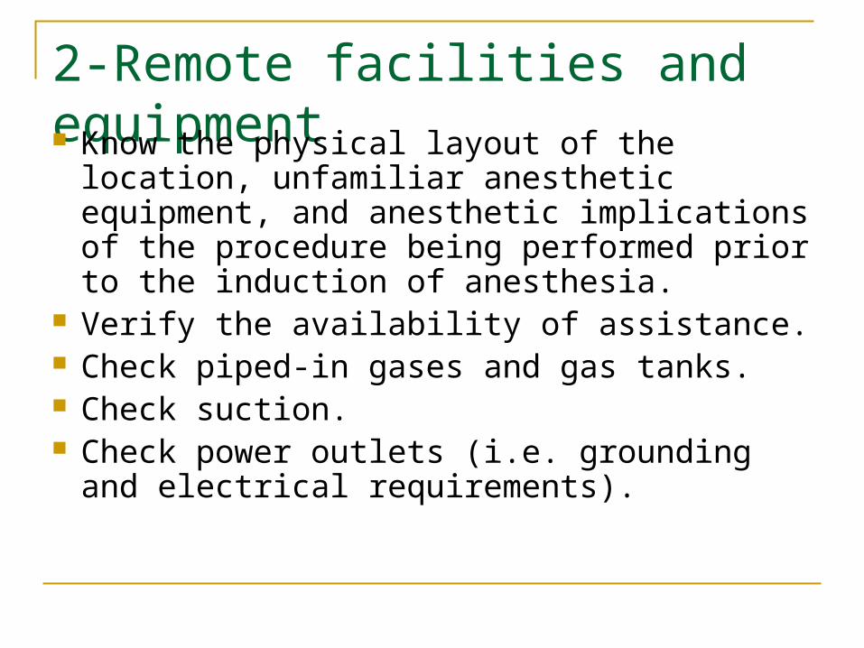
2-Remote facilities and equipment Know the physical layout of the location,
unfamiliar anesthetic equipment, and anesthetic implications of the procedure being performed prior to the induction of anesthesia.
Verify the availability of assistance. Check piped-in gases and gas tanks. Check suction. Check power outlets (i.e. grounding and
electrical requirements).
3-Remote personnel
Nurses and radiology techs are often less familiar with the management of anesthesia, therefore they are often unable to provide skilled assistance in an emergency
4-Remote recovery care
Patient must be medically stable before transport.
Patient must be accompanied to the recovery area.
Provisions for O2 delivery and monitoring on the transport cart are required.
Appropriate recovery facilities and staff must be provided.
Office-based anesthesia ASA and JCAHO guidelines
Employment of appropriately trained and credentialed anesthesia personnel.
Availability of properly maintained anesthesia equipment. Complete documentation of the care provided as required
at other surgical sites. Use of standard ASA monitoring. Provision of a PACU that is staffed by trained nursing
personnel. Availability of emergency equipment. Establishment of a written plan for emergency transport of
the patient to a comprehensive care center if a complication occurs.
Office-based anesthesia, (contd.) Often used for ENT and dental procedures. Patient requires a full preoperative workup. Potentially difficult airways are not good
candidates. Procedures often involve local anesthesia
plus IV sedation or light general anesthesia with a mask or LMA.
Agents of choice include: propofol, sevo, des, and N2O.
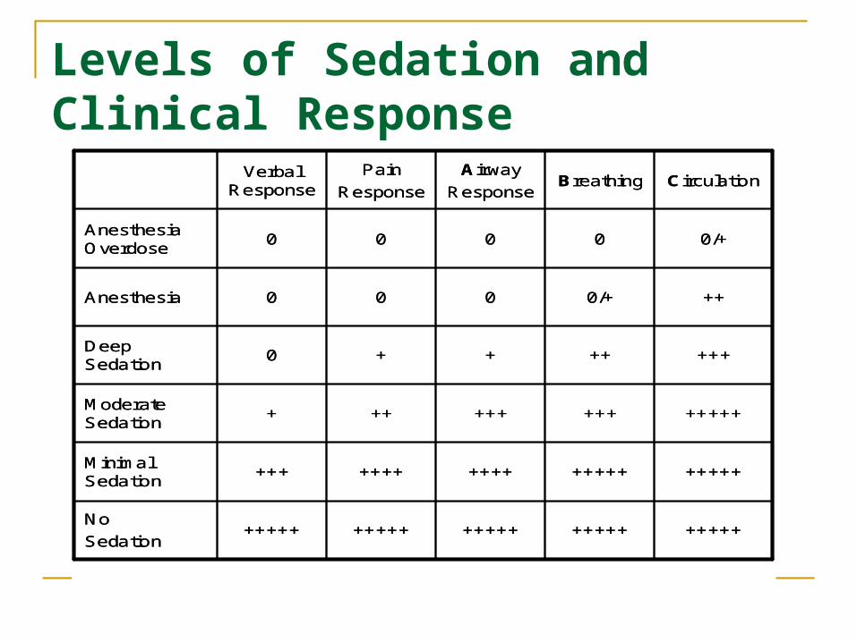
Levels of Sedation and Clinical Response
CirculationBreathingAirway
Response
Pain
ResponseVerbal
Response
+++++++++++++++++++++++++No
Sedation
+++++++++++++++++++++Minimal Sedation
++++++++++++++Moderate Sedation
+++++++0Deep Sedation
++0/+000Anesthesia
0/+0000Anesthesia Overdose
CirculationBreathingAirway
Response
Pain
ResponseVerbal
Response
+++++++++++++++++++++++++No
Sedation
+++++++++++++++++++++Minimal Sedation
++++++++++++++Moderate Sedation
+++++++0Deep Sedation
++0/+000Anesthesia
0/+0000Anesthesia Overdose
Options
Conscious sedation Monitored anesthesia Care (MAC) General anesthesia
Special ConsiderationsRadiology suite
Includes: US, CT, MRI, RFA, and neuro-coiling. The rooms are often crowded with bulky equipment. Patients are often required to hold still for long periods of
time (moderate sedation Vs monitored anesthesia care). Some patients still require anesthesia:
children, unconscious patients, movement disorders, adults with learning difficulties.
Special Considerations Radiology suite, contd. Unique hazard: radiation exposure.
Leukemia and fetal abnormalities. Dosimeters are required Lead aprons, thyroid shields, leaded glass
screens, and video monitoring.
Special Considerations Radiology suite, contd. Iodinated contrast media.
Older ionized contrast media were hyperosmolar and toxic.
Newer non-ionized contrast media have lower osmolality and improved side-effects.
Predisposing factors to adverse reactions from contrast media include a history of: bronchospasm, allergy, cardiac disease, hypovolemia, hematologic disease, renal dysfunction, extremes of age, anxiety, and medications (beta-blockers, aspirin, and NSAIDs).
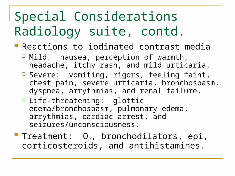
Special Considerations Radiology suite, contd. Reactions to iodinated contrast media.
Mild: nausea, perception of warmth, headache, itchy rash, and mild urticaria.
Severe: vomiting, rigors, feeling faint, chest pain, severe urticaria, bronchospasm, dyspnea, arrythmias, and renal failure.
Life-threatening: glottic edema/bronchospasm, pulmonary edema, arrythmias, cardiac arrest, and seizures/unconsciousness.
Treatment: O2, bronchodilators, epi, corticosteroids, and antihistamines.
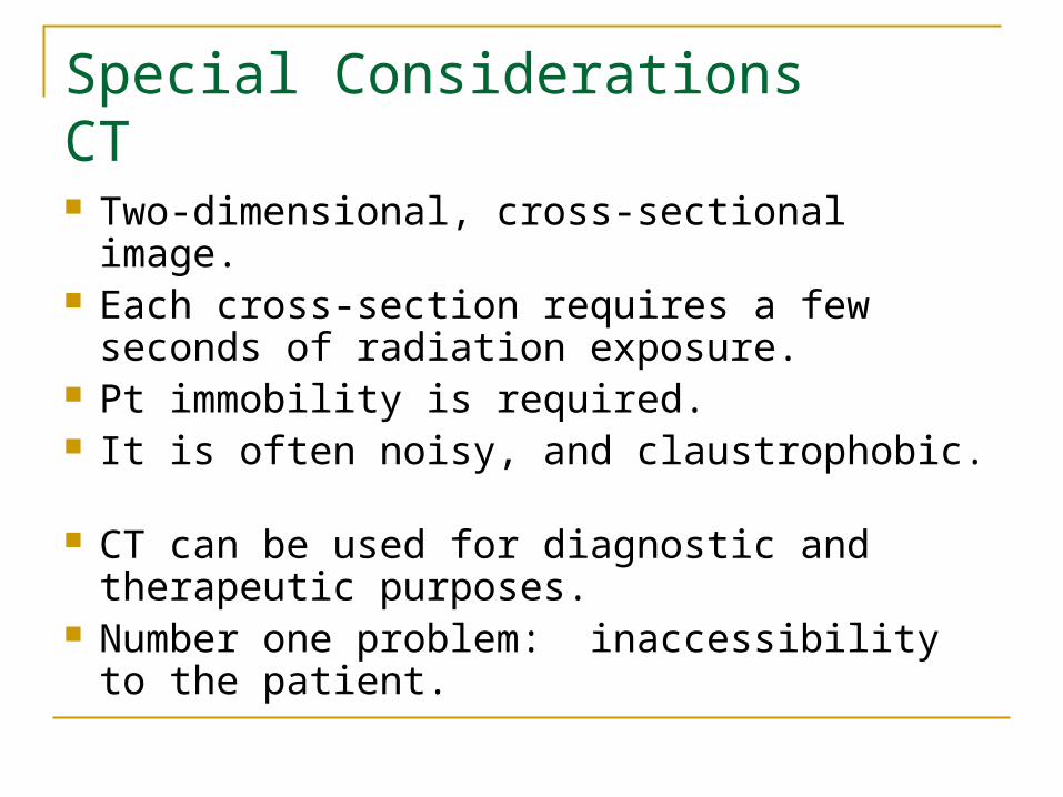
Special Considerations CT Two-dimensional, cross-sectional image. Each cross-section requires a few seconds of
radiation exposure. Pt immobility is required. It is often noisy, and claustrophobic. CT can be used for diagnostic and
therapeutic purposes. Number one problem: inaccessibility to the
patient.
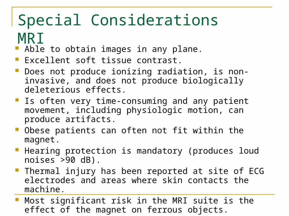
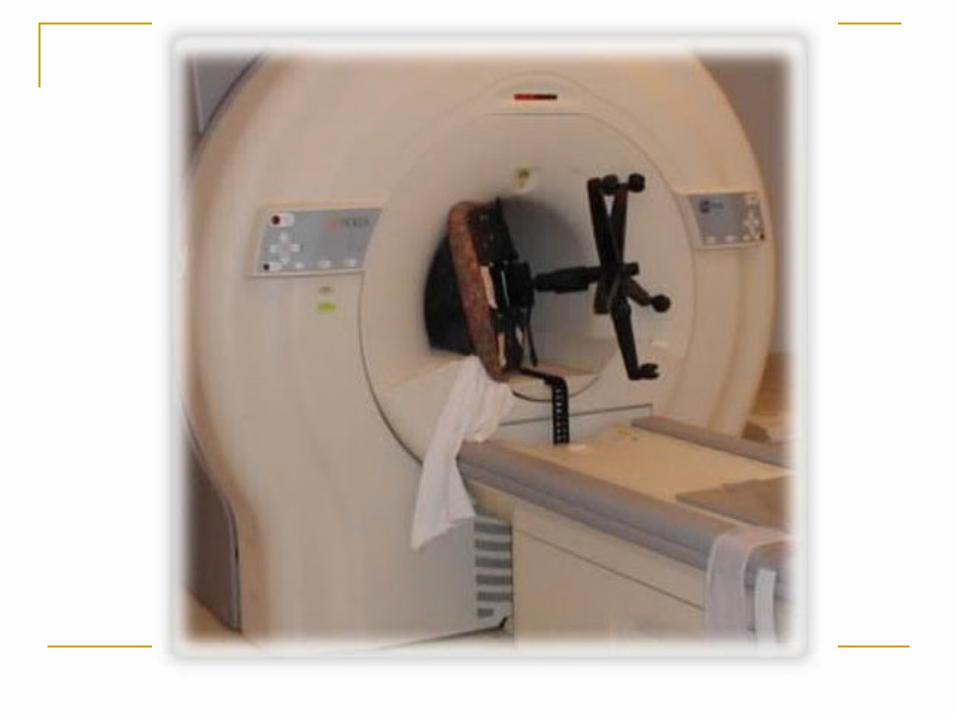
Special Considerations MRI
Able to obtain images in any plane. Excellent soft tissue contrast. Does not produce ionizing radiation, is non-invasive, and
does not produce biologically deleterious effects. Is often very time-consuming and any patient movement,
including physiologic motion, can produce artifacts. Obese patients can often not fit within the magnet. Hearing protection is mandatory (produces loud noises
>90 dB). Thermal injury has been reported at site of ECG
electrodes and areas where skin contacts the machine. Most significant risk in the MRI suite is the effect of the
magnet on ferrous objects.
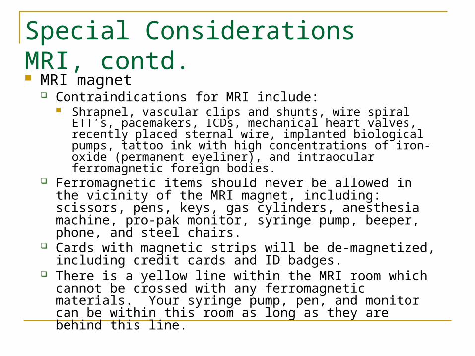
Special Considerations MRI, contd. MRI magnet
Contraindications for MRI include: Shrapnel, vascular clips and shunts, wire spiral ETT’s,
pacemakers, ICDs, mechanical heart valves, recently placed sternal wire, implanted biological pumps, tattoo ink with high concentrations of iron-oxide (permanent eyeliner), and intraocular ferromagnetic foreign bodies.
Ferromagnetic items should never be allowed in the vicinity of the MRI magnet, including: scissors, pens, keys, gas cylinders, anesthesia machine, pro-pak monitor, syringe pump, beeper, phone, and steel chairs.
Cards with magnetic strips will be de-magnetized, including credit cards and ID badges.
There is a yellow line within the MRI room which cannot be crossed with any ferromagnetic materials. Your syringe pump, pen, and monitor can be within this room as long as they are behind this line.
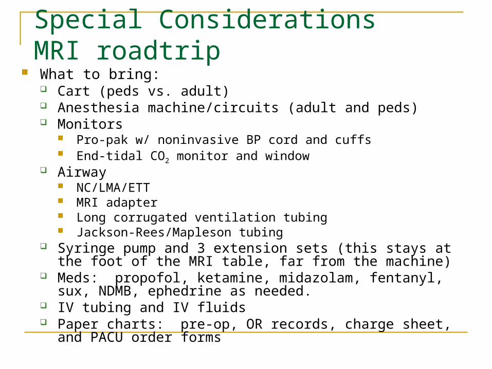
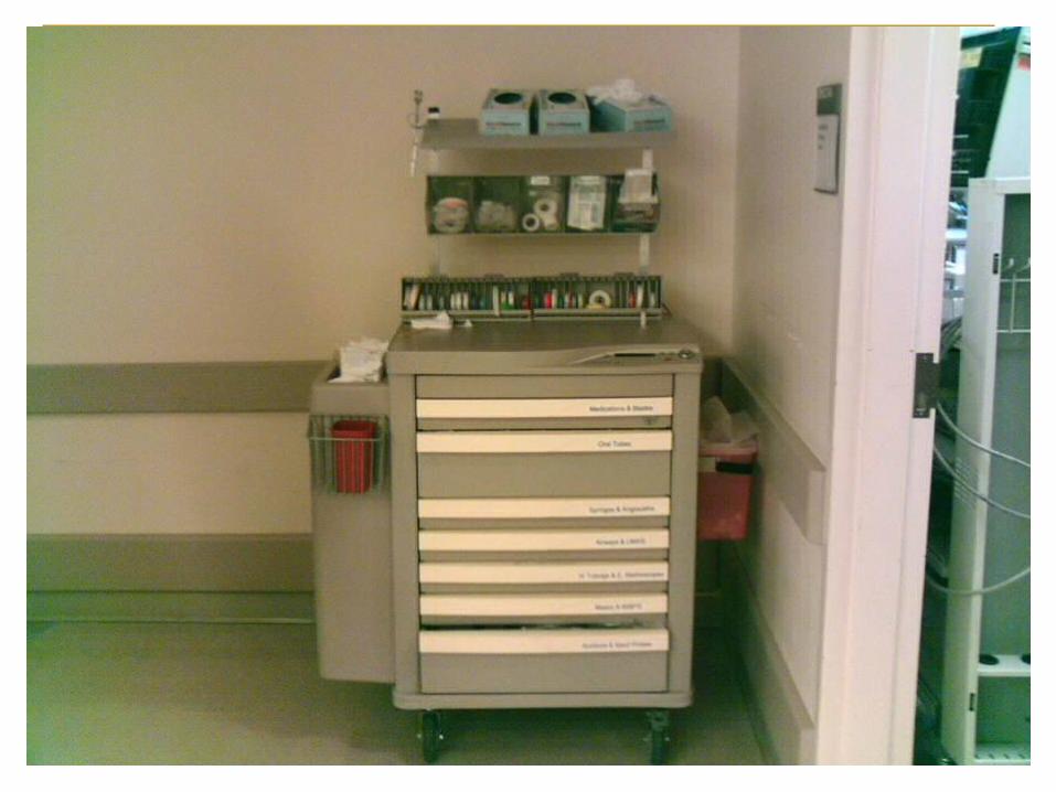
Special Considerations MRI roadtrip
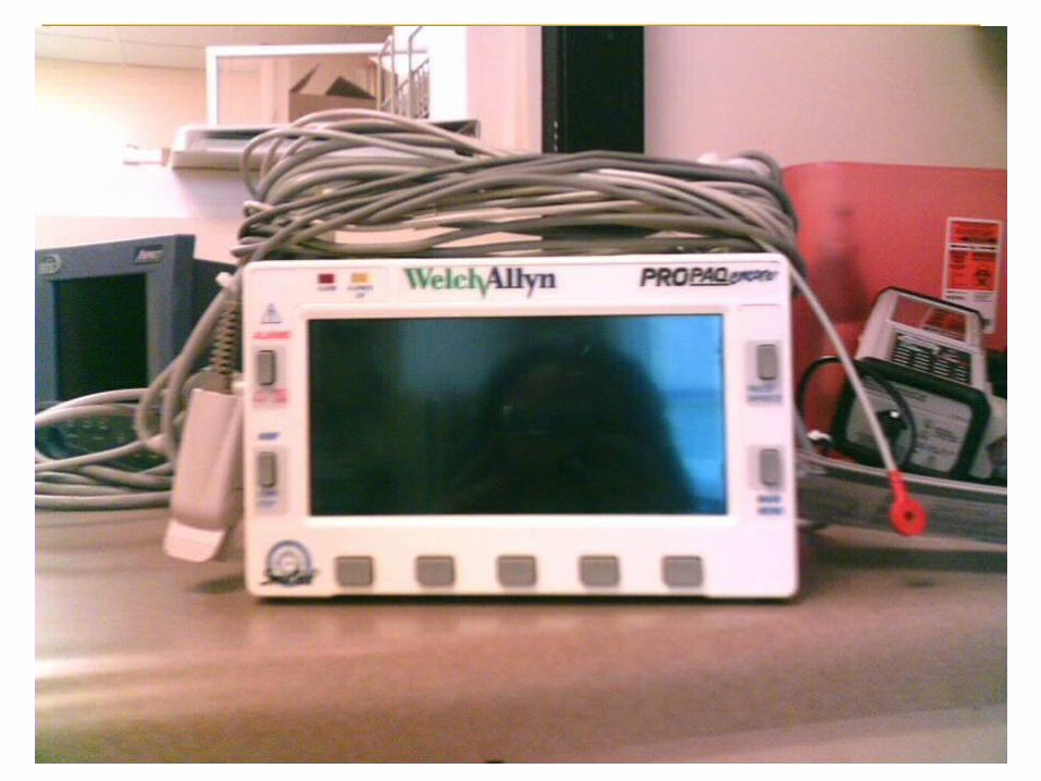
What to bring: Cart (peds vs. adult) Anesthesia machine/circuits (adult and peds) Monitors
Pro-pak w/ noninvasive BP cord and cuffs End-tidal CO2 monitor and window
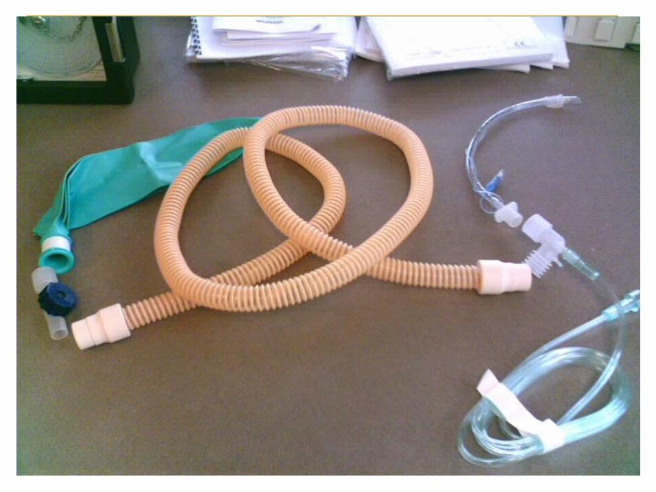
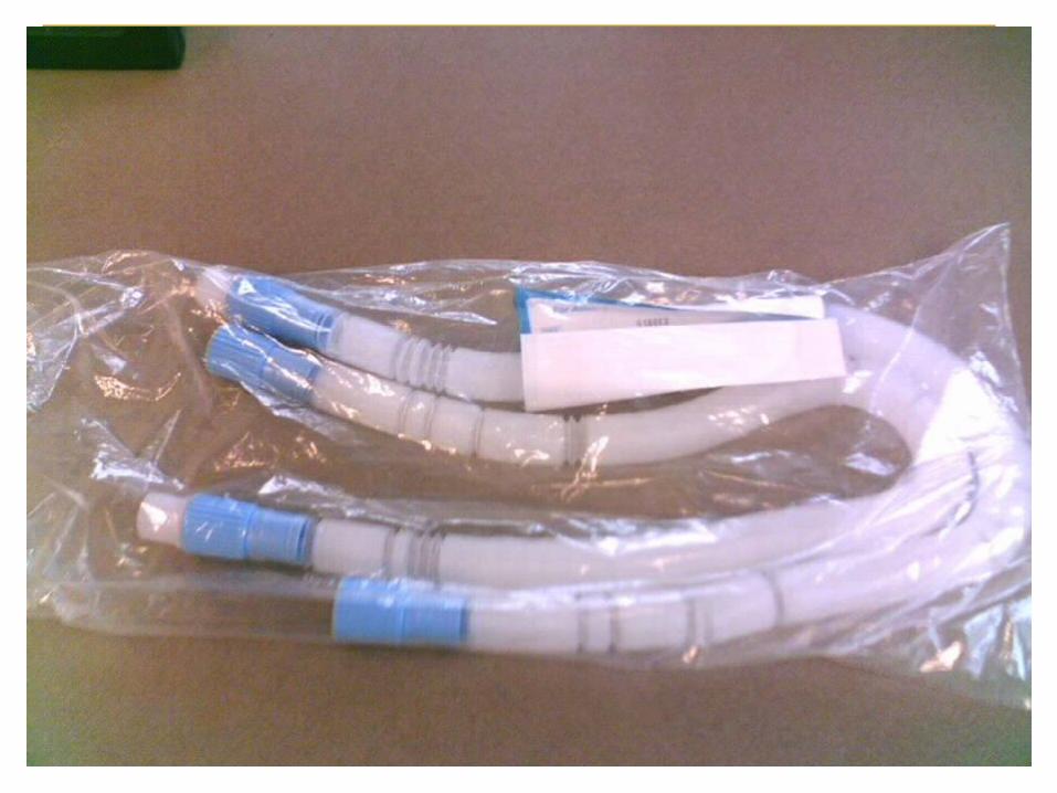
Airway NC/LMA/ETT MRI adapter Long corrugated ventilation tubing Jackson-Rees/Mapleson tubing
Syringe pump and 3 extension sets (this stays at the foot of the MRI table, far from the machine)
Meds: propofol, ketamine, midazolam, fentanyl, sux, NDMB, ephedrine as needed.
IV tubing and IV fluids Paper charts: pre-op, OR records, charge sheet, and PACU
order forms
MRI roadtrip, contd. Initial workup: vital signs, pre-op, set up your equipment in the
far corner of the holding area, and familiarize yourself with physical layout, location, verify availability of assistance, check gases, suction, and MRI monitors.
Induce the patient in the holding area on the MRI-safe cart, and then transport the patient to the MRI.
Do not take metal into the MRI room! Leave the monnitors, anesthesia machine, oxygen cylinder, etc.
outside of the room. Place the patient on the MRI table, and apply the MRI-
compatible monitors already available in the MRI suite. At the end of the case, take the patient back to the holding area
and extubate there. During the case, call angiography recovery room (yellow hallway)
to give warning for patient recovery (they use the same anesthesia post-op order forms).
Radiology RFA
Often done in CT but occasionally MRI. Kidney, lung, and liver. Currently requesting general anesthesia with ETT
secondary to prone positioning and the need to lay still for extended periods of time.
It is our job to check pressure points and padding. Radiology techs are not trained to be concerned.
Bring a face pillow in addition to the MRI road trip list.

Interventional Radiology
Embolization of cerebral and dural AVM’s, coiling of cerebral aneurysms, angioplasty of sclerotic lesions, and thrombolysis of acute thromboembolic stroke.
These procedures often require deliberate hypotension.
Radiologist may request rapid transition between deep sedation and an awake responsive state.
Cerebral Coiling In addition to the MRI road trip list you should bring:
Arterial line set up, extension tubing. Fluid warmer and Bair hugger/ lower body. Infusion pumps Medications: NTG, nipride, esmolol, labetalol,
heparin, and protamine Radiologist may request anything from deep IV
sedation to GA with ETT. Always have 2 large-gauge IV’s in place. One for drug
infusion and one for rapid fluid administration. Stay in constant communication with OR in case of an
emergency. Pt often transported to the ICU post-op.
Remote Cardiac Lab
Elective cardioversion Cart with emergency drugs. Etomidate or thiopental Standard monitoring Preoxygenate Give small incremental doses of etomidate or thiopental
until the eyelash reflex is abolished. Remove the mask immediately before the shock and
confirm no one is touching the pt. Ventilate with 100% O2 post-shock until consciousness is
regained. Consider RSI with ETT if high risk for aspiration.
Remote Cardiac Lab contd.
Cardiac RFA IV sedation with NC to GA with ETT depending on
the pt’s co-morbidities. Bring same supplies as MRI road trip. Take lots of propofol with you. Midazolam and fentanyl are used to titrate in
during the more painful parts of the procedure. (esp. the ablation)
Remote Cardiac Lab contd. Pacemaker/ ICD placement
Patients may be very sick, they may require GA.
(Consider the need for arterial line for BP monitoring). People have been known to code and require CPR.
GI endoscopy suite
Colonoscopy and upper GI scopes Pt’s are often MR, uncooperative or very sick
(bleeding esophageal varices, impaired liver function reducing drug metabolism) .
Take all of the equipment for the MRI road trip.
ECT Indications
Major depression Mania Certain forms of schizophrenia Parkinson’s syndrome
Contraindications Pheochromocytoma Increased ICP Recent CVA Cardiovascular conduction defects High risk pregnancy Aortic and cerebral aneurysms
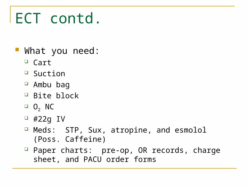
ECT contd.
What you need: Cart Suction Ambu bag Bite block O2 NC #22g IV Meds: STP, Sux, atropine, and esmolol (Poss. Caffeine) Paper charts: pre-op, OR records, charge sheet, and
PACU order forms
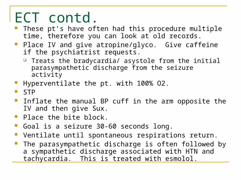
ECT contd. These pt’s have often had this procedure multiple time, therefore
you can look at old records. Place IV and give atropine/glyco. Give caffeine if the psychiatrist
requests. Treats the bradycardia/ asystole from the initial parasympathetic
discharge from the seizure activity Hyperventilate the pt. with 100% O2. STP Inflate the manual BP cuff in the arm opposite the IV and then
give Sux. Place the bite block. Goal is a seizure 30-60 seconds long. Ventilate until spontaneous respirations return. The parasympathetic discharge is often followed by a
sympathetic discharge associated with HTN and tachycardia. This is treated with esmolol.
Take Home Message
The standards of anesthesia care and patient monitoring are the same
regardless of location
THANK YOU