renal disease and pk/pd - ucla ctsi
TRANSCRIPT
Renal Disease and PK/PD
Anjay Rastogi MD PhD Division of Nephrology
Drugs and Kidneys Kidney is one of the major organ of drug elimination from the human body Renal disease and dialysis alters the pharmacokinetics and pharmacodynamics of most commonly used drugs Polypharmacy is common in renal disease patients with a median of 8 drugs being used Patients with renal disease on an average suffer from adverse effects as compared to the general population
Renal Disease and Drugs
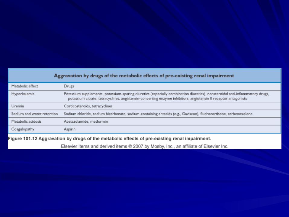
Decreased elimination Uremic effects Nephrotoxicity
Estimation of renal function Effect of renal disease on PK and PD Drug Nephrotoxicity Management of poisoning
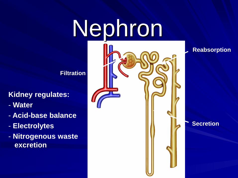
Nephron Filtration
Secretion
Reabsorption
Kidney regulates: - Water - Acid-base balance - Electrolytes - Nitrogenous waste excretion
6
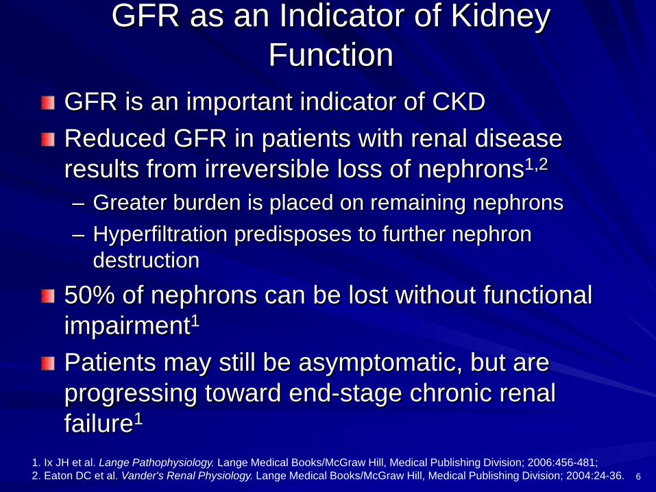
GFR as an Indicator of Kidney Function
GFR is an important indicator of CKD Reduced GFR in patients with renal disease results from irreversible loss of nephrons1,2
– Greater burden is placed on remaining nephrons – Hyperfiltration predisposes to further nephron
destruction 50% of nephrons can be lost without functional impairment1
Patients may still be asymptomatic, but are progressing toward end-stage chronic renal failure1
1. Ix JH et al. Lange Pathophysiology. Lange Medical Books/McGraw Hill, Medical Publishing Division; 2006:456-481; 2. Eaton DC et al. Vander's Renal Physiology. Lange Medical Books/McGraw Hill, Medical Publishing Division; 2004:24-36.
7
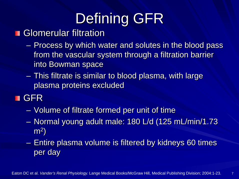
Defining GFR Glomerular filtration – Process by which water and solutes in the blood pass
from the vascular system through a filtration barrier into Bowman space
– This filtrate is similar to blood plasma, with large plasma proteins excluded
GFR – Volume of filtrate formed per unit of time – Normal young adult male: 180 L/d (125 mL/min/1.73
m2) – Entire plasma volume is filtered by kidneys 60 times
per day
Eaton DC et al. Vander’s Renal Physiology. Lange Medical Books/McGraw Hill, Medical Publishing Division; 2004:1-23.
8
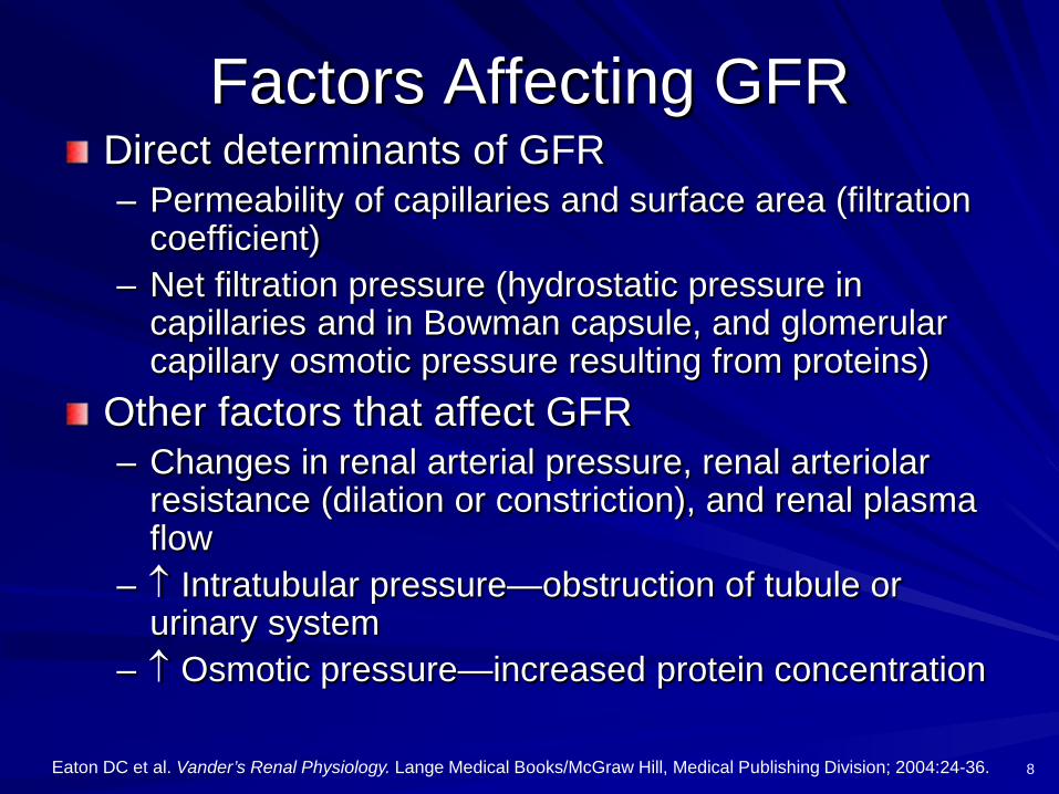
Factors Affecting GFR Direct determinants of GFR – Permeability of capillaries and surface area (filtration
coefficient) – Net filtration pressure (hydrostatic pressure in
capillaries and in Bowman capsule, and glomerular capillary osmotic pressure resulting from proteins)
Other factors that affect GFR – Changes in renal arterial pressure, renal arteriolar
resistance (dilation or constriction), and renal plasma flow
– ↑ Intratubular pressure—obstruction of tubule or urinary system
– ↑ Osmotic pressure—increased protein concentration
Eaton DC et al. Vander’s Renal Physiology. Lange Medical Books/McGraw Hill, Medical Publishing Division; 2004:24-36.
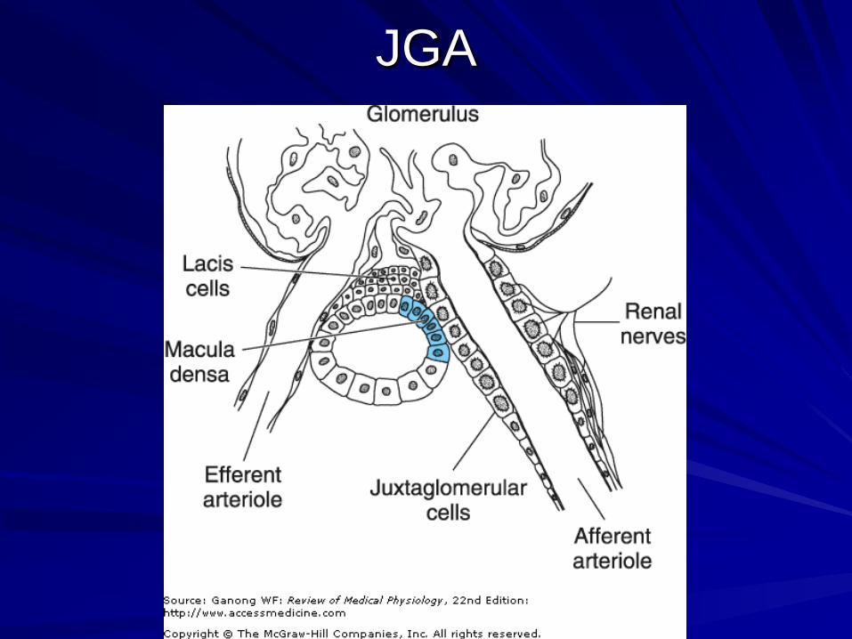
JGA
Estimation of GFR
Inulin clearance Iothalamate scans 24 hour urine collection eGFR Creatinine
Seru
m C
reat
inin
e (m
g/dL
)
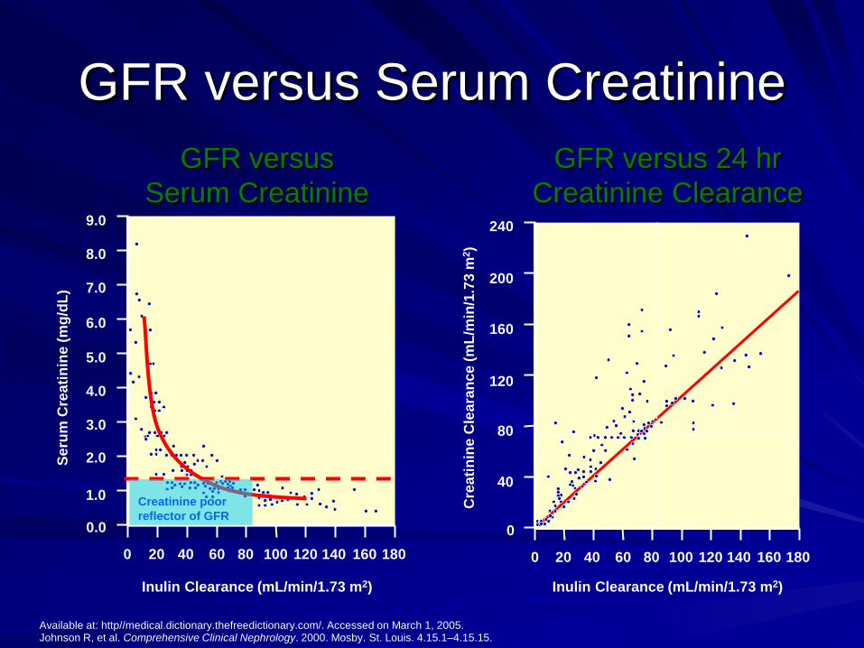
GFR versus Serum Creatinine
Available at: http//medical.dictionary.thefreedictionary.com/. Accessed on March 1, 2005. Johnson R, et al. Comprehensive Clinical Nephrology. 2000. Mosby. St. Louis. 4.15.1–4.15.15.
9.0
8.0
7.0
6.0
5.0
4.0
3.0
2.0
1.0
0.0
0 20 40 60 80 100 120 140 160 180
Inulin Clearance (mL/min/1.73 m2)
Creatinine poor reflector of GFR
GFR versus Serum Creatinine
GFR versus 24 hr Creatinine Clearance
240
200
160
120
80
40
0
Cre
atin
ine
Cle
aran
ce (m
L/m
in/1
.73
m2 )
0 20 40 60 80 100 120 140 160 180
Inulin Clearance (mL/min/1.73 m2)
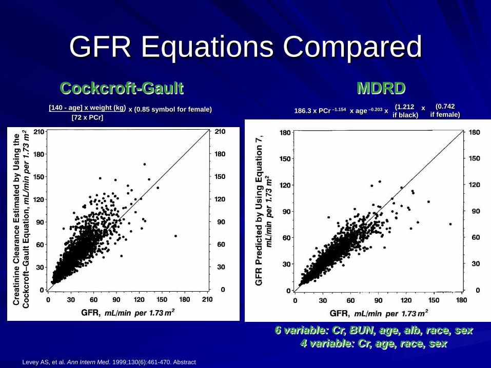
GFR Equations Compared
6 variable: Cr, BUN, age, alb, race, sex 4 variable: Cr, age, race, sex
Levey AS, et al. Ann Intern Med. 1999;130(6):461-470. Abstract
Cockcroft-Gault MDRD [140 - age] x weight (kg)
[72 x PCr] x (0.85 symbol for female) 186.3 x PCr –1.154 x age –0.203 x (0.742
if female) (1.212
if black) x
Pharmacokinetics
Pharmacokinetics
Absorption and bioavailability Drug distribution
Volume of distribution Protein binding
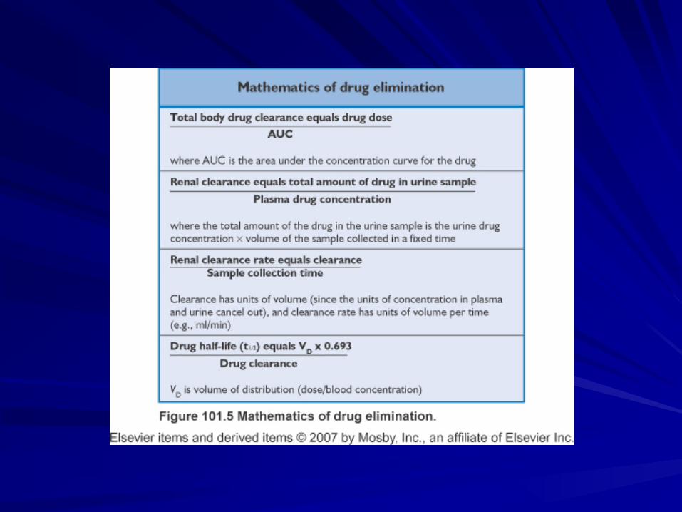
Biotransformation and drug metabolism Elimination
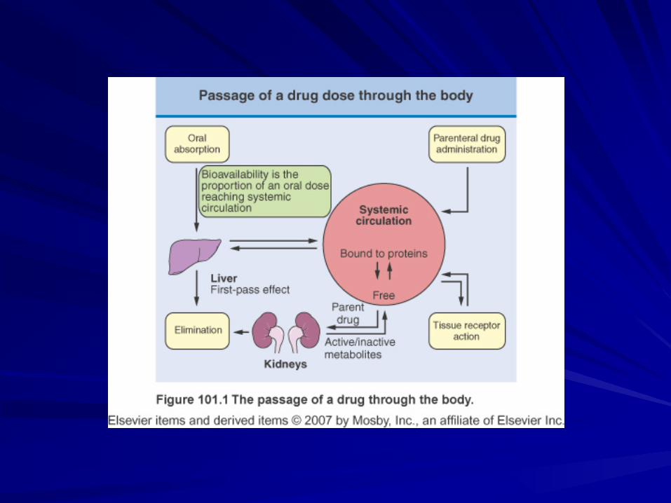
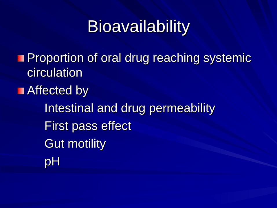
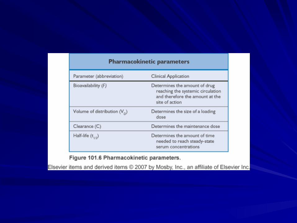
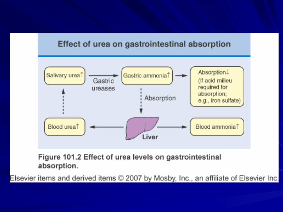
Bioavailability
Proportion of oral drug reaching systemic circulation Affected by
Intestinal and drug permeability First pass effect Gut motility pH
Plasma Protein Binding
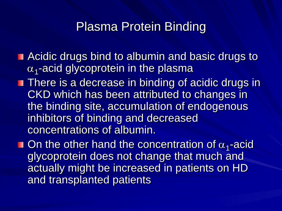
Acidic drugs bind to albumin and basic drugs to α1-acid glycoprotein in the plasma There is a decrease in binding of acidic drugs in CKD which has been attributed to changes in the binding site, accumulation of endogenous inhibitors of binding and decreased concentrations of albumin. On the other hand the concentration of α1-acid glycoprotein does not change that much and actually might be increased in patients on HD and transplanted patients
Nephrotic Syndrome
Complex interaction Hypoalbuminemia may result in lesser protein binding with more free drug in the plasma while at the same time there might be loss of albumin bound drug in the urine Some drugs are also known to produce adverse effects more readily in patients with nephrotic syndrome eg clofibrate can produce severe muscle necrosis
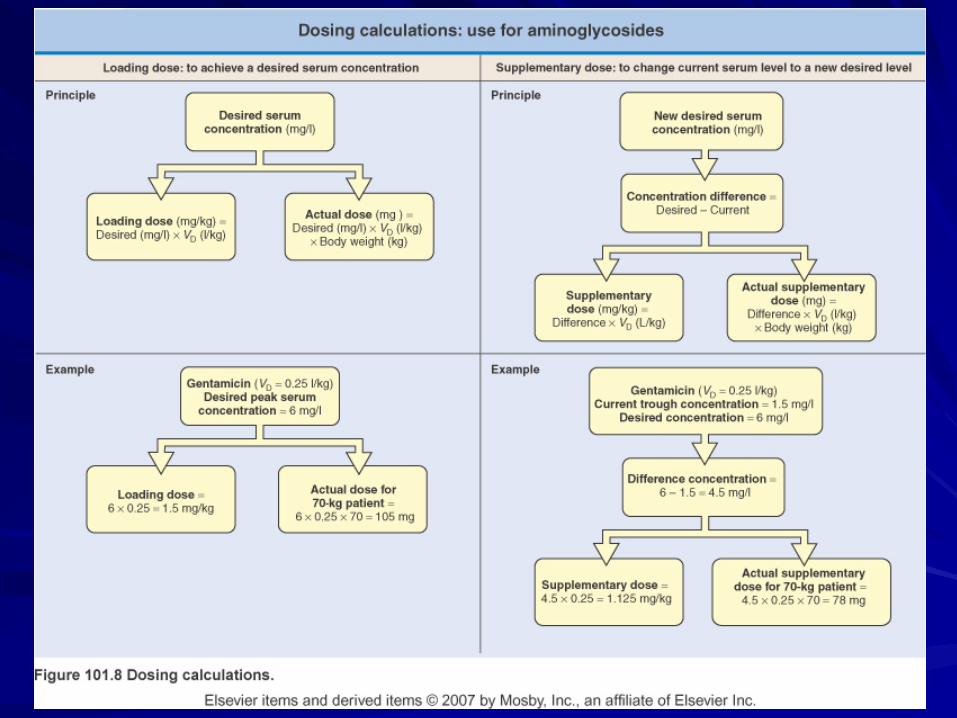
Volume of Distribution (VD)
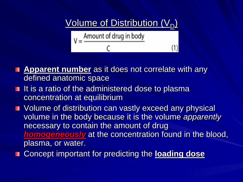
Apparent number as it does not correlate with any defined anatomic space It is a ratio of the administered dose to plasma concentration at equilibrium Volume of distribution can vastly exceed any physical volume in the body because it is the volume apparently necessary to contain the amount of drug homogeneously at the concentration found in the blood, plasma, or water. Concept important for predicting the loading dose
Factors affecting VD
Protein and tissue binding Volume status
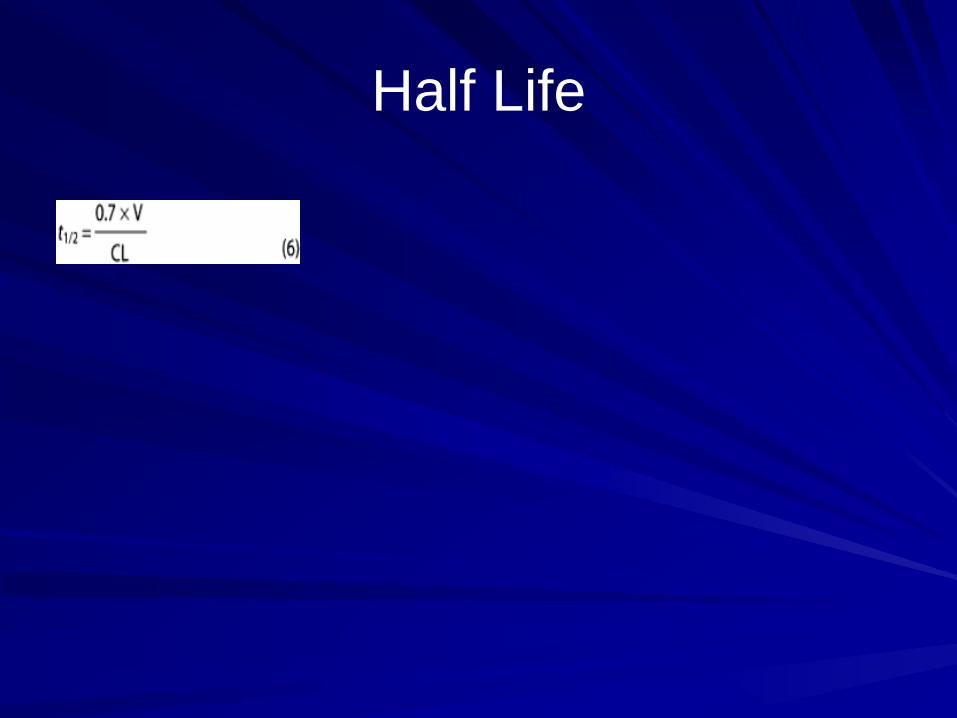
Half Life
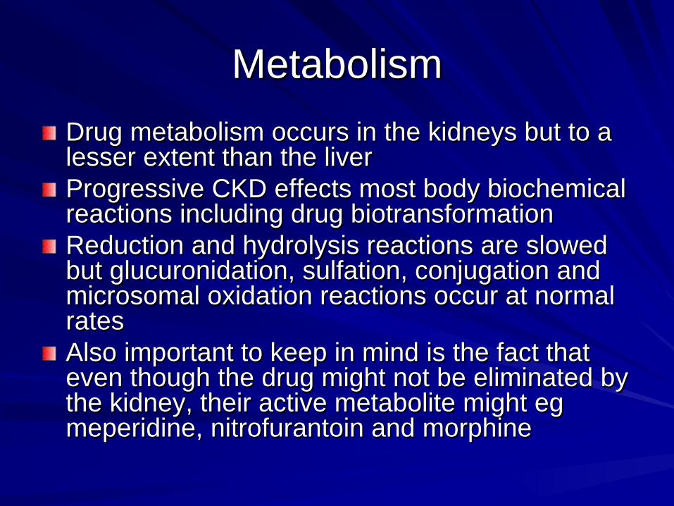
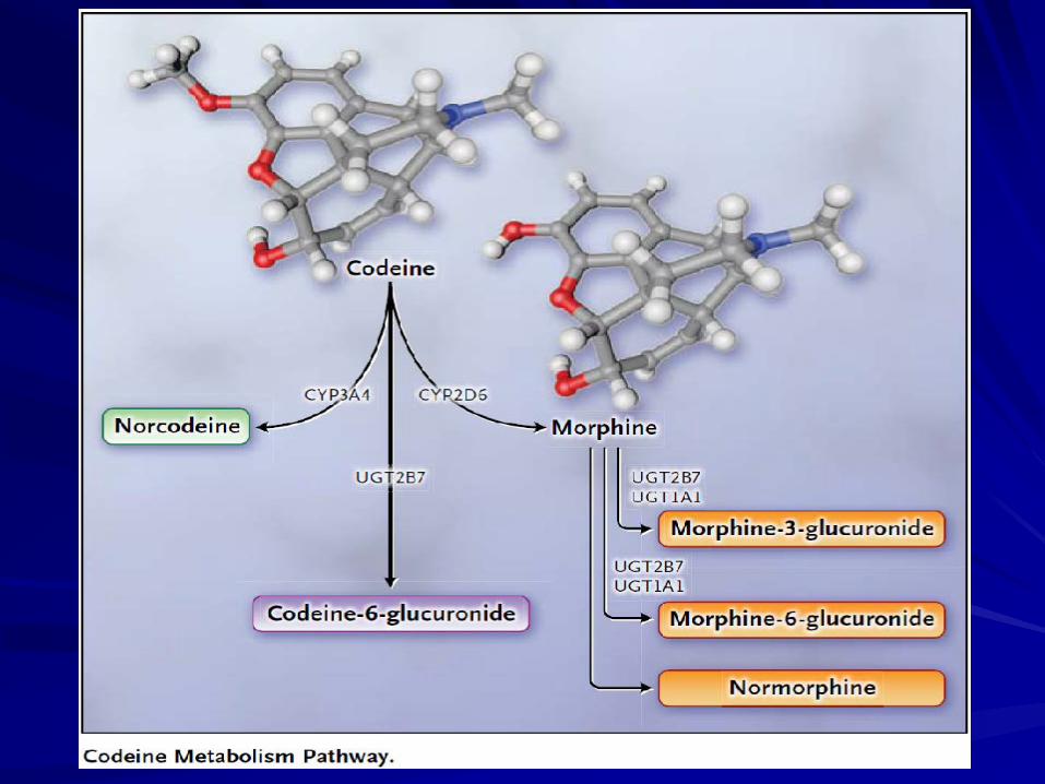
Metabolism Drug metabolism occurs in the kidneys but to a lesser extent than the liver Progressive CKD effects most body biochemical reactions including drug biotransformation Reduction and hydrolysis reactions are slowed but glucuronidation, sulfation, conjugation and microsomal oxidation reactions occur at normal rates Also important to keep in mind is the fact that even though the drug might not be eliminated by the kidney, their active metabolite might eg meperidine, nitrofurantoin and morphine
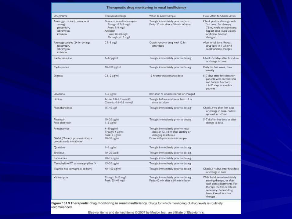
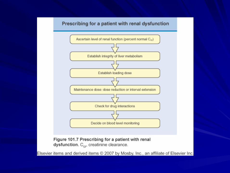
Dosing in a patient with renal insufficiency

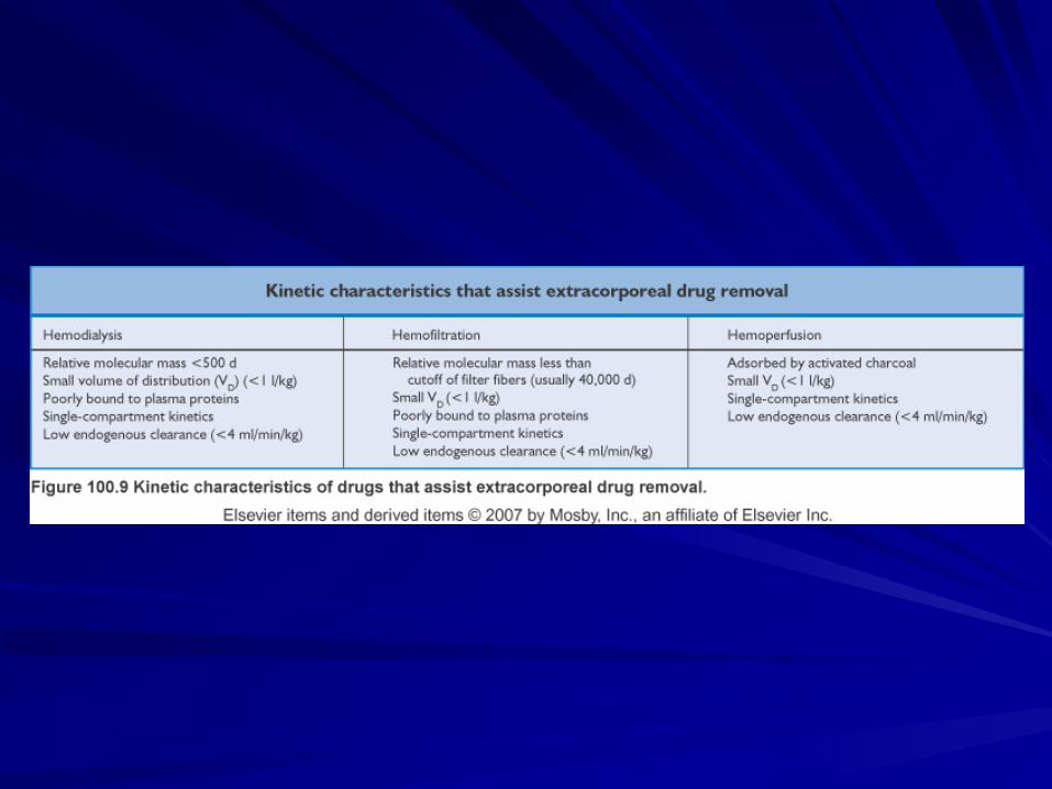
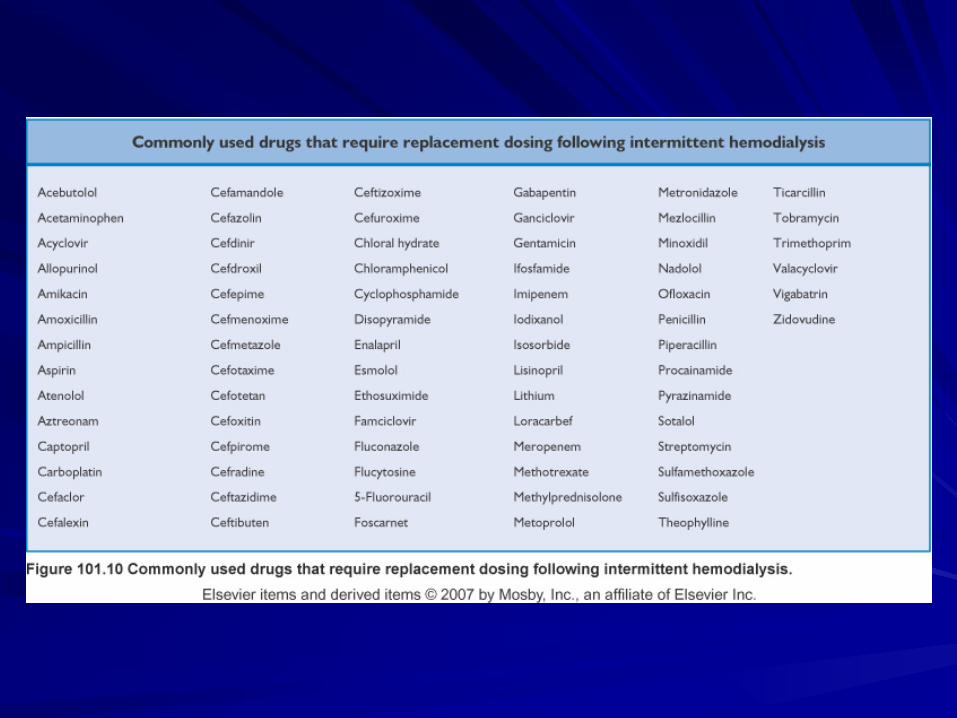
Extracorporeal Drug Losses
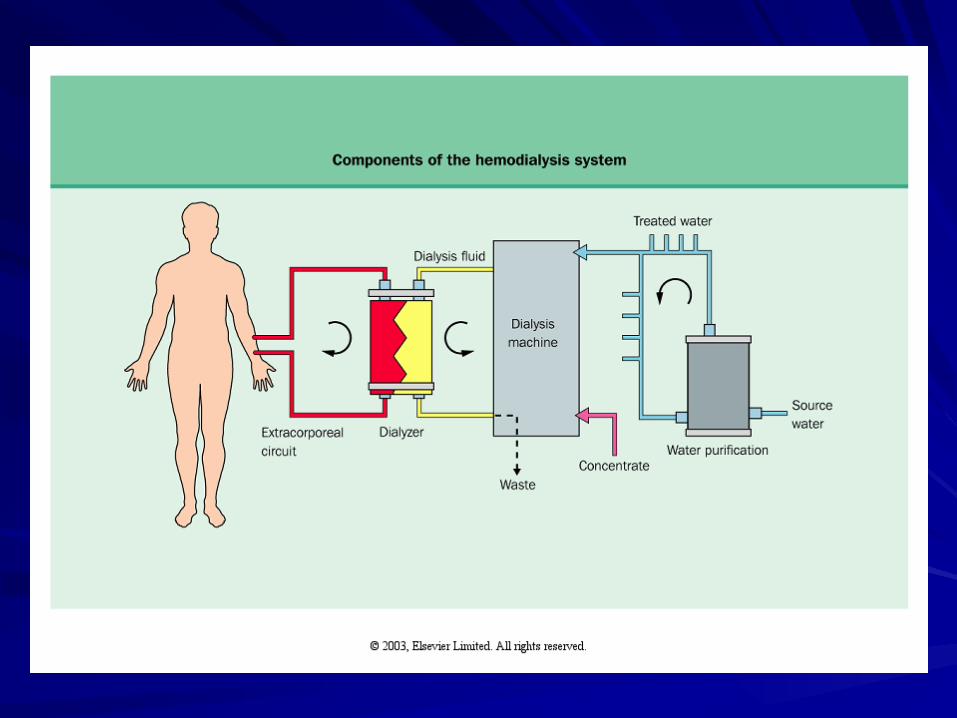
Dialysis
Dialysis is extracorporeal purification of blood Artificial Kidney
Dialysis
Diffusion of small molecules down their concentration gradient across a
semipermeable membrane
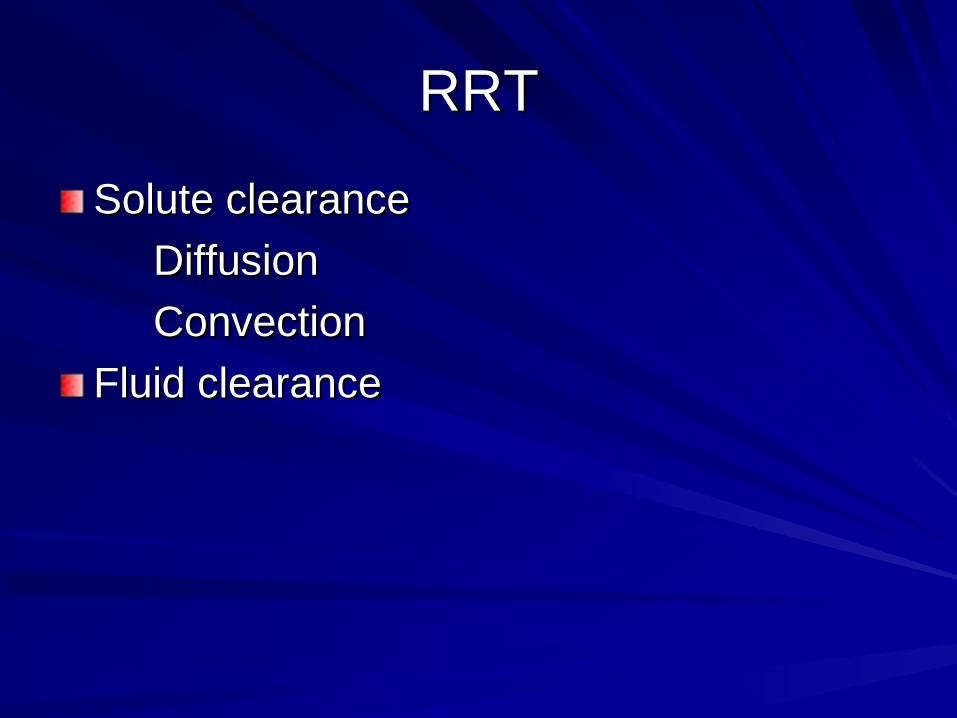
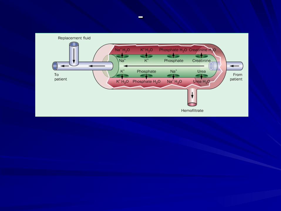
RRT
Solute clearance Diffusion Convection
Fluid clearance
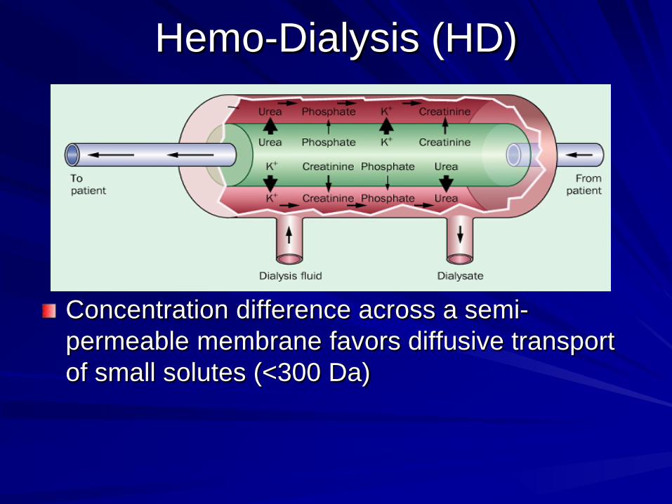
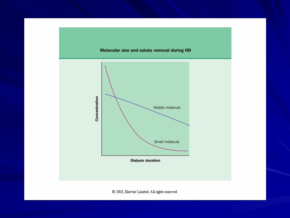
Hemo-Dialysis (HD)
Concentration difference across a semi-permeable membrane favors diffusive transport of small solutes (<300 Da)
Diffusion
Urea is used as the marker for small molecule diffusion during dialysis
Diffusive clearance is a function of Blood flow rate Membrane surface area Time
-
Ultra-filtration
Removal of water during dialysis from the patients circulation
HD – Transmembrane pressure gradient PD – Osmotically driven by glucose
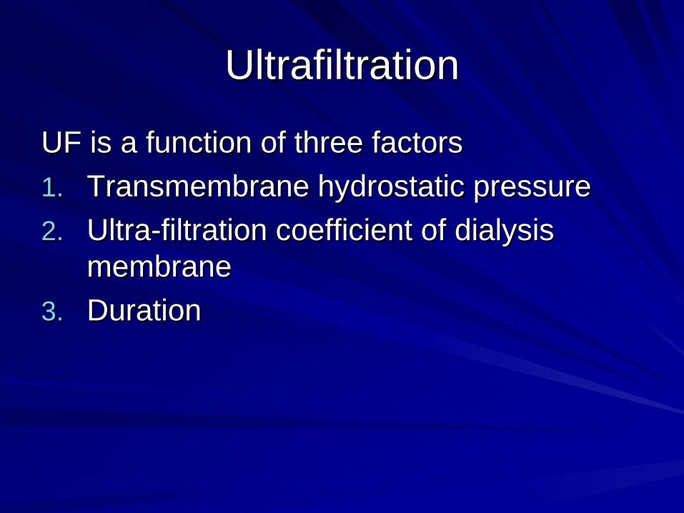
Ultrafiltration
UF is a function of three factors 1. Transmembrane hydrostatic pressure 2. Ultra-filtration coefficient of dialysis
membrane 3. Duration
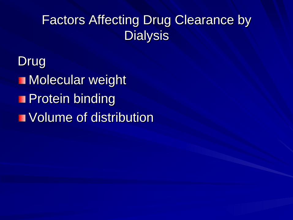
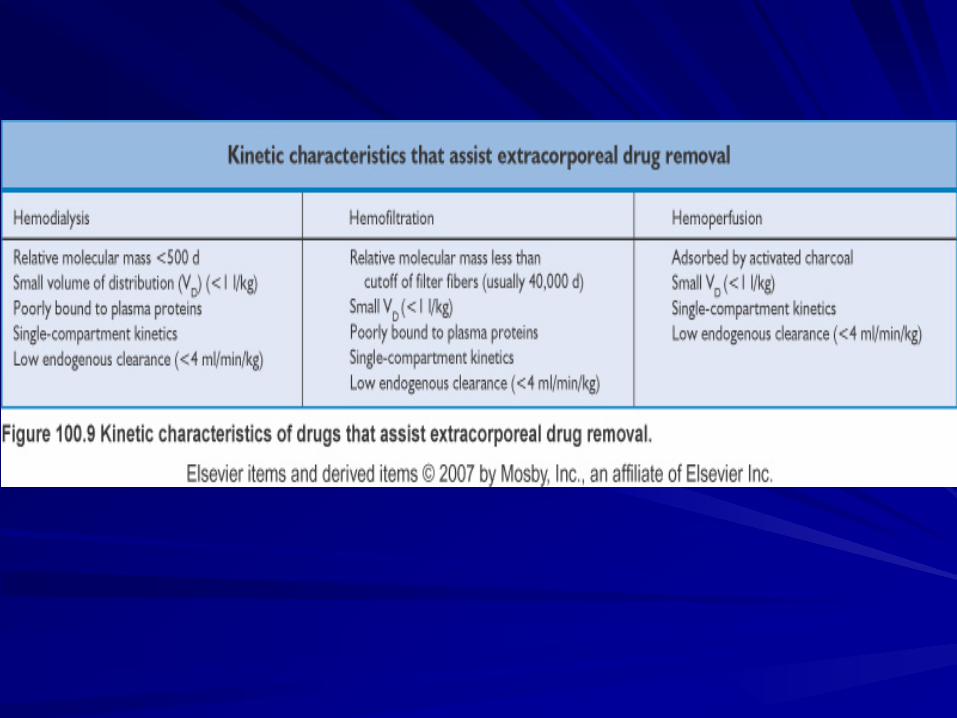
Factors Affecting Drug Clearance by Dialysis
Drug Molecular weight Protein binding Volume of distribution
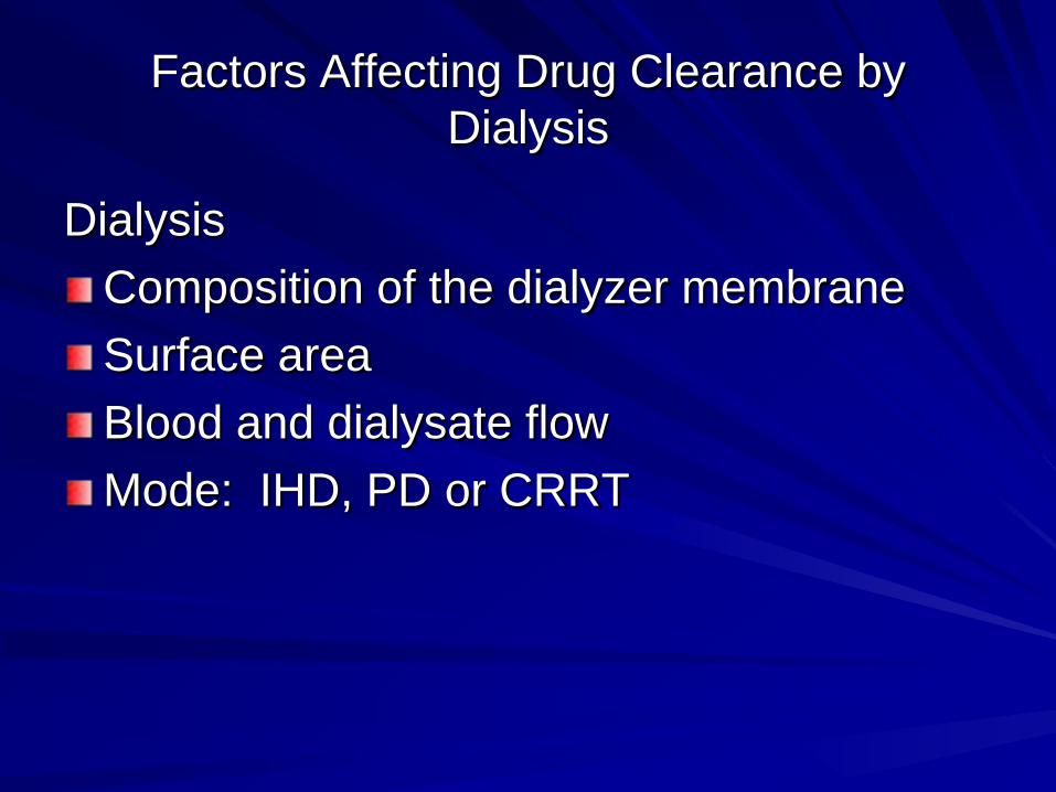
Factors Affecting Drug Clearance by Dialysis
Dialysis Composition of the dialyzer membrane Surface area Blood and dialysate flow Mode: IHD, PD or CRRT
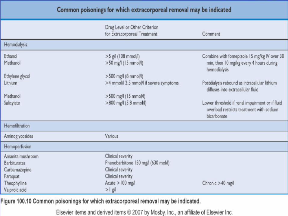
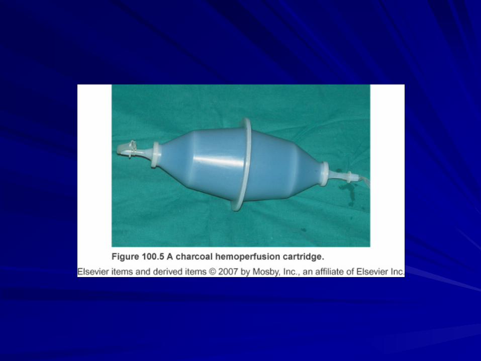
Hemoperfusion Extracorporeal form of treatment where large volumes of blood is passed over an adsorbent surface Activated carbon (irreversibly bound by van der Waals’ forces) and resins (not irreversibly bound) are the adsorbents most commonly used. Higher MW (100-40,000 daltons) are well adsorbed with charcoal having greater affinity for water soluble and resin for lipid soluble compounds Time factor

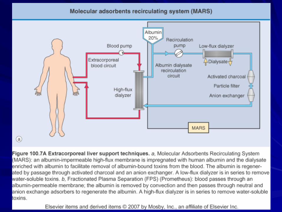
MARS
Molecular Adsorbent Recirculating System Effectively removes protein bound toxins
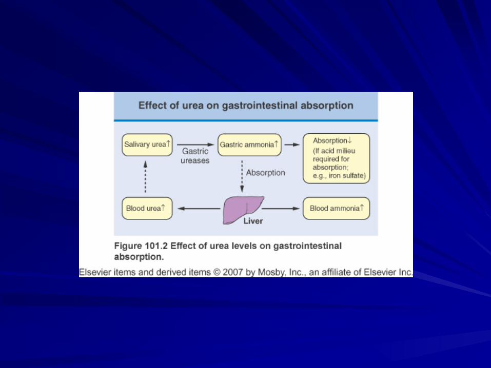
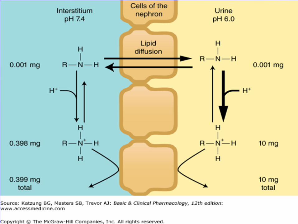
Diuresis at controlled pH
Nonionized drugs are lipid soluble and will diffuse through cell membranes relatively easily, promoting passive absorption of filtered drugs. By contrast, drugs in the ionized states are poor absorbed. Alteration in the urine pH can alter the absorption of weak acids and bases.
Nephrotoxicity
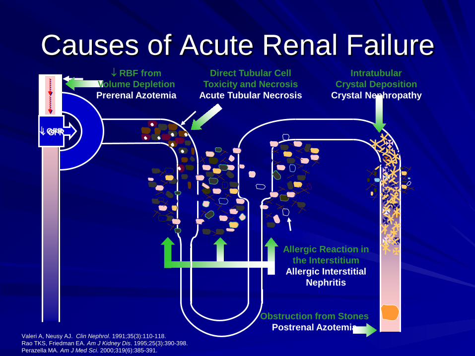
↓ GFR
Obstruction from Stones Postrenal Azotemia
Valeri A, Neusy AJ. Clin Nephrol. 1991;35(3):110-118. Rao TKS, Friedman EA. Am J Kidney Dis. 1995;25(3):390-398. Perazella MA. Am J Med Sci. 2000;319(6):385-391.
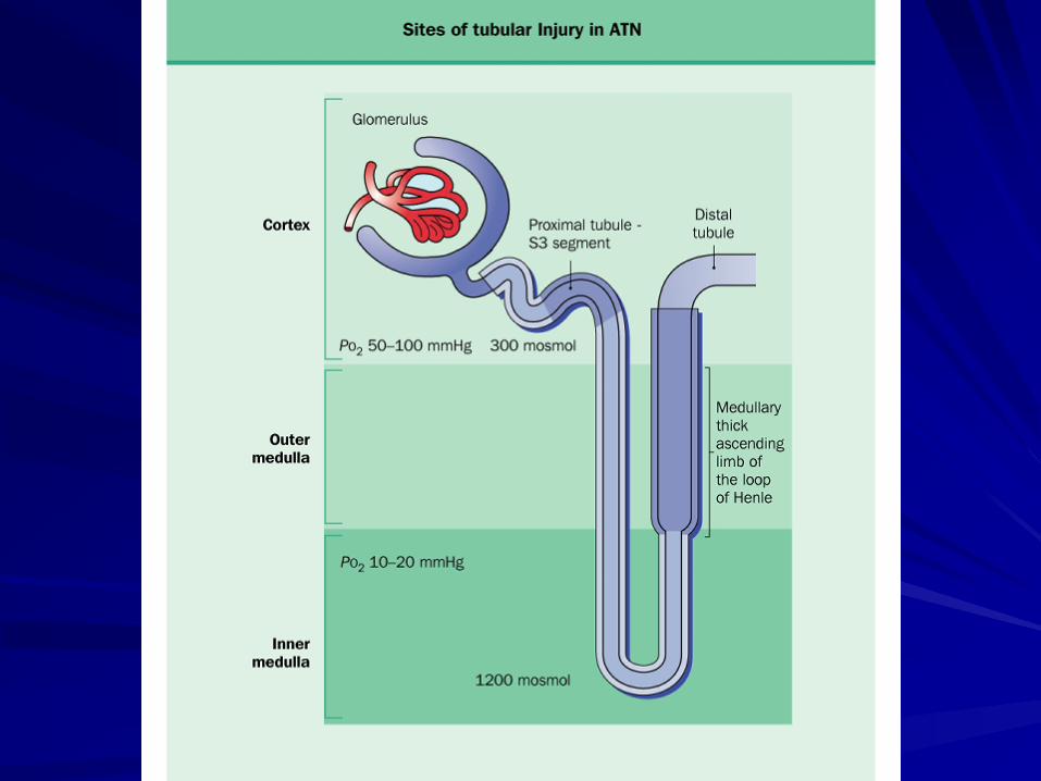
Causes of Acute Renal Failure Direct Tubular Cell
Toxicity and Necrosis Acute Tubular Necrosis
↓ RBF from Volume Depletion Prerenal Azotemia
Intratubular Crystal Deposition
Crystal Nephropathy
Allergic Reaction in the Interstitium
Allergic Interstitial Nephritis
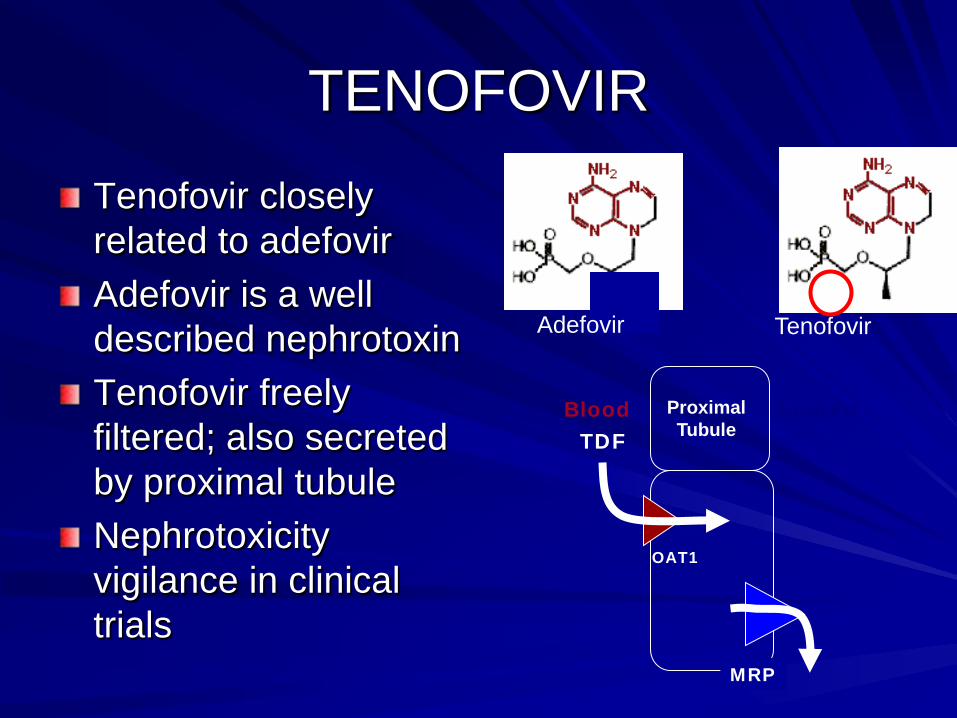
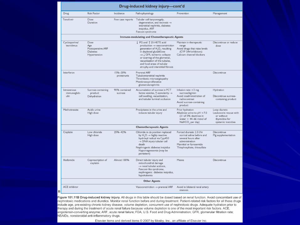
TENOFOVIR
Adefovir Tenofovir
Tenofovir closely related to adefovir Adefovir is a well described nephrotoxin Tenofovir freely filtered; also secreted by proximal tubule Nephrotoxicity vigilance in clinical trials
TDF Blood Lumen
OAT1
MRP
Proximal Tubule
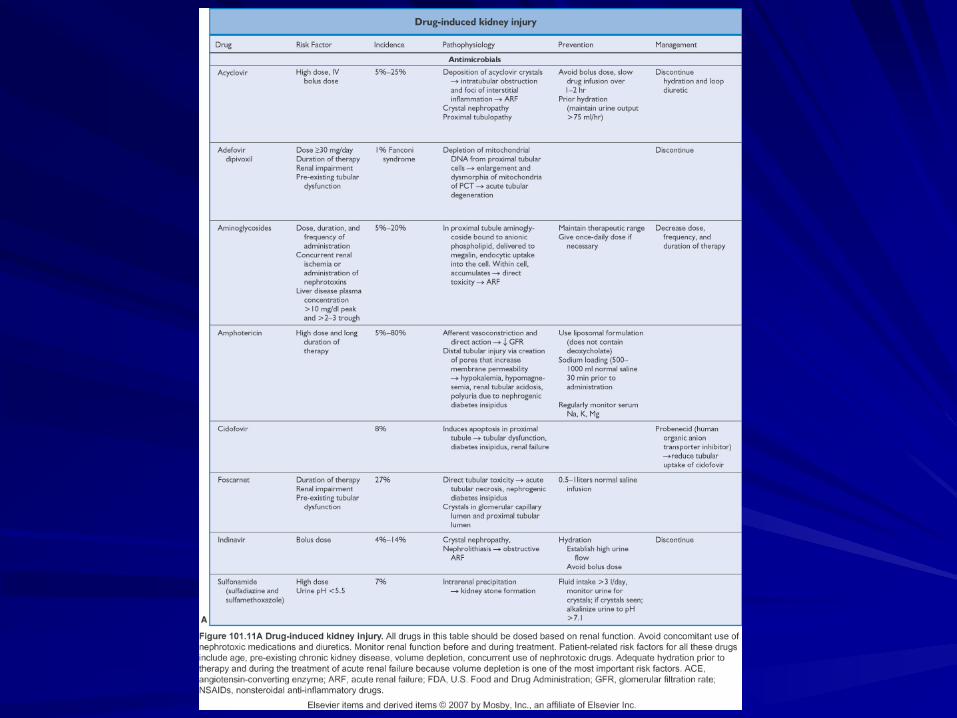

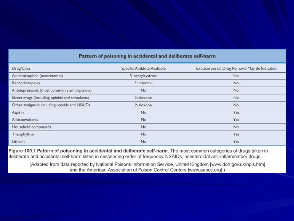
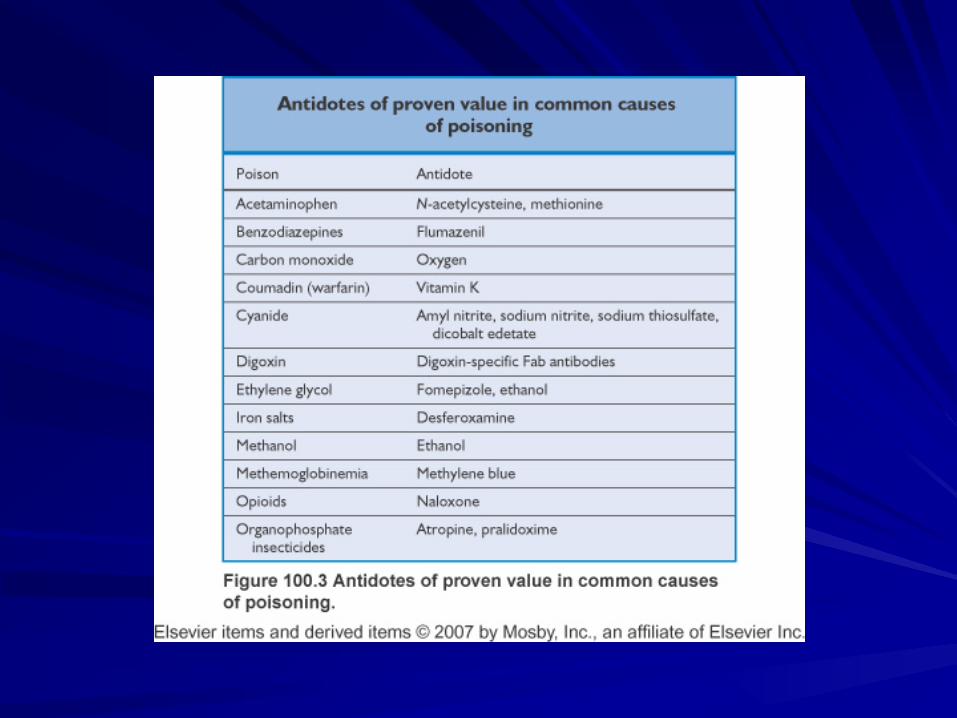
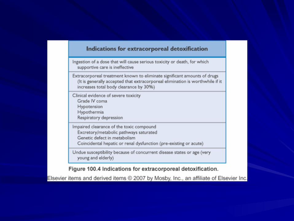
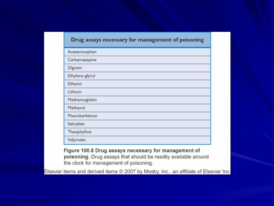
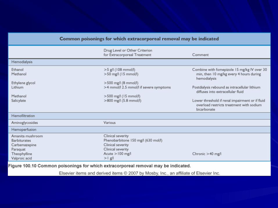
Toxicology