renal involvement in nail-patella syndrome: report of three cases
TRANSCRIPT
NEPHROLOGY - CASE REPORT
Renal involvement in nail-patella syndrome: reportof three cases
Puneet Sood Æ Maria C. Rojas Æ Zvi Talor
Received: 25 November 2008 / Accepted: 3 March 2009 / Published online: 19 March 2009
� Springer Science+Business Media, B.V. 2009
Nail-patella syndrome (NPS) or hereditary onycho-
osteodysplasia (HOOD) was originally described in
1820 by Chatelian, a patient with a triad of abnormal
nails, elbows, and knees. The first report describing the
family cluster of the disease was published in 1969 by
Beals and Eckhardt [1]. Case reports in the literature
regarding the nephropathy observed in NPS are varied,
including minimal change disease [2], recurrent uri-
nary tract infections, nephrolithiasis vesicoureteral
reflux, renal hypoplasia/atrophy, PAN-type vasculitis
[3], and Goodpasture’s syndrome [4].
Here we report three cases with their respective
pedigrees followed at our institution with evidence of
different types of renal manifestations.
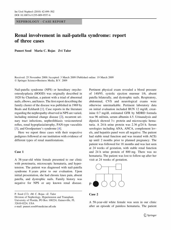
Case 1
A 38-year-old white female presented to our clinic
with proteinuria, microscopic hematuria, and hyper-
tension. The patient was diagnosed with nail-patella
syndrome 8 years prior to our evaluation. Upon
initial presentation, she had chronic knee pain, absent
patella, and dystrophic nails. Family history was
negative for NPS or any known renal disease.
Pertinent physical exam revealed a blood pressure
of 140/95, systolic ejection murmur 1/6, absent
patella bilaterally, and dystrophic nails. Respiratory,
abdominal, CVS and neurological exams were
otherwise unremarkable. Pertinent laboratory data
on initial evaluation included BUN 12 mg/dl, creat-
inine 0.7 mg/dl, estimated GFR by MDRD formula
was 96 ml/min, serum albumin 4.5. Urinanalysis and
dipstick showed 3? protein and microscopic hema-
turia. A 24-h urine protein was 2.36 g/24 h. Serum
serologies including ANA, ANCA, complement lev-
els, and hepatitis panel were all negative. The patient
had stable renal function and was treated with ACEi
up until 2 months prior to planned pregnancy. The
patient was followed for 18 months and was last seen
at 24 weeks of gestation, with stable renal function
and 24 h urine protein of 800 mg. There was no
hematuria. The patient was lost to follow-up after her
visit at 24 weeks of gestation.
P
Case 2
A 58-year-old white female was seen in our clinic
after an episode of painless hematuria. The patient
P. Sood (&) � M. C. Rojas � Z. Talor
Division of Nephrology, Hypertension and Transplant,
University of Florida, PO Box 100224, Gainesville, FL
32610-0224, USA
e-mail: [email protected]
123
Int Urol Nephrol (2010) 42:499–502
DOI 10.1007/s11255-009-9557-6

had previously been diagnosed with NPS. Her past
medical history was remarkable for deep venous
thrombosis of the right lower extremity, basal cell
carcinoma status post resection, and dyslipidemia.
Family history was positive for NPS in her father,
paternal grandfather, and four of her children.
Pertinent findings on physical exam were a blood
pressure of 150/78, dystrophic nails, absent patella
bilaterally, 1? lower extremity edema bilaterally, and
subluxation of the elbows. Pertinent laboratory data
on initial evaluation was a BUN 11 mg/dl, creatinine
0.8 mg/dl, albumin 4.3 g/dl, estimated 24-h protein
excretion based on a spot urine protein, and creatinine
ratio was 0.10 g. Urinalysis showed specific gravity
of 1.014, negative protein, and 23 RBC. Renal
ultrasound showed a right kidney of 11.2 cm and a
left kidney of 11.4 cm and no hydronephrosis or
structural lesion to explain the hematuria. The patient
has been followed for 2 years without any further
episodes of gross hematuria and has had a stable
baseline renal function and no proteinuria.
P
Case 3
A 27-year-old white female with a history of nail-
patella syndrome, grade IV vesicoureteral reflux,
status post left ureteral reimplantation in 1992, recur-
rent urinary tract infections, and non-nephrotic range
proteinuria. The patient was part of a large kindred of
NPS from her mother’s side. The patient was evaluated
in the adult nephrology clinic at 34 weeks of gestation.
Previously, she had been followed by pediatric
nephrology. Pertinent physical examination revealed
blood pressure of 110/60, dysmorphic nails, absent
patella bilaterally, pregnant abdominal exam, 2? pedal
edema bilaterally. Pertinent laboratory data included a
BUN 23 mg/dl, serum creatinine 0.7 mg/dl, albumin
2.7 g/dl. Twenty-four-hour urine collection revealed
proteinuria of 2.7 g/24 h. Renal ultrasound showed a
right kidney of 9.7 cm, left kidney size of 9.1 cm,
cortical scarring, and mild left hydronephrosis. She
had renal scarring of the left kidney on a dim-
ercaptosuccinic acid (DMSA) scan. No intervention
was done for proteinuria because of the patient’s
pregnant status. The patient was seen once more at
6 weeks after her uneventful pregnancy outcome. She
was found to have a proteinuria of 8 g based on a spot
urine exam. She did not have any dysmorphic RBCs on
her urianalysis and her blood pressure was in the
normal range. A renal biopsy was contemplated but the
patient was lost to follow-up after that visit.
P
Discussion
Nail-patella syndrome is a rare autosomal-dominant
pleiotropic genetic disorder with a prevalence of
1/500,000 and a high degree of penetrance but
variable expression [5]. The genetic abnormality
has been identified as a loss of function mutation in
LMX1B, and a transcription factor belonging to the
LIM homeodomain family localized in the long arm
of chromosome 9 (9q34) [6]. This transcription factor
is involved in the normal dorso-ventral patterning of
the limbs and normal development of glomerular
basement membrane in the kidney [7]. There is a
suggested role for LMX1B in the regulation of
collagen IV expression and in the transcriptional
regulation of podocyte specification and differentia-
tion [7]. Homozygous LMX1B knockout mice lack
patella and suffer from podocyte damage.
500 Int Urol Nephrol (2010) 42:499–502
123

Phenotypic characteristics of the NPS include
hypoplastic or absent patella, dystrophic fingernails
and toenails, dysplasia of the elbows, iliac horns,
hyperpigmentation of pupillary margins, heterochro-
mic iris with clover leaf deformity, microcornea,
glaucoma, and skin laxity [8]. Renal manifestations
of NPS were initially described by Hawkins and
Smith in 1950 with four of 21 affected individuals
showing clinical features of chronic glomerulone-
phritis [9]. Renal involvement in cases has been
reported as ranging from 25 to 60% and manifests as
proteinuria, microscopic hematuria, and hypertension
[8]. Proteinuria may present at any time from birth
onwards and may be intermittent. In a retrospective
study of 123 cases of NPS reported in the literature
by Meyrier et al., 60% of patients presented with
non-nephrotic range proteinuria. The proteinuria may
remain asymptomatic, remit spontaneously, or may
progress to nephrotic syndrome in less than 20% of
the patients [8, 10, 11]. In this study, 15% of the
patients progressed to ESRD. The severity of renal
impairment varies significantly among patients [8].
The presence of hematuria in NPS has been described
in 10–20% of patients [5, 7, 8]. Renal presentation
may occur for the first time during pregnancy. In the
study cohort of Meyrier et al., the incidence of pre-
eclampsia was found to be 29% as compared to 2–5%
described in pregnancies in general population [12].
In a cohort of eight Dutch affected families, seven
different mutations were identified. There was a high
incidence of renal disease in one of the families, with
the nephropathy sparing some family members and
with different spectrum of the severity of disease in
others [13]. To date, there is no correlation between
genotypic and phenotypic expression of the disease.
There are no specific light microscopy or immuno-
fluorescence features of NPS. Electron microscopy
shows irregular thickening of the glomerular basement
membrane with electron lucent areas, the so-called
‘‘moth-eaten appearance’’. Other histopathology find-
ings include podocyte foot process effacement,
fibrillary inclusions in the GBM, and diffuse hydropic
degeneration of tubular epithelium. The fibrils have
been identified as type III collagen [14].
There is no specific therapy for the renal involve-
ment in NPS. The use of RAAS blocking agents in
various settings of proteinuria has been discussed and
debated [15, 16]. We found a single case report of
successful treatment of proteinuria with combined
ACEi and ARB treatment for 2 years in a 7-year-old
pediatric patient with NPS [17]. Successful renal
transplantation has been reported [17, 18]. A case of
successful cadaveric renal transplant from a donor
with nail-patella syndrome has been reported in the
literature [18]. Genetic counseling is recommended
since it is an autosomal-dominant disease.
In summary, we present here three patients with
nail-patella syndrome, two of them with non-nephro-
tic range proteinuria, and one with microscopic
hematuria along with their respective pedigree anal-
ysis. Since some of these patients can end up with
loss of kidney function, regular clinical follow-up is
important.
References
1. Beals R, Eckhardt A (1969) Hereditary onycho-osteodys-
plasia. A report of nine kindreds. J Bone Joint Surg Am 51:
505–516
2. Hari P, Mantan M, Dinda A, Hari S, Bagga A (2006)
Steroid responsive nephrotic syndrome in a patient with
nail-patella syndrome. Pediatr Nephrol 21(8):1197–1199.
doi:10.1007/s00467-006-0154-y
3. Croock AD, Bashar KM, Powers JM (1987) Vasculitis and
renal disease in nail-patella syndrome: case report and
literature review. Ann Rheum Dis 46(7):562–565. doi:
10.1136/ard.46.7.562
4. Curtis JJ, Bhathena D, Leach RP, Galla JH, Lucas BA,
Luke RG (1976) Goodpasture’s syndrome in a patient with
the nail-patella syndrome. Am J Med 61(3):401–406. doi:
10.1016/0002-9343(76)90378-8
5. Sweeney E, Fryer A, Mountford R, Green A, McIntosh I
(2003) Nail-patella syndrome: a review of the phenotype
aided by developmental biology. J Med Genet 40:153–162.
doi:10.1136/jmg.40.3.153
6. Witzgall R (2008) How are podocytes affected in nail-
patella syndrome. Pediatr Nephrol 23(7):1017–1020. doi:
10.1007/s00467-007-0714-9
7. Bongers EM, Knoers NV (2003) From gene to disease; the
nail-patella syndrome and the LMX1B gene. Ned Tijdschr
Geneeskd 147(2):67–69
8. Meyrier A, Rizzo R, Gubler MC (1990) The nail-patella
syndrome. A review. J Nephrol 2:133–140
9. Sutcliffe NP, Cahsman SL, Savage COS, Fox JG, Boulton-
Jones JM (1989) Variability of the antigenicity of the
glomerular basement membrane in nail-patella syndrome.
Nephrol Dial Transplant 4:262–265
10. Daniel CRD, Osment LS, Noojin RO (1980) Triangular
lunulae. A clue to nail-patella syndrome. Arch Dermatol
116:448–449. doi:10.1001/archderm.116.4.448
11. Simila S, Vesa L, Wasz-Hockert O (1970) Hereditary
onyco-osteodysplasia (the nail-patella syndrome) with
nephrosis like renal disease in a new born. Pediatrics
46:61–65
Int Urol Nephrol (2010) 42:499–502 501
123

12. Skjaerven R, Wilcox AJ, Lie RT (2002) The interval
between pregnancies and risk of preeclampsia. N Engl
J Med 346(1):33–38
13. Knoers NV, Bongers EM, van Beersum SE, Lommen EJ,
van Bokhoven H, Hol FA (2000) Nail-patella syndrome.
Identification of mutations in the LMX1B gene in Dutch
families. J Am Soc Nephrol 11:1762–1766
14. Muth RG (1965) The nephropathy of hereditary osteo-
onychodysplasia. Ann Intern Med 62:1279–1285
15. Siamopoulos KC, Kalaitzidis RG (2008) Inhibition of
renin-angiotensin system and chronic kidney disease. Int
Urol Nephrol 40:1015–1025. doi:10.1007/s11255-008-
9424-x
16. Covic A, Gusbeth-Tatomir P, Goldsmith David JA (2007)
Current dilemmas in inhibiting the renal-angiotensin sys-
tem: do not forget real life. Int Urol Nephrol 39:571–576.
doi:10.1007/s11255-007-9211-0
17. Proesmans W, Van Dyck M, Devriendt K (2009) Nail-
patella syndrome, infantile nephrotic syndrome: complete
remission with antiproteinuric treatment. NDT Advance
Access. doi:10.1093/ndt/gfn725
18. Chan PC, Chan KW, Cheng IK, Chan MK (1988) Living
related renal transplantation in a patient with nail-patella
syndrome. Nephron 50:164–166. doi:10.1159/000185147
502 Int Urol Nephrol (2010) 42:499–502
123