renal pelvis renal cortex renal medulla minor calyx renal column renal pelivs ureter renal papilla...
TRANSCRIPT


Renalpelvis
Renal cortex
Renal medulla
Minor calyx
Renal column
Renal pelivs
Ureter
Renal papilla
Major calyx
Renal pyramid
Renal capsule
Nephrons
collecting duct
papilla
Renal cortex
Renal medulla
Boman's capsule
Renal tubule

Minor calyx
Major calyx
Renal pelvis
Renal artery
Renal vein
Renal papilla
Ureter
Renal pyramid
Renal column
Renal capsule
Renal medulla
Renal cortex

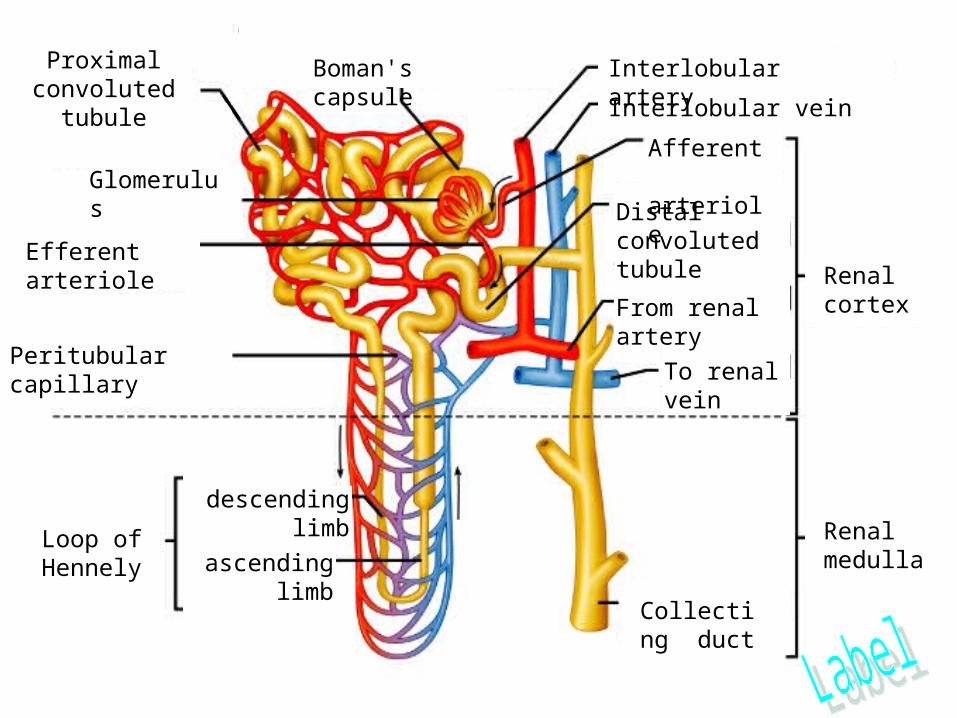
Proximal convoluted tubule
Glomerulus
Efferent arteriole
Peritubular capillary
Loop of Hennely
descending limb
ascending limb
Boman's capsule Interlobular artery
Interlobular vein
Afferent arteriole
Distal convoluted tubule
From renal artery
To renal vein
Collecting duct
Renal cortex
Renal medulla

Collecting duct
Distal convoluted tube
Loop of Henley
Vasa recta
Peritubular capillaries
Bowman’s capsule
Glomerulus
Proximal convoluted tube
Efferent arteriole Afferent arteriole
4243 37
4340
41
3635 34
3338 descending
39 ascending

The ureter is lined with transitional epithelium. The surrounding lamina propria and folded lumen allow for easy expansion of the lumen when necessary.
The bladder has transitional epithelium and a thick lamina propria to allow for expansion.
Red blood in, yellow urine out. The afferent arteriole is seen entering the glomerulus at the vascular pole (red arrows). The urinary pole (yellow arrow) is where the filtrate enters the tubule system (specifically, it enters the proximal convoluted tubule).


Credits
http://www.kumc.edu/instruction/medicine/anatomy/histoweb/urinary/urinary.htm
http://health.allrefer.com/pictures-images/