renal ultrasound - bmus
TRANSCRIPT

Renal Ultrasound (Basic Principles)
BMUS Study Day
Rosie Conlon Clinical Specialist Sonographer
NATIONAL REHABILITATION HOSPITAL
Dun laoghaire Dublin Sat 15th October 2016
WHY
ldquoBones can break muscles can atrophy glands can loaf about and even the brain can sleep without immediate danger to survival BUT when the kidneys failhellip Neither bone muscle gland nor brain could carry onrdquo
Homer William Smith
ldquoThe Evolution of the Kidneyrdquo Lectures on the Kidney (1943)
Renal Ultrasound (Basic Principles) and BMUS Study Case
PREPARATION
bull 500mls 1 hour before avoiding micturition
bull Catheter clamped 15 hours before
bull Fluids by PEG 15 hours before
bull Operator Dependant
bull Real Time
bull Reproducible
bull Non-invasive
bull Inspiration
WHAT - PROTOCOL
bull Both Kidneys
bull Urinary Bladder
bull +- Residual Volume
bull Pelvic Surveillance
bull Aorta
bull Local protocol pertinent to the population
bull Full bladder
The most important step in diagnosis is realising that it might exist
Renal Ultrasound (Basic Principles) and BMUS Study Case
RIGHT KIDNEY-TECHNIQUE
bull A 35-5 MHz probe is typically used to scan the kidney For the right kidney have the patient lie supine and place the probe in the right lower intercostal space in the midaxillary line Use the liver as your ldquoacoustic windowrdquo and aim the probe slightly posteriorly (toward the kidney) Gently rock the probe (up and down or side to side) to scan the entire kidney If needed you can have the patient inspire or exhale which allows for subtle movement of the kidney
bull Obtain longitudinal (long axis) and transverse (short axis) views
Renal Ultrasound (Basic Principles) and BMUS Study Case
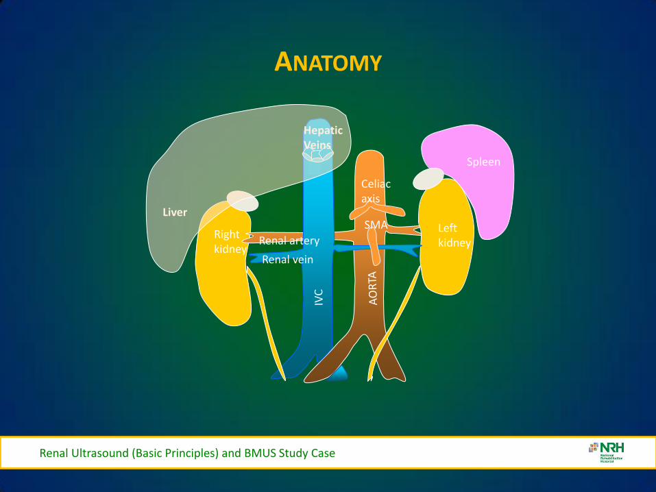
ANATOMY
Renal Ultrasound (Basic Principles) and BMUS Study Case
Celiac axis
SMA
Renal artery
Renal vein
Hepatic Veins
Right kidney
Left kidney
Liver
Spleen
NORMAL
Renal Ultrasound (Basic Principles) and BMUS Study Case
LEFT KIDNEY-TECHNIQUE
bull For the left kidney have the patient lie supine or in the right lateral decubitus position Place the probe in the lower intercostal space on the posterior axillary line The placement will be more cephalad and posterior than when visualizing the right kidney Again gently rock the probe to scan the entire kidney
bull Obtain longitudinal and transverse views
Renal Ultrasound (Basic Principles) and BMUS Study Case
APPROACH TO SCANNING
Renal Ultrasound (Basic Principles) and BMUS Study Case
bull Right kidney scanning approach anterior lateral posterior
bull Liver is the acoustic window
bull Left kidney requires a posterior approach through the spleen
bull Air-filled bowel impedes anterior scanning
I
LIVER STOMACH
IVC
AORTA K K
S
RENAL SCANNING APPROACHES
Renal Ultrasound (Basic Principles) and BMUS Study Case
NORMAL ANATOMY
Renal Ultrasound (Basic Principles) and BMUS Study Case
bull 9-12 cm long 4-5 cm wide 3-4 cm thick
bull Gerotarsquos fascia encloses kidney capsule perinephric fat
bull Sinus
ndash Hilum vessels nerves lymphatics ureter
ndash Pelvis major and minor calyces
bull Parenchyma surrounds the sinus
ndash Cortex site of urine formation contains nephrons
ndash Medulla contains pyramids that pass urine to minor calyces Columns of Bertin
separate pyramids
NORMAL ANATOMY
Renal Ultrasound (Basic Principles) and BMUS Study Case
Renal capsule Cortex
Medullary pyramids
Minor Calyx
Kidney Anatomy
Medulla
Sinus
Major Calyx
NORMAL ANATOMY
Renal Ultrasound (Basic Principles) and BMUS Study Case
bull Kidneys are retroperitoneal T12 - L4
bull Right kidney is lower than the left kidney
bull Right kidney is posterio-inferior to liver amp gallbladder
bull Left kidney is inferior-medial to the spleen
bull Adrenal glands are superior anterior medial to each kidney
THICKENED BLADDER WALL
Renal Ultrasound (Basic Principles) and BMUS Study Case
BLADDER VOLUME
Renal Ultrasound (Basic Principles) and BMUS Study Case
Ureteric Jets
Renal Ultrasound (Basic Principles) and BMUS Study Case
Male Anatomy
Prostate Gland Seminal Vesicles
OVER-HYDRATION
Pre-Micturition Post Micturition
Renal Ultrasound (Basic Principles) and BMUS Study Case
INTERVENTIONAL
VARIANTS
Renal Ultrasound (Basic Principles) and BMUS Study Case
bull Dromedary humps
ndash Lateral kidney bulge same echogenicity as the cortex
bull Hypertrophied column of Bertin
ndash Cortical tissue indents the renal sinus
bull Double collecting system
ndash Sinus divided by a hypertrophied column of Bertin
bull Horseshoe kidney
ndash Kidneys are connected usually at the lower pole
bull Renal ectopia
ndash One or both kidneys outside the normal renal fossa
VARIANTS
Pelvic Kidney
Dromedary Hump
Renal Ultrasound (Basic Principles) and BMUS Study Case
Renal Ultrasound (Basic Principles) and BMUS Study Case
EXTRA RENAL PELVIS
bull Extra Renal Pelvis refers to the presence of the renal pelvis outside the confines of the renal hilum It is a normal variant that is found in ~10 of the population
bull The renal pelvis is formed by all the major calyces An exta-renal pelvis usually appears dilated giving a false indication of an obstructive pathology
CT for clarification
bull Avoid confusion with hydroureter
or PUJ obstruction
Renal Ultrasound (Basic Principles) and BMUS Study Case
VARIANTS CONT
Renal Ultrasound (Basic Principles) and BMUS Study Case
HORSESHOE KIDNEY
Renal Ultrasound (Basic Principles) and BMUS Study Case
CROSS FUSED ECTOPIA
bull Cross fused ectopic kidney
bull The left kidney is fused to the lower pole of the right kidney
Renal Ultrasound (Basic Principles) and BMUS Study Case
RENAL CYSTS
Cyst 1 Cyst 2
Renal Ultrasound (Basic Principles) and BMUS Study Case
RENAL SCARRING
Renal Ultrasound (Basic Principles) and BMUS Study Case
MULTICYSTIC DYSPLASTIC KIDNEY
bull Right kidney in a new-born shows no normal renal parenchyma and the right renal fossa filled with multiple simple appearing cystic structures cw MCDK The cysts did NOT communicate
Renal Ultrasound (Basic Principles) and BMUS Study Case
ANGIOMYOLIPOMA
bull This is a homogeneous highly echogenic usually rounded lesion in the renal parenchyma containing blood vessels muscle tissue and fat as the name suggests They are usually solitary asymptomatic lesions found incidentally although larger lesions can haemorrhage causing haematuria and pain
Renal Ultrasound (Basic Principles) and BMUS Study Case
MULTIPLE ANGIOMYOLIPOMAS
bull Also associated with Tuberose sclerosis they tend to be multiple and bilateral
Renal Ultrasound (Basic Principles) and BMUS Study Case
OBSTRUCTIVE UROPATHY GRADING SYSTEM - SUBJECTIVE
bull Mild Minimal separation of calyces
bull Moderate Dilation of major and minor calyceal system
bull Severe Marked dilation of the renal pelvis and thinning of the renal parenchyma
Renal Ultrasound (Basic Principles) and BMUS Study Case
RANGE OF HYDRONEPHROSIS
Renal Ultrasound (Basic Principles) and BMUS Study Case
Normal Mild Moderate Severe
HYDRONEPHROSIS
Renal Ultrasound (Basic Principles) and BMUS Study Case
Intrinsic acquired
bull Renal lithiasis bull Neoplasm (renal ureteral bladder)
bull Papillary necrosis
bull Ureterocele
bull Blood clot
bull Neurogenic bladder
bull Anticholinergics
bull Pregnancy PID uterine prolapse)
bull Diuretics
bull Vesico-ureteral reflux
bull Diabetes insipidus
Intrinsic congenital
bull Stenosis (ureteral urethral meatal)
bull Adynamic ureter
bull Spinal cord defects
bull Duplication of the ureter
bull Ureterocele
HYDRONEPHROSIS
Mild Hydronephrosis
Renal Ultrasound (Basic Principles) and BMUS Study Case
HYDRONEPHROSIS
Moderate Hydronephrosis
Renal Ultrasound (Basic Principles) and BMUS Study Case
SEVERE HYDRONEPHROSIS
Renal Ultrasound (Basic Principles) and BMUS Study Case
Severe Hydronephrosis
SMALL KIDNEYS
bull Unilateral (may be bilateral)
ndash Chronic infections
ndash RAS Renal Artery Stenosis
ndash Hypoplastic Kidney
ndash Always bilateral
bull Chronic glomerulonephritis
bull Hypertensive nephropathy
bull Collagen Vascular Disease
Renal Ultrasound (Basic Principles) and BMUS Study Case
ENLARGED KIDNEYS
Always Unilateral
ndash Compensatory hypertrophy
ndash Bilateral or Unilateral
bull Renal Mass
bull Hydronephrosis
bull Renal vein thromboses
bull Lymphoma
bull Amyloidosis
Always Bilateral
ndash PCK Polycystic Kidney Disease
ndash AGN Acute glomerulonephritis
ndash Amyloidosis
Renal Ultrasound (Basic Principles) and BMUS Study Case
Urolithiasis
Renal Calculi- Bladder Calculi
US only
41
Fig 3
Fig 4 Fig 5
Fig 3 4 amp 5 ndash Bladder stones with posterior shadowing clearly seen at US in distended bladder but not apparent on X-ray
Staghorn Calculus
BLADDER DIVERTICULAE
Renal Ultrasound (Basic Principles) and BMUS Study Case
bull Diverticula of the bladder in
infants and children are common and occur when bladder mucosa protrudes through a congenital defect in the detrusor muscle wall Most are primary Those secondary to obstruction or neurogenic dysfunction are less common than previously thought
Renal Ultrasound (Basic Principles) and BMUS Study Case
Renal Ultrasound (Basic Principles) and BMUS Study Case
BLADDER CALCULI
Renal Ultrasound (Basic Principles) and BMUS Study Case
RENAL MASS
Renal Ultrasound (Basic Principles) and BMUS Study Case
bull Pyelonephritis sonographic appearance is most commonly normal but you may find hypoechoic cortex and loss of demarcation between the outer cortex and middle pyramids and columns of Bertin
bull Renal mass may have any echotexture (hyperechoic anechoic etc) and appear anywhere within the kidney
bull Transplant kidney a normal echotexture kidney typically in a pelvic location
TRANSITIONAL CELL CARCINOMA
bull Ultrasound images Large transitional cell carcinoma in the upper pole of the RK The changes are more subtle than those of renal cell carcinoma and the renal outline remains intact
bull Transitional cell carcinoma is the most common bladder tumour occurring less frequently in the collecting system of the kidney and the ureter It usually presents with haematuria while still small It is best diagnosed with cystoscopy
Renal Ultrasound (Basic Principles) and BMUS Study Case
HYDRONEPHROSIS DUE TO TCC URETER
Renal Ultrasound (Basic Principles) and BMUS Study Case
US AND CT RENAL CA
Renal Ultrasound (Basic Principles) and BMUS Study Case
RENAL CELL CARCINOMA
bull Large heterogeneous mass which enlarges and deforms the shape of the kidney (Fig below) The mass may contain areas of cystic degeneration andor calcification It has a predilection to spread into the ipsilateral renal vein and IVC
bull Colour Doppler usually reveals a disorganized and increased blood flow pattern within the mass with high velocities from the arterioverous shunts within the carcinoma
Renal Ultrasound (Basic Principles) and BMUS Study Case
bull Smaller RCCs can be hyperechoic and may be confused with benign angiomyolipoma The latter has well-defined borders whilst an RCC is illdefined
BURKITTS LYMPHOMA
Renal Ultrasound (Basic Principles) and BMUS Study Case
NEUROBLASTOMA
Renal Ultrasound (Basic Principles) and BMUS Study Case
RENAL TRAUMA
Renal Ultrasound (Basic Principles) and BMUS Study Case
POLYCYSTIC KIDNEYS
Renal Ultrasound (Basic Principles) and BMUS Study Case
RENAL TRANSPLANTS-SURGICAL TECHNIQUE
bull Usually heterotopic (placed in addition to the native diseased kidneys)
bull Positioned in the extra-peritoneal pouch in the iliac fossa (usually the right) anterior to the iliacus and psoas muscles
Renal Ultrasound (Basic Principles) and BMUS Study Case
THE ROLE OF ULTRASOUND
B Mode imaging
bull Morphological appearances
PC dilation
Peri-renal fluid collections
bull Doppler
Colour Power Perfusion
Spectral Doppler
Waveforms
bull Intervention
Guide Biopsy Procedures
Drain Fluid Collections
Placement of Nephrostomy
Tubes
Renal Ultrasound (Basic Principles) and BMUS Study Case
LS RENAL TRANSPLANT (USING EXTENDED FIELD OF VIEW)
Renal Ultrasound (Basic Principles) and BMUS Study Case
PERFUSION WITH POWER AND COLOUR DOPPLER
Renal Ultrasound (Basic Principles) and BMUS Study Case
BACKGROUND
The main purpose of regular urology review is to preserve renal function with the ultimate aim of a non obstructed urinary system
Urinary Tract Infections (UTI) are the most frequent medical complication in patients with Spinal Cord Injuries (SCI) and a major cause of morbidity
Renal Ultrasound (Basic Principles) and BMUS Study Case
X-RAY ONLY
Fig 1 amp 2 - US showing decompressed bladder around IDUC X-ray in same patient confirming large calculus not visualised on US
Fig 1 Fig 2
Renal Ultrasound (Basic Principles) and BMUS Study Case
US ONLY
Fig 3
Fig 4
Fig 5
Fig 3 4 amp 5 ndash Bladder stones with posterior shadowing clearly seen at US in distended bladder but not apparent on X-ray
Renal Ultrasound (Basic Principles) and BMUS Study Case
RESULTS
bull Bladder ultrasound alone diagnosed 36 bladder calculi compared to 18 diagnosed by X-ray alone
bull US and X-ray combined diagnosed 46
Renal Ultrasound (Basic Principles) and BMUS Study Case
BLADDER MUCUS
bull In one patient a focal mobile area of increased reflectivity was demonstrated posteriorly in the bladder No posterior acoustic shadowing was seen to suggest of calculus formation
bull On flexible cystoscopy no calculus identified only mucus seen
Renal Ultrasound (Basic Principles) and BMUS Study Case
CASE STUDY 1
Renal Ultrasound (Basic Principles) and BMUS Study Case
CASE STUDY 2-VHL (VON HIPPEL LINDAU SYNDROME)
bull renal lesions
ndash renal cell carcinoma(s) (RCCs) usually of the clear cell type
bull 70 lifetime risk
bull RCCs present at an earlier age in those with vHL
ndash renal cysts
bull can occur in up to 75 of cases
bull often tend to be bilateral and multiple
bull Prognosis is poor with a median survival of ~50 years with the most common cause of death being RCC and cerebellar haemangioblastomas
Renal Ultrasound (Basic Principles) and BMUS Study Case
Renal Ultrasound (Basic Principles) and BMUS Study Case
Case Study 3
Renal Ultrasound (Basic Principles) and BMUS Study Case
Renal Ultrasound (Basic Principles) and BMUS Study Case
PAGE KIDNEY
Renal Ultrasound (Basic Principles) and BMUS Study Case
THANK YOU FOR YOUR ATTENTION
Renal Ultrasound (Basic Principles) and BMUS Study Case
Rosie Conlon rosieconlonnrhie

WHY
ldquoBones can break muscles can atrophy glands can loaf about and even the brain can sleep without immediate danger to survival BUT when the kidneys failhellip Neither bone muscle gland nor brain could carry onrdquo
Homer William Smith
ldquoThe Evolution of the Kidneyrdquo Lectures on the Kidney (1943)
Renal Ultrasound (Basic Principles) and BMUS Study Case
PREPARATION
bull 500mls 1 hour before avoiding micturition
bull Catheter clamped 15 hours before
bull Fluids by PEG 15 hours before
bull Operator Dependant
bull Real Time
bull Reproducible
bull Non-invasive
bull Inspiration
WHAT - PROTOCOL
bull Both Kidneys
bull Urinary Bladder
bull +- Residual Volume
bull Pelvic Surveillance
bull Aorta
bull Local protocol pertinent to the population
bull Full bladder
The most important step in diagnosis is realising that it might exist
Renal Ultrasound (Basic Principles) and BMUS Study Case
RIGHT KIDNEY-TECHNIQUE
bull A 35-5 MHz probe is typically used to scan the kidney For the right kidney have the patient lie supine and place the probe in the right lower intercostal space in the midaxillary line Use the liver as your ldquoacoustic windowrdquo and aim the probe slightly posteriorly (toward the kidney) Gently rock the probe (up and down or side to side) to scan the entire kidney If needed you can have the patient inspire or exhale which allows for subtle movement of the kidney
bull Obtain longitudinal (long axis) and transverse (short axis) views
Renal Ultrasound (Basic Principles) and BMUS Study Case
ANATOMY
Renal Ultrasound (Basic Principles) and BMUS Study Case
Celiac axis
SMA
Renal artery
Renal vein
Hepatic Veins
Right kidney
Left kidney
Liver
Spleen
NORMAL
Renal Ultrasound (Basic Principles) and BMUS Study Case
LEFT KIDNEY-TECHNIQUE
bull For the left kidney have the patient lie supine or in the right lateral decubitus position Place the probe in the lower intercostal space on the posterior axillary line The placement will be more cephalad and posterior than when visualizing the right kidney Again gently rock the probe to scan the entire kidney
bull Obtain longitudinal and transverse views
Renal Ultrasound (Basic Principles) and BMUS Study Case
APPROACH TO SCANNING
Renal Ultrasound (Basic Principles) and BMUS Study Case
bull Right kidney scanning approach anterior lateral posterior
bull Liver is the acoustic window
bull Left kidney requires a posterior approach through the spleen
bull Air-filled bowel impedes anterior scanning
I
LIVER STOMACH
IVC
AORTA K K
S
RENAL SCANNING APPROACHES
Renal Ultrasound (Basic Principles) and BMUS Study Case
NORMAL ANATOMY
Renal Ultrasound (Basic Principles) and BMUS Study Case
bull 9-12 cm long 4-5 cm wide 3-4 cm thick
bull Gerotarsquos fascia encloses kidney capsule perinephric fat
bull Sinus
ndash Hilum vessels nerves lymphatics ureter
ndash Pelvis major and minor calyces
bull Parenchyma surrounds the sinus
ndash Cortex site of urine formation contains nephrons
ndash Medulla contains pyramids that pass urine to minor calyces Columns of Bertin
separate pyramids
NORMAL ANATOMY
Renal Ultrasound (Basic Principles) and BMUS Study Case
Renal capsule Cortex
Medullary pyramids
Minor Calyx
Kidney Anatomy
Medulla
Sinus
Major Calyx
NORMAL ANATOMY
Renal Ultrasound (Basic Principles) and BMUS Study Case
bull Kidneys are retroperitoneal T12 - L4
bull Right kidney is lower than the left kidney
bull Right kidney is posterio-inferior to liver amp gallbladder
bull Left kidney is inferior-medial to the spleen
bull Adrenal glands are superior anterior medial to each kidney
THICKENED BLADDER WALL
Renal Ultrasound (Basic Principles) and BMUS Study Case
BLADDER VOLUME
Renal Ultrasound (Basic Principles) and BMUS Study Case
Ureteric Jets
Renal Ultrasound (Basic Principles) and BMUS Study Case
Male Anatomy
Prostate Gland Seminal Vesicles
OVER-HYDRATION
Pre-Micturition Post Micturition
Renal Ultrasound (Basic Principles) and BMUS Study Case
INTERVENTIONAL
VARIANTS
Renal Ultrasound (Basic Principles) and BMUS Study Case
bull Dromedary humps
ndash Lateral kidney bulge same echogenicity as the cortex
bull Hypertrophied column of Bertin
ndash Cortical tissue indents the renal sinus
bull Double collecting system
ndash Sinus divided by a hypertrophied column of Bertin
bull Horseshoe kidney
ndash Kidneys are connected usually at the lower pole
bull Renal ectopia
ndash One or both kidneys outside the normal renal fossa
VARIANTS
Pelvic Kidney
Dromedary Hump
Renal Ultrasound (Basic Principles) and BMUS Study Case
Renal Ultrasound (Basic Principles) and BMUS Study Case
EXTRA RENAL PELVIS
bull Extra Renal Pelvis refers to the presence of the renal pelvis outside the confines of the renal hilum It is a normal variant that is found in ~10 of the population
bull The renal pelvis is formed by all the major calyces An exta-renal pelvis usually appears dilated giving a false indication of an obstructive pathology
CT for clarification
bull Avoid confusion with hydroureter
or PUJ obstruction
Renal Ultrasound (Basic Principles) and BMUS Study Case
VARIANTS CONT
Renal Ultrasound (Basic Principles) and BMUS Study Case
HORSESHOE KIDNEY
Renal Ultrasound (Basic Principles) and BMUS Study Case
CROSS FUSED ECTOPIA
bull Cross fused ectopic kidney
bull The left kidney is fused to the lower pole of the right kidney
Renal Ultrasound (Basic Principles) and BMUS Study Case
RENAL CYSTS
Cyst 1 Cyst 2
Renal Ultrasound (Basic Principles) and BMUS Study Case
RENAL SCARRING
Renal Ultrasound (Basic Principles) and BMUS Study Case
MULTICYSTIC DYSPLASTIC KIDNEY
bull Right kidney in a new-born shows no normal renal parenchyma and the right renal fossa filled with multiple simple appearing cystic structures cw MCDK The cysts did NOT communicate
Renal Ultrasound (Basic Principles) and BMUS Study Case
ANGIOMYOLIPOMA
bull This is a homogeneous highly echogenic usually rounded lesion in the renal parenchyma containing blood vessels muscle tissue and fat as the name suggests They are usually solitary asymptomatic lesions found incidentally although larger lesions can haemorrhage causing haematuria and pain
Renal Ultrasound (Basic Principles) and BMUS Study Case
MULTIPLE ANGIOMYOLIPOMAS
bull Also associated with Tuberose sclerosis they tend to be multiple and bilateral
Renal Ultrasound (Basic Principles) and BMUS Study Case
OBSTRUCTIVE UROPATHY GRADING SYSTEM - SUBJECTIVE
bull Mild Minimal separation of calyces
bull Moderate Dilation of major and minor calyceal system
bull Severe Marked dilation of the renal pelvis and thinning of the renal parenchyma
Renal Ultrasound (Basic Principles) and BMUS Study Case
RANGE OF HYDRONEPHROSIS
Renal Ultrasound (Basic Principles) and BMUS Study Case
Normal Mild Moderate Severe
HYDRONEPHROSIS
Renal Ultrasound (Basic Principles) and BMUS Study Case
Intrinsic acquired
bull Renal lithiasis bull Neoplasm (renal ureteral bladder)
bull Papillary necrosis
bull Ureterocele
bull Blood clot
bull Neurogenic bladder
bull Anticholinergics
bull Pregnancy PID uterine prolapse)
bull Diuretics
bull Vesico-ureteral reflux
bull Diabetes insipidus
Intrinsic congenital
bull Stenosis (ureteral urethral meatal)
bull Adynamic ureter
bull Spinal cord defects
bull Duplication of the ureter
bull Ureterocele
HYDRONEPHROSIS
Mild Hydronephrosis
Renal Ultrasound (Basic Principles) and BMUS Study Case
HYDRONEPHROSIS
Moderate Hydronephrosis
Renal Ultrasound (Basic Principles) and BMUS Study Case
SEVERE HYDRONEPHROSIS
Renal Ultrasound (Basic Principles) and BMUS Study Case
Severe Hydronephrosis
SMALL KIDNEYS
bull Unilateral (may be bilateral)
ndash Chronic infections
ndash RAS Renal Artery Stenosis
ndash Hypoplastic Kidney
ndash Always bilateral
bull Chronic glomerulonephritis
bull Hypertensive nephropathy
bull Collagen Vascular Disease
Renal Ultrasound (Basic Principles) and BMUS Study Case
ENLARGED KIDNEYS
Always Unilateral
ndash Compensatory hypertrophy
ndash Bilateral or Unilateral
bull Renal Mass
bull Hydronephrosis
bull Renal vein thromboses
bull Lymphoma
bull Amyloidosis
Always Bilateral
ndash PCK Polycystic Kidney Disease
ndash AGN Acute glomerulonephritis
ndash Amyloidosis
Renal Ultrasound (Basic Principles) and BMUS Study Case
Urolithiasis
Renal Calculi- Bladder Calculi
US only
41
Fig 3
Fig 4 Fig 5
Fig 3 4 amp 5 ndash Bladder stones with posterior shadowing clearly seen at US in distended bladder but not apparent on X-ray
Staghorn Calculus
BLADDER DIVERTICULAE
Renal Ultrasound (Basic Principles) and BMUS Study Case
bull Diverticula of the bladder in
infants and children are common and occur when bladder mucosa protrudes through a congenital defect in the detrusor muscle wall Most are primary Those secondary to obstruction or neurogenic dysfunction are less common than previously thought
Renal Ultrasound (Basic Principles) and BMUS Study Case
Renal Ultrasound (Basic Principles) and BMUS Study Case
BLADDER CALCULI
Renal Ultrasound (Basic Principles) and BMUS Study Case
RENAL MASS
Renal Ultrasound (Basic Principles) and BMUS Study Case
bull Pyelonephritis sonographic appearance is most commonly normal but you may find hypoechoic cortex and loss of demarcation between the outer cortex and middle pyramids and columns of Bertin
bull Renal mass may have any echotexture (hyperechoic anechoic etc) and appear anywhere within the kidney
bull Transplant kidney a normal echotexture kidney typically in a pelvic location
TRANSITIONAL CELL CARCINOMA
bull Ultrasound images Large transitional cell carcinoma in the upper pole of the RK The changes are more subtle than those of renal cell carcinoma and the renal outline remains intact
bull Transitional cell carcinoma is the most common bladder tumour occurring less frequently in the collecting system of the kidney and the ureter It usually presents with haematuria while still small It is best diagnosed with cystoscopy
Renal Ultrasound (Basic Principles) and BMUS Study Case
HYDRONEPHROSIS DUE TO TCC URETER
Renal Ultrasound (Basic Principles) and BMUS Study Case
US AND CT RENAL CA
Renal Ultrasound (Basic Principles) and BMUS Study Case
RENAL CELL CARCINOMA
bull Large heterogeneous mass which enlarges and deforms the shape of the kidney (Fig below) The mass may contain areas of cystic degeneration andor calcification It has a predilection to spread into the ipsilateral renal vein and IVC
bull Colour Doppler usually reveals a disorganized and increased blood flow pattern within the mass with high velocities from the arterioverous shunts within the carcinoma
Renal Ultrasound (Basic Principles) and BMUS Study Case
bull Smaller RCCs can be hyperechoic and may be confused with benign angiomyolipoma The latter has well-defined borders whilst an RCC is illdefined
BURKITTS LYMPHOMA
Renal Ultrasound (Basic Principles) and BMUS Study Case
NEUROBLASTOMA
Renal Ultrasound (Basic Principles) and BMUS Study Case
RENAL TRAUMA
Renal Ultrasound (Basic Principles) and BMUS Study Case
POLYCYSTIC KIDNEYS
Renal Ultrasound (Basic Principles) and BMUS Study Case
RENAL TRANSPLANTS-SURGICAL TECHNIQUE
bull Usually heterotopic (placed in addition to the native diseased kidneys)
bull Positioned in the extra-peritoneal pouch in the iliac fossa (usually the right) anterior to the iliacus and psoas muscles
Renal Ultrasound (Basic Principles) and BMUS Study Case
THE ROLE OF ULTRASOUND
B Mode imaging
bull Morphological appearances
PC dilation
Peri-renal fluid collections
bull Doppler
Colour Power Perfusion
Spectral Doppler
Waveforms
bull Intervention
Guide Biopsy Procedures
Drain Fluid Collections
Placement of Nephrostomy
Tubes
Renal Ultrasound (Basic Principles) and BMUS Study Case
LS RENAL TRANSPLANT (USING EXTENDED FIELD OF VIEW)
Renal Ultrasound (Basic Principles) and BMUS Study Case
PERFUSION WITH POWER AND COLOUR DOPPLER
Renal Ultrasound (Basic Principles) and BMUS Study Case
BACKGROUND
The main purpose of regular urology review is to preserve renal function with the ultimate aim of a non obstructed urinary system
Urinary Tract Infections (UTI) are the most frequent medical complication in patients with Spinal Cord Injuries (SCI) and a major cause of morbidity
Renal Ultrasound (Basic Principles) and BMUS Study Case
X-RAY ONLY
Fig 1 amp 2 - US showing decompressed bladder around IDUC X-ray in same patient confirming large calculus not visualised on US
Fig 1 Fig 2
Renal Ultrasound (Basic Principles) and BMUS Study Case
US ONLY
Fig 3
Fig 4
Fig 5
Fig 3 4 amp 5 ndash Bladder stones with posterior shadowing clearly seen at US in distended bladder but not apparent on X-ray
Renal Ultrasound (Basic Principles) and BMUS Study Case
RESULTS
bull Bladder ultrasound alone diagnosed 36 bladder calculi compared to 18 diagnosed by X-ray alone
bull US and X-ray combined diagnosed 46
Renal Ultrasound (Basic Principles) and BMUS Study Case
BLADDER MUCUS
bull In one patient a focal mobile area of increased reflectivity was demonstrated posteriorly in the bladder No posterior acoustic shadowing was seen to suggest of calculus formation
bull On flexible cystoscopy no calculus identified only mucus seen
Renal Ultrasound (Basic Principles) and BMUS Study Case
CASE STUDY 1
Renal Ultrasound (Basic Principles) and BMUS Study Case
CASE STUDY 2-VHL (VON HIPPEL LINDAU SYNDROME)
bull renal lesions
ndash renal cell carcinoma(s) (RCCs) usually of the clear cell type
bull 70 lifetime risk
bull RCCs present at an earlier age in those with vHL
ndash renal cysts
bull can occur in up to 75 of cases
bull often tend to be bilateral and multiple
bull Prognosis is poor with a median survival of ~50 years with the most common cause of death being RCC and cerebellar haemangioblastomas
Renal Ultrasound (Basic Principles) and BMUS Study Case
Renal Ultrasound (Basic Principles) and BMUS Study Case
Case Study 3
Renal Ultrasound (Basic Principles) and BMUS Study Case
Renal Ultrasound (Basic Principles) and BMUS Study Case
PAGE KIDNEY
Renal Ultrasound (Basic Principles) and BMUS Study Case
THANK YOU FOR YOUR ATTENTION
Renal Ultrasound (Basic Principles) and BMUS Study Case
Rosie Conlon rosieconlonnrhie
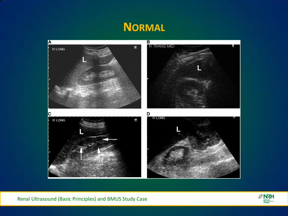
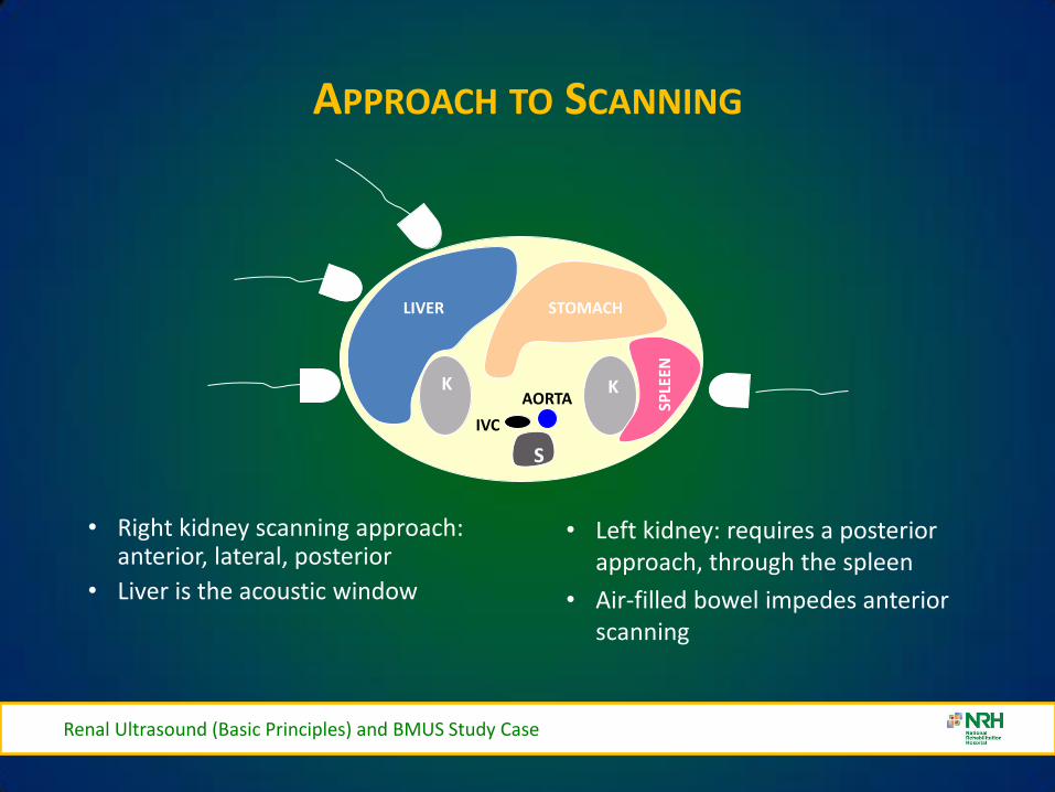
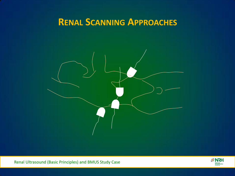
NORMAL ANATOMY
Renal Ultrasound (Basic Principles) and BMUS Study Case
bull 9-12 cm long 4-5 cm wide 3-4 cm thick
bull Gerotarsquos fascia encloses kidney capsule perinephric fat
bull Sinus
ndash Hilum vessels nerves lymphatics ureter
ndash Pelvis major and minor calyces
bull Parenchyma surrounds the sinus
ndash Cortex site of urine formation contains nephrons
ndash Medulla contains pyramids that pass urine to minor calyces Columns of Bertin
separate pyramids
NORMAL ANATOMY
Renal Ultrasound (Basic Principles) and BMUS Study Case
Renal capsule Cortex
Medullary pyramids
Minor Calyx
Kidney Anatomy
Medulla
Sinus
Major Calyx
NORMAL ANATOMY
Renal Ultrasound (Basic Principles) and BMUS Study Case
bull Kidneys are retroperitoneal T12 - L4
bull Right kidney is lower than the left kidney
bull Right kidney is posterio-inferior to liver amp gallbladder
bull Left kidney is inferior-medial to the spleen
bull Adrenal glands are superior anterior medial to each kidney
THICKENED BLADDER WALL
Renal Ultrasound (Basic Principles) and BMUS Study Case
BLADDER VOLUME
Renal Ultrasound (Basic Principles) and BMUS Study Case
Ureteric Jets
Renal Ultrasound (Basic Principles) and BMUS Study Case
Male Anatomy
Prostate Gland Seminal Vesicles
OVER-HYDRATION
Pre-Micturition Post Micturition
Renal Ultrasound (Basic Principles) and BMUS Study Case
INTERVENTIONAL
VARIANTS
Renal Ultrasound (Basic Principles) and BMUS Study Case
bull Dromedary humps
ndash Lateral kidney bulge same echogenicity as the cortex
bull Hypertrophied column of Bertin
ndash Cortical tissue indents the renal sinus
bull Double collecting system
ndash Sinus divided by a hypertrophied column of Bertin
bull Horseshoe kidney
ndash Kidneys are connected usually at the lower pole
bull Renal ectopia
ndash One or both kidneys outside the normal renal fossa
VARIANTS
Pelvic Kidney
Dromedary Hump
Renal Ultrasound (Basic Principles) and BMUS Study Case
Renal Ultrasound (Basic Principles) and BMUS Study Case
EXTRA RENAL PELVIS
bull Extra Renal Pelvis refers to the presence of the renal pelvis outside the confines of the renal hilum It is a normal variant that is found in ~10 of the population
bull The renal pelvis is formed by all the major calyces An exta-renal pelvis usually appears dilated giving a false indication of an obstructive pathology
CT for clarification
bull Avoid confusion with hydroureter
or PUJ obstruction
Renal Ultrasound (Basic Principles) and BMUS Study Case
VARIANTS CONT
Renal Ultrasound (Basic Principles) and BMUS Study Case
HORSESHOE KIDNEY
Renal Ultrasound (Basic Principles) and BMUS Study Case
CROSS FUSED ECTOPIA
bull Cross fused ectopic kidney
bull The left kidney is fused to the lower pole of the right kidney
Renal Ultrasound (Basic Principles) and BMUS Study Case
RENAL CYSTS
Cyst 1 Cyst 2
Renal Ultrasound (Basic Principles) and BMUS Study Case
RENAL SCARRING
Renal Ultrasound (Basic Principles) and BMUS Study Case
MULTICYSTIC DYSPLASTIC KIDNEY
bull Right kidney in a new-born shows no normal renal parenchyma and the right renal fossa filled with multiple simple appearing cystic structures cw MCDK The cysts did NOT communicate
Renal Ultrasound (Basic Principles) and BMUS Study Case
ANGIOMYOLIPOMA
bull This is a homogeneous highly echogenic usually rounded lesion in the renal parenchyma containing blood vessels muscle tissue and fat as the name suggests They are usually solitary asymptomatic lesions found incidentally although larger lesions can haemorrhage causing haematuria and pain
Renal Ultrasound (Basic Principles) and BMUS Study Case
MULTIPLE ANGIOMYOLIPOMAS
bull Also associated with Tuberose sclerosis they tend to be multiple and bilateral
Renal Ultrasound (Basic Principles) and BMUS Study Case
OBSTRUCTIVE UROPATHY GRADING SYSTEM - SUBJECTIVE
bull Mild Minimal separation of calyces
bull Moderate Dilation of major and minor calyceal system
bull Severe Marked dilation of the renal pelvis and thinning of the renal parenchyma
Renal Ultrasound (Basic Principles) and BMUS Study Case
RANGE OF HYDRONEPHROSIS
Renal Ultrasound (Basic Principles) and BMUS Study Case
Normal Mild Moderate Severe
HYDRONEPHROSIS
Renal Ultrasound (Basic Principles) and BMUS Study Case
Intrinsic acquired
bull Renal lithiasis bull Neoplasm (renal ureteral bladder)
bull Papillary necrosis
bull Ureterocele
bull Blood clot
bull Neurogenic bladder
bull Anticholinergics
bull Pregnancy PID uterine prolapse)
bull Diuretics
bull Vesico-ureteral reflux
bull Diabetes insipidus
Intrinsic congenital
bull Stenosis (ureteral urethral meatal)
bull Adynamic ureter
bull Spinal cord defects
bull Duplication of the ureter
bull Ureterocele
HYDRONEPHROSIS
Mild Hydronephrosis
Renal Ultrasound (Basic Principles) and BMUS Study Case
HYDRONEPHROSIS
Moderate Hydronephrosis
Renal Ultrasound (Basic Principles) and BMUS Study Case
SEVERE HYDRONEPHROSIS
Renal Ultrasound (Basic Principles) and BMUS Study Case
Severe Hydronephrosis
SMALL KIDNEYS
bull Unilateral (may be bilateral)
ndash Chronic infections
ndash RAS Renal Artery Stenosis
ndash Hypoplastic Kidney
ndash Always bilateral
bull Chronic glomerulonephritis
bull Hypertensive nephropathy
bull Collagen Vascular Disease
Renal Ultrasound (Basic Principles) and BMUS Study Case
ENLARGED KIDNEYS
Always Unilateral
ndash Compensatory hypertrophy
ndash Bilateral or Unilateral
bull Renal Mass
bull Hydronephrosis
bull Renal vein thromboses
bull Lymphoma
bull Amyloidosis
Always Bilateral
ndash PCK Polycystic Kidney Disease
ndash AGN Acute glomerulonephritis
ndash Amyloidosis
Renal Ultrasound (Basic Principles) and BMUS Study Case
Urolithiasis
Renal Calculi- Bladder Calculi
US only
41
Fig 3
Fig 4 Fig 5
Fig 3 4 amp 5 ndash Bladder stones with posterior shadowing clearly seen at US in distended bladder but not apparent on X-ray
Staghorn Calculus
BLADDER DIVERTICULAE
Renal Ultrasound (Basic Principles) and BMUS Study Case
bull Diverticula of the bladder in
infants and children are common and occur when bladder mucosa protrudes through a congenital defect in the detrusor muscle wall Most are primary Those secondary to obstruction or neurogenic dysfunction are less common than previously thought
Renal Ultrasound (Basic Principles) and BMUS Study Case
Renal Ultrasound (Basic Principles) and BMUS Study Case
BLADDER CALCULI
Renal Ultrasound (Basic Principles) and BMUS Study Case
RENAL MASS
Renal Ultrasound (Basic Principles) and BMUS Study Case
bull Pyelonephritis sonographic appearance is most commonly normal but you may find hypoechoic cortex and loss of demarcation between the outer cortex and middle pyramids and columns of Bertin
bull Renal mass may have any echotexture (hyperechoic anechoic etc) and appear anywhere within the kidney
bull Transplant kidney a normal echotexture kidney typically in a pelvic location
TRANSITIONAL CELL CARCINOMA
bull Ultrasound images Large transitional cell carcinoma in the upper pole of the RK The changes are more subtle than those of renal cell carcinoma and the renal outline remains intact
bull Transitional cell carcinoma is the most common bladder tumour occurring less frequently in the collecting system of the kidney and the ureter It usually presents with haematuria while still small It is best diagnosed with cystoscopy
Renal Ultrasound (Basic Principles) and BMUS Study Case
HYDRONEPHROSIS DUE TO TCC URETER
Renal Ultrasound (Basic Principles) and BMUS Study Case
US AND CT RENAL CA
Renal Ultrasound (Basic Principles) and BMUS Study Case
RENAL CELL CARCINOMA
bull Large heterogeneous mass which enlarges and deforms the shape of the kidney (Fig below) The mass may contain areas of cystic degeneration andor calcification It has a predilection to spread into the ipsilateral renal vein and IVC
bull Colour Doppler usually reveals a disorganized and increased blood flow pattern within the mass with high velocities from the arterioverous shunts within the carcinoma
Renal Ultrasound (Basic Principles) and BMUS Study Case
bull Smaller RCCs can be hyperechoic and may be confused with benign angiomyolipoma The latter has well-defined borders whilst an RCC is illdefined
BURKITTS LYMPHOMA
Renal Ultrasound (Basic Principles) and BMUS Study Case
NEUROBLASTOMA
Renal Ultrasound (Basic Principles) and BMUS Study Case
RENAL TRAUMA
Renal Ultrasound (Basic Principles) and BMUS Study Case
POLYCYSTIC KIDNEYS
Renal Ultrasound (Basic Principles) and BMUS Study Case
RENAL TRANSPLANTS-SURGICAL TECHNIQUE
bull Usually heterotopic (placed in addition to the native diseased kidneys)
bull Positioned in the extra-peritoneal pouch in the iliac fossa (usually the right) anterior to the iliacus and psoas muscles
Renal Ultrasound (Basic Principles) and BMUS Study Case
THE ROLE OF ULTRASOUND
B Mode imaging
bull Morphological appearances
PC dilation
Peri-renal fluid collections
bull Doppler
Colour Power Perfusion
Spectral Doppler
Waveforms
bull Intervention
Guide Biopsy Procedures
Drain Fluid Collections
Placement of Nephrostomy
Tubes
Renal Ultrasound (Basic Principles) and BMUS Study Case
LS RENAL TRANSPLANT (USING EXTENDED FIELD OF VIEW)
Renal Ultrasound (Basic Principles) and BMUS Study Case
PERFUSION WITH POWER AND COLOUR DOPPLER
Renal Ultrasound (Basic Principles) and BMUS Study Case
BACKGROUND
The main purpose of regular urology review is to preserve renal function with the ultimate aim of a non obstructed urinary system
Urinary Tract Infections (UTI) are the most frequent medical complication in patients with Spinal Cord Injuries (SCI) and a major cause of morbidity
Renal Ultrasound (Basic Principles) and BMUS Study Case
X-RAY ONLY
Fig 1 amp 2 - US showing decompressed bladder around IDUC X-ray in same patient confirming large calculus not visualised on US
Fig 1 Fig 2
Renal Ultrasound (Basic Principles) and BMUS Study Case
US ONLY
Fig 3
Fig 4
Fig 5
Fig 3 4 amp 5 ndash Bladder stones with posterior shadowing clearly seen at US in distended bladder but not apparent on X-ray
Renal Ultrasound (Basic Principles) and BMUS Study Case
RESULTS
bull Bladder ultrasound alone diagnosed 36 bladder calculi compared to 18 diagnosed by X-ray alone
bull US and X-ray combined diagnosed 46
Renal Ultrasound (Basic Principles) and BMUS Study Case
BLADDER MUCUS
bull In one patient a focal mobile area of increased reflectivity was demonstrated posteriorly in the bladder No posterior acoustic shadowing was seen to suggest of calculus formation
bull On flexible cystoscopy no calculus identified only mucus seen
Renal Ultrasound (Basic Principles) and BMUS Study Case
CASE STUDY 1
Renal Ultrasound (Basic Principles) and BMUS Study Case
CASE STUDY 2-VHL (VON HIPPEL LINDAU SYNDROME)
bull renal lesions
ndash renal cell carcinoma(s) (RCCs) usually of the clear cell type
bull 70 lifetime risk
bull RCCs present at an earlier age in those with vHL
ndash renal cysts
bull can occur in up to 75 of cases
bull often tend to be bilateral and multiple
bull Prognosis is poor with a median survival of ~50 years with the most common cause of death being RCC and cerebellar haemangioblastomas
Renal Ultrasound (Basic Principles) and BMUS Study Case
Renal Ultrasound (Basic Principles) and BMUS Study Case
Case Study 3
Renal Ultrasound (Basic Principles) and BMUS Study Case
Renal Ultrasound (Basic Principles) and BMUS Study Case
PAGE KIDNEY
Renal Ultrasound (Basic Principles) and BMUS Study Case
THANK YOU FOR YOUR ATTENTION
Renal Ultrasound (Basic Principles) and BMUS Study Case
Rosie Conlon rosieconlonnrhie

NORMAL ANATOMY
Renal Ultrasound (Basic Principles) and BMUS Study Case
Renal capsule Cortex
Medullary pyramids
Minor Calyx
Kidney Anatomy
Medulla
Sinus
Major Calyx
NORMAL ANATOMY
Renal Ultrasound (Basic Principles) and BMUS Study Case
bull Kidneys are retroperitoneal T12 - L4
bull Right kidney is lower than the left kidney
bull Right kidney is posterio-inferior to liver amp gallbladder
bull Left kidney is inferior-medial to the spleen
bull Adrenal glands are superior anterior medial to each kidney
THICKENED BLADDER WALL
Renal Ultrasound (Basic Principles) and BMUS Study Case
BLADDER VOLUME
Renal Ultrasound (Basic Principles) and BMUS Study Case
Ureteric Jets
Renal Ultrasound (Basic Principles) and BMUS Study Case
Male Anatomy
Prostate Gland Seminal Vesicles
OVER-HYDRATION
Pre-Micturition Post Micturition
Renal Ultrasound (Basic Principles) and BMUS Study Case
INTERVENTIONAL
VARIANTS
Renal Ultrasound (Basic Principles) and BMUS Study Case
bull Dromedary humps
ndash Lateral kidney bulge same echogenicity as the cortex
bull Hypertrophied column of Bertin
ndash Cortical tissue indents the renal sinus
bull Double collecting system
ndash Sinus divided by a hypertrophied column of Bertin
bull Horseshoe kidney
ndash Kidneys are connected usually at the lower pole
bull Renal ectopia
ndash One or both kidneys outside the normal renal fossa
VARIANTS
Pelvic Kidney
Dromedary Hump
Renal Ultrasound (Basic Principles) and BMUS Study Case
Renal Ultrasound (Basic Principles) and BMUS Study Case
EXTRA RENAL PELVIS
bull Extra Renal Pelvis refers to the presence of the renal pelvis outside the confines of the renal hilum It is a normal variant that is found in ~10 of the population
bull The renal pelvis is formed by all the major calyces An exta-renal pelvis usually appears dilated giving a false indication of an obstructive pathology
CT for clarification
bull Avoid confusion with hydroureter
or PUJ obstruction
Renal Ultrasound (Basic Principles) and BMUS Study Case
VARIANTS CONT
Renal Ultrasound (Basic Principles) and BMUS Study Case
HORSESHOE KIDNEY
Renal Ultrasound (Basic Principles) and BMUS Study Case
CROSS FUSED ECTOPIA
bull Cross fused ectopic kidney
bull The left kidney is fused to the lower pole of the right kidney
Renal Ultrasound (Basic Principles) and BMUS Study Case
RENAL CYSTS
Cyst 1 Cyst 2
Renal Ultrasound (Basic Principles) and BMUS Study Case
RENAL SCARRING
Renal Ultrasound (Basic Principles) and BMUS Study Case
MULTICYSTIC DYSPLASTIC KIDNEY
bull Right kidney in a new-born shows no normal renal parenchyma and the right renal fossa filled with multiple simple appearing cystic structures cw MCDK The cysts did NOT communicate
Renal Ultrasound (Basic Principles) and BMUS Study Case
ANGIOMYOLIPOMA
bull This is a homogeneous highly echogenic usually rounded lesion in the renal parenchyma containing blood vessels muscle tissue and fat as the name suggests They are usually solitary asymptomatic lesions found incidentally although larger lesions can haemorrhage causing haematuria and pain
Renal Ultrasound (Basic Principles) and BMUS Study Case
MULTIPLE ANGIOMYOLIPOMAS
bull Also associated with Tuberose sclerosis they tend to be multiple and bilateral
Renal Ultrasound (Basic Principles) and BMUS Study Case
OBSTRUCTIVE UROPATHY GRADING SYSTEM - SUBJECTIVE
bull Mild Minimal separation of calyces
bull Moderate Dilation of major and minor calyceal system
bull Severe Marked dilation of the renal pelvis and thinning of the renal parenchyma
Renal Ultrasound (Basic Principles) and BMUS Study Case
RANGE OF HYDRONEPHROSIS
Renal Ultrasound (Basic Principles) and BMUS Study Case
Normal Mild Moderate Severe
HYDRONEPHROSIS
Renal Ultrasound (Basic Principles) and BMUS Study Case
Intrinsic acquired
bull Renal lithiasis bull Neoplasm (renal ureteral bladder)
bull Papillary necrosis
bull Ureterocele
bull Blood clot
bull Neurogenic bladder
bull Anticholinergics
bull Pregnancy PID uterine prolapse)
bull Diuretics
bull Vesico-ureteral reflux
bull Diabetes insipidus
Intrinsic congenital
bull Stenosis (ureteral urethral meatal)
bull Adynamic ureter
bull Spinal cord defects
bull Duplication of the ureter
bull Ureterocele
HYDRONEPHROSIS
Mild Hydronephrosis
Renal Ultrasound (Basic Principles) and BMUS Study Case
HYDRONEPHROSIS
Moderate Hydronephrosis
Renal Ultrasound (Basic Principles) and BMUS Study Case
SEVERE HYDRONEPHROSIS
Renal Ultrasound (Basic Principles) and BMUS Study Case
Severe Hydronephrosis
SMALL KIDNEYS
bull Unilateral (may be bilateral)
ndash Chronic infections
ndash RAS Renal Artery Stenosis
ndash Hypoplastic Kidney
ndash Always bilateral
bull Chronic glomerulonephritis
bull Hypertensive nephropathy
bull Collagen Vascular Disease
Renal Ultrasound (Basic Principles) and BMUS Study Case
ENLARGED KIDNEYS
Always Unilateral
ndash Compensatory hypertrophy
ndash Bilateral or Unilateral
bull Renal Mass
bull Hydronephrosis
bull Renal vein thromboses
bull Lymphoma
bull Amyloidosis
Always Bilateral
ndash PCK Polycystic Kidney Disease
ndash AGN Acute glomerulonephritis
ndash Amyloidosis
Renal Ultrasound (Basic Principles) and BMUS Study Case
Urolithiasis
Renal Calculi- Bladder Calculi
US only
41
Fig 3
Fig 4 Fig 5
Fig 3 4 amp 5 ndash Bladder stones with posterior shadowing clearly seen at US in distended bladder but not apparent on X-ray
Staghorn Calculus
BLADDER DIVERTICULAE
Renal Ultrasound (Basic Principles) and BMUS Study Case
bull Diverticula of the bladder in
infants and children are common and occur when bladder mucosa protrudes through a congenital defect in the detrusor muscle wall Most are primary Those secondary to obstruction or neurogenic dysfunction are less common than previously thought
Renal Ultrasound (Basic Principles) and BMUS Study Case
Renal Ultrasound (Basic Principles) and BMUS Study Case
BLADDER CALCULI
Renal Ultrasound (Basic Principles) and BMUS Study Case
RENAL MASS
Renal Ultrasound (Basic Principles) and BMUS Study Case
bull Pyelonephritis sonographic appearance is most commonly normal but you may find hypoechoic cortex and loss of demarcation between the outer cortex and middle pyramids and columns of Bertin
bull Renal mass may have any echotexture (hyperechoic anechoic etc) and appear anywhere within the kidney
bull Transplant kidney a normal echotexture kidney typically in a pelvic location
TRANSITIONAL CELL CARCINOMA
bull Ultrasound images Large transitional cell carcinoma in the upper pole of the RK The changes are more subtle than those of renal cell carcinoma and the renal outline remains intact
bull Transitional cell carcinoma is the most common bladder tumour occurring less frequently in the collecting system of the kidney and the ureter It usually presents with haematuria while still small It is best diagnosed with cystoscopy
Renal Ultrasound (Basic Principles) and BMUS Study Case
HYDRONEPHROSIS DUE TO TCC URETER
Renal Ultrasound (Basic Principles) and BMUS Study Case
US AND CT RENAL CA
Renal Ultrasound (Basic Principles) and BMUS Study Case
RENAL CELL CARCINOMA
bull Large heterogeneous mass which enlarges and deforms the shape of the kidney (Fig below) The mass may contain areas of cystic degeneration andor calcification It has a predilection to spread into the ipsilateral renal vein and IVC
bull Colour Doppler usually reveals a disorganized and increased blood flow pattern within the mass with high velocities from the arterioverous shunts within the carcinoma
Renal Ultrasound (Basic Principles) and BMUS Study Case
bull Smaller RCCs can be hyperechoic and may be confused with benign angiomyolipoma The latter has well-defined borders whilst an RCC is illdefined
BURKITTS LYMPHOMA
Renal Ultrasound (Basic Principles) and BMUS Study Case
NEUROBLASTOMA
Renal Ultrasound (Basic Principles) and BMUS Study Case
RENAL TRAUMA
Renal Ultrasound (Basic Principles) and BMUS Study Case
POLYCYSTIC KIDNEYS
Renal Ultrasound (Basic Principles) and BMUS Study Case
RENAL TRANSPLANTS-SURGICAL TECHNIQUE
bull Usually heterotopic (placed in addition to the native diseased kidneys)
bull Positioned in the extra-peritoneal pouch in the iliac fossa (usually the right) anterior to the iliacus and psoas muscles
Renal Ultrasound (Basic Principles) and BMUS Study Case
THE ROLE OF ULTRASOUND
B Mode imaging
bull Morphological appearances
PC dilation
Peri-renal fluid collections
bull Doppler
Colour Power Perfusion
Spectral Doppler
Waveforms
bull Intervention
Guide Biopsy Procedures
Drain Fluid Collections
Placement of Nephrostomy
Tubes
Renal Ultrasound (Basic Principles) and BMUS Study Case
LS RENAL TRANSPLANT (USING EXTENDED FIELD OF VIEW)
Renal Ultrasound (Basic Principles) and BMUS Study Case
PERFUSION WITH POWER AND COLOUR DOPPLER
Renal Ultrasound (Basic Principles) and BMUS Study Case
BACKGROUND
The main purpose of regular urology review is to preserve renal function with the ultimate aim of a non obstructed urinary system
Urinary Tract Infections (UTI) are the most frequent medical complication in patients with Spinal Cord Injuries (SCI) and a major cause of morbidity
Renal Ultrasound (Basic Principles) and BMUS Study Case
X-RAY ONLY
Fig 1 amp 2 - US showing decompressed bladder around IDUC X-ray in same patient confirming large calculus not visualised on US
Fig 1 Fig 2
Renal Ultrasound (Basic Principles) and BMUS Study Case
US ONLY
Fig 3
Fig 4
Fig 5
Fig 3 4 amp 5 ndash Bladder stones with posterior shadowing clearly seen at US in distended bladder but not apparent on X-ray
Renal Ultrasound (Basic Principles) and BMUS Study Case
RESULTS
bull Bladder ultrasound alone diagnosed 36 bladder calculi compared to 18 diagnosed by X-ray alone
bull US and X-ray combined diagnosed 46
Renal Ultrasound (Basic Principles) and BMUS Study Case
BLADDER MUCUS
bull In one patient a focal mobile area of increased reflectivity was demonstrated posteriorly in the bladder No posterior acoustic shadowing was seen to suggest of calculus formation
bull On flexible cystoscopy no calculus identified only mucus seen
Renal Ultrasound (Basic Principles) and BMUS Study Case
CASE STUDY 1
Renal Ultrasound (Basic Principles) and BMUS Study Case
CASE STUDY 2-VHL (VON HIPPEL LINDAU SYNDROME)
bull renal lesions
ndash renal cell carcinoma(s) (RCCs) usually of the clear cell type
bull 70 lifetime risk
bull RCCs present at an earlier age in those with vHL
ndash renal cysts
bull can occur in up to 75 of cases
bull often tend to be bilateral and multiple
bull Prognosis is poor with a median survival of ~50 years with the most common cause of death being RCC and cerebellar haemangioblastomas
Renal Ultrasound (Basic Principles) and BMUS Study Case
Renal Ultrasound (Basic Principles) and BMUS Study Case
Case Study 3
Renal Ultrasound (Basic Principles) and BMUS Study Case
Renal Ultrasound (Basic Principles) and BMUS Study Case
PAGE KIDNEY
Renal Ultrasound (Basic Principles) and BMUS Study Case
THANK YOU FOR YOUR ATTENTION
Renal Ultrasound (Basic Principles) and BMUS Study Case
Rosie Conlon rosieconlonnrhie
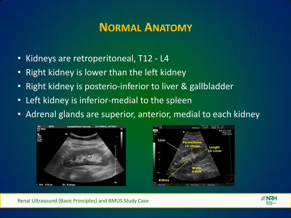
NORMAL ANATOMY
Renal Ultrasound (Basic Principles) and BMUS Study Case
bull Kidneys are retroperitoneal T12 - L4
bull Right kidney is lower than the left kidney
bull Right kidney is posterio-inferior to liver amp gallbladder
bull Left kidney is inferior-medial to the spleen
bull Adrenal glands are superior anterior medial to each kidney
THICKENED BLADDER WALL
Renal Ultrasound (Basic Principles) and BMUS Study Case
BLADDER VOLUME
Renal Ultrasound (Basic Principles) and BMUS Study Case
Ureteric Jets
Renal Ultrasound (Basic Principles) and BMUS Study Case
Male Anatomy
Prostate Gland Seminal Vesicles
OVER-HYDRATION
Pre-Micturition Post Micturition
Renal Ultrasound (Basic Principles) and BMUS Study Case
INTERVENTIONAL
VARIANTS
Renal Ultrasound (Basic Principles) and BMUS Study Case
bull Dromedary humps
ndash Lateral kidney bulge same echogenicity as the cortex
bull Hypertrophied column of Bertin
ndash Cortical tissue indents the renal sinus
bull Double collecting system
ndash Sinus divided by a hypertrophied column of Bertin
bull Horseshoe kidney
ndash Kidneys are connected usually at the lower pole
bull Renal ectopia
ndash One or both kidneys outside the normal renal fossa
VARIANTS
Pelvic Kidney
Dromedary Hump
Renal Ultrasound (Basic Principles) and BMUS Study Case
Renal Ultrasound (Basic Principles) and BMUS Study Case
EXTRA RENAL PELVIS
bull Extra Renal Pelvis refers to the presence of the renal pelvis outside the confines of the renal hilum It is a normal variant that is found in ~10 of the population
bull The renal pelvis is formed by all the major calyces An exta-renal pelvis usually appears dilated giving a false indication of an obstructive pathology
CT for clarification
bull Avoid confusion with hydroureter
or PUJ obstruction
Renal Ultrasound (Basic Principles) and BMUS Study Case
VARIANTS CONT
Renal Ultrasound (Basic Principles) and BMUS Study Case
HORSESHOE KIDNEY
Renal Ultrasound (Basic Principles) and BMUS Study Case
CROSS FUSED ECTOPIA
bull Cross fused ectopic kidney
bull The left kidney is fused to the lower pole of the right kidney
Renal Ultrasound (Basic Principles) and BMUS Study Case
RENAL CYSTS
Cyst 1 Cyst 2
Renal Ultrasound (Basic Principles) and BMUS Study Case
RENAL SCARRING
Renal Ultrasound (Basic Principles) and BMUS Study Case
MULTICYSTIC DYSPLASTIC KIDNEY
bull Right kidney in a new-born shows no normal renal parenchyma and the right renal fossa filled with multiple simple appearing cystic structures cw MCDK The cysts did NOT communicate
Renal Ultrasound (Basic Principles) and BMUS Study Case
ANGIOMYOLIPOMA
bull This is a homogeneous highly echogenic usually rounded lesion in the renal parenchyma containing blood vessels muscle tissue and fat as the name suggests They are usually solitary asymptomatic lesions found incidentally although larger lesions can haemorrhage causing haematuria and pain
Renal Ultrasound (Basic Principles) and BMUS Study Case
MULTIPLE ANGIOMYOLIPOMAS
bull Also associated with Tuberose sclerosis they tend to be multiple and bilateral
Renal Ultrasound (Basic Principles) and BMUS Study Case
OBSTRUCTIVE UROPATHY GRADING SYSTEM - SUBJECTIVE
bull Mild Minimal separation of calyces
bull Moderate Dilation of major and minor calyceal system
bull Severe Marked dilation of the renal pelvis and thinning of the renal parenchyma
Renal Ultrasound (Basic Principles) and BMUS Study Case
RANGE OF HYDRONEPHROSIS
Renal Ultrasound (Basic Principles) and BMUS Study Case
Normal Mild Moderate Severe
HYDRONEPHROSIS
Renal Ultrasound (Basic Principles) and BMUS Study Case
Intrinsic acquired
bull Renal lithiasis bull Neoplasm (renal ureteral bladder)
bull Papillary necrosis
bull Ureterocele
bull Blood clot
bull Neurogenic bladder
bull Anticholinergics
bull Pregnancy PID uterine prolapse)
bull Diuretics
bull Vesico-ureteral reflux
bull Diabetes insipidus
Intrinsic congenital
bull Stenosis (ureteral urethral meatal)
bull Adynamic ureter
bull Spinal cord defects
bull Duplication of the ureter
bull Ureterocele
HYDRONEPHROSIS
Mild Hydronephrosis
Renal Ultrasound (Basic Principles) and BMUS Study Case
HYDRONEPHROSIS
Moderate Hydronephrosis
Renal Ultrasound (Basic Principles) and BMUS Study Case
SEVERE HYDRONEPHROSIS
Renal Ultrasound (Basic Principles) and BMUS Study Case
Severe Hydronephrosis
SMALL KIDNEYS
bull Unilateral (may be bilateral)
ndash Chronic infections
ndash RAS Renal Artery Stenosis
ndash Hypoplastic Kidney
ndash Always bilateral
bull Chronic glomerulonephritis
bull Hypertensive nephropathy
bull Collagen Vascular Disease
Renal Ultrasound (Basic Principles) and BMUS Study Case
ENLARGED KIDNEYS
Always Unilateral
ndash Compensatory hypertrophy
ndash Bilateral or Unilateral
bull Renal Mass
bull Hydronephrosis
bull Renal vein thromboses
bull Lymphoma
bull Amyloidosis
Always Bilateral
ndash PCK Polycystic Kidney Disease
ndash AGN Acute glomerulonephritis
ndash Amyloidosis
Renal Ultrasound (Basic Principles) and BMUS Study Case
Urolithiasis
Renal Calculi- Bladder Calculi
US only
41
Fig 3
Fig 4 Fig 5
Fig 3 4 amp 5 ndash Bladder stones with posterior shadowing clearly seen at US in distended bladder but not apparent on X-ray
Staghorn Calculus
BLADDER DIVERTICULAE
Renal Ultrasound (Basic Principles) and BMUS Study Case
bull Diverticula of the bladder in
infants and children are common and occur when bladder mucosa protrudes through a congenital defect in the detrusor muscle wall Most are primary Those secondary to obstruction or neurogenic dysfunction are less common than previously thought
Renal Ultrasound (Basic Principles) and BMUS Study Case
Renal Ultrasound (Basic Principles) and BMUS Study Case
BLADDER CALCULI
Renal Ultrasound (Basic Principles) and BMUS Study Case
RENAL MASS
Renal Ultrasound (Basic Principles) and BMUS Study Case
bull Pyelonephritis sonographic appearance is most commonly normal but you may find hypoechoic cortex and loss of demarcation between the outer cortex and middle pyramids and columns of Bertin
bull Renal mass may have any echotexture (hyperechoic anechoic etc) and appear anywhere within the kidney
bull Transplant kidney a normal echotexture kidney typically in a pelvic location
TRANSITIONAL CELL CARCINOMA
bull Ultrasound images Large transitional cell carcinoma in the upper pole of the RK The changes are more subtle than those of renal cell carcinoma and the renal outline remains intact
bull Transitional cell carcinoma is the most common bladder tumour occurring less frequently in the collecting system of the kidney and the ureter It usually presents with haematuria while still small It is best diagnosed with cystoscopy
Renal Ultrasound (Basic Principles) and BMUS Study Case
HYDRONEPHROSIS DUE TO TCC URETER
Renal Ultrasound (Basic Principles) and BMUS Study Case
US AND CT RENAL CA
Renal Ultrasound (Basic Principles) and BMUS Study Case
RENAL CELL CARCINOMA
bull Large heterogeneous mass which enlarges and deforms the shape of the kidney (Fig below) The mass may contain areas of cystic degeneration andor calcification It has a predilection to spread into the ipsilateral renal vein and IVC
bull Colour Doppler usually reveals a disorganized and increased blood flow pattern within the mass with high velocities from the arterioverous shunts within the carcinoma
Renal Ultrasound (Basic Principles) and BMUS Study Case
bull Smaller RCCs can be hyperechoic and may be confused with benign angiomyolipoma The latter has well-defined borders whilst an RCC is illdefined
BURKITTS LYMPHOMA
Renal Ultrasound (Basic Principles) and BMUS Study Case
NEUROBLASTOMA
Renal Ultrasound (Basic Principles) and BMUS Study Case
RENAL TRAUMA
Renal Ultrasound (Basic Principles) and BMUS Study Case
POLYCYSTIC KIDNEYS
Renal Ultrasound (Basic Principles) and BMUS Study Case
RENAL TRANSPLANTS-SURGICAL TECHNIQUE
bull Usually heterotopic (placed in addition to the native diseased kidneys)
bull Positioned in the extra-peritoneal pouch in the iliac fossa (usually the right) anterior to the iliacus and psoas muscles
Renal Ultrasound (Basic Principles) and BMUS Study Case
THE ROLE OF ULTRASOUND
B Mode imaging
bull Morphological appearances
PC dilation
Peri-renal fluid collections
bull Doppler
Colour Power Perfusion
Spectral Doppler
Waveforms
bull Intervention
Guide Biopsy Procedures
Drain Fluid Collections
Placement of Nephrostomy
Tubes
Renal Ultrasound (Basic Principles) and BMUS Study Case
LS RENAL TRANSPLANT (USING EXTENDED FIELD OF VIEW)
Renal Ultrasound (Basic Principles) and BMUS Study Case
PERFUSION WITH POWER AND COLOUR DOPPLER
Renal Ultrasound (Basic Principles) and BMUS Study Case
BACKGROUND
The main purpose of regular urology review is to preserve renal function with the ultimate aim of a non obstructed urinary system
Urinary Tract Infections (UTI) are the most frequent medical complication in patients with Spinal Cord Injuries (SCI) and a major cause of morbidity
Renal Ultrasound (Basic Principles) and BMUS Study Case
X-RAY ONLY
Fig 1 amp 2 - US showing decompressed bladder around IDUC X-ray in same patient confirming large calculus not visualised on US
Fig 1 Fig 2
Renal Ultrasound (Basic Principles) and BMUS Study Case
US ONLY
Fig 3
Fig 4
Fig 5
Fig 3 4 amp 5 ndash Bladder stones with posterior shadowing clearly seen at US in distended bladder but not apparent on X-ray
Renal Ultrasound (Basic Principles) and BMUS Study Case
RESULTS
bull Bladder ultrasound alone diagnosed 36 bladder calculi compared to 18 diagnosed by X-ray alone
bull US and X-ray combined diagnosed 46
Renal Ultrasound (Basic Principles) and BMUS Study Case
BLADDER MUCUS
bull In one patient a focal mobile area of increased reflectivity was demonstrated posteriorly in the bladder No posterior acoustic shadowing was seen to suggest of calculus formation
bull On flexible cystoscopy no calculus identified only mucus seen
Renal Ultrasound (Basic Principles) and BMUS Study Case
CASE STUDY 1
Renal Ultrasound (Basic Principles) and BMUS Study Case
CASE STUDY 2-VHL (VON HIPPEL LINDAU SYNDROME)
bull renal lesions
ndash renal cell carcinoma(s) (RCCs) usually of the clear cell type
bull 70 lifetime risk
bull RCCs present at an earlier age in those with vHL
ndash renal cysts
bull can occur in up to 75 of cases
bull often tend to be bilateral and multiple
bull Prognosis is poor with a median survival of ~50 years with the most common cause of death being RCC and cerebellar haemangioblastomas
Renal Ultrasound (Basic Principles) and BMUS Study Case
Renal Ultrasound (Basic Principles) and BMUS Study Case
Case Study 3
Renal Ultrasound (Basic Principles) and BMUS Study Case
Renal Ultrasound (Basic Principles) and BMUS Study Case
PAGE KIDNEY
Renal Ultrasound (Basic Principles) and BMUS Study Case
THANK YOU FOR YOUR ATTENTION
Renal Ultrasound (Basic Principles) and BMUS Study Case
Rosie Conlon rosieconlonnrhie
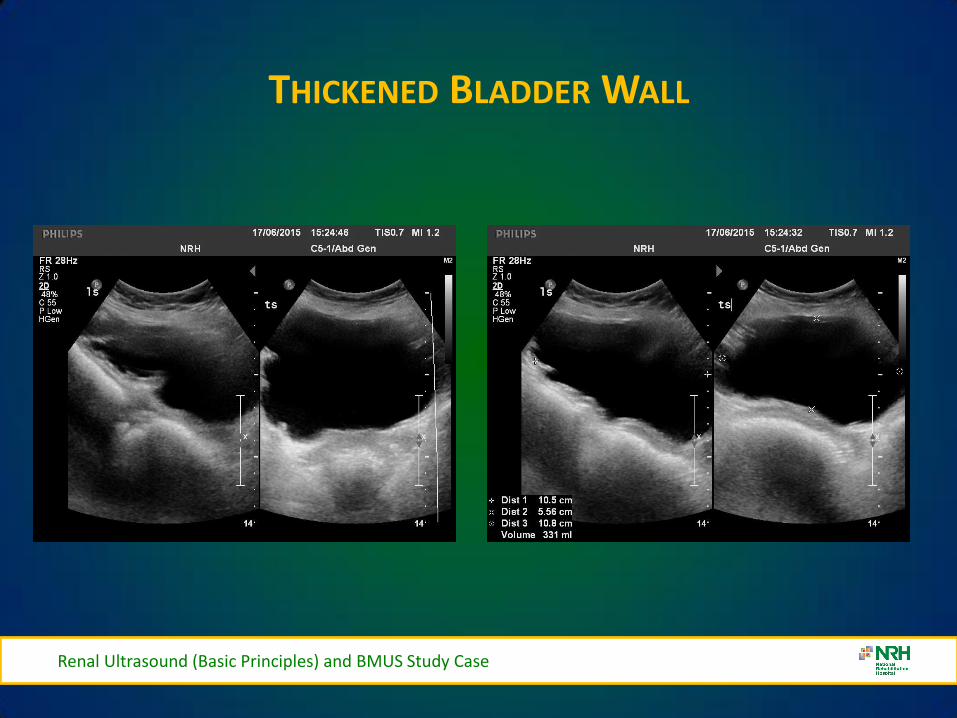
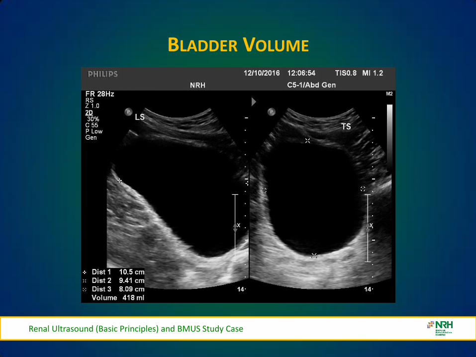
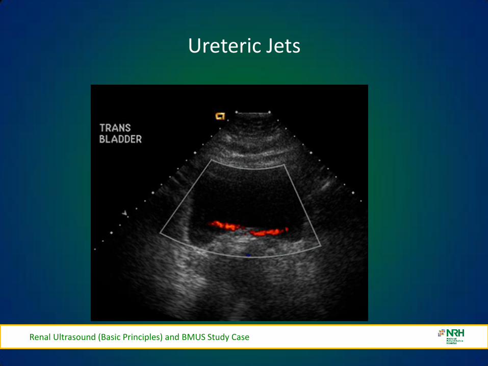
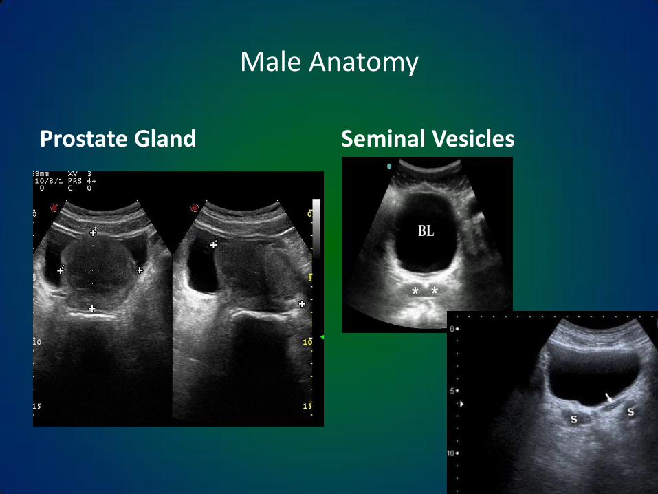
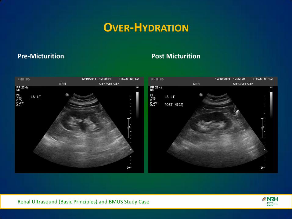
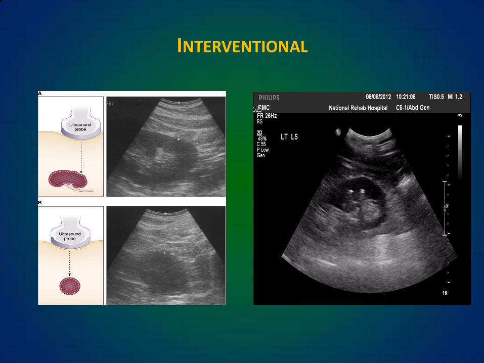

VARIANTS
Renal Ultrasound (Basic Principles) and BMUS Study Case
bull Dromedary humps
ndash Lateral kidney bulge same echogenicity as the cortex
bull Hypertrophied column of Bertin
ndash Cortical tissue indents the renal sinus
bull Double collecting system
ndash Sinus divided by a hypertrophied column of Bertin
bull Horseshoe kidney
ndash Kidneys are connected usually at the lower pole
bull Renal ectopia
ndash One or both kidneys outside the normal renal fossa
VARIANTS
Pelvic Kidney
Dromedary Hump
Renal Ultrasound (Basic Principles) and BMUS Study Case
Renal Ultrasound (Basic Principles) and BMUS Study Case
EXTRA RENAL PELVIS
bull Extra Renal Pelvis refers to the presence of the renal pelvis outside the confines of the renal hilum It is a normal variant that is found in ~10 of the population
bull The renal pelvis is formed by all the major calyces An exta-renal pelvis usually appears dilated giving a false indication of an obstructive pathology
CT for clarification
bull Avoid confusion with hydroureter
or PUJ obstruction
Renal Ultrasound (Basic Principles) and BMUS Study Case
VARIANTS CONT
Renal Ultrasound (Basic Principles) and BMUS Study Case
HORSESHOE KIDNEY
Renal Ultrasound (Basic Principles) and BMUS Study Case
CROSS FUSED ECTOPIA
bull Cross fused ectopic kidney
bull The left kidney is fused to the lower pole of the right kidney
Renal Ultrasound (Basic Principles) and BMUS Study Case
RENAL CYSTS
Cyst 1 Cyst 2
Renal Ultrasound (Basic Principles) and BMUS Study Case
RENAL SCARRING
Renal Ultrasound (Basic Principles) and BMUS Study Case
MULTICYSTIC DYSPLASTIC KIDNEY
bull Right kidney in a new-born shows no normal renal parenchyma and the right renal fossa filled with multiple simple appearing cystic structures cw MCDK The cysts did NOT communicate
Renal Ultrasound (Basic Principles) and BMUS Study Case
ANGIOMYOLIPOMA
bull This is a homogeneous highly echogenic usually rounded lesion in the renal parenchyma containing blood vessels muscle tissue and fat as the name suggests They are usually solitary asymptomatic lesions found incidentally although larger lesions can haemorrhage causing haematuria and pain
Renal Ultrasound (Basic Principles) and BMUS Study Case
MULTIPLE ANGIOMYOLIPOMAS
bull Also associated with Tuberose sclerosis they tend to be multiple and bilateral
Renal Ultrasound (Basic Principles) and BMUS Study Case
OBSTRUCTIVE UROPATHY GRADING SYSTEM - SUBJECTIVE
bull Mild Minimal separation of calyces
bull Moderate Dilation of major and minor calyceal system
bull Severe Marked dilation of the renal pelvis and thinning of the renal parenchyma
Renal Ultrasound (Basic Principles) and BMUS Study Case
RANGE OF HYDRONEPHROSIS
Renal Ultrasound (Basic Principles) and BMUS Study Case
Normal Mild Moderate Severe
HYDRONEPHROSIS
Renal Ultrasound (Basic Principles) and BMUS Study Case
Intrinsic acquired
bull Renal lithiasis bull Neoplasm (renal ureteral bladder)
bull Papillary necrosis
bull Ureterocele
bull Blood clot
bull Neurogenic bladder
bull Anticholinergics
bull Pregnancy PID uterine prolapse)
bull Diuretics
bull Vesico-ureteral reflux
bull Diabetes insipidus
Intrinsic congenital
bull Stenosis (ureteral urethral meatal)
bull Adynamic ureter
bull Spinal cord defects
bull Duplication of the ureter
bull Ureterocele
HYDRONEPHROSIS
Mild Hydronephrosis
Renal Ultrasound (Basic Principles) and BMUS Study Case
HYDRONEPHROSIS
Moderate Hydronephrosis
Renal Ultrasound (Basic Principles) and BMUS Study Case
SEVERE HYDRONEPHROSIS
Renal Ultrasound (Basic Principles) and BMUS Study Case
Severe Hydronephrosis
SMALL KIDNEYS
bull Unilateral (may be bilateral)
ndash Chronic infections
ndash RAS Renal Artery Stenosis
ndash Hypoplastic Kidney
ndash Always bilateral
bull Chronic glomerulonephritis
bull Hypertensive nephropathy
bull Collagen Vascular Disease
Renal Ultrasound (Basic Principles) and BMUS Study Case
ENLARGED KIDNEYS
Always Unilateral
ndash Compensatory hypertrophy
ndash Bilateral or Unilateral
bull Renal Mass
bull Hydronephrosis
bull Renal vein thromboses
bull Lymphoma
bull Amyloidosis
Always Bilateral
ndash PCK Polycystic Kidney Disease
ndash AGN Acute glomerulonephritis
ndash Amyloidosis
Renal Ultrasound (Basic Principles) and BMUS Study Case
Urolithiasis
Renal Calculi- Bladder Calculi
US only
41
Fig 3
Fig 4 Fig 5
Fig 3 4 amp 5 ndash Bladder stones with posterior shadowing clearly seen at US in distended bladder but not apparent on X-ray
Staghorn Calculus
BLADDER DIVERTICULAE
Renal Ultrasound (Basic Principles) and BMUS Study Case
bull Diverticula of the bladder in
infants and children are common and occur when bladder mucosa protrudes through a congenital defect in the detrusor muscle wall Most are primary Those secondary to obstruction or neurogenic dysfunction are less common than previously thought
Renal Ultrasound (Basic Principles) and BMUS Study Case
Renal Ultrasound (Basic Principles) and BMUS Study Case
BLADDER CALCULI
Renal Ultrasound (Basic Principles) and BMUS Study Case
RENAL MASS
Renal Ultrasound (Basic Principles) and BMUS Study Case
bull Pyelonephritis sonographic appearance is most commonly normal but you may find hypoechoic cortex and loss of demarcation between the outer cortex and middle pyramids and columns of Bertin
bull Renal mass may have any echotexture (hyperechoic anechoic etc) and appear anywhere within the kidney
bull Transplant kidney a normal echotexture kidney typically in a pelvic location
TRANSITIONAL CELL CARCINOMA
bull Ultrasound images Large transitional cell carcinoma in the upper pole of the RK The changes are more subtle than those of renal cell carcinoma and the renal outline remains intact
bull Transitional cell carcinoma is the most common bladder tumour occurring less frequently in the collecting system of the kidney and the ureter It usually presents with haematuria while still small It is best diagnosed with cystoscopy
Renal Ultrasound (Basic Principles) and BMUS Study Case
HYDRONEPHROSIS DUE TO TCC URETER
Renal Ultrasound (Basic Principles) and BMUS Study Case
US AND CT RENAL CA
Renal Ultrasound (Basic Principles) and BMUS Study Case
RENAL CELL CARCINOMA
bull Large heterogeneous mass which enlarges and deforms the shape of the kidney (Fig below) The mass may contain areas of cystic degeneration andor calcification It has a predilection to spread into the ipsilateral renal vein and IVC
bull Colour Doppler usually reveals a disorganized and increased blood flow pattern within the mass with high velocities from the arterioverous shunts within the carcinoma
Renal Ultrasound (Basic Principles) and BMUS Study Case
bull Smaller RCCs can be hyperechoic and may be confused with benign angiomyolipoma The latter has well-defined borders whilst an RCC is illdefined
BURKITTS LYMPHOMA
Renal Ultrasound (Basic Principles) and BMUS Study Case
NEUROBLASTOMA
Renal Ultrasound (Basic Principles) and BMUS Study Case
RENAL TRAUMA
Renal Ultrasound (Basic Principles) and BMUS Study Case
POLYCYSTIC KIDNEYS
Renal Ultrasound (Basic Principles) and BMUS Study Case
RENAL TRANSPLANTS-SURGICAL TECHNIQUE
bull Usually heterotopic (placed in addition to the native diseased kidneys)
bull Positioned in the extra-peritoneal pouch in the iliac fossa (usually the right) anterior to the iliacus and psoas muscles
Renal Ultrasound (Basic Principles) and BMUS Study Case
THE ROLE OF ULTRASOUND
B Mode imaging
bull Morphological appearances
PC dilation
Peri-renal fluid collections
bull Doppler
Colour Power Perfusion
Spectral Doppler
Waveforms
bull Intervention
Guide Biopsy Procedures
Drain Fluid Collections
Placement of Nephrostomy
Tubes
Renal Ultrasound (Basic Principles) and BMUS Study Case
LS RENAL TRANSPLANT (USING EXTENDED FIELD OF VIEW)
Renal Ultrasound (Basic Principles) and BMUS Study Case
PERFUSION WITH POWER AND COLOUR DOPPLER
Renal Ultrasound (Basic Principles) and BMUS Study Case
BACKGROUND
The main purpose of regular urology review is to preserve renal function with the ultimate aim of a non obstructed urinary system
Urinary Tract Infections (UTI) are the most frequent medical complication in patients with Spinal Cord Injuries (SCI) and a major cause of morbidity
Renal Ultrasound (Basic Principles) and BMUS Study Case
X-RAY ONLY
Fig 1 amp 2 - US showing decompressed bladder around IDUC X-ray in same patient confirming large calculus not visualised on US
Fig 1 Fig 2
Renal Ultrasound (Basic Principles) and BMUS Study Case
US ONLY
Fig 3
Fig 4
Fig 5
Fig 3 4 amp 5 ndash Bladder stones with posterior shadowing clearly seen at US in distended bladder but not apparent on X-ray
Renal Ultrasound (Basic Principles) and BMUS Study Case
RESULTS
bull Bladder ultrasound alone diagnosed 36 bladder calculi compared to 18 diagnosed by X-ray alone
bull US and X-ray combined diagnosed 46
Renal Ultrasound (Basic Principles) and BMUS Study Case
BLADDER MUCUS
bull In one patient a focal mobile area of increased reflectivity was demonstrated posteriorly in the bladder No posterior acoustic shadowing was seen to suggest of calculus formation
bull On flexible cystoscopy no calculus identified only mucus seen
Renal Ultrasound (Basic Principles) and BMUS Study Case
CASE STUDY 1
Renal Ultrasound (Basic Principles) and BMUS Study Case
CASE STUDY 2-VHL (VON HIPPEL LINDAU SYNDROME)
bull renal lesions
ndash renal cell carcinoma(s) (RCCs) usually of the clear cell type
bull 70 lifetime risk
bull RCCs present at an earlier age in those with vHL
ndash renal cysts
bull can occur in up to 75 of cases
bull often tend to be bilateral and multiple
bull Prognosis is poor with a median survival of ~50 years with the most common cause of death being RCC and cerebellar haemangioblastomas
Renal Ultrasound (Basic Principles) and BMUS Study Case
Renal Ultrasound (Basic Principles) and BMUS Study Case
Case Study 3
Renal Ultrasound (Basic Principles) and BMUS Study Case
Renal Ultrasound (Basic Principles) and BMUS Study Case
PAGE KIDNEY
Renal Ultrasound (Basic Principles) and BMUS Study Case
THANK YOU FOR YOUR ATTENTION
Renal Ultrasound (Basic Principles) and BMUS Study Case
Rosie Conlon rosieconlonnrhie
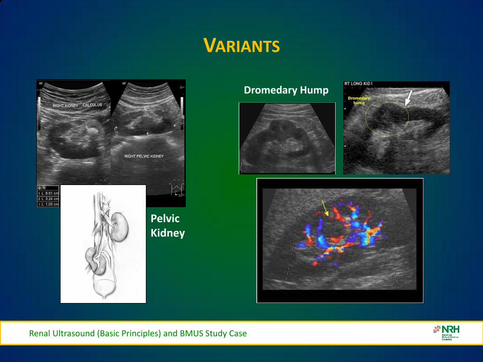
VARIANTS
Pelvic Kidney
Dromedary Hump
Renal Ultrasound (Basic Principles) and BMUS Study Case
Renal Ultrasound (Basic Principles) and BMUS Study Case
EXTRA RENAL PELVIS
bull Extra Renal Pelvis refers to the presence of the renal pelvis outside the confines of the renal hilum It is a normal variant that is found in ~10 of the population
bull The renal pelvis is formed by all the major calyces An exta-renal pelvis usually appears dilated giving a false indication of an obstructive pathology
CT for clarification
bull Avoid confusion with hydroureter
or PUJ obstruction
Renal Ultrasound (Basic Principles) and BMUS Study Case
VARIANTS CONT
Renal Ultrasound (Basic Principles) and BMUS Study Case
HORSESHOE KIDNEY
Renal Ultrasound (Basic Principles) and BMUS Study Case
CROSS FUSED ECTOPIA
bull Cross fused ectopic kidney
bull The left kidney is fused to the lower pole of the right kidney
Renal Ultrasound (Basic Principles) and BMUS Study Case
RENAL CYSTS
Cyst 1 Cyst 2
Renal Ultrasound (Basic Principles) and BMUS Study Case
RENAL SCARRING
Renal Ultrasound (Basic Principles) and BMUS Study Case
MULTICYSTIC DYSPLASTIC KIDNEY
bull Right kidney in a new-born shows no normal renal parenchyma and the right renal fossa filled with multiple simple appearing cystic structures cw MCDK The cysts did NOT communicate
Renal Ultrasound (Basic Principles) and BMUS Study Case
ANGIOMYOLIPOMA
bull This is a homogeneous highly echogenic usually rounded lesion in the renal parenchyma containing blood vessels muscle tissue and fat as the name suggests They are usually solitary asymptomatic lesions found incidentally although larger lesions can haemorrhage causing haematuria and pain
Renal Ultrasound (Basic Principles) and BMUS Study Case
MULTIPLE ANGIOMYOLIPOMAS
bull Also associated with Tuberose sclerosis they tend to be multiple and bilateral
Renal Ultrasound (Basic Principles) and BMUS Study Case
OBSTRUCTIVE UROPATHY GRADING SYSTEM - SUBJECTIVE
bull Mild Minimal separation of calyces
bull Moderate Dilation of major and minor calyceal system
bull Severe Marked dilation of the renal pelvis and thinning of the renal parenchyma
Renal Ultrasound (Basic Principles) and BMUS Study Case
RANGE OF HYDRONEPHROSIS
Renal Ultrasound (Basic Principles) and BMUS Study Case
Normal Mild Moderate Severe
HYDRONEPHROSIS
Renal Ultrasound (Basic Principles) and BMUS Study Case
Intrinsic acquired
bull Renal lithiasis bull Neoplasm (renal ureteral bladder)
bull Papillary necrosis
bull Ureterocele
bull Blood clot
bull Neurogenic bladder
bull Anticholinergics
bull Pregnancy PID uterine prolapse)
bull Diuretics
bull Vesico-ureteral reflux
bull Diabetes insipidus
Intrinsic congenital
bull Stenosis (ureteral urethral meatal)
bull Adynamic ureter
bull Spinal cord defects
bull Duplication of the ureter
bull Ureterocele
HYDRONEPHROSIS
Mild Hydronephrosis
Renal Ultrasound (Basic Principles) and BMUS Study Case
HYDRONEPHROSIS
Moderate Hydronephrosis
Renal Ultrasound (Basic Principles) and BMUS Study Case
SEVERE HYDRONEPHROSIS
Renal Ultrasound (Basic Principles) and BMUS Study Case
Severe Hydronephrosis
SMALL KIDNEYS
bull Unilateral (may be bilateral)
ndash Chronic infections
ndash RAS Renal Artery Stenosis
ndash Hypoplastic Kidney
ndash Always bilateral
bull Chronic glomerulonephritis
bull Hypertensive nephropathy
bull Collagen Vascular Disease
Renal Ultrasound (Basic Principles) and BMUS Study Case
ENLARGED KIDNEYS
Always Unilateral
ndash Compensatory hypertrophy
ndash Bilateral or Unilateral
bull Renal Mass
bull Hydronephrosis
bull Renal vein thromboses
bull Lymphoma
bull Amyloidosis
Always Bilateral
ndash PCK Polycystic Kidney Disease
ndash AGN Acute glomerulonephritis
ndash Amyloidosis
Renal Ultrasound (Basic Principles) and BMUS Study Case
Urolithiasis
Renal Calculi- Bladder Calculi
US only
41
Fig 3
Fig 4 Fig 5
Fig 3 4 amp 5 ndash Bladder stones with posterior shadowing clearly seen at US in distended bladder but not apparent on X-ray
Staghorn Calculus
BLADDER DIVERTICULAE
Renal Ultrasound (Basic Principles) and BMUS Study Case
bull Diverticula of the bladder in
infants and children are common and occur when bladder mucosa protrudes through a congenital defect in the detrusor muscle wall Most are primary Those secondary to obstruction or neurogenic dysfunction are less common than previously thought
Renal Ultrasound (Basic Principles) and BMUS Study Case
Renal Ultrasound (Basic Principles) and BMUS Study Case
BLADDER CALCULI
Renal Ultrasound (Basic Principles) and BMUS Study Case
RENAL MASS
Renal Ultrasound (Basic Principles) and BMUS Study Case
bull Pyelonephritis sonographic appearance is most commonly normal but you may find hypoechoic cortex and loss of demarcation between the outer cortex and middle pyramids and columns of Bertin
bull Renal mass may have any echotexture (hyperechoic anechoic etc) and appear anywhere within the kidney
bull Transplant kidney a normal echotexture kidney typically in a pelvic location
TRANSITIONAL CELL CARCINOMA
bull Ultrasound images Large transitional cell carcinoma in the upper pole of the RK The changes are more subtle than those of renal cell carcinoma and the renal outline remains intact
bull Transitional cell carcinoma is the most common bladder tumour occurring less frequently in the collecting system of the kidney and the ureter It usually presents with haematuria while still small It is best diagnosed with cystoscopy
Renal Ultrasound (Basic Principles) and BMUS Study Case
HYDRONEPHROSIS DUE TO TCC URETER
Renal Ultrasound (Basic Principles) and BMUS Study Case
US AND CT RENAL CA
Renal Ultrasound (Basic Principles) and BMUS Study Case
RENAL CELL CARCINOMA
bull Large heterogeneous mass which enlarges and deforms the shape of the kidney (Fig below) The mass may contain areas of cystic degeneration andor calcification It has a predilection to spread into the ipsilateral renal vein and IVC
bull Colour Doppler usually reveals a disorganized and increased blood flow pattern within the mass with high velocities from the arterioverous shunts within the carcinoma
Renal Ultrasound (Basic Principles) and BMUS Study Case
bull Smaller RCCs can be hyperechoic and may be confused with benign angiomyolipoma The latter has well-defined borders whilst an RCC is illdefined
BURKITTS LYMPHOMA
Renal Ultrasound (Basic Principles) and BMUS Study Case
NEUROBLASTOMA
Renal Ultrasound (Basic Principles) and BMUS Study Case
RENAL TRAUMA
Renal Ultrasound (Basic Principles) and BMUS Study Case
POLYCYSTIC KIDNEYS
Renal Ultrasound (Basic Principles) and BMUS Study Case
RENAL TRANSPLANTS-SURGICAL TECHNIQUE
bull Usually heterotopic (placed in addition to the native diseased kidneys)
bull Positioned in the extra-peritoneal pouch in the iliac fossa (usually the right) anterior to the iliacus and psoas muscles
Renal Ultrasound (Basic Principles) and BMUS Study Case
THE ROLE OF ULTRASOUND
B Mode imaging
bull Morphological appearances
PC dilation
Peri-renal fluid collections
bull Doppler
Colour Power Perfusion
Spectral Doppler
Waveforms
bull Intervention
Guide Biopsy Procedures
Drain Fluid Collections
Placement of Nephrostomy
Tubes
Renal Ultrasound (Basic Principles) and BMUS Study Case
LS RENAL TRANSPLANT (USING EXTENDED FIELD OF VIEW)
Renal Ultrasound (Basic Principles) and BMUS Study Case
PERFUSION WITH POWER AND COLOUR DOPPLER
Renal Ultrasound (Basic Principles) and BMUS Study Case
BACKGROUND
The main purpose of regular urology review is to preserve renal function with the ultimate aim of a non obstructed urinary system
Urinary Tract Infections (UTI) are the most frequent medical complication in patients with Spinal Cord Injuries (SCI) and a major cause of morbidity
Renal Ultrasound (Basic Principles) and BMUS Study Case
X-RAY ONLY
Fig 1 amp 2 - US showing decompressed bladder around IDUC X-ray in same patient confirming large calculus not visualised on US
Fig 1 Fig 2
Renal Ultrasound (Basic Principles) and BMUS Study Case
US ONLY
Fig 3
Fig 4
Fig 5
Fig 3 4 amp 5 ndash Bladder stones with posterior shadowing clearly seen at US in distended bladder but not apparent on X-ray
Renal Ultrasound (Basic Principles) and BMUS Study Case
RESULTS
bull Bladder ultrasound alone diagnosed 36 bladder calculi compared to 18 diagnosed by X-ray alone
bull US and X-ray combined diagnosed 46
Renal Ultrasound (Basic Principles) and BMUS Study Case
BLADDER MUCUS
bull In one patient a focal mobile area of increased reflectivity was demonstrated posteriorly in the bladder No posterior acoustic shadowing was seen to suggest of calculus formation
bull On flexible cystoscopy no calculus identified only mucus seen
Renal Ultrasound (Basic Principles) and BMUS Study Case
CASE STUDY 1
Renal Ultrasound (Basic Principles) and BMUS Study Case
CASE STUDY 2-VHL (VON HIPPEL LINDAU SYNDROME)
bull renal lesions
ndash renal cell carcinoma(s) (RCCs) usually of the clear cell type
bull 70 lifetime risk
bull RCCs present at an earlier age in those with vHL
ndash renal cysts
bull can occur in up to 75 of cases
bull often tend to be bilateral and multiple
bull Prognosis is poor with a median survival of ~50 years with the most common cause of death being RCC and cerebellar haemangioblastomas
Renal Ultrasound (Basic Principles) and BMUS Study Case
Renal Ultrasound (Basic Principles) and BMUS Study Case
Case Study 3
Renal Ultrasound (Basic Principles) and BMUS Study Case
Renal Ultrasound (Basic Principles) and BMUS Study Case
PAGE KIDNEY
Renal Ultrasound (Basic Principles) and BMUS Study Case
THANK YOU FOR YOUR ATTENTION
Renal Ultrasound (Basic Principles) and BMUS Study Case
Rosie Conlon rosieconlonnrhie

Renal Ultrasound (Basic Principles) and BMUS Study Case
EXTRA RENAL PELVIS
bull Extra Renal Pelvis refers to the presence of the renal pelvis outside the confines of the renal hilum It is a normal variant that is found in ~10 of the population
bull The renal pelvis is formed by all the major calyces An exta-renal pelvis usually appears dilated giving a false indication of an obstructive pathology
CT for clarification
bull Avoid confusion with hydroureter
or PUJ obstruction
Renal Ultrasound (Basic Principles) and BMUS Study Case
VARIANTS CONT
Renal Ultrasound (Basic Principles) and BMUS Study Case
HORSESHOE KIDNEY
Renal Ultrasound (Basic Principles) and BMUS Study Case
CROSS FUSED ECTOPIA
bull Cross fused ectopic kidney
bull The left kidney is fused to the lower pole of the right kidney
Renal Ultrasound (Basic Principles) and BMUS Study Case
RENAL CYSTS
Cyst 1 Cyst 2
Renal Ultrasound (Basic Principles) and BMUS Study Case
RENAL SCARRING
Renal Ultrasound (Basic Principles) and BMUS Study Case
MULTICYSTIC DYSPLASTIC KIDNEY
bull Right kidney in a new-born shows no normal renal parenchyma and the right renal fossa filled with multiple simple appearing cystic structures cw MCDK The cysts did NOT communicate
Renal Ultrasound (Basic Principles) and BMUS Study Case
ANGIOMYOLIPOMA
bull This is a homogeneous highly echogenic usually rounded lesion in the renal parenchyma containing blood vessels muscle tissue and fat as the name suggests They are usually solitary asymptomatic lesions found incidentally although larger lesions can haemorrhage causing haematuria and pain
Renal Ultrasound (Basic Principles) and BMUS Study Case
MULTIPLE ANGIOMYOLIPOMAS
bull Also associated with Tuberose sclerosis they tend to be multiple and bilateral
Renal Ultrasound (Basic Principles) and BMUS Study Case
OBSTRUCTIVE UROPATHY GRADING SYSTEM - SUBJECTIVE
bull Mild Minimal separation of calyces
bull Moderate Dilation of major and minor calyceal system
bull Severe Marked dilation of the renal pelvis and thinning of the renal parenchyma
Renal Ultrasound (Basic Principles) and BMUS Study Case
RANGE OF HYDRONEPHROSIS
Renal Ultrasound (Basic Principles) and BMUS Study Case
Normal Mild Moderate Severe
HYDRONEPHROSIS
Renal Ultrasound (Basic Principles) and BMUS Study Case
Intrinsic acquired
bull Renal lithiasis bull Neoplasm (renal ureteral bladder)
bull Papillary necrosis
bull Ureterocele
bull Blood clot
bull Neurogenic bladder
bull Anticholinergics
bull Pregnancy PID uterine prolapse)
bull Diuretics
bull Vesico-ureteral reflux
bull Diabetes insipidus
Intrinsic congenital
bull Stenosis (ureteral urethral meatal)
bull Adynamic ureter
bull Spinal cord defects
bull Duplication of the ureter
bull Ureterocele
HYDRONEPHROSIS
Mild Hydronephrosis
Renal Ultrasound (Basic Principles) and BMUS Study Case
HYDRONEPHROSIS
Moderate Hydronephrosis
Renal Ultrasound (Basic Principles) and BMUS Study Case
SEVERE HYDRONEPHROSIS
Renal Ultrasound (Basic Principles) and BMUS Study Case
Severe Hydronephrosis
SMALL KIDNEYS
bull Unilateral (may be bilateral)
ndash Chronic infections
ndash RAS Renal Artery Stenosis
ndash Hypoplastic Kidney
ndash Always bilateral
bull Chronic glomerulonephritis
bull Hypertensive nephropathy
bull Collagen Vascular Disease
Renal Ultrasound (Basic Principles) and BMUS Study Case
ENLARGED KIDNEYS
Always Unilateral
ndash Compensatory hypertrophy
ndash Bilateral or Unilateral
bull Renal Mass
bull Hydronephrosis
bull Renal vein thromboses
bull Lymphoma
bull Amyloidosis
Always Bilateral
ndash PCK Polycystic Kidney Disease
ndash AGN Acute glomerulonephritis
ndash Amyloidosis
Renal Ultrasound (Basic Principles) and BMUS Study Case
Urolithiasis
Renal Calculi- Bladder Calculi
US only
41
Fig 3
Fig 4 Fig 5
Fig 3 4 amp 5 ndash Bladder stones with posterior shadowing clearly seen at US in distended bladder but not apparent on X-ray
Staghorn Calculus
BLADDER DIVERTICULAE
Renal Ultrasound (Basic Principles) and BMUS Study Case
bull Diverticula of the bladder in
infants and children are common and occur when bladder mucosa protrudes through a congenital defect in the detrusor muscle wall Most are primary Those secondary to obstruction or neurogenic dysfunction are less common than previously thought
Renal Ultrasound (Basic Principles) and BMUS Study Case
Renal Ultrasound (Basic Principles) and BMUS Study Case
BLADDER CALCULI
Renal Ultrasound (Basic Principles) and BMUS Study Case
RENAL MASS
Renal Ultrasound (Basic Principles) and BMUS Study Case
bull Pyelonephritis sonographic appearance is most commonly normal but you may find hypoechoic cortex and loss of demarcation between the outer cortex and middle pyramids and columns of Bertin
bull Renal mass may have any echotexture (hyperechoic anechoic etc) and appear anywhere within the kidney
bull Transplant kidney a normal echotexture kidney typically in a pelvic location
TRANSITIONAL CELL CARCINOMA
bull Ultrasound images Large transitional cell carcinoma in the upper pole of the RK The changes are more subtle than those of renal cell carcinoma and the renal outline remains intact
bull Transitional cell carcinoma is the most common bladder tumour occurring less frequently in the collecting system of the kidney and the ureter It usually presents with haematuria while still small It is best diagnosed with cystoscopy
Renal Ultrasound (Basic Principles) and BMUS Study Case
HYDRONEPHROSIS DUE TO TCC URETER
Renal Ultrasound (Basic Principles) and BMUS Study Case
US AND CT RENAL CA
Renal Ultrasound (Basic Principles) and BMUS Study Case
RENAL CELL CARCINOMA
bull Large heterogeneous mass which enlarges and deforms the shape of the kidney (Fig below) The mass may contain areas of cystic degeneration andor calcification It has a predilection to spread into the ipsilateral renal vein and IVC
bull Colour Doppler usually reveals a disorganized and increased blood flow pattern within the mass with high velocities from the arterioverous shunts within the carcinoma
Renal Ultrasound (Basic Principles) and BMUS Study Case
bull Smaller RCCs can be hyperechoic and may be confused with benign angiomyolipoma The latter has well-defined borders whilst an RCC is illdefined
BURKITTS LYMPHOMA
Renal Ultrasound (Basic Principles) and BMUS Study Case
NEUROBLASTOMA
Renal Ultrasound (Basic Principles) and BMUS Study Case
RENAL TRAUMA
Renal Ultrasound (Basic Principles) and BMUS Study Case
POLYCYSTIC KIDNEYS
Renal Ultrasound (Basic Principles) and BMUS Study Case
RENAL TRANSPLANTS-SURGICAL TECHNIQUE
bull Usually heterotopic (placed in addition to the native diseased kidneys)
bull Positioned in the extra-peritoneal pouch in the iliac fossa (usually the right) anterior to the iliacus and psoas muscles
Renal Ultrasound (Basic Principles) and BMUS Study Case
THE ROLE OF ULTRASOUND
B Mode imaging
bull Morphological appearances
PC dilation
Peri-renal fluid collections
bull Doppler
Colour Power Perfusion
Spectral Doppler
Waveforms
bull Intervention
Guide Biopsy Procedures
Drain Fluid Collections
Placement of Nephrostomy
Tubes
Renal Ultrasound (Basic Principles) and BMUS Study Case
LS RENAL TRANSPLANT (USING EXTENDED FIELD OF VIEW)
Renal Ultrasound (Basic Principles) and BMUS Study Case
PERFUSION WITH POWER AND COLOUR DOPPLER
Renal Ultrasound (Basic Principles) and BMUS Study Case
BACKGROUND
The main purpose of regular urology review is to preserve renal function with the ultimate aim of a non obstructed urinary system
Urinary Tract Infections (UTI) are the most frequent medical complication in patients with Spinal Cord Injuries (SCI) and a major cause of morbidity
Renal Ultrasound (Basic Principles) and BMUS Study Case
X-RAY ONLY
Fig 1 amp 2 - US showing decompressed bladder around IDUC X-ray in same patient confirming large calculus not visualised on US
Fig 1 Fig 2
Renal Ultrasound (Basic Principles) and BMUS Study Case
US ONLY
Fig 3
Fig 4
Fig 5
Fig 3 4 amp 5 ndash Bladder stones with posterior shadowing clearly seen at US in distended bladder but not apparent on X-ray
Renal Ultrasound (Basic Principles) and BMUS Study Case
RESULTS
bull Bladder ultrasound alone diagnosed 36 bladder calculi compared to 18 diagnosed by X-ray alone
bull US and X-ray combined diagnosed 46
Renal Ultrasound (Basic Principles) and BMUS Study Case
BLADDER MUCUS
bull In one patient a focal mobile area of increased reflectivity was demonstrated posteriorly in the bladder No posterior acoustic shadowing was seen to suggest of calculus formation
bull On flexible cystoscopy no calculus identified only mucus seen
Renal Ultrasound (Basic Principles) and BMUS Study Case
CASE STUDY 1
Renal Ultrasound (Basic Principles) and BMUS Study Case
CASE STUDY 2-VHL (VON HIPPEL LINDAU SYNDROME)
bull renal lesions
ndash renal cell carcinoma(s) (RCCs) usually of the clear cell type
bull 70 lifetime risk
bull RCCs present at an earlier age in those with vHL
ndash renal cysts
bull can occur in up to 75 of cases
bull often tend to be bilateral and multiple
bull Prognosis is poor with a median survival of ~50 years with the most common cause of death being RCC and cerebellar haemangioblastomas
Renal Ultrasound (Basic Principles) and BMUS Study Case
Renal Ultrasound (Basic Principles) and BMUS Study Case
Case Study 3
Renal Ultrasound (Basic Principles) and BMUS Study Case
Renal Ultrasound (Basic Principles) and BMUS Study Case
PAGE KIDNEY
Renal Ultrasound (Basic Principles) and BMUS Study Case
THANK YOU FOR YOUR ATTENTION
Renal Ultrasound (Basic Principles) and BMUS Study Case
Rosie Conlon rosieconlonnrhie
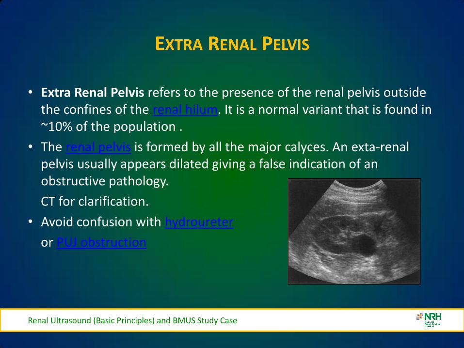
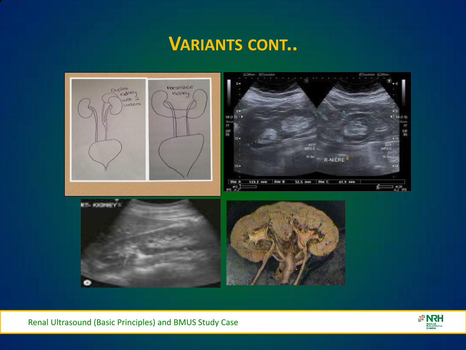
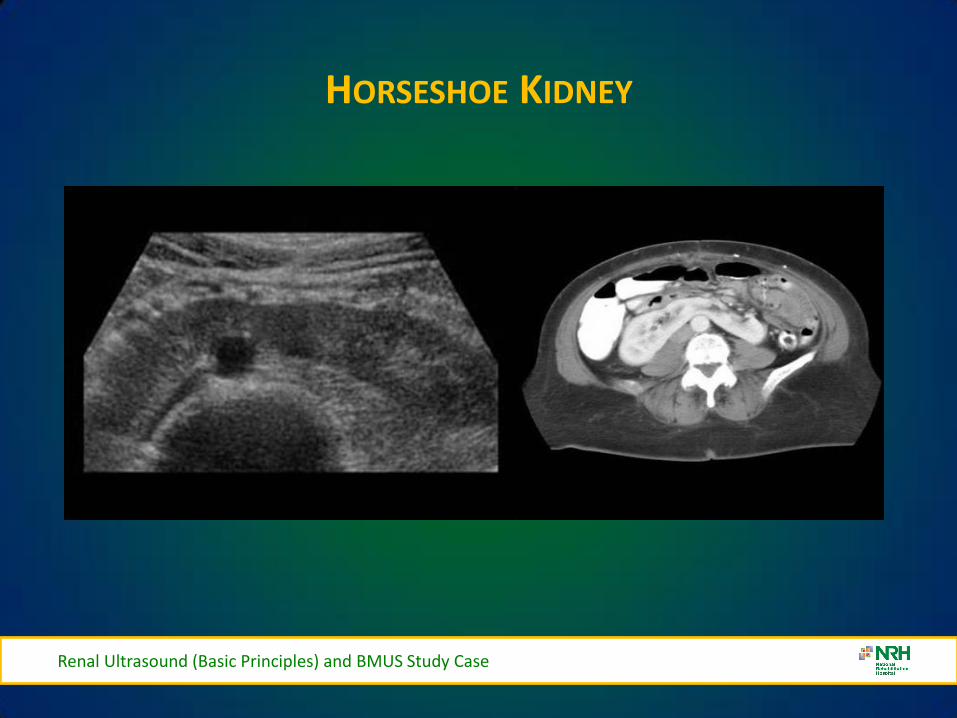
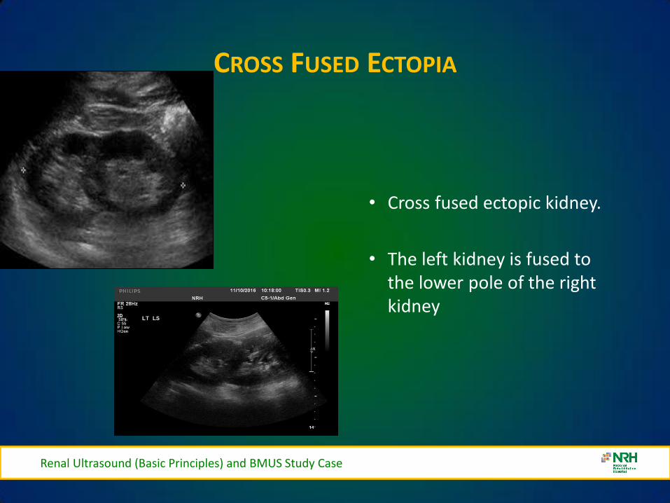
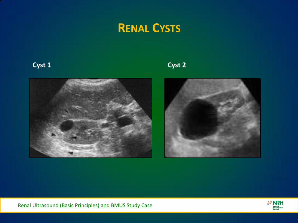
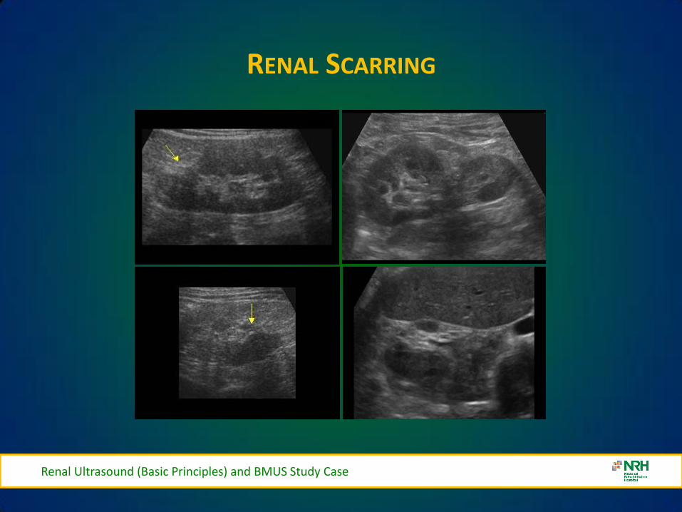
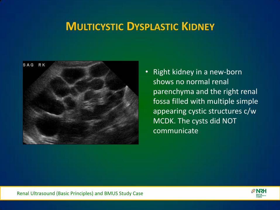
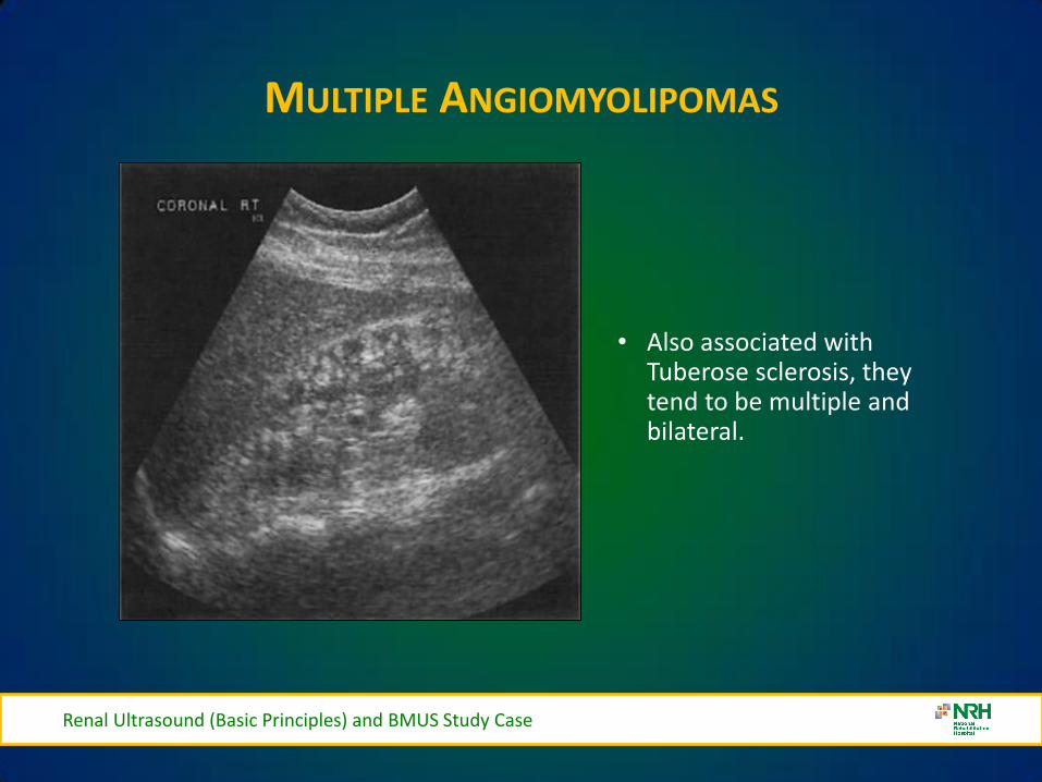
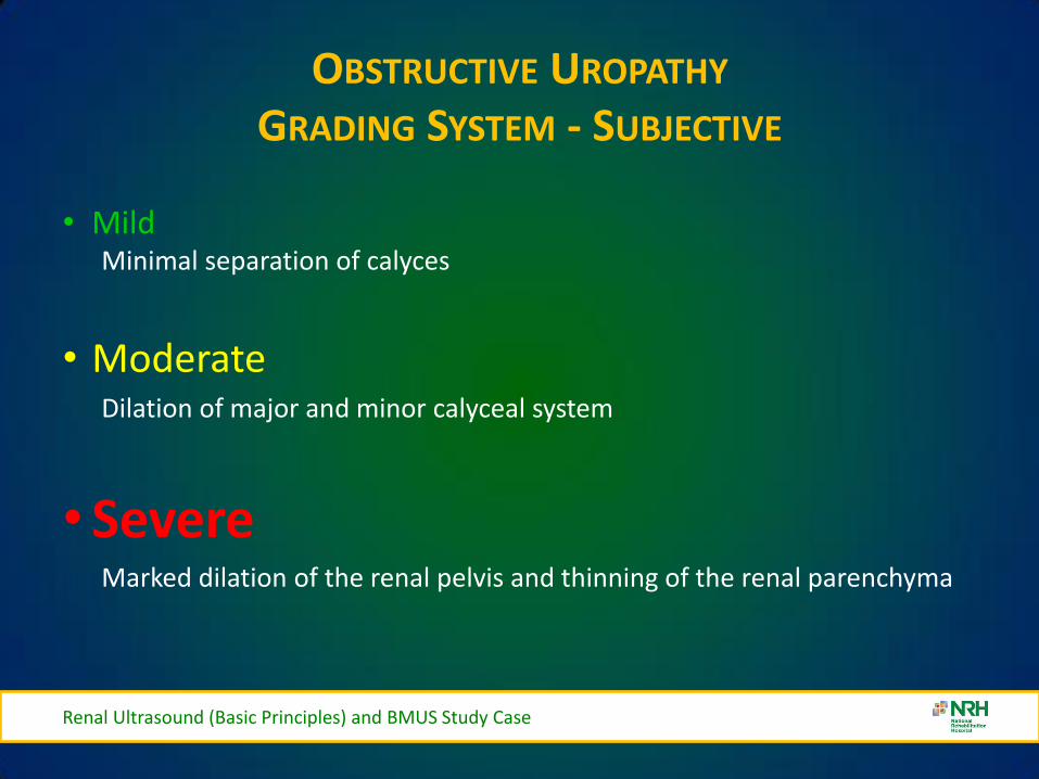
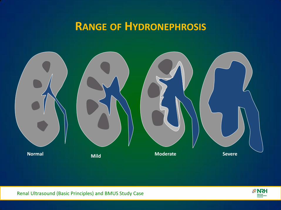
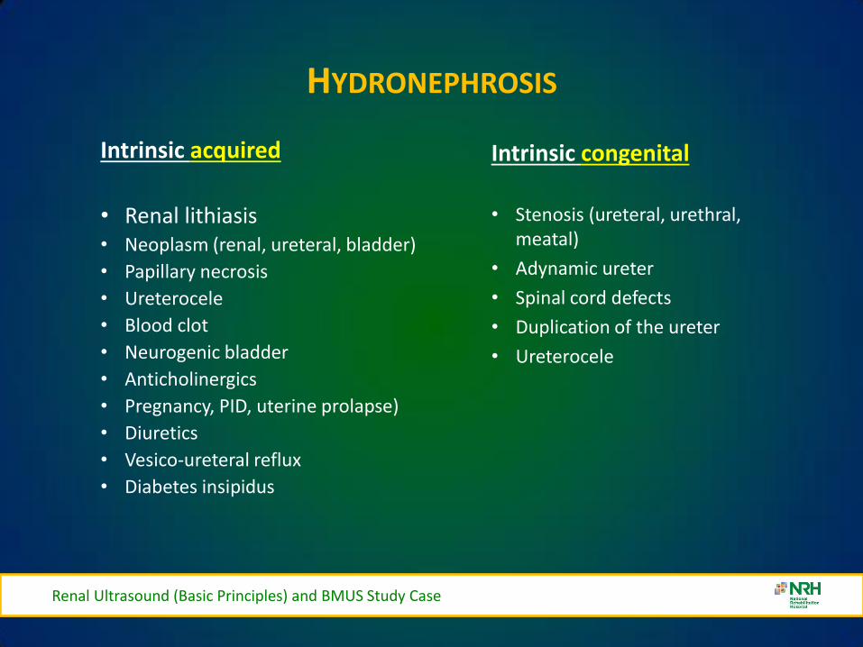
HYDRONEPHROSIS
Renal Ultrasound (Basic Principles) and BMUS Study Case
Intrinsic acquired
bull Renal lithiasis bull Neoplasm (renal ureteral bladder)
bull Papillary necrosis
bull Ureterocele
bull Blood clot
bull Neurogenic bladder
bull Anticholinergics
bull Pregnancy PID uterine prolapse)
bull Diuretics
bull Vesico-ureteral reflux
bull Diabetes insipidus
Intrinsic congenital
bull Stenosis (ureteral urethral meatal)
bull Adynamic ureter
bull Spinal cord defects
bull Duplication of the ureter
bull Ureterocele
HYDRONEPHROSIS
Mild Hydronephrosis
Renal Ultrasound (Basic Principles) and BMUS Study Case
HYDRONEPHROSIS
Moderate Hydronephrosis
Renal Ultrasound (Basic Principles) and BMUS Study Case
SEVERE HYDRONEPHROSIS
Renal Ultrasound (Basic Principles) and BMUS Study Case
Severe Hydronephrosis
SMALL KIDNEYS
bull Unilateral (may be bilateral)
ndash Chronic infections
ndash RAS Renal Artery Stenosis
ndash Hypoplastic Kidney
ndash Always bilateral
bull Chronic glomerulonephritis
bull Hypertensive nephropathy
bull Collagen Vascular Disease
Renal Ultrasound (Basic Principles) and BMUS Study Case
ENLARGED KIDNEYS
Always Unilateral
ndash Compensatory hypertrophy
ndash Bilateral or Unilateral
bull Renal Mass
bull Hydronephrosis
bull Renal vein thromboses
bull Lymphoma
bull Amyloidosis
Always Bilateral
ndash PCK Polycystic Kidney Disease
ndash AGN Acute glomerulonephritis
ndash Amyloidosis
Renal Ultrasound (Basic Principles) and BMUS Study Case
Urolithiasis
Renal Calculi- Bladder Calculi
US only
41
Fig 3
Fig 4 Fig 5
Fig 3 4 amp 5 ndash Bladder stones with posterior shadowing clearly seen at US in distended bladder but not apparent on X-ray
Staghorn Calculus
BLADDER DIVERTICULAE
Renal Ultrasound (Basic Principles) and BMUS Study Case
bull Diverticula of the bladder in
infants and children are common and occur when bladder mucosa protrudes through a congenital defect in the detrusor muscle wall Most are primary Those secondary to obstruction or neurogenic dysfunction are less common than previously thought
Renal Ultrasound (Basic Principles) and BMUS Study Case
Renal Ultrasound (Basic Principles) and BMUS Study Case
BLADDER CALCULI
Renal Ultrasound (Basic Principles) and BMUS Study Case
RENAL MASS
Renal Ultrasound (Basic Principles) and BMUS Study Case
bull Pyelonephritis sonographic appearance is most commonly normal but you may find hypoechoic cortex and loss of demarcation between the outer cortex and middle pyramids and columns of Bertin
bull Renal mass may have any echotexture (hyperechoic anechoic etc) and appear anywhere within the kidney
bull Transplant kidney a normal echotexture kidney typically in a pelvic location
TRANSITIONAL CELL CARCINOMA
bull Ultrasound images Large transitional cell carcinoma in the upper pole of the RK The changes are more subtle than those of renal cell carcinoma and the renal outline remains intact
bull Transitional cell carcinoma is the most common bladder tumour occurring less frequently in the collecting system of the kidney and the ureter It usually presents with haematuria while still small It is best diagnosed with cystoscopy
Renal Ultrasound (Basic Principles) and BMUS Study Case
HYDRONEPHROSIS DUE TO TCC URETER
Renal Ultrasound (Basic Principles) and BMUS Study Case
US AND CT RENAL CA
Renal Ultrasound (Basic Principles) and BMUS Study Case
RENAL CELL CARCINOMA
bull Large heterogeneous mass which enlarges and deforms the shape of the kidney (Fig below) The mass may contain areas of cystic degeneration andor calcification It has a predilection to spread into the ipsilateral renal vein and IVC
bull Colour Doppler usually reveals a disorganized and increased blood flow pattern within the mass with high velocities from the arterioverous shunts within the carcinoma
Renal Ultrasound (Basic Principles) and BMUS Study Case
bull Smaller RCCs can be hyperechoic and may be confused with benign angiomyolipoma The latter has well-defined borders whilst an RCC is illdefined
BURKITTS LYMPHOMA
Renal Ultrasound (Basic Principles) and BMUS Study Case
NEUROBLASTOMA
Renal Ultrasound (Basic Principles) and BMUS Study Case
RENAL TRAUMA
Renal Ultrasound (Basic Principles) and BMUS Study Case
POLYCYSTIC KIDNEYS
Renal Ultrasound (Basic Principles) and BMUS Study Case
RENAL TRANSPLANTS-SURGICAL TECHNIQUE
bull Usually heterotopic (placed in addition to the native diseased kidneys)
bull Positioned in the extra-peritoneal pouch in the iliac fossa (usually the right) anterior to the iliacus and psoas muscles
Renal Ultrasound (Basic Principles) and BMUS Study Case
THE ROLE OF ULTRASOUND
B Mode imaging
bull Morphological appearances
PC dilation
Peri-renal fluid collections
bull Doppler
Colour Power Perfusion
Spectral Doppler
Waveforms
bull Intervention
Guide Biopsy Procedures
Drain Fluid Collections
Placement of Nephrostomy
Tubes
Renal Ultrasound (Basic Principles) and BMUS Study Case
LS RENAL TRANSPLANT (USING EXTENDED FIELD OF VIEW)
Renal Ultrasound (Basic Principles) and BMUS Study Case
PERFUSION WITH POWER AND COLOUR DOPPLER
Renal Ultrasound (Basic Principles) and BMUS Study Case
BACKGROUND
The main purpose of regular urology review is to preserve renal function with the ultimate aim of a non obstructed urinary system
Urinary Tract Infections (UTI) are the most frequent medical complication in patients with Spinal Cord Injuries (SCI) and a major cause of morbidity
Renal Ultrasound (Basic Principles) and BMUS Study Case
X-RAY ONLY
Fig 1 amp 2 - US showing decompressed bladder around IDUC X-ray in same patient confirming large calculus not visualised on US
Fig 1 Fig 2
Renal Ultrasound (Basic Principles) and BMUS Study Case
US ONLY
Fig 3
Fig 4
Fig 5
Fig 3 4 amp 5 ndash Bladder stones with posterior shadowing clearly seen at US in distended bladder but not apparent on X-ray
Renal Ultrasound (Basic Principles) and BMUS Study Case
RESULTS
bull Bladder ultrasound alone diagnosed 36 bladder calculi compared to 18 diagnosed by X-ray alone
bull US and X-ray combined diagnosed 46
Renal Ultrasound (Basic Principles) and BMUS Study Case
BLADDER MUCUS
bull In one patient a focal mobile area of increased reflectivity was demonstrated posteriorly in the bladder No posterior acoustic shadowing was seen to suggest of calculus formation
bull On flexible cystoscopy no calculus identified only mucus seen
Renal Ultrasound (Basic Principles) and BMUS Study Case
CASE STUDY 1
Renal Ultrasound (Basic Principles) and BMUS Study Case
CASE STUDY 2-VHL (VON HIPPEL LINDAU SYNDROME)
bull renal lesions
ndash renal cell carcinoma(s) (RCCs) usually of the clear cell type
bull 70 lifetime risk
bull RCCs present at an earlier age in those with vHL
ndash renal cysts
bull can occur in up to 75 of cases
bull often tend to be bilateral and multiple
bull Prognosis is poor with a median survival of ~50 years with the most common cause of death being RCC and cerebellar haemangioblastomas
Renal Ultrasound (Basic Principles) and BMUS Study Case
Renal Ultrasound (Basic Principles) and BMUS Study Case
Case Study 3
Renal Ultrasound (Basic Principles) and BMUS Study Case
Renal Ultrasound (Basic Principles) and BMUS Study Case
PAGE KIDNEY
Renal Ultrasound (Basic Principles) and BMUS Study Case
THANK YOU FOR YOUR ATTENTION
Renal Ultrasound (Basic Principles) and BMUS Study Case
Rosie Conlon rosieconlonnrhie
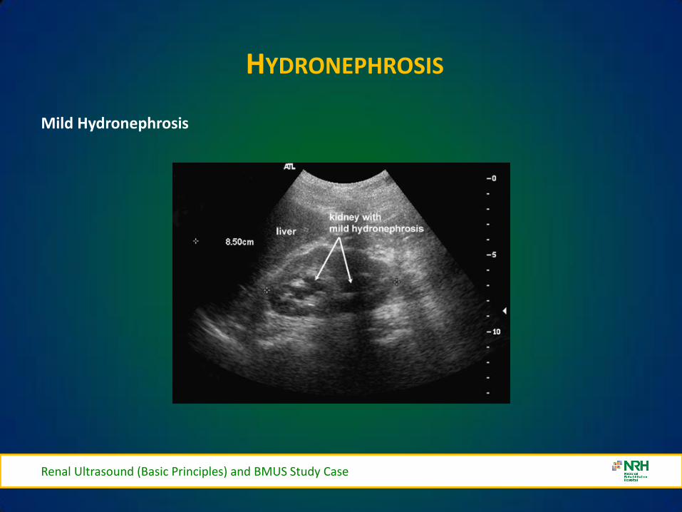
HYDRONEPHROSIS
Mild Hydronephrosis
Renal Ultrasound (Basic Principles) and BMUS Study Case
HYDRONEPHROSIS
Moderate Hydronephrosis
Renal Ultrasound (Basic Principles) and BMUS Study Case
SEVERE HYDRONEPHROSIS
Renal Ultrasound (Basic Principles) and BMUS Study Case
Severe Hydronephrosis
SMALL KIDNEYS
bull Unilateral (may be bilateral)
ndash Chronic infections
ndash RAS Renal Artery Stenosis
ndash Hypoplastic Kidney
ndash Always bilateral
bull Chronic glomerulonephritis
bull Hypertensive nephropathy
bull Collagen Vascular Disease
Renal Ultrasound (Basic Principles) and BMUS Study Case
ENLARGED KIDNEYS
Always Unilateral
ndash Compensatory hypertrophy
ndash Bilateral or Unilateral
bull Renal Mass
bull Hydronephrosis
bull Renal vein thromboses
bull Lymphoma
bull Amyloidosis
Always Bilateral
ndash PCK Polycystic Kidney Disease
ndash AGN Acute glomerulonephritis
ndash Amyloidosis
Renal Ultrasound (Basic Principles) and BMUS Study Case
Urolithiasis
Renal Calculi- Bladder Calculi
US only
41
Fig 3
Fig 4 Fig 5
Fig 3 4 amp 5 ndash Bladder stones with posterior shadowing clearly seen at US in distended bladder but not apparent on X-ray
Staghorn Calculus
BLADDER DIVERTICULAE
Renal Ultrasound (Basic Principles) and BMUS Study Case
bull Diverticula of the bladder in
infants and children are common and occur when bladder mucosa protrudes through a congenital defect in the detrusor muscle wall Most are primary Those secondary to obstruction or neurogenic dysfunction are less common than previously thought
Renal Ultrasound (Basic Principles) and BMUS Study Case
Renal Ultrasound (Basic Principles) and BMUS Study Case
BLADDER CALCULI
Renal Ultrasound (Basic Principles) and BMUS Study Case
RENAL MASS
Renal Ultrasound (Basic Principles) and BMUS Study Case
bull Pyelonephritis sonographic appearance is most commonly normal but you may find hypoechoic cortex and loss of demarcation between the outer cortex and middle pyramids and columns of Bertin
bull Renal mass may have any echotexture (hyperechoic anechoic etc) and appear anywhere within the kidney
bull Transplant kidney a normal echotexture kidney typically in a pelvic location
TRANSITIONAL CELL CARCINOMA
bull Ultrasound images Large transitional cell carcinoma in the upper pole of the RK The changes are more subtle than those of renal cell carcinoma and the renal outline remains intact
bull Transitional cell carcinoma is the most common bladder tumour occurring less frequently in the collecting system of the kidney and the ureter It usually presents with haematuria while still small It is best diagnosed with cystoscopy
Renal Ultrasound (Basic Principles) and BMUS Study Case
HYDRONEPHROSIS DUE TO TCC URETER
Renal Ultrasound (Basic Principles) and BMUS Study Case
US AND CT RENAL CA
Renal Ultrasound (Basic Principles) and BMUS Study Case
RENAL CELL CARCINOMA
bull Large heterogeneous mass which enlarges and deforms the shape of the kidney (Fig below) The mass may contain areas of cystic degeneration andor calcification It has a predilection to spread into the ipsilateral renal vein and IVC
bull Colour Doppler usually reveals a disorganized and increased blood flow pattern within the mass with high velocities from the arterioverous shunts within the carcinoma
Renal Ultrasound (Basic Principles) and BMUS Study Case
bull Smaller RCCs can be hyperechoic and may be confused with benign angiomyolipoma The latter has well-defined borders whilst an RCC is illdefined
BURKITTS LYMPHOMA
Renal Ultrasound (Basic Principles) and BMUS Study Case
NEUROBLASTOMA
Renal Ultrasound (Basic Principles) and BMUS Study Case
RENAL TRAUMA
Renal Ultrasound (Basic Principles) and BMUS Study Case
POLYCYSTIC KIDNEYS
Renal Ultrasound (Basic Principles) and BMUS Study Case
RENAL TRANSPLANTS-SURGICAL TECHNIQUE
bull Usually heterotopic (placed in addition to the native diseased kidneys)
bull Positioned in the extra-peritoneal pouch in the iliac fossa (usually the right) anterior to the iliacus and psoas muscles
Renal Ultrasound (Basic Principles) and BMUS Study Case
THE ROLE OF ULTRASOUND
B Mode imaging
bull Morphological appearances
PC dilation
Peri-renal fluid collections
bull Doppler
Colour Power Perfusion
Spectral Doppler
Waveforms
bull Intervention
Guide Biopsy Procedures
Drain Fluid Collections
Placement of Nephrostomy
Tubes
Renal Ultrasound (Basic Principles) and BMUS Study Case
LS RENAL TRANSPLANT (USING EXTENDED FIELD OF VIEW)
Renal Ultrasound (Basic Principles) and BMUS Study Case
PERFUSION WITH POWER AND COLOUR DOPPLER
Renal Ultrasound (Basic Principles) and BMUS Study Case
BACKGROUND
The main purpose of regular urology review is to preserve renal function with the ultimate aim of a non obstructed urinary system
Urinary Tract Infections (UTI) are the most frequent medical complication in patients with Spinal Cord Injuries (SCI) and a major cause of morbidity
Renal Ultrasound (Basic Principles) and BMUS Study Case
X-RAY ONLY
Fig 1 amp 2 - US showing decompressed bladder around IDUC X-ray in same patient confirming large calculus not visualised on US
Fig 1 Fig 2
Renal Ultrasound (Basic Principles) and BMUS Study Case
US ONLY
Fig 3
Fig 4
Fig 5
Fig 3 4 amp 5 ndash Bladder stones with posterior shadowing clearly seen at US in distended bladder but not apparent on X-ray
Renal Ultrasound (Basic Principles) and BMUS Study Case
RESULTS
bull Bladder ultrasound alone diagnosed 36 bladder calculi compared to 18 diagnosed by X-ray alone
bull US and X-ray combined diagnosed 46
Renal Ultrasound (Basic Principles) and BMUS Study Case
BLADDER MUCUS
bull In one patient a focal mobile area of increased reflectivity was demonstrated posteriorly in the bladder No posterior acoustic shadowing was seen to suggest of calculus formation
bull On flexible cystoscopy no calculus identified only mucus seen
Renal Ultrasound (Basic Principles) and BMUS Study Case
CASE STUDY 1
Renal Ultrasound (Basic Principles) and BMUS Study Case
CASE STUDY 2-VHL (VON HIPPEL LINDAU SYNDROME)
bull renal lesions
ndash renal cell carcinoma(s) (RCCs) usually of the clear cell type
bull 70 lifetime risk
bull RCCs present at an earlier age in those with vHL
ndash renal cysts
bull can occur in up to 75 of cases
bull often tend to be bilateral and multiple
bull Prognosis is poor with a median survival of ~50 years with the most common cause of death being RCC and cerebellar haemangioblastomas
Renal Ultrasound (Basic Principles) and BMUS Study Case
Renal Ultrasound (Basic Principles) and BMUS Study Case
Case Study 3
Renal Ultrasound (Basic Principles) and BMUS Study Case
Renal Ultrasound (Basic Principles) and BMUS Study Case
PAGE KIDNEY
Renal Ultrasound (Basic Principles) and BMUS Study Case
THANK YOU FOR YOUR ATTENTION
Renal Ultrasound (Basic Principles) and BMUS Study Case
Rosie Conlon rosieconlonnrhie
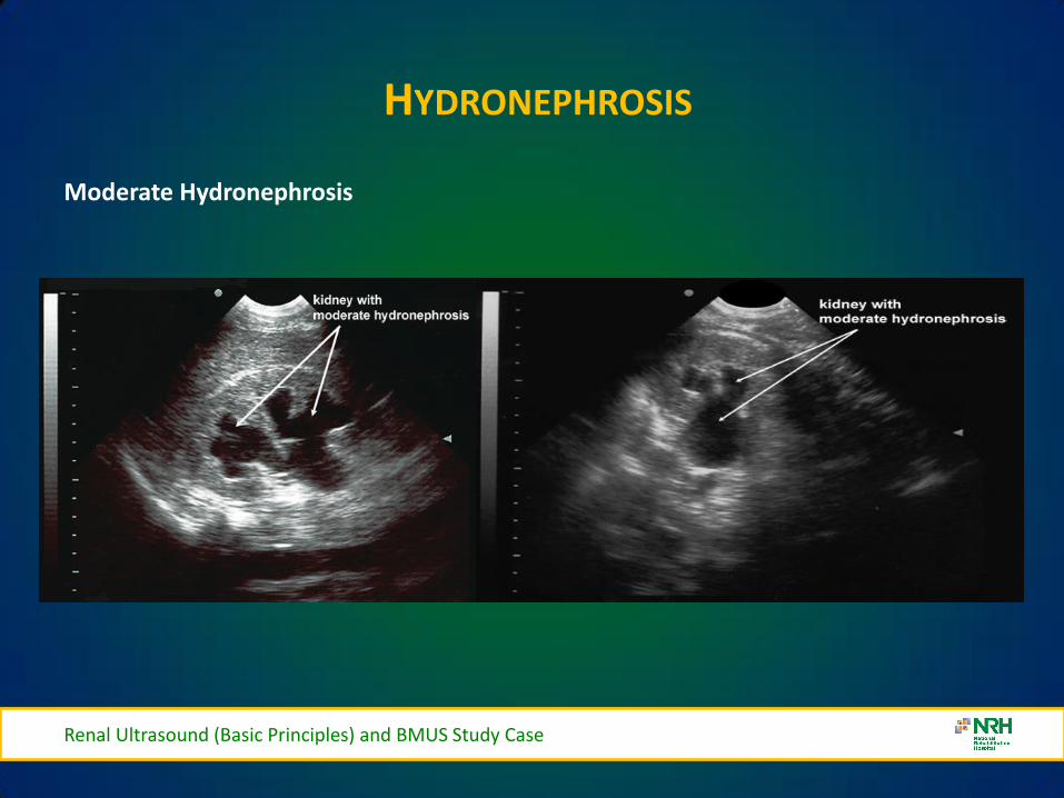
HYDRONEPHROSIS
Moderate Hydronephrosis
Renal Ultrasound (Basic Principles) and BMUS Study Case
SEVERE HYDRONEPHROSIS
Renal Ultrasound (Basic Principles) and BMUS Study Case
Severe Hydronephrosis
SMALL KIDNEYS
bull Unilateral (may be bilateral)
ndash Chronic infections
ndash RAS Renal Artery Stenosis
ndash Hypoplastic Kidney
ndash Always bilateral
bull Chronic glomerulonephritis
bull Hypertensive nephropathy
bull Collagen Vascular Disease
Renal Ultrasound (Basic Principles) and BMUS Study Case
ENLARGED KIDNEYS
Always Unilateral
ndash Compensatory hypertrophy
ndash Bilateral or Unilateral
bull Renal Mass
bull Hydronephrosis
bull Renal vein thromboses
bull Lymphoma
bull Amyloidosis
Always Bilateral
ndash PCK Polycystic Kidney Disease
ndash AGN Acute glomerulonephritis
ndash Amyloidosis
Renal Ultrasound (Basic Principles) and BMUS Study Case
Urolithiasis
Renal Calculi- Bladder Calculi
US only
41
Fig 3
Fig 4 Fig 5
Fig 3 4 amp 5 ndash Bladder stones with posterior shadowing clearly seen at US in distended bladder but not apparent on X-ray
Staghorn Calculus
BLADDER DIVERTICULAE
Renal Ultrasound (Basic Principles) and BMUS Study Case
bull Diverticula of the bladder in
infants and children are common and occur when bladder mucosa protrudes through a congenital defect in the detrusor muscle wall Most are primary Those secondary to obstruction or neurogenic dysfunction are less common than previously thought
Renal Ultrasound (Basic Principles) and BMUS Study Case
Renal Ultrasound (Basic Principles) and BMUS Study Case
BLADDER CALCULI
Renal Ultrasound (Basic Principles) and BMUS Study Case
RENAL MASS
Renal Ultrasound (Basic Principles) and BMUS Study Case
bull Pyelonephritis sonographic appearance is most commonly normal but you may find hypoechoic cortex and loss of demarcation between the outer cortex and middle pyramids and columns of Bertin
bull Renal mass may have any echotexture (hyperechoic anechoic etc) and appear anywhere within the kidney
bull Transplant kidney a normal echotexture kidney typically in a pelvic location
TRANSITIONAL CELL CARCINOMA
bull Ultrasound images Large transitional cell carcinoma in the upper pole of the RK The changes are more subtle than those of renal cell carcinoma and the renal outline remains intact
bull Transitional cell carcinoma is the most common bladder tumour occurring less frequently in the collecting system of the kidney and the ureter It usually presents with haematuria while still small It is best diagnosed with cystoscopy
Renal Ultrasound (Basic Principles) and BMUS Study Case
HYDRONEPHROSIS DUE TO TCC URETER
Renal Ultrasound (Basic Principles) and BMUS Study Case
US AND CT RENAL CA
Renal Ultrasound (Basic Principles) and BMUS Study Case
RENAL CELL CARCINOMA
bull Large heterogeneous mass which enlarges and deforms the shape of the kidney (Fig below) The mass may contain areas of cystic degeneration andor calcification It has a predilection to spread into the ipsilateral renal vein and IVC
bull Colour Doppler usually reveals a disorganized and increased blood flow pattern within the mass with high velocities from the arterioverous shunts within the carcinoma
Renal Ultrasound (Basic Principles) and BMUS Study Case
bull Smaller RCCs can be hyperechoic and may be confused with benign angiomyolipoma The latter has well-defined borders whilst an RCC is illdefined
BURKITTS LYMPHOMA
Renal Ultrasound (Basic Principles) and BMUS Study Case
NEUROBLASTOMA
Renal Ultrasound (Basic Principles) and BMUS Study Case
RENAL TRAUMA
Renal Ultrasound (Basic Principles) and BMUS Study Case
POLYCYSTIC KIDNEYS
Renal Ultrasound (Basic Principles) and BMUS Study Case
RENAL TRANSPLANTS-SURGICAL TECHNIQUE
bull Usually heterotopic (placed in addition to the native diseased kidneys)
bull Positioned in the extra-peritoneal pouch in the iliac fossa (usually the right) anterior to the iliacus and psoas muscles
Renal Ultrasound (Basic Principles) and BMUS Study Case
THE ROLE OF ULTRASOUND
B Mode imaging
bull Morphological appearances
PC dilation
Peri-renal fluid collections
bull Doppler
Colour Power Perfusion
Spectral Doppler
Waveforms
bull Intervention
Guide Biopsy Procedures
Drain Fluid Collections
Placement of Nephrostomy
Tubes
Renal Ultrasound (Basic Principles) and BMUS Study Case
LS RENAL TRANSPLANT (USING EXTENDED FIELD OF VIEW)
Renal Ultrasound (Basic Principles) and BMUS Study Case
PERFUSION WITH POWER AND COLOUR DOPPLER
Renal Ultrasound (Basic Principles) and BMUS Study Case
BACKGROUND
The main purpose of regular urology review is to preserve renal function with the ultimate aim of a non obstructed urinary system
Urinary Tract Infections (UTI) are the most frequent medical complication in patients with Spinal Cord Injuries (SCI) and a major cause of morbidity
Renal Ultrasound (Basic Principles) and BMUS Study Case
X-RAY ONLY
Fig 1 amp 2 - US showing decompressed bladder around IDUC X-ray in same patient confirming large calculus not visualised on US
Fig 1 Fig 2
Renal Ultrasound (Basic Principles) and BMUS Study Case
US ONLY
Fig 3
Fig 4
Fig 5
Fig 3 4 amp 5 ndash Bladder stones with posterior shadowing clearly seen at US in distended bladder but not apparent on X-ray
Renal Ultrasound (Basic Principles) and BMUS Study Case
RESULTS
bull Bladder ultrasound alone diagnosed 36 bladder calculi compared to 18 diagnosed by X-ray alone
bull US and X-ray combined diagnosed 46
Renal Ultrasound (Basic Principles) and BMUS Study Case
BLADDER MUCUS
bull In one patient a focal mobile area of increased reflectivity was demonstrated posteriorly in the bladder No posterior acoustic shadowing was seen to suggest of calculus formation
bull On flexible cystoscopy no calculus identified only mucus seen
Renal Ultrasound (Basic Principles) and BMUS Study Case
CASE STUDY 1
Renal Ultrasound (Basic Principles) and BMUS Study Case
CASE STUDY 2-VHL (VON HIPPEL LINDAU SYNDROME)
bull renal lesions
ndash renal cell carcinoma(s) (RCCs) usually of the clear cell type
bull 70 lifetime risk
bull RCCs present at an earlier age in those with vHL
ndash renal cysts
bull can occur in up to 75 of cases
bull often tend to be bilateral and multiple
bull Prognosis is poor with a median survival of ~50 years with the most common cause of death being RCC and cerebellar haemangioblastomas
Renal Ultrasound (Basic Principles) and BMUS Study Case
Renal Ultrasound (Basic Principles) and BMUS Study Case
Case Study 3
Renal Ultrasound (Basic Principles) and BMUS Study Case
Renal Ultrasound (Basic Principles) and BMUS Study Case
PAGE KIDNEY
Renal Ultrasound (Basic Principles) and BMUS Study Case
THANK YOU FOR YOUR ATTENTION
Renal Ultrasound (Basic Principles) and BMUS Study Case
Rosie Conlon rosieconlonnrhie
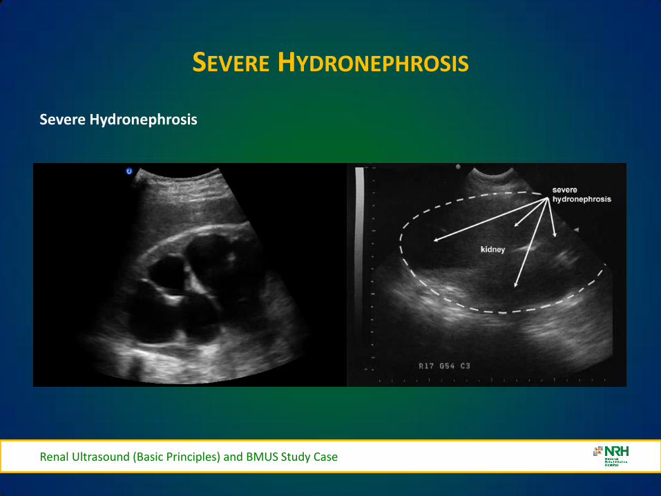
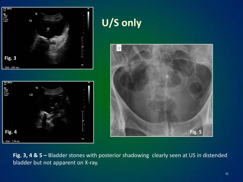
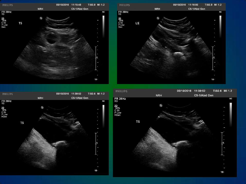
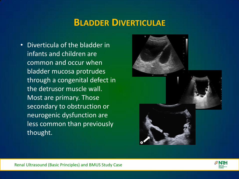
Renal Ultrasound (Basic Principles) and BMUS Study Case
Renal Ultrasound (Basic Principles) and BMUS Study Case
BLADDER CALCULI
Renal Ultrasound (Basic Principles) and BMUS Study Case
RENAL MASS
Renal Ultrasound (Basic Principles) and BMUS Study Case
bull Pyelonephritis sonographic appearance is most commonly normal but you may find hypoechoic cortex and loss of demarcation between the outer cortex and middle pyramids and columns of Bertin
bull Renal mass may have any echotexture (hyperechoic anechoic etc) and appear anywhere within the kidney
bull Transplant kidney a normal echotexture kidney typically in a pelvic location
TRANSITIONAL CELL CARCINOMA
bull Ultrasound images Large transitional cell carcinoma in the upper pole of the RK The changes are more subtle than those of renal cell carcinoma and the renal outline remains intact
bull Transitional cell carcinoma is the most common bladder tumour occurring less frequently in the collecting system of the kidney and the ureter It usually presents with haematuria while still small It is best diagnosed with cystoscopy
Renal Ultrasound (Basic Principles) and BMUS Study Case
HYDRONEPHROSIS DUE TO TCC URETER
Renal Ultrasound (Basic Principles) and BMUS Study Case
US AND CT RENAL CA
Renal Ultrasound (Basic Principles) and BMUS Study Case
RENAL CELL CARCINOMA
bull Large heterogeneous mass which enlarges and deforms the shape of the kidney (Fig below) The mass may contain areas of cystic degeneration andor calcification It has a predilection to spread into the ipsilateral renal vein and IVC
bull Colour Doppler usually reveals a disorganized and increased blood flow pattern within the mass with high velocities from the arterioverous shunts within the carcinoma
Renal Ultrasound (Basic Principles) and BMUS Study Case
bull Smaller RCCs can be hyperechoic and may be confused with benign angiomyolipoma The latter has well-defined borders whilst an RCC is illdefined
BURKITTS LYMPHOMA
Renal Ultrasound (Basic Principles) and BMUS Study Case
NEUROBLASTOMA
Renal Ultrasound (Basic Principles) and BMUS Study Case
RENAL TRAUMA
Renal Ultrasound (Basic Principles) and BMUS Study Case
POLYCYSTIC KIDNEYS
Renal Ultrasound (Basic Principles) and BMUS Study Case
RENAL TRANSPLANTS-SURGICAL TECHNIQUE
bull Usually heterotopic (placed in addition to the native diseased kidneys)
bull Positioned in the extra-peritoneal pouch in the iliac fossa (usually the right) anterior to the iliacus and psoas muscles
Renal Ultrasound (Basic Principles) and BMUS Study Case
THE ROLE OF ULTRASOUND
B Mode imaging
bull Morphological appearances
PC dilation
Peri-renal fluid collections
bull Doppler
Colour Power Perfusion
Spectral Doppler
Waveforms
bull Intervention
Guide Biopsy Procedures
Drain Fluid Collections
Placement of Nephrostomy
Tubes
Renal Ultrasound (Basic Principles) and BMUS Study Case
LS RENAL TRANSPLANT (USING EXTENDED FIELD OF VIEW)
Renal Ultrasound (Basic Principles) and BMUS Study Case
PERFUSION WITH POWER AND COLOUR DOPPLER
Renal Ultrasound (Basic Principles) and BMUS Study Case
BACKGROUND
The main purpose of regular urology review is to preserve renal function with the ultimate aim of a non obstructed urinary system
Urinary Tract Infections (UTI) are the most frequent medical complication in patients with Spinal Cord Injuries (SCI) and a major cause of morbidity
Renal Ultrasound (Basic Principles) and BMUS Study Case
X-RAY ONLY
Fig 1 amp 2 - US showing decompressed bladder around IDUC X-ray in same patient confirming large calculus not visualised on US
Fig 1 Fig 2
Renal Ultrasound (Basic Principles) and BMUS Study Case
US ONLY
Fig 3
Fig 4
Fig 5
Fig 3 4 amp 5 ndash Bladder stones with posterior shadowing clearly seen at US in distended bladder but not apparent on X-ray
Renal Ultrasound (Basic Principles) and BMUS Study Case
RESULTS
bull Bladder ultrasound alone diagnosed 36 bladder calculi compared to 18 diagnosed by X-ray alone
bull US and X-ray combined diagnosed 46
Renal Ultrasound (Basic Principles) and BMUS Study Case
BLADDER MUCUS
bull In one patient a focal mobile area of increased reflectivity was demonstrated posteriorly in the bladder No posterior acoustic shadowing was seen to suggest of calculus formation
bull On flexible cystoscopy no calculus identified only mucus seen
Renal Ultrasound (Basic Principles) and BMUS Study Case
CASE STUDY 1
Renal Ultrasound (Basic Principles) and BMUS Study Case
CASE STUDY 2-VHL (VON HIPPEL LINDAU SYNDROME)
bull renal lesions
ndash renal cell carcinoma(s) (RCCs) usually of the clear cell type
bull 70 lifetime risk
bull RCCs present at an earlier age in those with vHL
ndash renal cysts
bull can occur in up to 75 of cases
bull often tend to be bilateral and multiple
bull Prognosis is poor with a median survival of ~50 years with the most common cause of death being RCC and cerebellar haemangioblastomas
Renal Ultrasound (Basic Principles) and BMUS Study Case
Renal Ultrasound (Basic Principles) and BMUS Study Case
Case Study 3
Renal Ultrasound (Basic Principles) and BMUS Study Case
Renal Ultrasound (Basic Principles) and BMUS Study Case
PAGE KIDNEY
Renal Ultrasound (Basic Principles) and BMUS Study Case
THANK YOU FOR YOUR ATTENTION
Renal Ultrasound (Basic Principles) and BMUS Study Case
Rosie Conlon rosieconlonnrhie
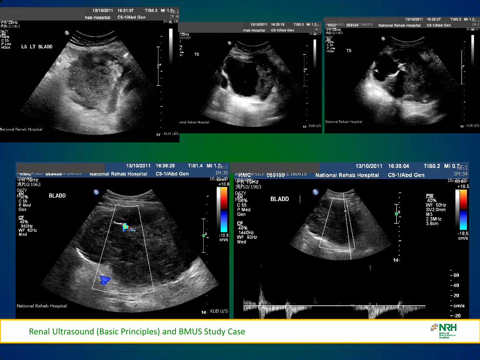
Renal Ultrasound (Basic Principles) and BMUS Study Case
BLADDER CALCULI
Renal Ultrasound (Basic Principles) and BMUS Study Case
RENAL MASS
Renal Ultrasound (Basic Principles) and BMUS Study Case
bull Pyelonephritis sonographic appearance is most commonly normal but you may find hypoechoic cortex and loss of demarcation between the outer cortex and middle pyramids and columns of Bertin
bull Renal mass may have any echotexture (hyperechoic anechoic etc) and appear anywhere within the kidney
bull Transplant kidney a normal echotexture kidney typically in a pelvic location
TRANSITIONAL CELL CARCINOMA
bull Ultrasound images Large transitional cell carcinoma in the upper pole of the RK The changes are more subtle than those of renal cell carcinoma and the renal outline remains intact
bull Transitional cell carcinoma is the most common bladder tumour occurring less frequently in the collecting system of the kidney and the ureter It usually presents with haematuria while still small It is best diagnosed with cystoscopy
Renal Ultrasound (Basic Principles) and BMUS Study Case
HYDRONEPHROSIS DUE TO TCC URETER
Renal Ultrasound (Basic Principles) and BMUS Study Case
US AND CT RENAL CA
Renal Ultrasound (Basic Principles) and BMUS Study Case
RENAL CELL CARCINOMA
bull Large heterogeneous mass which enlarges and deforms the shape of the kidney (Fig below) The mass may contain areas of cystic degeneration andor calcification It has a predilection to spread into the ipsilateral renal vein and IVC
bull Colour Doppler usually reveals a disorganized and increased blood flow pattern within the mass with high velocities from the arterioverous shunts within the carcinoma
Renal Ultrasound (Basic Principles) and BMUS Study Case
bull Smaller RCCs can be hyperechoic and may be confused with benign angiomyolipoma The latter has well-defined borders whilst an RCC is illdefined
BURKITTS LYMPHOMA
Renal Ultrasound (Basic Principles) and BMUS Study Case
NEUROBLASTOMA
Renal Ultrasound (Basic Principles) and BMUS Study Case
RENAL TRAUMA
Renal Ultrasound (Basic Principles) and BMUS Study Case
POLYCYSTIC KIDNEYS
Renal Ultrasound (Basic Principles) and BMUS Study Case
RENAL TRANSPLANTS-SURGICAL TECHNIQUE
bull Usually heterotopic (placed in addition to the native diseased kidneys)
bull Positioned in the extra-peritoneal pouch in the iliac fossa (usually the right) anterior to the iliacus and psoas muscles
Renal Ultrasound (Basic Principles) and BMUS Study Case
THE ROLE OF ULTRASOUND
B Mode imaging
bull Morphological appearances
PC dilation
Peri-renal fluid collections
bull Doppler
Colour Power Perfusion
Spectral Doppler
Waveforms
bull Intervention
Guide Biopsy Procedures
Drain Fluid Collections
Placement of Nephrostomy
Tubes
Renal Ultrasound (Basic Principles) and BMUS Study Case
LS RENAL TRANSPLANT (USING EXTENDED FIELD OF VIEW)
Renal Ultrasound (Basic Principles) and BMUS Study Case
PERFUSION WITH POWER AND COLOUR DOPPLER
Renal Ultrasound (Basic Principles) and BMUS Study Case
BACKGROUND
The main purpose of regular urology review is to preserve renal function with the ultimate aim of a non obstructed urinary system
Urinary Tract Infections (UTI) are the most frequent medical complication in patients with Spinal Cord Injuries (SCI) and a major cause of morbidity
Renal Ultrasound (Basic Principles) and BMUS Study Case
X-RAY ONLY
Fig 1 amp 2 - US showing decompressed bladder around IDUC X-ray in same patient confirming large calculus not visualised on US
Fig 1 Fig 2
Renal Ultrasound (Basic Principles) and BMUS Study Case
US ONLY
Fig 3
Fig 4
Fig 5
Fig 3 4 amp 5 ndash Bladder stones with posterior shadowing clearly seen at US in distended bladder but not apparent on X-ray
Renal Ultrasound (Basic Principles) and BMUS Study Case
RESULTS
bull Bladder ultrasound alone diagnosed 36 bladder calculi compared to 18 diagnosed by X-ray alone
bull US and X-ray combined diagnosed 46
Renal Ultrasound (Basic Principles) and BMUS Study Case
BLADDER MUCUS
bull In one patient a focal mobile area of increased reflectivity was demonstrated posteriorly in the bladder No posterior acoustic shadowing was seen to suggest of calculus formation
bull On flexible cystoscopy no calculus identified only mucus seen
Renal Ultrasound (Basic Principles) and BMUS Study Case
CASE STUDY 1
Renal Ultrasound (Basic Principles) and BMUS Study Case
CASE STUDY 2-VHL (VON HIPPEL LINDAU SYNDROME)
bull renal lesions
ndash renal cell carcinoma(s) (RCCs) usually of the clear cell type
bull 70 lifetime risk
bull RCCs present at an earlier age in those with vHL
ndash renal cysts
bull can occur in up to 75 of cases
bull often tend to be bilateral and multiple
bull Prognosis is poor with a median survival of ~50 years with the most common cause of death being RCC and cerebellar haemangioblastomas
Renal Ultrasound (Basic Principles) and BMUS Study Case
Renal Ultrasound (Basic Principles) and BMUS Study Case
Case Study 3
Renal Ultrasound (Basic Principles) and BMUS Study Case
Renal Ultrasound (Basic Principles) and BMUS Study Case
PAGE KIDNEY
Renal Ultrasound (Basic Principles) and BMUS Study Case
THANK YOU FOR YOUR ATTENTION
Renal Ultrasound (Basic Principles) and BMUS Study Case
Rosie Conlon rosieconlonnrhie
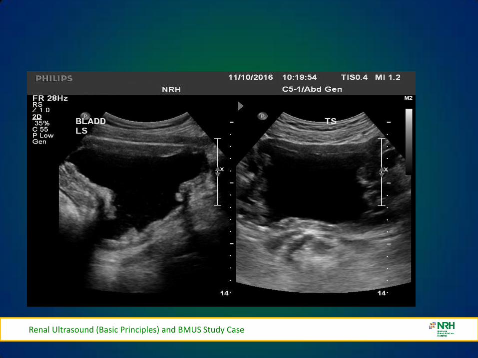
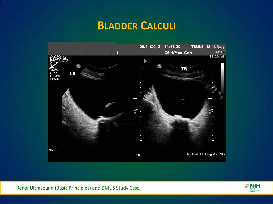
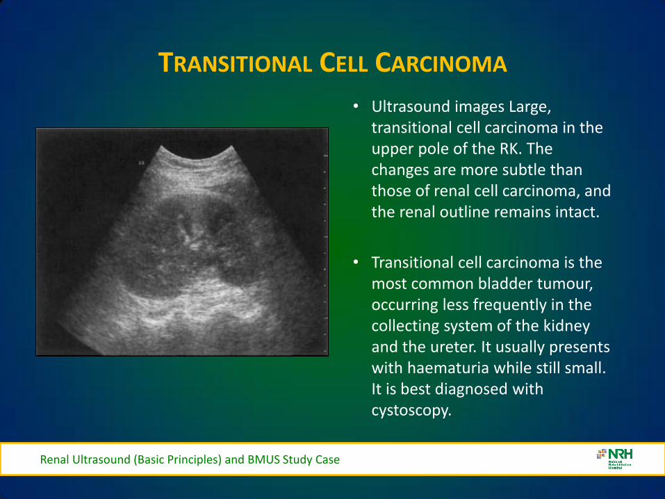
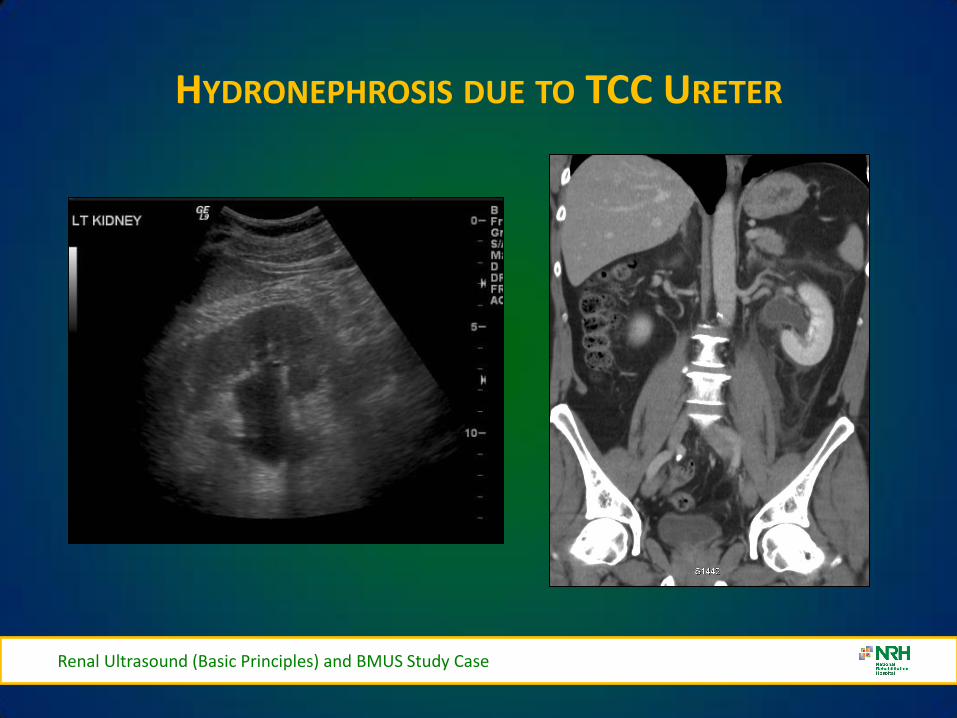
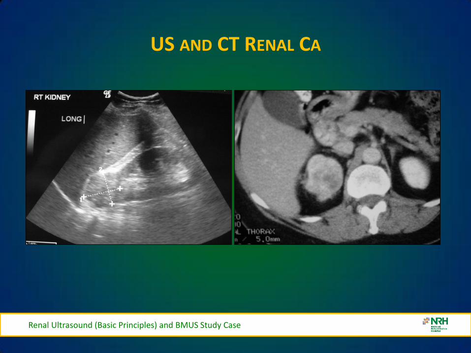
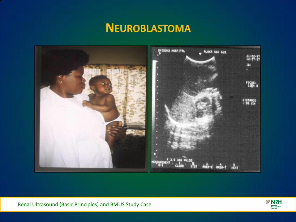
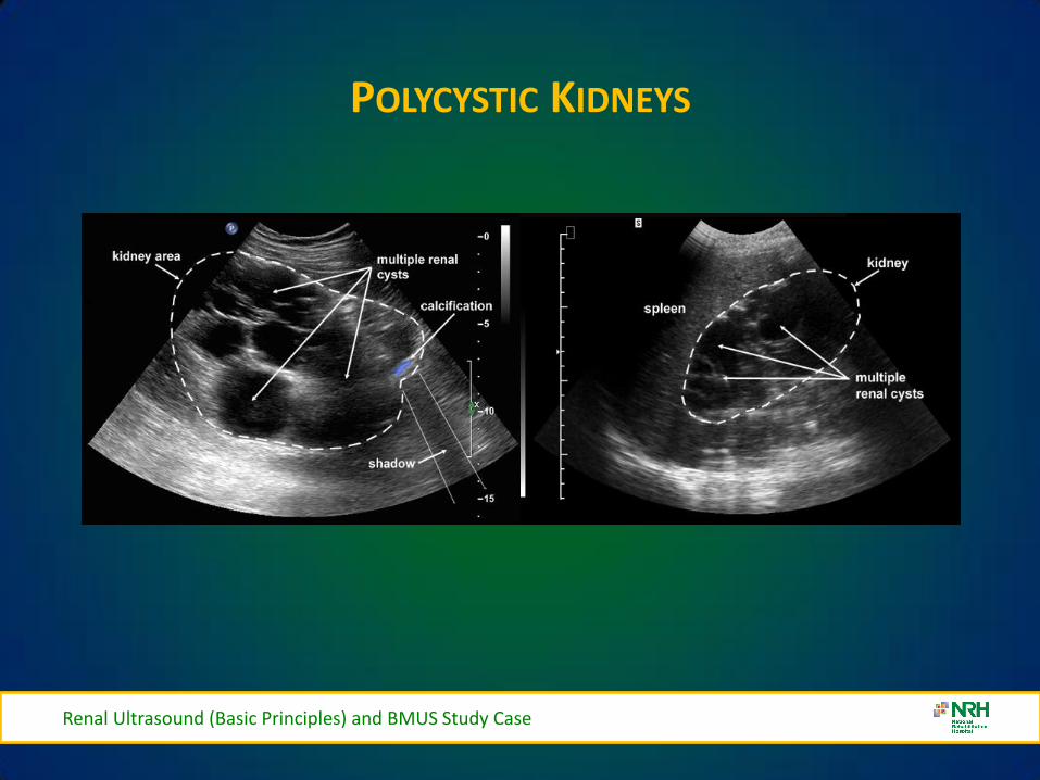
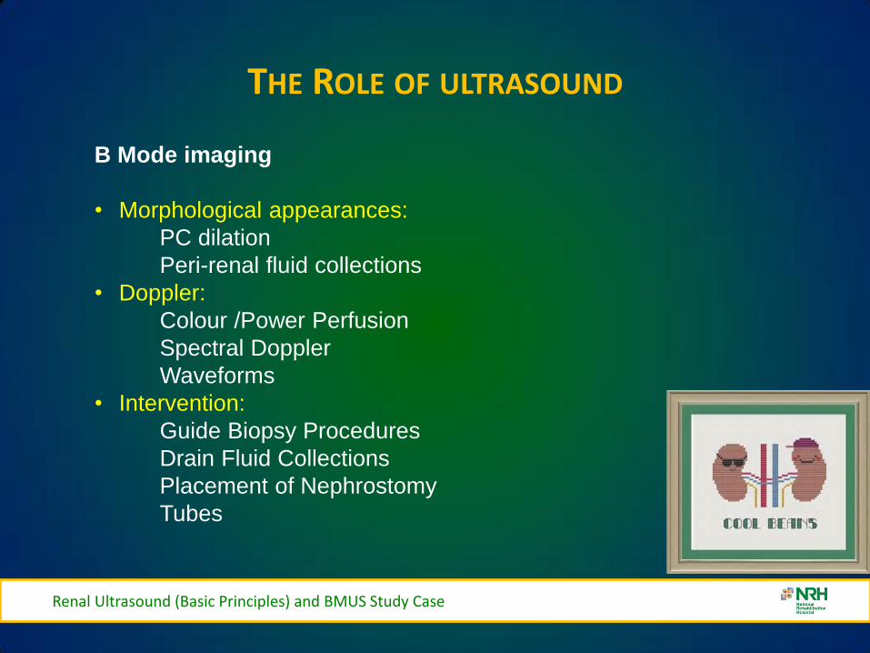
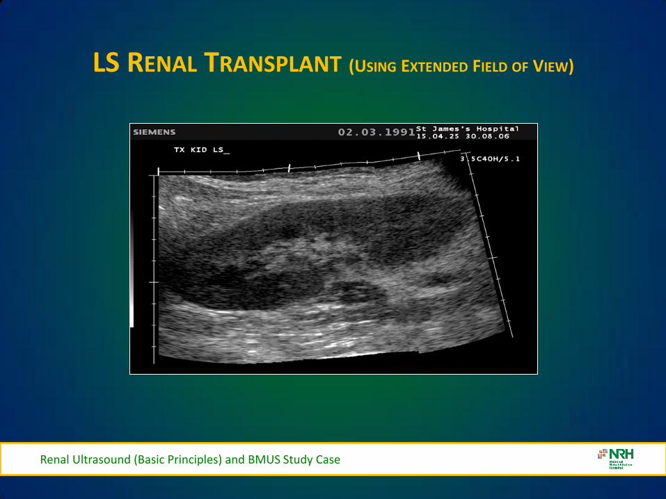
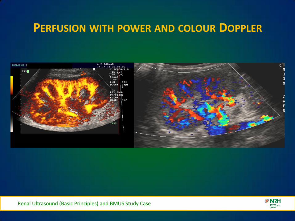
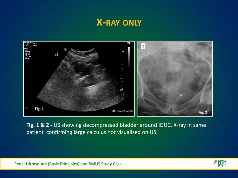
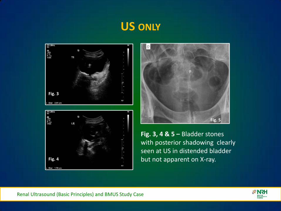
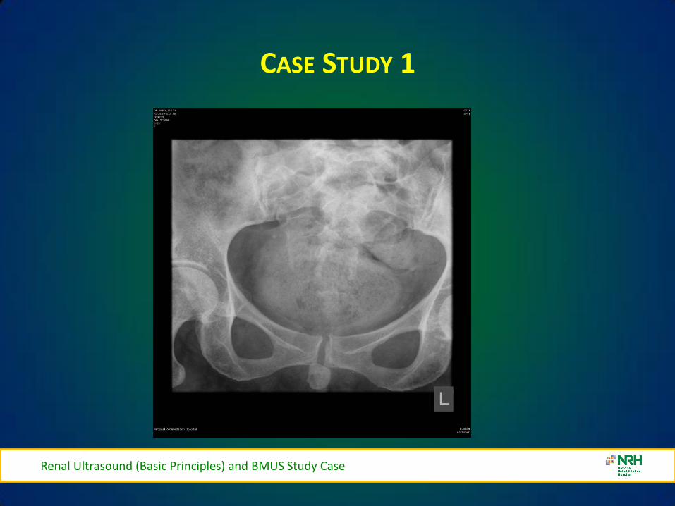

Renal Ultrasound (Basic Principles) and BMUS Study Case
Case Study 3
Renal Ultrasound (Basic Principles) and BMUS Study Case
Renal Ultrasound (Basic Principles) and BMUS Study Case
PAGE KIDNEY
Renal Ultrasound (Basic Principles) and BMUS Study Case
THANK YOU FOR YOUR ATTENTION
Renal Ultrasound (Basic Principles) and BMUS Study Case
Rosie Conlon rosieconlonnrhie
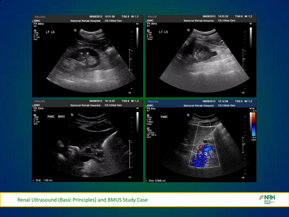
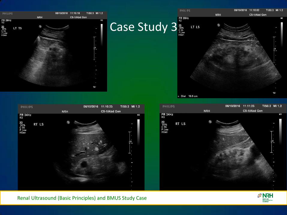
Renal Ultrasound (Basic Principles) and BMUS Study Case
PAGE KIDNEY
Renal Ultrasound (Basic Principles) and BMUS Study Case
THANK YOU FOR YOUR ATTENTION
Renal Ultrasound (Basic Principles) and BMUS Study Case
Rosie Conlon rosieconlonnrhie
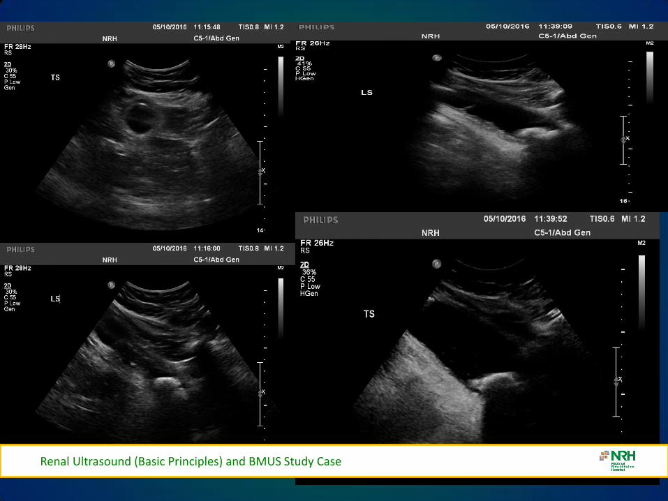
PAGE KIDNEY
Renal Ultrasound (Basic Principles) and BMUS Study Case
THANK YOU FOR YOUR ATTENTION
Renal Ultrasound (Basic Principles) and BMUS Study Case
Rosie Conlon rosieconlonnrhie
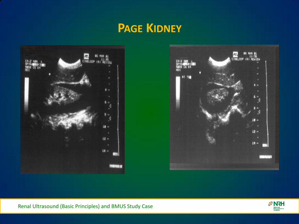
THANK YOU FOR YOUR ATTENTION
Renal Ultrasound (Basic Principles) and BMUS Study Case
Rosie Conlon rosieconlonnrhie