report of the regional workshop on the identification of … preparation of this document this...
TRANSCRIPT

Report of the
REGIONAL WORKSHOP ON THE IDENTIFICATION OF DEEP-SEA CARTILAGINOUS FISHES OF THE INDIAN OCEAN Albion, Mauritius, 10–13 June 2014
FAO Fisheries and
Aquaculture Report
FIRF/R1091 (En)
ISSN 2070-6987

Cover and report photographs: All photographs courtesy of Dr Dave Ebert.
FAO Fisheries and Aquaculture Report No. 1091 FIRF/R1091 (En)
Report of the
REGIONAL WORKSHOP ON THE IDENTIFICATION OF DEEP-SEA CARTILAGINOUS FISHES
OF THE INDIAN OCEAN
Albion, Mauritius, 10–13 June 2014
FOOD AND AGRICULTURE ORGANIZATION OF THE UNITED NATIONS
Rome, 2015

The designations employed and the presentation of material in this information product do not imply the expression of any opinion whatsoever on the part of the Food and Agriculture Organization of the United Nations (FAO) concerning the legal or development status of any country, territory, city or area or of its authorities, or concerning the delimitation of its frontiers or boundaries. The mention of specific companies or products of manufacturers, whether or not these have been patented, does not imply that these have been endorsed or recommended by FAO in preference to others of a similar nature that are not mentioned. The views expressed in this information product are those of the author(s) and do not necessarily reflect the views or policies of FAO. E-ISBN 978-92-5-108665-0 © FAO, 2015 FAO encourages the use, reproduction and dissemination of material in this information product. Except where otherwise indicated, material may be copied, downloaded and printed for private study, research and teaching purposes, or for use in non-commercial products or services, provided that appropriate acknowledgement of FAO as the source and copyright holder is given and that FAO’s endorsement of users’ views, products or services is not implied in any way. All requests for translation and adaptation rights, and for resale and other commercial use rights should be made via www.fao.org/contact-us/licence-request or addressed to [email protected]. FAO information products are available on the FAO website (www.fao.org/publications) and can be purchased through [email protected].

iii
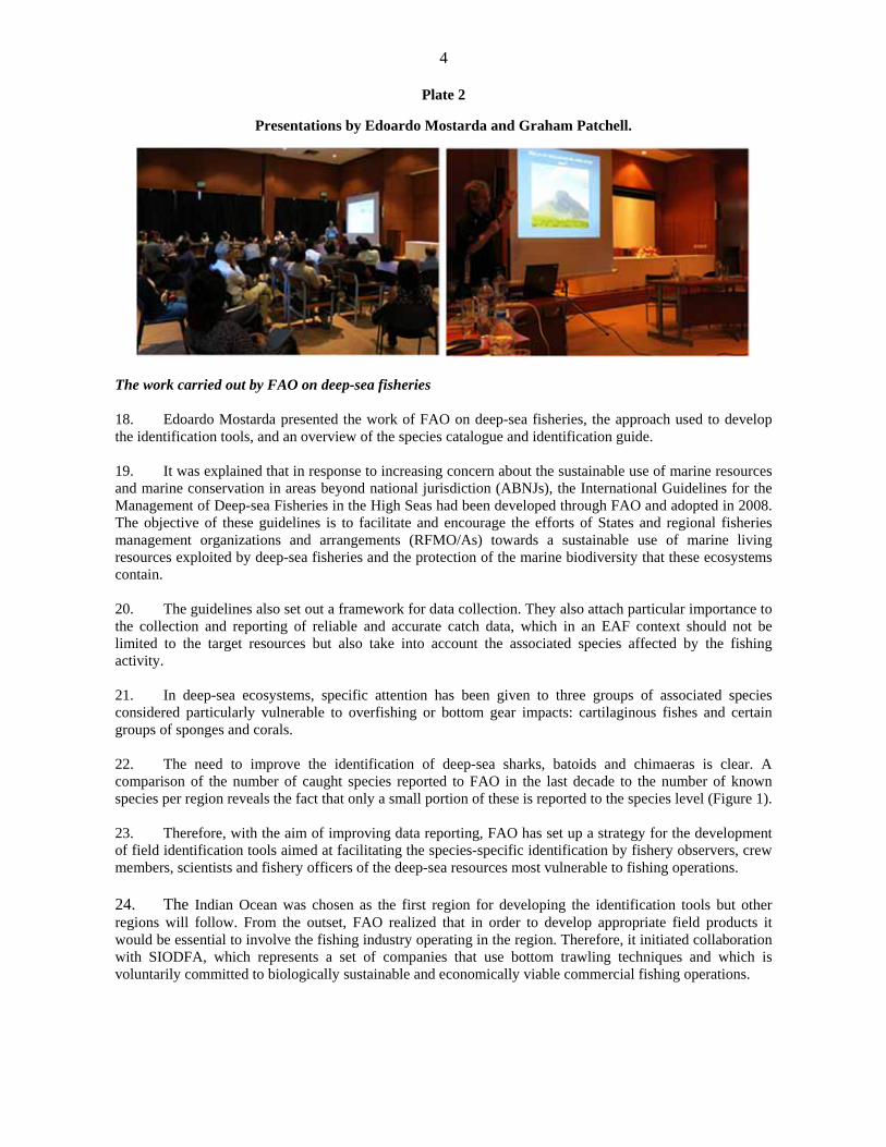
Preparation of this document
This report gives a full account of the regional workshop “Identification of Deep-sea Cartilaginous Fishes of the Indian Ocean”, which was held at the Albion Fisheries Research Center, Albion, Mauritius, from 10 to 13 June 2014. The workshop was funded by two projects: “Support to the Implementation of the International Guidelines on the Management of Deep-Sea Fisheries in the High Seas” (GCP/GLO/323/NOR); and “Fisheries Management and Marine Conservation within a Changing Ecosystem Context” (GCP/INT/253/JPN). It was aimed at improving the capabilities of scientists from countries bordering the Indian Ocean to identify a range of deep-sea cartilaginous fish species caught in the region by using an established species identification process and through the correct use of the identification tools developed by the FAO Deep-sea Fisheries and FishFinder Programmes. The main objectives of the workshop were to improve the participants’ knowledge on: (i) the anatomical features and taxonomy of the orders of deep-sea sharks occurring in the Indian Ocean; (ii) the use of the taxonomic keys included in the reference text material (e.g. FAO catalogues and identification guides); and (iii) the methods for processing and identifying a selection of deep-sea shark specimens. In addition, a biological data collection protocol was illustrated, thus allowing for improved reporting of shark specimens. The report provides a record of the presentations, lectures and of the practical sessions held during the workshop.
FAO. 2015. Report of the Regional Workshop on the Identification of Deep-sea Cartilaginous Fishes of the Indian Ocean. Albion, Mauritius, 10–13 June 2014. FAO Fisheries and Aquaculture Report No. 1091. Rome. 41 pp.
Abstract
The regional workshop “Identification of Deep-sea Cartilaginous Fishes of the Indian Ocean” was held in Albion, Mauritius, from 10 to 13 June 2014. It was attended by 19 participants from a wide range of countries and fields of expertise, including taxonomy and bioecology of cartilaginous fishes. The general objective of the workshop was to improve the capabilities of scientists from countries bordering the Indian Ocean to identify a range of deep-sea cartilaginous fish species caught in the region. The participants were introduced to the anatomical features and taxonomy of the orders of deep-sea sharks occurring in the Indian Ocean, to the use of the taxonomic keys included in the reference text material (e.g. FAO catalogues and identification guides) and to the methodologies of processing and identifying a selection of specimens. In addition, a biological data collection protocol was illustrated, thus allowing for improved reporting of shark specimens.

v
Contents
Preparation of this document ............................................................................................................................ iii
Abstract ............................................................................................................................................................ iii
Contents ............................................................................................................................................................. v
Abbreviations and acronyms ............................................................................................................................ iv
Report of the training workshop ........................................................................................................................ 1
Opening of the workshop .............................................................................................................................. 1
Background information ............................................................................................................................... 2
The work carried out by FAO to improve the identification of marine organisms ................................... 2
The work carried out by the Southern Indian Ocean Deepsea Fishers Association (SIODFA) ............... 3
The work carried out by FAO on deep-sea fisheries ................................................................................ 4
The Indian Ocean chondrichthyan fauna .................................................................................................. 5
Session 1 – Lecture: the order Squaliformes ................................................................................................. 6
Session 1 – Practicum: identification of deep-sea shark species ................................................................... 8
Session 2 – Lecture: methodologies of biological data collection in the field ............................................ 10
Photographic documentation .................................................................................................................. 11
Taking genetic samples ........................................................................................................................... 11
Length data ............................................................................................................................................. 12
Maturity ranking and reproduction parameters ....................................................................................... 12
Spiral valve count ................................................................................................................................... 12
Stomach contents analysis ...................................................................................................................... 12
Preservation, tagging and bagging of a specimen ................................................................................... 13
Session 2 – Practicum: identification and biological data collection on deep-sea shark species ................ 13
Session 3 – Lecture: the order Carcharhiniformes ...................................................................................... 14
Session 3 – Practicum: identification and biological data collection on deep-sea shark species ................ 14
APPENDIX 1: List of Participants .................................................................................................................. 17
APPENDIX 2: Agenda .................................................................................................................................... 19
APPENDIX 3: Shark ID sheet - First Encounter Form ................................................................................... 21
APPENDIX 4: Photographing specimens for identification ............................................................................. 22
APPENDIX 5: Taking Genetic Samples ......................................................................................................... 23
APPENDIX 6: Taking length data ................................................................................................................... 26
APPENDIX 7: Maturity ranking and measurements....................................................................................... 27
APPENDIX 8: Spiral Valve Count ................................................................................................................. 29
APPENDIX 9: Stomach contents analysis ...................................................................................................... 30
APPENDIX 10: Vertebrae and Spines ............................................................................................................ 31
APPENDIX 11: Saving and preservation of unknown, rare, or strange specimens ........................................ 33
APPENDIX 12: Tagging and bagging a specimen or sample ......................................................................... 34
APPENDIX 13: Cartilaginous fish data collection protocol ........................................................................... 35
APPENDIX 14: A protocol for conducting at-sea research aboard a commercial trawler……………………..36

vi
Abbreviations and acronyms
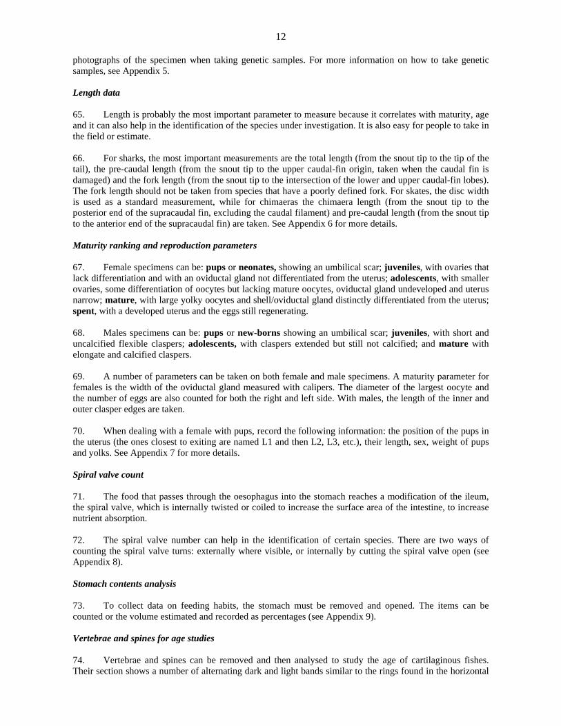
ABNJ areas beyond national jurisdiction
AOS acoustic-optical system
BPA benthic protected area
EAF ecosystem approach to fisheries
IUCN International Union for Conservation of Nature
RFMO/A regional fisheries management organization/arrangement
SIODFA Southern Indian Ocean Deepsea Fishers Association

1
Report of the training workshop
Opening of the workshop 1. Within the framework of the FAO Deep-sea High Seas Fisheries Programme, a regional workshop “Identification of Deep-sea Cartilaginous fishes of the Indian Ocean” was held at the Albion Fisheries Research Center, Albion, Mauritius, from 10 to 13 June 2014. 2. The workshop was attended by 19 scientists representing a wide range of countries facing the Indian Ocean (Appendix 1). 3. The opening ceremony, which was held on the afternoon of 10 June, was chaired by Mr Beeharry, Divisional Scientific Officer of the Ministry of Fisheries of Mauritius. Mr. Beeharry introduced Mr. Chooramun, Assistant Director of Fisheries (Fisheries Research, Management, Development and Training) of the Ministry of Fisheries of Mauritius, who welcomed the participants and highlighted the importance of the training workshop to the scientific community of the region and to Mauritius in particular. Mr. Chooramun expressed his honour at having the workshop held in Mauritius and wished all the participants a successful meeting and pleasant stay in Mauritius. 4. Subsequently, Dr. Edoardo Mostarda, FAO consultant for the Deep-sea High-Seas Fisheries and FishFinder Programmes, welcomed the participants and thanked both the Mauritian authorities for making available the Albion Fisheries Research Center, and all the people who had contributed to the realization of the workshop.
Plate 1
Opening ceremony.
5. The agenda (Appendix 2) and objectives of the workshop were introduced by Dr. Dave Ebert, Moss Landing Marine Laboratories. It was explained that the general objective of the workshop was to train scientists from countries bordering the Indian Ocean in the identification of deep-sea cartilaginous fishes and sample processing. 6. The participants were told that a number of specific objectives were to be reached by the end of the workshop, such as:
Learning the anatomical features of the deep-sea species belonging to the main shark orders occurring in the Indian Ocean.

2
Using the taxonomic keys included in the reference text material (e.g. FAO catalogue and identification guide).
Processing and identifying a number of deep-sea shark species.
Understanding how, and being able, to: (i) take basic measurements; (ii) take biological samples, i.e. for age, growth and molecular studies; (iii) take photographs; and (iv) preserve specimens in the field.
Background information The work carried out by FAO to improve the identification of marine organisms 7. The first presentation was delivered by Edoardo Mostarda (Plate 2). He clarified the role of FAO in conservation and fisheries management issues, the importance of systematics in fisheries management, and the activities of the FAO FishFinder Programme. 8. It was explained that, in order to achieve food security for all, FAO deals with all the practices that provide a source of food, income and livelihood to people around the world. In particular, it was highlighted how important fish is as a source of food, providing 20 percent of animal protein to about 3 billion people with a yearly production of more than 150 million tonnes. However, the depletion of numerous stocks underlines the growing need for improved management including policy, planning, data collection, research, laws, enforcement and regional cooperation. 9. The work of the FAO Fisheries and Aquaculture Department centres on the “sustainable management and use of fisheries and aquaculture resources”. The department strongly endorses the development and implementation of an ecosystem approach to fisheries (EAF) including mainstreaming biodiversity and ecosystem concerns in fisheries management. Greater awareness is placed on the effects of fishing also on non-target species and habitats, and the department is also committed to ensuring biodiversity conservation. Fishery-induced changes can influence the structure and functioning of marine ecosystems and generate a loss of productivity and stability of entire ecosystems. 10. It was stressed that proper identification of species is an important component of fishery resources management and biodiversity preservation. That is, it is difficult to manage what is unknown and to detect changes in the species composition of an area if no clear picture is available of what is present. 11. Two examples of the implications of erroneous management actions based on inaccurate species identification were presented. In the 1970s, fishery managers in the Unites States of America planned to use data on the Brazilian Spanish mackerel to manage the population occurring in their waters. However, Collette et al. (1978)1 showed that the Brazilian population represented a distinct species (Scomberomorus brasiliensis). The species found in United States waters is S. maculatus, a much smaller fish, reaching a maximum size of 77 cm fork length compared with 125 cm in S. brasiliensis and it matures at a smaller size (Collette and Nauen, 1983)2. Managing the United States species with the Brazilian data could have resulted in unnecessary economic impacts on the fishing industry and inadequate conservation measures for S. maculatus. 12. The yellowfin tuna was known by 27 names until Gibbs and Collette (1967)3 showed that all referred to a single worldwide species. Much time and money has been unnecessarily spent gathering meristic, morphometric, anatomical, distributional, and life-history data on tunas in each area where they were found.
1 Collette, B.B., Russo, J.L. & Zavala-Camin, L.A. 1978. Scomberomorus brasiliensis, a new species of Spanish mackerel from the western Atlantic. Fish. Bull., 76: 273–280. 2 Collette, B.B. & Nauen, C.E. 1983. FAO Species Catalogue. Vol. 2, Scombrids of the World. An annotated and illustrated catalogue of tunas, mackerels, bonitos and related species known to date. FAO Fish. Syn. No. 125(2). Rome, FAO. 137 pp. (also available at www.fao.org/docrep/009/ac478e/ac478e00.htm). 3 Gibbs, R.H. Jr. & Collette, B.B. 1967. Comparative anatomy and systematics of the tunas, genus Thunnus. Fish. Bull., 66(1): 65–130.

3
After Gibbs and Collette’s finding, managers have been able to use data on food or larval development from other regions to aid them in their work. 13. The main issues that make identification of fish in fisheries often difficult were addressed. First, species descriptions are published in a large variety of specialized journals usually unavailable to the fishery workers, and many of them are based on a few museum specimens rather than on representative samples of fresh material. Moreover, fishery data collectors have to identify many species at the same time and to do this they need multi-specific identification guides, which are often lacking. Finally, nomenclature is often not available for most regions or countries, and the fishery workers are confronted with a wide, confusing and often contradictory spectrum of names, most of them of dubious value and furthermore information on distribution, habitat, biology, and fisheries of individual species are all too often lacking entirely. 14. In order to improve the quality of fishery statistics, FAO developed (in 1972) the Species Identification and Data Programme (now called FishFinder Programme). It was stressed that, after 40 years, there is still a need to improve marine resources identification in many regions where the percentage of catches reported to the species level is still low. The objectives of the programme are to improve the identification of marine organisms of actual and potential interest to fisheries by developing and disseminating tools to facilitate species identification in fisheries and by providing a global and coherent system of scientific and common nomenclature. Priority is assigned to resources of major commercial importance or threatened species and to developing countries/regions facing difficulties in species identification. The main activities of the programme are: securing the best and most up-to-date information (calling upon knowledgeable specialists in taxonomy); compiling information on species distribution in order to produce distribution maps; drawing reliable and accurate illustrations of marine organisms and their anatomical details; and to producing and distributing, through different media, species identification information for fishery purposes. The principal outputs of the programme are publications such as species catalogues, regional catalogues, field guides, pocket guides, CD–ROMS, synopses, fact sheets available on the Web, species distribution maps and scientific illustrations. The work carried out by the Southern Indian Ocean Deepsea Fishers Association (SIODFA) 15. Subsequently, the floor was given to Mr Graham Patchell, from the Sealord Group, New Zealand, and chief scientist for the Southern Indian Ocean Deepsea Fishers Association (SIODFA). He presented the work that the deep-sea fishing industry is carrying out in the southern Indian Ocean (Plate 2). 16. Mr Patchell explained that the seafood industry had to be proactive to ensure sustainable fisheries. The Sealord Group, prior to carrying out any fishing 15 years ago, mapped out the entire habitat, more than 160 000 km². Since then, it has been collecting information for the maintenance and prediction of biodiversity in the Indian Ocean. It has also been collaborating with FAO and the International Union for Conservation of Nature (IUCN) to set up the first network of benthic protected areas (BPAs) in the high seas. The southwest Indian Ridge has been habitat mapped with side-scan sonar, giving a clear understanding of what the habitat looks like and allowing for the setting up of a number of BPAs where fishing cannot take place. The FV Will Watch currently carries out acoustic surveys and collects records of any shark, coral and sponge bycatch. However, the identification at the species level of individuals belonging to the latter groups has been particularly difficult. Therefore, the development of the identification tools by FAO has been very helpful. 17. A general description was provided of the multifrequency acoustic-optical system (AOS) attached to the headline of the deep-water demersal trawl net used in the Indian Ocean. This system uses acoustics at different frequencies and integrated habitat monitoring camera systems to help monitor and thus protect ocean habitats and vulnerable species. Footage showing deep-sea sharks among orange roughy schools was shown. This technology could be used to collect real-time data during the haul, enabling the impacts of the net on particular species or habitats to be avoided or minimized.
4
Plate 2
Presentations by Edoardo Mostarda and Graham Patchell.
The work carried out by FAO on deep-sea fisheries 18. Edoardo Mostarda presented the work of FAO on deep-sea fisheries, the approach used to develop the identification tools, and an overview of the species catalogue and identification guide. 19. It was explained that in response to increasing concern about the sustainable use of marine resources and marine conservation in areas beyond national jurisdiction (ABNJs), the International Guidelines for the Management of Deep-sea Fisheries in the High Seas had been developed through FAO and adopted in 2008. The objective of these guidelines is to facilitate and encourage the efforts of States and regional fisheries management organizations and arrangements (RFMO/As) towards a sustainable use of marine living resources exploited by deep-sea fisheries and the protection of the marine biodiversity that these ecosystems contain. 20. The guidelines also set out a framework for data collection. They also attach particular importance to the collection and reporting of reliable and accurate catch data, which in an EAF context should not be limited to the target resources but also take into account the associated species affected by the fishing activity. 21. In deep-sea ecosystems, specific attention has been given to three groups of associated species considered particularly vulnerable to overfishing or bottom gear impacts: cartilaginous fishes and certain groups of sponges and corals. 22. The need to improve the identification of deep-sea sharks, batoids and chimaeras is clear. A comparison of the number of caught species reported to FAO in the last decade to the number of known species per region reveals the fact that only a small portion of these is reported to the species level (Figure 1). 23. Therefore, with the aim of improving data reporting, FAO has set up a strategy for the development of field identification tools aimed at facilitating the species-specific identification by fishery observers, crew members, scientists and fishery officers of the deep-sea resources most vulnerable to fishing operations. 24. The Indian Ocean was chosen as the first region for developing the identification tools but other regions will follow. From the outset, FAO realized that in order to develop appropriate field products it would be essential to involve the fishing industry operating in the region. Therefore, it initiated collaboration with SIODFA, which represents a set of companies that use bottom trawling techniques and which is voluntarily committed to biologically sustainable and economically viable commercial fishing operations.

5
Figure 1
Number of deep-sea cartilaginous fish species reported to FAO and known to occur in the Indian Ocean
25. Subsequently, FAO organized a regional workshop in Mauritius attended by taxonomists, onboard observers, and industry representatives with the aim of drafting recommendations for the development of appropriate field products. Particular consideration was given to: (i) the working environment of the different types of fishing vessels operating in the Indian Ocean deep sea; (ii) the level of expertise of the users involved in data collection; and (iii) reported experiences of crew members and fishery observers. Hence, the guide was geared towards use by the crew, who were generally recognized to be knowledgeable about the species they deal with and interested in knowing more and being engaged in scientific work. 26. An identification guide was developed based on the conclusions and recommendations agreed upon by participants. The guide includes: full species accounts for 36 shark species selected as being those more difficult to identify and/or commonly caught; simplified accounts for 16 shark species that have very particular characteristics and/or are rarely caught; and short accounts of 52 shark species that could be misidentified with more common species occurring in the area. In total, the guide includes 104 of the 117 species known to occur in the Indian Ocean. 27. Finally, the structure of the guide and species catalogue was illustrated to the participants. The Indian Ocean chondrichthyan fauna 28. Dave Ebert presented his work entitled “Deep-sea Indian Ocean sharks: biodiversity & identification of charismatic predators” (Plate 3). 29. The public’s perception of sharks often conjures up images of a large fearsome toothy predator, with its large dorsal fin cutting through the water surface. However, the reality is that sharks come in a variety of sizes and shapes, from the whale shark (Rhincodon typus), the world’s largest fish, to the dwarf pygmy sharks (Squaliolus spp.), and these enigmatic fishes occupy most marine, and some freshwater, habitats. In addition, the batoids and chimaeras, along with the sharks, form a distinctive group of fishes collectively referred to as the chondrichthyans. There are more than 500 species of sharks, along with almost 650 batoid and 50 chimaera species, bringing the overall total to about 1 200 species of sharks and shark-like fishes. 30. In terms of identification, the diversity of sharks and their relatives has increased markedly in the past decade with more than 200 new species described since 2000. Since 2007, 147 new species have been described, an average of 24.5 new species per year. This represents almost 20 percent of all shark species

6
that have been described, and compares with about 200 species that described in the previous 30 years (1970–1999). Most of the new species discovered in the past decade have come from the Indo-Australian region, followed by the southern African and western North Pacific regions. Many of these newly discovered species are deep-sea inhabitants, mostly at depths in excess of 200 m. The discovery of new species combined with the taxonomic resolution of species complexes has led to a scientific renaissance in chondrichthyan taxonomy, and it highlights the importance of taxonomy for their proper identification and management. 31. The Indian Ocean is one of the more diverse regions for chondrichthyans, but is insufficiently studied, especially the deep sea. Some 123 sharks, 63 batoids and 18 chimaeras occur in the Indian Ocean deep sea, representing about 17 percent of all known chondrichthyan species. The majority of species are within three major orders (Squaliformes, Carcharhiniformes and Rajiformes), and mostly within nine families, including the Squalidae, Centrophoridae, Etmopteridae, Somniosidae, Dalatiidae, Scyliorhinidae, Arhychobatidae, Rajidae, and Chimaeridae; these families collectively comprise 162 (79.4 percent) of all Indian Ocean deep-sea chondrichthyans. 32. The family Rajidae is the most diverse with 39 species, followed by the Scyliorhinidae with 34 species, and the Chimaeridae, Centrophoridae and Etmopteridae, with 18, 16 and 16 species each, respectively. Of these totals, about 23.7 percent (29 species) of sharks, 31.7 percent (20 species) of batoids, and 44.4 percent (8 species) of chimaeras have only been described since 2002. The number of species found between the Eastern and Western Indian Ocean, FAO Areas 51 and 57, is remarkably similar in numbers to the 80 shark species found in each region, 32 (FAO Area 57) and 34 (FAO Area 51) batoid, and 12 (FAO Area 57) and 9 (FAO Area 51) chimaera species. However, only about one-third of sharks, and less than 5 percent of batoids and 17 percent of chimaeras occur in both FAO Areas, highlighting a high degree of regional endemism within the Indian Ocean deep sea. Finally, much of the research and new species identification in the Indian Ocean deep sea have occurred off Australia and Indonesia, and in the southwestern area off South Africa, Mozambique, and some of the seamounts on the southern Madagascar Ridge. The northern Indian Ocean, especially off east Africa, including many of the Western Indian Ocean islands, the Arabian Sea, and the Bay of Bengal, still remains relatively unexplored.
Plate 3
Dave Ebert presenting a general overview on the Indian Ocean deep-sea chondrichthyan fauna.
Session 1 – Lecture: the order Squaliformes 33. Dave Ebert introduced the participants to the main features that characterize the class Chondrichthyes and its main subgroups e.g. Elasmobranchii (sharks, rays and skates) and Holocephali (chimaeras). These differ from the Osteichthyes or bony fish in having a cartilaginous skeleton, male paired

7
copulatory organs (claspers) for internal fertilization of eggs, placoid scales, teeth not fused to jaws (only to connective tissue), lipid (squalene) filled livers, soft and unsegmented fin rays (ceratotrichia), no swim bladder and lung, and high concentrations of urea in blood (for osmoregualtion). 34. Sharks (Selachii) are an easily identifiable group of more than 500 species worldwide and characterized by having 5–7 gill openings and the pectoral fins not attached to the head. Most shark species have 2 dorsal fins, with the exception of the members of the order Hexanchiformes, which have a single dorsal fin and also 6 or 7 gill openings. A number of species, which are mostly deep-sea, have spines in front of either one or both dorsal fins. The spines can be very prominent, and thus visible, or difficult to detect. Sharks also have paired pelvic fins and an anal fin, which is lacking in the order Squaliformes, one of the most important groups of deep-sea sharks, which was subsequently presented in detail. 35. The participants were asked to follow the guide to the orders included in the identification guide to reach the order Squaliformes. The main characters that lead to this order are the presence of 5–7 gill slits, the absence of an anal fin, the body shape not being ray-like, and the shape of the snout, which is short and not saw-like. 36. The main characteristics of the Squaliformes were highlighted. This order, the members of which are commonly known as dogfish sharks, is a large and varied group of more than 130 species occurring worldwide at all depths. It includes the only shark species found at high latitudes by the poles, and it shows its greater diversity in deep water. The species belonging to this group can range in size from dwarf to very large, and all reproduce by viviparity with yolk-sac, and have litters of between 2 and 50. Their long gestation periods (18–24 months) and low fecundity may lead to a high vulnerability to fishing. 37. The Squaliformes is represented in the Indian Ocean deep-sea by seven families. The participants were asked to follow the dichotomous key included in the identification guide to reach the different families. 38. The bramble sharks (family Echinorhinidae), are represented in the Indian Ocean by two species, Echinorhinus brucus and E. cookei. Both are large and sluggish sharks with stout cylindrical bodies, large and thornlike denticles, a broad flat head and small dorsal fins with no spines. The key distinguishing features that allows for separating these two species were highlighted. 39. The rough sharks (Oxynotidae) are represented in the Indian Ocean deep sea by at least one species (Oxynotus bruniensis) and possibly a second species (O. centrina). These sharks have a compressed body, triangular in cross-section, very high dorsal fins with spines, and rough skin. Their biology is poorly known and their distribution is scattered. 40. The dogfish sharks (Squalidae) with two genera, Cirrhigaleus and Squalus, and with 2 and 11 species occurring in the Indian Ocean deep sea, have similar teeth in both jaws and a subterminal notch absent from the caudal fin. The two genera differ in the length of the secondary lobe of the anterior nasal flap, which is larger in Cirrhigaleus than in Squalus. The importance of taking photographs of the underside of head was stressed, in order to be able to compare the distance from snout tip to nostril with that from nostril to front of upper labial furrow. This character is important for separating species within the genus Squalus. 41. The gulper sharks (Centrophoridae) are comprised of two genera, Deania and Centrophorus, with 3 and 10 species reported from the Indian Ocean, respectively. These species tend to have large green eyes, upper teeth relatively broad and bladelike, lower teeth that are low and wide, and dorsal fins with spines. The genus Centrophorus is one of the most taxonomically complex and confusing elasmobranch groups. For example, three species, C. granulosus, C. acus and C. niaukang, described from different regions were found

8
only recently to be different life stages of a single species, i.e. C. granulosus (White et al., 20134). The two genera can be separated by looking at the snout length. 42. The lanternsharks (Etmopteridae) characterized by the presence of black markings and light organs on the flanks, caudal fin and underside of body, are represented in the Indian Ocean deep sea by 2 genera, Centroscyllium and Etmopterus, with 2 and 13 species, respectively. The best characters used to identify these species are the shape, length and position of the markings (when visible), and the shape and arrangement of the dermal denticles along the dorsal surface of head, trunk and caudal peduncle. 43. The sleeper sharks (Somniosidae) are a relatively small family represented in the Indian Ocean deep sea by 6 genera and 8 species. Most of the species are of moderately large size and their uniform dark brown to black coloration makes them look alike. The characters that are used to separate the different species are the shape of the teeth, the presence or absence of small dorsal-fin spines, the length of the snout and the shape of the lower teeth. 44. The kitefin sharks (Dalatiidae) are a group of deep-sea sharks with more oceanic habits. Five genera and six species occur in the deep waters of the Indian Ocean. Most of the species are caught occasionally. However, the kitefin shark Dalatias licha is widespread and frequently caught. This species can be identified by its very short preoral snout length, lips thick and pleated, and lower teeth with strongly serrated edges. Session 1 – Practicum: identification of deep-sea shark species 45. After the morning lecture, the laboratory work began in an open space of the Albion Fisheries Research Center set up with four metallic tables and two large bins containing the specimens to be identified. It was explained that it had only been possible to collect shark specimens, thus limiting the lectures and hands-on sessions to this chondrichthyan group. 46. The participants were divided into 4 groups of 4–5 people. Each group gathered around one of the tables equipped with a measuring board and laboratory tools such as scissors, forceps, scalpels and calipers (Plate 4). 47. Each group was supervised by an instructor, and one participant had the task of writing down on an identification sheet (see Appendix 3) the characters of the species under investigation. 48. The participants were tasked with the identification of three species: Etmopterus granulosus, Centroselachus crepidater and Dalatias licha. 49. The instructors made sure that the participants used the keys to the orders and families included in the identification guide. 50. Each species was measured (total length), sexed and photographed. The instructors showed how to take other important measurements, such as mouth width, snout length, dorsal-fin height and width, and they clarified every doubt expressed by the participants regarding particular external anatomical features, e.g. denticles (shape and arrangement), teeth (shape and counting of the rows), and dorsal-fin spines (origin and length). 51. By using the identification guide, each group was able to identify the three species correctly. However, the support of the instructors was necessary in order to clarify some characters whose interpretation was subjective. One of the more difficult characters to understand was the shape of the head, which is used to separate the members of the Dalatiidae from the Somniosidae. When going through the guide to the families, the participants had to decide whether the specimen they had in front of them, the
4 White, W.T., Ebert, D.A., Naylor, G.J.P., Ho, H.-C., Clerkin, P., Veríssimo, A. & Cotton, C.F. 2013. Revision of the genus Centrophorus (Squaliformes: Centrophoridae): Part 1—Redescription of Centrophorus granulosus (Bloch & Schneider), a senior synonym of C. acus Garman and C. niaukang Teng. Zootaxa, 3752: 35–72.

9
kitefin shark Dalatias licha (Plate 4) belonging to the family Dalatiidae, had a moderately broad and somewhat flattened-conical head (Somniosidae) or a narrow and rounded-conical head (Dalatiidae).
Plate 4
Practical sessions.
52. The same problem arose when they had to identify the longnose velvet dogfish Centroselachus crepidater (Plate 5), belonging to the family Somniosidae.
Plate 5
Detail of the head shape of Dalatias licha (left) and Centroselachus crepidater (right).

10
53. Finally, the participants needed the help of the instructors in the identification of the southern lanternshark Etmopterus granulosus (Plate 6). The main problem was the recognition of the black markings and light organs on the flanks and tail that are characteristic of the lanternsharks (Etmopteridae). Moreover, other characters were difficult to understand such as the arrangement of the denticles and the length of the flank and caudal markings.
Plate 6
Identification of Etmopterus granulosus
54. After the practical session, the groups were asked to meet and review the work done during the day and to present the results to the other participants. Each instructor helped the participants to outline the main characters observed for each species, and one representative was nominated to present these results (Plate 7).
Plate 7
Presentation of the day’s work by the participants.
Session 2 – Lecture: methodologies of biological data collection in the field 55. Dave Ebert and Paul Clerkin made a presentation on the methodologies of data collection from sharks in the field. Each participant was provided with a set of guidelines edited by Paul Clerkin summarizing these methodologies (see Appendixes 4–12).

11
The following topics were covered: Photographic documentation 56. Taking photographs of shark specimens is important as it can allow other experts to properly identify a species. Photographs are easy to share and usually show the individual in the best possible conditions. 57. A series of photographs must be taken of each shark specimen. In particular, the following photographs should be taken:
A full lateral view of the specimen facing to the left. It is important to have the entire shark in the picture, avoiding cutting parts of the caudal fin and snout. Make sure that this photograph shows a straight-on view and not a ventral or dorsal view. An effort should be made to have the surface of the dorsal fins parallel to the ground.
A lateral, dorsal and ventral view of the head from the snout tip to the gills.
A close up of the pelvic-fin region, dorsal fins (both in the same photograph in order to compare the relative size and shape of the fins), caudal fin and pectoral fin. A photograph of the ventral side including both the pectoral and pelvic fins can be useful.
A close-up of the mouth region showing the labial furrows.
58. Additional photographs should be taken to show any unusual or descriptive feature, e.g. teeth, denticles. 59. Every photograph should include a ruler or an object of known size in order to have a metric scale. It should also include an identification tag with information such as the species name (when identified), the date, sex and ID number. For more information on how to take photographs, see Appendix 4. Taking genetic samples 60. Genetics is a useful tool in defining a species as distinct on a molecular level. There are three different ways of taking a tissue sample for molecular studies, depending on whether:
the specimen has to be discarded;
the specimen has to be preserved and become a voucher specimen;
the specimen is alive.
61. If the specimen has to be discarded, the best place to take a sample from is the liver, the largest organ in the body. Another option is the muscle tissue close to the vertebral spine. 62. If the specimen has to remain undamaged, the tissue sample should be taken from the area just behind the pelvic fins and anus. 63. From live specimens, it is best to take a clip of tissue from the pectoral fin or from the muscle tissue just below the dorsal fin. 64. When taking a tissue sample, make sure to sterilize the equipment (scissors, forceps, scalpels, etc.). Do not handle tissues with bare hands (this in order not to contaminate the sample). Take a sample the size of a pea and cut it into three slivers. Put each sample in a vial with pure 100 percent ethanol. Shake the vial well to allow the ethanol to penetrate into the tissue, and make sure you record a number on the vial that matches the number on your data sheet and thus the photographs, morphometric data, etc. It is very important to take
12
photographs of the specimen when taking genetic samples. For more information on how to take genetic samples, see Appendix 5. Length data 65. Length is probably the most important parameter to measure because it correlates with maturity, age and it can also help in the identification of the species under investigation. It is also easy for people to take in the field or estimate. 66. For sharks, the most important measurements are the total length (from the snout tip to the tip of the tail), the pre-caudal length (from the snout tip to the upper caudal-fin origin, taken when the caudal fin is damaged) and the fork length (from the snout tip to the intersection of the lower and upper caudal-fin lobes). The fork length should not be taken from species that have a poorly defined fork. For skates, the disc width is used as a standard measurement, while for chimaeras the chimaera length (from the snout tip to the posterior end of the supracaudal fin, excluding the caudal filament) and pre-caudal length (from the snout tip to the anterior end of the supracaudal fin) are taken. See Appendix 6 for more details. Maturity ranking and reproduction parameters 67. Female specimens can be: pups or neonates, showing an umbilical scar; juveniles, with ovaries that lack differentiation and with an oviductal gland not differentiated from the uterus; adolescents, with smaller ovaries, some differentiation of oocytes but lacking mature oocytes, oviductal gland undeveloped and uterus narrow; mature, with large yolky oocytes and shell/oviductal gland distinctly differentiated from the uterus; spent, with a developed uterus and the eggs still regenerating. 68. Males specimens can be: pups or new-borns showing an umbilical scar; juveniles, with short and uncalcified flexible claspers; adolescents, with claspers extended but still not calcified; and mature with elongate and calcified claspers. 69. A number of parameters can be taken on both female and male specimens. A maturity parameter for females is the width of the oviductal gland measured with calipers. The diameter of the largest oocyte and the number of eggs are also counted for both the right and left side. With males, the length of the inner and outer clasper edges are taken. 70. When dealing with a female with pups, record the following information: the position of the pups in the uterus (the ones closest to exiting are named L1 and then L2, L3, etc.), their length, sex, weight of pups and yolks. See Appendix 7 for more details. Spiral valve count 71. The food that passes through the oesophagus into the stomach reaches a modification of the ileum, the spiral valve, which is internally twisted or coiled to increase the surface area of the intestine, to increase nutrient absorption. 72. The spiral valve number can help in the identification of certain species. There are two ways of counting the spiral valve turns: externally where visible, or internally by cutting the spiral valve open (see Appendix 8). Stomach contents analysis 73. To collect data on feeding habits, the stomach must be removed and opened. The items can be counted or the volume estimated and recorded as percentages (see Appendix 9). Vertebrae and spines for age studies 74. Vertebrae and spines can be removed and then analysed to study the age of cartilaginous fishes. Their section shows a number of alternating dark and light bands similar to the rings found in the horizontal

13
cross-section of trees. Often, the rings represent the changing of the seasons (mainly winter and summer), but other times the rings are annual. The collection of samples every month allows for the determination of the interval between the bands. 75. About ten vertebrae must be removed from the vertebral spine region at the first dorsal- and pectoral-fin level. Spines should be taken from directly in front of each dorsal fin and cut out with a “V” cut. Both vertebrae and spines should be tagged (identifying number corresponding to all the recorded data), bagged and frozen. For more details, see Appendix 10. 76. The laboratory procedure for sectioning and reading the vertebrae and spines was described. Preservation, tagging and bagging of a specimen 77. The specimens that are to be kept must not be dissected. They must be injected with 100 percent formalin in a number of different spots along the side of the body and in the abdominal cavity. After injecting the specimen, it should be soaked in a 10 percent formalin/water solution for up to two weeks, but also a couple of days should be sufficient. Protective gear (long gloves, eye protection glasses, gas mask, protective clothes) must be used as formalin is highly toxic. The soaked specimens must be rinsed in water, wrapped in cloth, bagged, tagged and shipped to the research group. For long-term preservation, the specimen treated with formalin should be kept in a 70 percent ethanol/water solution. For more details, see Appendixes 11 and 12. Data collection protocols 78. A step-by-step data collection protocol for cartilaginous fish species and one specifically for conducting at-sea research aboard a commercial trawler were provided (see Appendixes 13 and 14). Session 2 – Practicum: identification and biological data collection on deep-sea shark species 79. The participants were tasked with the identification of four species: Deania calcea, Centroscymnus owstonii, Proscymnodon plunketii and Centrophorus granulosus. 80. The identification of the two centrophorids, the birdbeak dogfish Deania calcea and gulper shark Centrophorus granulosus was relatively easy, with all participants being able to reach the family level by looking at the different shapes of the upper and lower teeth. Other key characters for the former species, such as the snout being extremely long and flattened allowed for its identification, while C. granulosus was identified by looking at the shape of the dermal denticles and the shape of the pectoral fins. 81. The roughskin dogfish Centroscymnus owstonii and plunket shark Proscymnodon plunketi were more difficult to identify. The character (shape of the head) included in the key to the families that allows for the separation of the Somniosidae from the Dalatiidae was again of difficult interpretation. 82. The participants were able to reach the genus level in the case of Centroscymnus but struggled to determine whether the specimen was C. coelolepis or C. owstonii. Indeed, some of the details included in the guide did not help to separate the two species unambiguously. For example, the shape of the denticles, which was similar to a teardrop in the specimen under investigation, was not depicted in either species account. Moreover, the difference in the preoral length shown by the illustration of the underside of head was not so pronounced. 83. The plunket shark was more distinctive with its large and stocky body and the dermal denticles being of a very characteristic shape. Moreover, the specimen measured more than 140 cm, a size only reached by this species. 84. After the identification phase, the participants practised taking photographs based on the indications provided during the morning lecture.

14
85. Paul Clerkin showed all participants how to take biological samples from one of the specimens (Plate 8). The plunket shark was dissected and the different organs were shown and described. Then the maturity stage was determined, oviductal gland measured, vertebrae and spines removed, spiral valve turns counted, tissue sample taken, and stomach contents analysed. 86. After the demonstration, each group performed a standard biological data collection procedure on the other specimens under the supervision of the instructors.
Plate 8
Demonstration of biological data collection methodologies.
87. Again, the groups were asked to meet and review the work done during the day and to present the results to the other participants. Each instructor helped the participants to outline the main characters observed for each species, and one representative was nominated to present these results. Session 3 – Lecture: the order Carcharhiniformes 88. Dave Ebert illustrated the main features of the order Carcharhiniformes. The guide to the families and the diagnostic characters of the main species included in the identification guide were described. 89. Particular attention was given to the species of the genus Apristurus, belonging to the catsharks (Scyliorhinidae). These species are of very difficult identification. For this reason, a dedicated section presenting the general characters of the members of this genus was included in the identification guide. The species of the genus Apristurus are characterized by a flattened and spatulate head, very long labial furrows, rear-sited dorsal fins and a uniform coloration. They can be divided in three species groupings (Apristurus longicephalus-group; A. brunneus-group; and A. spongiceps group) based on a number of differences. The characters of a species representative of each grouping were presented. 90. The characteristics of the other most common and important catshark species were illustrated. Session 3 – Practical session: identification and biological data collection on deep-sea shark species 91. The participants continued practising the biological sample collection procedures on a number of specimens. All samples were preserved for further processing. 92. Three specimens of an Apristurus species were shown and their main characters described. Then, each group was asked to identify the species, at first by assigning them to one of the above mentioned subgroups, and then by looking at the species representative for that group and its similar species.

15
93. The participants were able to place the specimens within the Apristurus brunneus group. The Apristurus longicephalus group was ruled out after measuring the length to nostrils, which was shorter than 6.4 percent of total length. Then, the upper and lower labial furrows were measured to determine whether the specimens belonged to the A. spongiceps-group or A. brunneus group. The latter group was chosen as the specimens had the upper labial furrows longer than their lowers and a slender body. 94. The species representative of the Apristurus brunneus group in the identification guide is Apristurus melanoasper. By looking at the diagnostic characters of this species, it was not possible to exclude it. Moreover, by going through the accounts of the other five members of the A. brunneus group, it was not possible to assign the specimens to one of these species. However, based on the long interdorsal space, the three specimens were tentatively identified as A. cf. saldanha (Plate 9).
Plate 9
Specimens of Apristurus cf. saldanha being identified.
95. After the final practical session, two presentations on topics related to the species and ecosystems subject of the workshop, were delivered. 96. Mr. Daley presented the results of his PhD research project focusing on movement-based conservation strategies for deep-water sharks. The research uses passive acoustic telemetry to monitor the home range and essential habitat of Centrophorus species with the purpose of evaluating the possibility of defining areas closed to fisheries. 97. Mr. Valinassab presented the work done on the Iranian side of the Oman Sea to estimate the biomass of lanternfish (Myctophidae), given its potential importance for fishmeal production. 98. The participants showed great interest in the topics of the workshop, and their active participation enabled its success. Many of them asked to be involved in a second workshop focusing on skates and chimaeras, whose inclusion had been planned did not occur owing to the impossibility of collecting specimens. 99. Finally, Edoardo Mostarda informed the participants that in order to allow them to follow up, and with the aim of providing a forum where they could easily share information, publications and photographs and ask the experts questions on the workshop topics, an online discussion group would be set up in the following weeks. 100. The training workshop closed with a group photograph (Plate 10).

16
Plate 10
Group photograph.

17
Appendix 1 List of participants
KALLI VALAPPIL AKHILESH Central Marine Fisheries Research Institute Kerala, India E-mail: [email protected] AYSHA AKHTAR Institute of Marine Sciences and Fisheries, Chittagong University Chittagong, Bangladesh E-mail: [email protected] AHMAD ALI SEAFDEC/MFRDMD Kuala Terengganu, Terengganu Malaysia E-mail: [email protected] KHADEEJA ALI Marine Research Centre Malé, Maldives E-mail: [email protected] SURAJ BACHA GIAN Mauritius Oceanography Institute Quatre-Bornes, Mauritius E-mail: [email protected] DEVANAND BOLAKY Albion Fisheries Research Center Ministry of Fisheries Albion, Mauritius E-mail: [email protected] LUVNA CAUSSY Albion Fisheries Research Center Ministry of Fisheries Albion, Mauritius E-mail: [email protected] LIM SHEUNG CLIVY Albion Fisheries Research Center Ministry of Fisheries Albion, Mauritius E-mail: [email protected] DHARMENDRA DEGAMBUR Albion Fisheries Research Center Albion, Mauritius E-mail: [email protected]
KURSHID ELAHEEBOCUS Albion Fisheries Research Center Ministry of Fisheries Albion, Mauritius E-mail: [email protected] FAHMI Research Centre for Oceanography Indonesian Institute of Sciences (LIPI) Jl. Ancol, Timur Jakarta, Indonesia E-mail: [email protected] TASSAPON KRAJANGDARA Andaman Sea Fisheries Research and Development Center Phuket, Thailand E-mail: [email protected] RANWEL NELSON MBUKWAH Deep sea Fishing Authority Zanzibar, United Republic of Tanzania E-mail: [email protected] RODNEY MELANIE Seychelles Fishing Authority Mahe, Seychelles E-mail: [email protected] BARBARA PALHA DE SOUSA Instituto Nacional de Investigacao Pesqueira (IIP) Maputo, Mozambique E-mail: [email protected] SYLVIO PERRINE Fisheries Research & Training Unit Rodrigues, Mauritius E-mail: [email protected] DINALI RANMADUGALA National Aquatic Resources Research and Development Agency (NARA) Sri Lanka E-mail: [email protected] HASINARIVO NODIER RAVELOSON Direction Régionale de la Pêche Atsinanana Tamatave, Madagascar E-mail: [email protected] TOORAJ VALINASSAB Iranian Fisheries Research Organization Teheran, Iran (Islamic Republic of) E-mail: [email protected]

18
List of instructors
PAUL CLERKIN Moss Landing Marine Laboratories Moss Landing, CA, United States of America E-mail: [email protected] ROSS DALEY CSIRO, Marine and Atmospheric Research Hobart, Tasmania, Australia E-mail: [email protected] DAVE EBERT Moss Landing Marine Laboratories Moss Landing, CA, United States of America E-mail: [email protected]
GRAHAM PATCHELL Resource Manager Sealord Group Ltd. Nelson, New Zealand E-mail: [email protected] FAO Food and Agriculture Organization of the United Nations Viale delle Terme di Caracalla 00153 Rome, Italy EDOARDO MOSTARDA Fishery expert Marine and Inland Fisheries Service E-mail: [email protected]

19
Appendix 2 Agenda
Day 1: Tuesday 10 June 2014
14:00
Welcome address by Mauritius Authorities Self-introduction of participants Overview of the workshop objectives, activities and expected outputs The deep-sea fishery industry perspective in the Indian Ocean Importance of information gathering and role in fisheries management
15:30
Current knowledge on deep sea cartilaginous fishes biodiversity in the Indian Ocean The FAO deep-sea fisheries programme: activities, approach used and future projects Wrap up discussion
16:30 Day Closure
Day 2: Wednesday 11 June 2014 09:00
Lectures:
Identification of major groups of deep-sea chondrichthyans
10:45
Practical session:
Identification of genera and species
12:30 Lunch
13:15
Practical session:
Continue with identification of genera and species
15:00
Wrap up discussion: questions, clarifications, review of day’s work
16:30 Day Closure
Day 3: Thursday 12 June 2014 09:00
Lectures:
Introduction to taking photographs and saving specimens in the field Taking and storing biological samples/ what happens then?
10:45
Practical session:
Identification of genera and species
12:30 Lunch

20
13:15
Practical session:
Continue with identification of genera and species Biological samples (taking samples in the field [basics] and on shore [detailed]
15:00
Wrap up discussion: questions, clarifications, review of day’s work
16:30 Day Closure
Day 4: Friday 13 June 2014 09:00
Lecture:
Identification of major groups of deep-sea Chondrichthyans
11:00
Practical session:
Practice taking basic measurements and biological samples Practice taking photographs and saving specimens Review day’s practicum with participants processing specimens, e.g. identification of the
species, recording of basic measurements, photographs, collection and preservation of biological samples.
12:30 Lunch
13:30
Wrap up discussion: questions, clarifications, review of day’s work
15:30
Wrap up discussion: questions, clarifications, review of day’s work (Continued)
16:30 Closing of the workshop
21
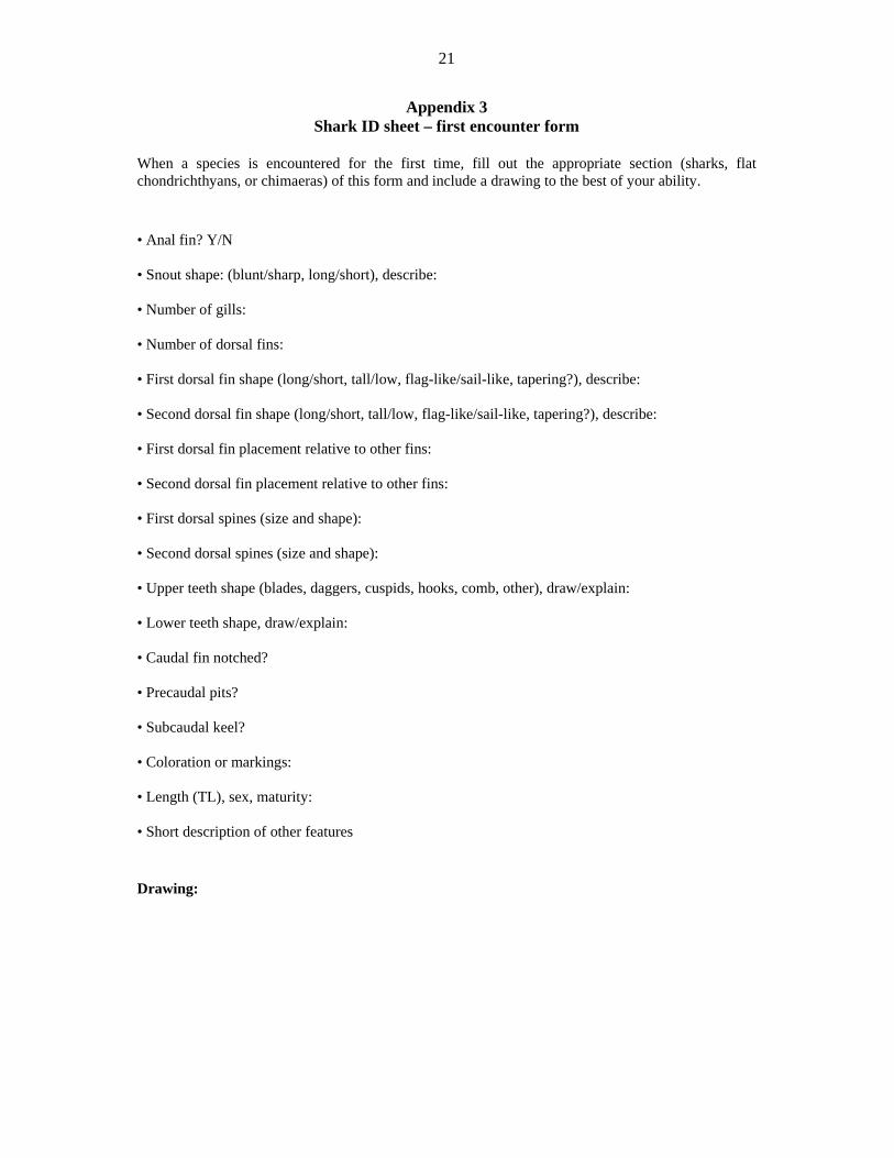
Appendix 3 Shark ID sheet – first encounter form
When a species is encountered for the first time, fill out the appropriate section (sharks, flat chondrichthyans, or chimaeras) of this form and include a drawing to the best of your ability. • Anal fin? Y/N
• Snout shape: (blunt/sharp, long/short), describe:
• Number of gills:
• Number of dorsal fins:
• First dorsal fin shape (long/short, tall/low, flag-like/sail-like, tapering?), describe:
• Second dorsal fin shape (long/short, tall/low, flag-like/sail-like, tapering?), describe:
• First dorsal fin placement relative to other fins:
• Second dorsal fin placement relative to other fins:
• First dorsal spines (size and shape):
• Second dorsal spines (size and shape):
• Upper teeth shape (blades, daggers, cuspids, hooks, comb, other), draw/explain:
• Lower teeth shape, draw/explain:
• Caudal fin notched?
• Precaudal pits?
• Subcaudal keel?
• Coloration or markings:
• Length (TL), sex, maturity:
• Short description of other features
Drawing:

22
Appendix 4 Photographing specimens for identification
Experience over many years has shown that the identification of sharks and rays can be problematic, especially with similar-looking species. Rare species are sometimes encountered and, if possible, these specimens, in addition to being photographed fresh, should be saved and forwarded to experts for possible identification. This can benefit the observers, regional agencies, and scientists (most of whom are interested in these observations), but are not usually at sea. Taking photographs for facilitating identification: If possible, try and place a ruler or other measuring scale alongside the specimen; if no ruler, is available, then some other object to show a size relationship. A handwritten label is desirable. It should include a number, the date, location, and other relevant capture information, and may include the person’s name. Plain coloured or an artificial background contrasting the specimen’s colour is fine. Sharks: Take photographs in lateral view and in total length, and dorsal and ventral views; if possible, with the fins erected and spread. Add close-ups of details that catch your eye, e.g. lateral and ventral view of head to gill openings or to origin of pectoral fins, mouth-nasal region, the jaws with dentition and scale -over detail, individual fins, interdorsal ridge, and colour marks or patterns. Close-ups of the teeth are also helpful, especially for sharks of the genus Carcharhinus.

23
Appendix 5 Taking genetic samples
General information
Genetic samples are used in many research projects. The softer the tissue, the easier it is for geneticists to extract DNA from the sample. All of the body’s tissues contain DNA. However, the three most-commonly sampled tissues are muscle, liver and fin. The type of tissue selected is based on sampling conditions and collection circumstances. Things to remember when collecting genetic samples:
Sterilize all working surfaces (tools, cutting surfaces, gloves, etc.) to avoid contamination
with other genetic material. Rinse everything vigorously in running water. There should be no visible impurities (tissue, oil or coloration). All tools must be cleaned directly before collecting a genetic sample, as the act of cutting into the shark exposes the tools to contaminants (stomach contents and material from other animals).
Do not handle tissues with bare hands. (You have DNA too!) You must wear gloves.
Use a clean blade, dermal punch, and forceps to remove a pea-sized sample, and place it in a numbered genetics vial.
Genetics vials must be filled with 100 percent pure ethanol with no derivatives.
Once the tissue is in the vial, replace the lid and shake the vial vigorously to diffuse the ethanol into the tissue.
Record the vial number on your deck sheet and place the vial in a cold area (a refrigerator is best), but make sure it is not cold enough to freeze. Freezing and thawing of ice crystals will damage the sample.
Scientists need to be able to verify your sample. If you are not keeping the specimen (as a voucher specimen) from which the tissue was collected, then you MUST take photographs of the specimen (see photo-documenting section). Tissues without documentation (identification number AND verification [either photographs or the voucher specimen]) are useless.
Specific information
The following information describes the procedure for collecting genetic samples from: 1) sharks, 2) rays or skates, and 3) chimaeras. In each case, the procedure is explained for collection from a specimen intended to be: a) discarded, b) kept as a voucher specimen, or c) returned to the ocean alive.
1) SHARKS a) Collection from a shark to be discarded
The primary location from which to collect a genetic sample is the liver. The liver is a soft tissue and easy for geneticist to process. Make an incision along the length of the body from pectoral fin to pelvic fin, then cut dorsally from each end of that incision. Fold the resulting flap of flesh up to open the body cavity. A shallow incision along the inside of the dorsal surface will prevent the flap of muscle from returning to its original position. The liver is the largest organ in a shark’s body. It is brown, green or tan, and appears as two large lobes running the length of the body cavity (Plate A5.1). Select a clean, healthy-looking section of the liver and excise a pea-sized sample. If the specimen is intended for discard and a muscle sample is to be collected, muscle tissue from the trunk can be accessed by making an incision down the length of the body and then making incisions

24
dorsally at the pectoral and pelvic fins. An incision along the muscle penetrates the peritoneum and ensures clean tissue for collection.
Plate A5.1 Tissue collection from a shark to be discarded
b) Collection from a shark to be kept as a specimen
If a specimen is designated for collection, taking muscle tissue will minimize damage to the specimen while supplying valuable genetic information. The most discrete location from which to collect a sample is just posterior of the pelvic fins, on the ventral surface of the body. As this area is hidden by the pelvic fins, the damage is less noticeable (Plate A5.2). First, be certain this area is free of any contaminating materials, then make a small incision in line with the body about 1–2 cm in length (or about the width of a fingernail). Use a sterilized knife or scalpel and a pair of forceps to remove a pea-sized sample of muscle tissue. If you have a dermal punch, insert it into the incision and scoop out a genetic sample by angling the dermal punch and gouging out a pea-sized sample of muscle tissue.
Plate A5.2 Tissue collection from a shark to be kept as a specimen

25
c) Collection from a live shark
A dermal punch is the preferred method for collecting a genetic sample from a live animal. This is because it yields a better tissue sample and is less damaging to the animal. When using a dermal punch on a live shark, the best location from which to obtain a muscle tissue sample is the trunk area just below the dorsal fin (Plate A5.3). This is the easiest area to sample and is the least damaging to the shark.
Plate A5.3 Collection of a tissue sample from trunk of a live shark
A fin clip produces genetic material that is very hard and, therefore, difficult for geneticists to process. It should only be used when taking a genetic sample from a live animal and a dermal punch is not available. To take a fin clip from a shark, you will need a strong blade and/or clippers. A scalpel will not work. Make two cuts into the pectoral fin in a V-shape to remove a pea-sized wedge (Plate A5.4). As the animal is alive, remember to stay clear of its mouth.
Plate A5.4 Collection of a tissue sample from dorsal fin of a live shark

26
Appendix 6 Taking length data

27
Appendix 7 Maturity ranking and measurements
Pregnant females have embryos in their uteri. Spent females have small eggs in their ovaries, and small oviducal glands, which diminish once the eggs have finished passing through them. However, the uteri are still dilated and pendulously hanging from the body cavity. The uteri of spent females are empty and have thin walls.
Numeric ranking system of maturity classes (modified from Ebert, 19965)
0 = unborn pup with mother; 1 =new born; 2 = juvenile; 3 = adolescent; 4 = adult;
5 = pregnant (female only); 6 = spent (female only)
5 Ebert, D.A. 1996. Biology of the sevengill shark, Notorynchus cepedianus (Peron,1807), in the temperate coastal waters of southern Africa. So. Afr. J. mar. Sci. 17:93-103.
Spent = developed uterus, ovaries atrophied
Neonates are new born and free swimming. They still
have an umbilical scar.
Pups are those individuals that are not ready to be born. They can be either inside their mother or spontaneously aborted due to the stress of capture. Pups are not free swimming. In viviparous (live-bearing) species, pups usually still have external yoke sacs. In oviparous (egg-laying) species, pups might still be in their egg cases.
Juveniles = lack differentiation of ovaries
Juveniles = lack differentiation of ovaries
Mature = large oocytes
Pregnant
28
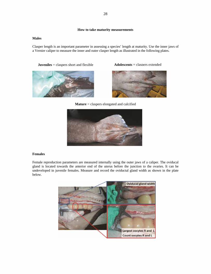
How to take maturity measurements
Males Clasper length is an important parameter in assessing a species’ length at maturity. Use the inner jaws of a Vernier caliper to measure the inner and outer clasper length as illustrated in the following plates.
Mature = claspers elongated and calcified
Females Female reproduction parameters are measured internally using the outer jaws of a caliper. The oviducal gland is located towards the anterior end of the uterus before the junction to the ovaries. It can be undeveloped in juvenile females. Measure and record the oviductal gland width as shown in the plate below.
Adolescents = claspers extended Juveniles = claspers short and flexible

29
Appendix 8 Spiral valve count
The spiral valve is an organ attached to the stomach that increases the surface area over which food passes. The number of “turns” in the spiral valve can be species-specific and is helpful in identifying species. If time permits, count the number of spiral valve turns and record this spiral valve count (SVC) on the deck sheet. The spiral valve is attached ventral-posteriorly to the stomach and ends attached to the vent (B in plate). Often, the SVC can be taken by counting the bands seen through the wall of the spiral valve (B in plate). If these bands are not easily seen from outside of the spiral valve, an SVC can be taken by cutting along the length of the spiral valve and counting the turns or spirals (A in plate). Record the SVC on the deck sheet under “SVC” for the appropriate specimen.

30
Appendix 9 Stomach contents analysis
After removing and dissecting the stomach, its content should be analysed and the prey items counted (see some examples below). The volume of food constituents can also be estimated by expressing the various items as a percentage of the total volume. This method of analysis is subjective in nature, and the investigator’s personal bias is likely to influence the results greatly.
Estimated volume: 60 percent bony fish; 30 percent shark; 10 percent squid.
1 fish
1 fish
3 fish
1 squid
1 shark
1 fish

31
Appendix 10 Vertebrae and spines
Sharks lack bones, but they do have hard structures namely vertebrae and fin spines. These vertebrae and fin spines can be cross-sectioned and their rings can be counted in order to estimate their age (just like counting tree rings). Vertebrae and spines are not required from every specimen. The most important thing is to collect vertebrae and spines from a variety of sizes and, therefore, different ages. As males and females often grow at different rates, it is important to collect vertebrae and spines from a mix of males and females. For these reasons, sizes are divided into 10 cm bins: 0–10 cm, >10–20 cm, >20–30 cm, and so on. For each species encountered, collect vertebrae and spines from up to five males and five females for each size bin. Once you collect a vertebrae and spine sample, record it (by tally) on the inventory form for the appropriate species, sex, and size bin (see protocol). Vertebrae and spines do not have to be collected from: catsharks, chimaeras, or hexanchiformes. Some species do not have fin spines, but can still be aged through their vertebrae. For these species, only collect vertebrae. In order for vertebrae and spine samples to be useful, you must also take and record length data. ALL fin material must be removed from fin spines. Collection from sharks Collect vertebrae from the shark’s torso area, roughly between the pectoral fins (see figure). This is most easily done by cutting into the body cavity and excising about 10 vertebrae through the roof of the body cavity (see accompanying plate). Cutting through the vertebrae can be difficult, and it is helpful to cut through the small space in between the individual discs (the spinal stenosis). Trim excess flesh away from vertebrae, but leave some muscle as a tissue sample for heavy-metal analysis. Tag and bag vertebrae with the same unique serial number assigned to the specimen from which the vertebrae were taken, and freeze them. Collect spines from the same specimens from which you have collected vertebrae. Take spines from directly in front of each dorsal fin and cut them out with a “V” cut. It is easiest to cut AGAINST the denticles (posterior to anterior). As the spines often extend deep into the muscle, it is best to cut all the way down to the vertebrae. If one spine is damaged, discard it and only keep the undamaged spine. If both spines are damaged, discard them both and use a different candidate as a specimen for spines. ALL fin material must be removed from fin spines. Freeze spines in the same bag as the associated vertebrae. Spines and their associated vertebrae can share the same unique serial number.

32
Collection from skates and rays
Collect vertebrae from the torso area of the skate/ray, roughly between the wings, under the nuchal thorns (see figure). This can be done by simply cutting a rectangle out of the back around the nuchal thorns and excising about ten vertebrae. Cutting through the vertebrae can be difficult, and it is helpful to cut through the small space in between the individual discs (the spinal stenosis). Trim excess flesh away from vertebrae, but leave some muscle as a tissue sample for heavy-metal analysis. Tag and bag vertebrae with the same a unique serial number assigned to the specimen from which the vertebrae were taken, and freeze them. Spines and thorns do not need to be taken from skates or rays.

33
Appendix 11 Saving and preservation of unknown, rare or strange specimens
In addition to taking photographs first of the fresh specimen, preserving and forwarding such individuals may be very important for science. These may document, for example, the first geographic records, the first records of small young or fully grown adults in a given location, or you may even have found a species so far unknown to science. At sea, after first photographing the specimen, send, if possible, a photograph or series of digital photographs to someone (e.g. a scientist) to further check the identification of the specimen and determine whether it should be saved. Once a further determination has been made on its possible identification, and it has been determined to save the specimen, it should be preserved by wrapping it in a plastic bag and deep-freezing it. Any associated information (see above) should be included along with the specimen. Use thick waterproof and leakage proof plastic bags or boxes for storage. If it is not possible to send digital photographs from sea, the specimen should be saved. Once back in port, the specimen should remain frozen until someone, preferably from a marine or fishery institute, zoological institute, or museum, and knowledgeable about the possible identification of the specimen, can further examine it. Once a determination has been made to save it, a tissue sample (about 2–5 g) should be removed and preserved in a vial of 100 percent ethanol. The entire specimen, assuming it is not too large, should then be preserved first in 10 percent formalin. A bin should then be set up in a well-ventilated (the liquid and gas are very toxic) facility and using a dilute concentrated formalin 1:9 with water. If possible, using a syringe, some formalin (100 percent) should be injected into the belly cavity, or a small cut can be made through the belly to allow penetration of formalin to the innards to prevent rotting inside the belly cavity. The storage bin can be outside in a secure area, but under cover and protected against the outside elements. Once preserved, the specimen can be shipped to a regional expert for further examination, and it may become part of the fish collection of a national or major international museum. Where to inject the shark with 100 percent formalin:

34
Appendix 12 Tagging and bagging a specimen or sample
All collected material for scientific use requires documentation and a unique serial number. Material can include full specimens, stomach contents, vertebrae and spines, as well as other biological material. Any material collected should be documented on the deck sheet in the row designated for the specimen. Each animal should have its own haul code (haul number and fish count number) and a unique serial number for redundancy (see deck sheet section).
• The haul code is a composite of the haul number and the fish count number for that haul. Example: while working up haul 137, the fifteenth specimen you record has the haul code 137-15.
• Unique serial numbers are serial numbers on pre-printed on the tags.
Full specimens: Place a tag through the fleshy part of the caudal fin. Do not put the tag through the fin radials. Tags put through fin radials often tear out and are lost. Record the tag serial number on the deck sheet. Place the specimen in a plastic bag along with any notes about the specimen (use a blank deck sheet for this purpose), and close the bag securely. Place the specimen in the freezer. Genetic samples: Each genetic sample vial has its own unique vial number. After collecting a genetic sample and placing it in a vial, record the vial number on the deck sheet. Place the vials in a refrigerator, but make sure that the samples do not freeze. Freezing and thawing of the tissue will result in degradation of the tissue sample. Other: Vertebrae, spines, parasites or anything unusual should be collected and placed in a bag tagged with the unique serial number of the specimen from which the sample was taken. If multiple samples are taken from the same specimen, the same unique serial number may be used (hand written on a tag in pencil). Each specimen should have its own bag with its own tag. Vertebrae and spines from the same individual can be kept in the same bag and may share a tag. Record tag numbers on the deck sheet. Place tagged and bagged samples in the freezer.

35
Appendix 13 Cartilaginous fish data collection protocol
For every chondrichthyan encountered:
• Identify the animal as a shark, skate, ray, or chimaera.
• Sex the animal.
• Take length data.
• Take additional notes.
• Proceed to process based on specimen type (voucher, non-voucher, live).
For voucher specimens:
• Rank maturity (externally only).
• Take lengths.
• Take genetic tissue and photograph (ALWAYS REQUIRED!).
• Tag and bag.
For non-voucher specimens:
• Rank maturity.
• Take lengths.
• Take genetic tissue and photograph (six males and six females per species).
• Take maturity measurements.
• Check stomach contents.
• Take spiral valve count.
• Take vertebrae and spines (five from each 10 cm size bin for each sex per species).
For live specimens:
• Take photograph.
• Take lengths.
• Record sex.
• Rank maturity (externally only).
• Take genetic sample.
36
Appendix 14 A protocol for conducting at-sea research aboard a commercial trawler
A) Identification numbers At-sea research involves collecting many specimens and samples, recording many types of data, and documenting the record with many photographs. Because of the large amount of biological material and data involved, it is critically important that researchers employ a fool-proof cross-referencing system for identifying and correlating all this information. Specimens and samples that cannot be linked to associated data are of little use. The following numbering system is designed to track all data, specimens and samples as simply as possible while minimizing the likelihood of errors. 1) Haul code A haul code is a combination of a haul number and a fish number. a) A haul number is the number the vessel assigns to each haul. Haul numbers occur in numerical succession and run consecutively for the duration of the expedition, e.g. the vessel’s first haul is haul number 1, the second haul is haul number 2, etc. Using a haul number is more practical than using a haul date, as there might be several hauls each day. b) A fish number is the numbered order in which a specimen from a haul is examined, e.g. the fifth specimen examined from a haul is fish number 5. Example of haul code: The second specimen examined from haul #132 would have haul code 132-2. A haul code gives a one-of-a-kind identification number to every fish: (i) from which data are taken (species, sex, length, location, etc.); (ii) that is collected as a whole specimen; (iii) from which a sample (vertebrae, spine, egg, embryo, genetics) is taken; or (iv) that is photographed. A haul code is assigned to EVERY individual fish from which ANY data are collected. Integrating the genus and species names into the code might appear useful, but has potential for introducing error or uncertainty, thereby risking data utility. For this reason, it is discouraged. 2) Unique serial number A unique serial number is used to identify each specimen and each sample, regardless of species. Serial numbers have five digits and proceed from 20000 to 99999 and are pre-printed on tags used to label bags containing specimens and samples. Serial numbers represent a redundant, backup system for identifying specimens and samples, and for matching them with data and photographs. As such, serial numbers serve as a built-in failsafe for correlating data. 3) Vial number A vial number is a unique number used exclusively for the identification of a genetic sample. These numbers are pre-printed on vials. Vial numbers proceed from 10001 to 19999. IDENTIFICATION NUMBERS (HAUL CODE, SERIAL NUMBER, AND VIAL NUMBER) MUST BE RECORDED ON THE DECK SHEET FOR EACH HAUL.
B) Forms 1) Deck sheet Deck sheets are used to record raw data. These sheets are printed on both sides of waterproof paper.
37
Deck sheets include space to record:
species, sex, and count;
specimen data (length, maturity, etc.);
haul data (haul number, location, time of net deployment and retrieval, fishing depth, gear type, and bottom depth);
notes on diet or other data/events associated with a haul.
2) Tag and bag form Tag and bag forms have a pre-printed unique serial number and spaces for recording haul code and vial numbers associated with the bag’s contents. These forms are used for tracking whole specimens, as well as spines and vertebrae samples. Tag and bag forms are printed on waterproof paper and should be tagged on the specimen. 3) Vessel haul form This form is used to transcribe haul data (location, depth, fishing time, etc.) from the ship’s log notebook in the wheelhouse. 4) First encounter form This form is used whenever a species is encountered for the first time. Fill out this form and include a drawing (or drawings) to aid in proper identification and future species verification. 5) Inventory form This is a form for maintaining an updated inventory checklist of the chondrichthyans (sharks, skates, chimaeras) encountered from each size range and sex and for documenting the kind of samples collected. See inventory form. Keep a waterproof paper copy of this form at the work station to facilitate updating samples. An electronic copy of the inventory form should be updated daily. 6) Daily log The daily log is an electronic document used for recording field notes of unusual or abnormal events from hauls, and other information that might be useful when further processing specimens, samples and data. Maintain this log as a Word document. Make log entries daily. C) Procedures 1) Haul data Haul data must be kept for all hauls.
a) Record haul data on each deck sheet, include haul number, and – with the captain’s permission – location, time (based on a 24-hour clock) of net deployment and retrieval, as well as fishing depth, gear type, and bottom depth. b) If access to this information is not available, record the haul number, and other haul data will be retrieved at a later date.

38
2) Specimen identification Identify every chondrichthyan to species (or the lowest taxa possible).
a) Every specimen should have its own row on the deck sheet and at a minimum have entries for (i) species, (ii) haul code, and (iii) sex. b) For hauls with more than 100 individuals of the same species, a count for males and a count for females may be recorded under the “notes” section. In such cases, recording the haul number is sufficient. It is not necessary to record complete haul codes that include individual fish numbers.
3) Specimen collection (“tag and bag”) Collect 12 whole specimens (6 males and 6 females) of each species of chondrichthyans.
a) Collect specimens from as wide a size range (small, medium, and large) as possible.
b) Photograph specimens while fresh. In the photograph, include a tag with the haul code and the photograph sequence number. Example: the first photograph of haul code 244-23 will be 244-23 (1), the second photograph of the same specimen will be 244-23 (2), the third, 244-23 (3), etc. Include a ruler in the photograph for reference.
c) Record sex and length data.
d) Assign a haul code and a unique serial number to each collected specimen and record the specimen’s sex and length data on a deck sheet.
e) Most wanted: tag and bag all species on the “most wanted” posters.
4) Basic biological data
a) Record length data and maturity ranking/scan for every chondrichthyan possible.
b) This includes: (i) length (TL and PC); (ii) disc width, disc length, and total length for skates and rays; (iii) sex; and (iv) a maturity scan code (0–4 for males, and 0–6 for females).
c) Photograph male and female representatives of each maturity stage.
d) Collect pregnant females as described below.
5) Reproductive and maturity parameters
a) Record inner and outer clasper lengths from males.
b) Take oviductal gland width from as many females as possible.
c) Take oocyte count and largest oocyte width from each ovary from as many females as possible. Left and right oocyte counts and widths each have data boxes on the deck sheet.
6) Tissue samples
a) Take tissue samples when the collection of a whole specimen is prohibited by its size or when it is healthy enough to be returned to the sea.
b) Take tissue samples from up to 10 specimens of each species (5 per sex).
c) Take tissue samples with a dermal punch from live specimens being returned to sea. Use a knife to excise tissue samples from dead specimens.
d) The primary tissue from which to take a sample is the liver.

39
e) Take precautions to ensure there is no cross-contamination of genetic material.
f) Collect a pea-sized piece of tissue, slice it into 4 or 5 slivers, and store the slivers in an ethanol-filled vial with a pre-assigned vial number.
g) Record identifying vial numbers on deck sheet.
h) Every tissue sample REQUIRES a series of photographs associated with it to later confirm identification.
7) Photo-documentation
a) Photograph all observed chondrichthyans species, and, if/when time permits, also photograph deep-sea bony fishes. Use a cloth background when possible. Remember to include a tag with the haul code and a ruler in the photograph as references. See “Photo-documentation protocol.”
b) Every specimen from which a genetic sample is taken REQUIRES photo-documentation.
c) Bony fish: Only include full lateral photograph and any other views that seem important for the specific species. Record the haul number so the fish can later be matched up with location data. Collect at least one specimen of each species of deep-sea bony fish encountered.
8) Pregnant females Collect pregnant females and egg cases when practical; otherwise collect as many data as possible.
a) If the female is too large to be collected, take a tissue sample along with length data, and tag and bag the embryos in a single bag, and record the unique serial number on the deck sheet.
b) Record embryos position in each uterus. “Right” and “left” refers to a female’s right and left, record with an “R” or “L”. The numbering of embryos starts at the cloaca and moves along the uterus away from the cloaca. Position “1” designates the pup closest to the cloaca. For example, the pup in a female’s right uterus closest to the cloaca (next in line to be born) is recorded as “R1”. The fifth pup (4 away from the cloaca) in the left uterus is recorded as “L5”.
c) If neither the mother nor embryos can be kept, collect biological data from both the mother (length, tissue sample) and embryos (sex, length, position in each uterus, and tissue samples).
d) Always photograph pregnant females and embryos regardless of whether or not they are kept.
e) If a pregnant female is collected, her eggs and embryos may be placed in the same plastic bag as the female, and everything may be assigned the same unique serial number.
f) If data are collected on pups (sex, length, position in uterus), the pups all have same the same haul code as the mother but each is assigned its own unique serial number.
9) Vertebrae
a) Collect vertebrae from the chondrichthyan’s torso area (roughly between the pectoral fins).
b) A vertebrae sample should have about ten vertebrae.
c) For each species, collect vertebrae samples from 5 males and 5 females per 10 cm size bin.
d) Trim excess flesh away from vertebrae, but leave some muscle tissue for heavy metal analysis.
e) Tag and bag vertebrae with a unique serial number, and freeze.
f) For species with spines, see item 10 below.
g) Collect vertebrae from a small subset (about 10) of each species encountered (on the vertebrae wanted list).

40
10) Spines Take spine samples in connection with vertebrae samples in species possessing spines.
a) Take spines from directly in front of each dorsal fin, and cut out with a “V” cut.
b) Collect both spines and freeze them in the same bag as the vertebrae.
c) When bagged together, spines and vertebrae should share the same unique serial number.
d) If one spine is damaged, discard it and only keep the undamaged spine. If both spines are damaged, discard them both and use a different specimen.
e) Collect ten pairs of spines and trim excess flesh from around the spine. Make certain you remove all of the fin.
f) For each species, collect spines from 5 males and 5 females per 10 cm size bin.
11) Diet As time permits, record general field notes on stomach contents.
a) Record diet data whenever there are stomach contents.
b) Record diet as general composition of stomach contents or count (e.g. about 2 fish, 5 squid) and as the percentage of stomach volume (60 percent fish, 40 percent squid).
c) Record diet data in the “notes” section of the deck sheet for the associated haul.
d) Photograph unusual stomach contents.
e) If unusual, diets and stomachs may be tagged and bagged.
12) Special data For any deceased specimen too large to collect:
a) Take extensive photographs.
b) Record detailed reproduction data from females.
c) Collect any pups or egg cases from pregnant females.
d) Collect claspers from males.
e) Take diet data.
f) Photo-document EVERYTHING!
13) Coral Collect representative coral samples.
a) Record volume of coral in hauls on deck sheet.
b) Photograph specimens.
c) This should be done when practical as time permits.

41
14) Extra tissue Collect tissue samples to investigate mercury levels and/or other pollutant levels. Include excess tissue in the vertebrae and spine samples collected, but be sure not to include any fin material. 15) Inventory list Maintain and update a checklist/inventory of chondrichthyans (sharks, skates, chimaeras) encountered, and document the kind of samples taken.
a) The inventory list should document which chondrichthyans have been encountered from each size range and sex, and what kind of data was taken on them.
b) Update electronic copy of inventory form on a daily basis.
c) Keep a waterproof paper copy at the work station in order to keep track of samples.
16) Daily log Keep a daily log detailing abnormal events from hauls and information that will be helpful later when further processing the samples and data. Maintain the log as a Word document. Record a minimum of one entry per day. 17) First encounter Fill out a first encounter sheet every time a species is encountered for the first time. Fill in the check boxes and include a drawing (or drawings) to aid in proper identification and future species verification.

The regional workshop “Identification of Deep-sea Cartilaginous Fishes of the Indian
Ocean” was held in Albion, Mauritius, from 10 to 13 June 2014. It was attended by 19 participants from a wide range of countries and fields of expertise, including taxonomy and bioecology of cartilaginous fishes. The general objective of the workshop was to improve the capabilities of scientists from countries bordering the Indian Ocean to identify a range of deep-sea cartilaginous fish species caught in the region. The participants were introduced to the anatomical features and taxonomy of the orders of deep-sea sharks occurring in the Indian Ocean, to the use of the taxonomic keys included in the reference text material (e.g. FAO catalogues and identification guides) and to the methodologies of processing and identifying a selection of specimens. In addition, a biological data collection protocol was illustrated, thus allowing for improved reporting of shark specimens.
I4241E/1/03.15
ISBN 978-92-5-108665-0
9 7 8 9 2 5 1 0 8 6 6 5 0
ISSN 2070-6987