reports on the free-living platyhelminthes from australia
TRANSCRIPT

2004 Zoological Society of JapanZOOLOGICAL SCIENCE
21
: 333–341 (2004)
Reports on the Free-Living Platyhelminthes from Australia:Typhloplanoida, with the Description of Three New Taxa
Wim Willems
1
*, Tom Artois
1
, Wouter Vermin
1
, Thierry Backeljau
2
and Ernest Schockaert
1
1
Limburgs Universitair Centrum (LUC), Dept. SBG, Centre for Environmental Sciences,Research Group Biodiversity, Phylogeny and Population Studies,
University Campus, Building D, B-3590 Diepenbeek, Belgium
2
Royal Belgian Institute of Natural Sciences, Dept. Invertebrates,Vautierstraat 29, B-1000 Brussels, Belgium
ABSTRACT
—Five typhloplanoids from the Australian East Coast are reported, three of them new to sci-ence. Two taxa are members of Promesostomidae:
Vauclusia conica
n.g. n.sp., characterised by a cone-shaped stylet, the presence of a female bursa and a very long, partially-swollen female duct;
Brinkman-niella
australiensis
n.sp. has a funnel-shaped stylet with a smooth distal tip.
Pilamonila bimascula
n.g. n.sp.is a representative of the Solenopharyngidae, characterised by a stylet within a cirrus. The known speciesfound are
Ceratopera axi
and
Ptychopera scutulifer
.
Key words:
taxonomy, ‘Turbellaria’, Promesostomidae, Solenopharyngidae, Trigonostomidae
INTRODUCTION
Relatively few free-living platyhelminth species fromAustralia, excluding Polycladida, have been reported anddescribed. To date, most of the microturbellarians are Pros-eriata, with 41 species reported and described (Curini-Gal-letti 1997, 1998; Curini-Galletti and Cannon, 1995, 1996a, b,1997; Martens and Curini-Galletti, 1989; Curini-Galletti
etal.
, 2002; Faubel and Rohde, 1998). Also known are fourspecies of Macrostomida (Faubel, Blome and Cannon,1994; Sluys, 1986), a single representative of Kalyptorhyn-chia (the polycystidid
Gyratrix hermaphroditus
Ehrenberg,1831, which seems to be a complex of sibling species(Curini-Galletti and Puccinelli, 1990, 1998)), five species ofDalyellioida (of which only one,
Luriculus australiensis
Faubel
et al.
, 1994, is from a marine habitat (Faubel, Rohdeand Watson, 1994; Hartenstein and Dwine, 2000; Hochbergand Cannon, 2001, 2002a; Schmarda, 1859)) and 15 spe-cies of Typhloplanoida. Among the latter, ten species arefrom freshwater habitats (Hochberg and Cannon, 2002a;Kolasa and Schwartz, 1988; Noreña-Janssen and Faubel,1992; Schmarda, 1859). The five marine typhloplanoids are:
Magnetia queenslandica
Hochberg and Cannon, 2002 (seeHochberg and Cannon, 2002b), and four species of
Trigo-nostomum
(Willems
et al
., in press). However, in an ecolog-
ical study on tropical intertidal sediments Dittman (1991)recognised 108 different species, 16 of which were Typhlo-planoida. In this contribution we report on five more typhlo-planoids, of which three are new to science and two areknown from localities outside Australia (
Ceratopera axi
(Riedl, 1954) Den Hartog, 1964 and
Ptychopera scutulifer
Ehlers and Ax, 1974), bringing the total number of namedmarine typhloplanoids to ten for Australia. They have allbeen found on the East Coast in areas around Townsville,Brisbane (North Stradbroke Island), Sydney and betweenByron Bay and Coffs Harbour.
Next to their scientific names according to the Linneansystem (and the International Code on Zoological Nomen-clature (International Commission on Zoological Nomencla-ture, 1999)), we also propose a converted name for eachspecies in the phylogenetic system (Phylocode; http://www.ohio.edu/phylocode) following the system proposed byArtois (2001).
MATERIAL AND METHODS
The specimens for this study were collected during two sepa-rate expeditions: the first in August-September 1996 by Tom Artoisand Ernest Schockaert (ES) and the second by ES in September-November 1997. The animals were extracted from the sediment orfrom algae using the MgCl
2
-decantation method (see Schockaert,1996), studied alive and whole mounted with lactophenol. Remain-ing specimens, if any, were fixed in marine Bouin’s solution, embed-ded in paraffin, serially sectioned (4
µ
m sections) and stained withHeidenhain’s iron haematoxylin, using eosin as a counterstain.
* Corresponding author: Tel. +32-11-268383;FAX. +32-11-268301.E-mail: [email protected]

W. Willems
et al
.334
Camera lucida drawings of hard parts were made, using Nomarskiinterference. Drawings without a scale bar are freehand. Measure-ments of hard parts are taken axially, unless indicated otherwise.The positions of the gonopore and organs, and the measurementsof the pharynx are expressed in percentages of the total bodylength (distances from the anterior tip of the body).
The type material of the new species will be deposited in thecollections of the Queensland Museum, Brisbane, Australia.Voucher specimens of
Ceratopera axi
(Riedl, 1954) Den Hartog,1964 from California and the Falklands, were loaned from theSwedish Museum of Natural History in Stockholm (SMNH). A wholemount of
Ptychopera scutulifer
Ehlers and Ax, 1974 from Somaliais present in the collections of the LUC (Diepenbeek, Belgium).
Abbreviations used in the figures
b: brain; bs: bursal stalk; cg: caudal glands; cga: common genitalatrium; ci: cirrus; cil: cilia; de: ejaculatory duct; e: eye; ecm: externalcircular muscle; elm: external longitudinal muscle; fb: female bursa;fd: female duct; fg: female glands; gg: prostate glands; gm: glandsof Minot; gp: common genital pore; i: intestine; icm: internal circularmuscle; id: insemination duct; ilm: internal longitudinal muscle; lm:longitudinal muscle; m: mouth; ma: male atrium; od: oviduct; ov:ovary; pc: prepharyngeal cavity; pg: pharynx glands; ph: pharynx;pl: pharynx lumen; ppt: pharynx protractors; rg: rostral glands; rh:rhabdite; rm: radial muscle; s: stylet; sph: sphincter; t: testis; v: vasdeferens; vd: vitelloduct; vg: prostate vesicle; vit: vitellaria; vs: sem-inal vesicle; y, z: features described in respective text.
TAXONOMIC ACCOUNT
TRIGONOSTOMIDAE GRAFF, 1905
sensu
DEN HARTOG, 1964
Ceratopera
Den Hartog, 1964
Ceratopera axi
(Riedl, 1954) Den Hartog, 1964
ceratopera-axi
(Riedl, 1954) Den Hartog, 1964
Proxenetes axi
Riedl, 1954
Ceratopera bifida
Ehlers and Ax, 1974
Locality in Australia.
Arrawarra (New South Wales): on small,shell-shaped brown algae (
Pedina
sp
.)
(29/08/1996) and on
Pavonina
-like algae (27/08/1996) in intertidal rockpools.
Known distribution.
Gulf of Naples and Sicily (Riedl, 1954);Galapagos (Ehlers and Ax, 1974); Falkland Islands and Cal-ifornia (Karling, 1986); Weddell Sea and La Réunion (Artois
et al
. 2000).
Material.
Observations on live, mature specimens and twowhole mounts (one from each new locality). Whole mountsfrom Falkland and California (collections of SMNH).
Remarks
. Following Karling (1986) and Artois
et al
. (2000),we consider
C. bifida
Ehlers and Ax, 1974 a junior synonymof
C. axi
(Riedl, 1954) Den Hartog, 1964. The stylet of oneof the Australian specimens is 70
µ
m long (measured alongthe axis of the stylet; 56
µ
m if measured from top to bottomas in Ehlers and Ax, 1974). The bursal appendage of thisspecimen is 94
µ
m long and splits distally. Neither part couldbe measured in the second specimen. In comparison withother populations (see Table 1), the Australian specimenhas the smallest stylet.
Ptychopera
Den Hartog, 1964
Ptychopera scutulifer
Ehlers and Ax, 1974
ptychopera-scutulifer
Ehlers and Ax, 1974
Localities in Australia.
Arrawarra (New South Wales): south-ern part of the beach, on
Sargassum
-like algae in a largepermanent pool at the beginning of a mass of rocks (27/08/1996). North Stradbroke Island, Amity Point (Queensland):in muddy sediment from amongst mangroves (14/08/1996).
Known distribution.
Galapagos (Ehlers and Ax, 1974);Somalia (Schockaert and Martens, 1985).
Material.
Two mature specimens studied alive and mounted(one from each new locality). A whole mount of a specimenfrom Somalia (collections of LUC).
Remarks.
The specimen from Stradbroke Island has a 54
µ
m-long stylet (measured axially). The sclerotised part ofthe afferent female duct (Ehlers and Ax, 1974: ductus sper-maticus) between the swollen part (Ehlers and Ax, 1974:receptaculum seminis) and the atrial bursa (Ehlers and Ax,1974: bursa copulatrix) is 49
µ
m long and is bent over 90
°
.These data correspond with the measurements on speci-mens from the Galapagos (38–40
µ
m-long stylet, axiallymeasured; 23
µ
m-long sclerotised part of the afferent duct;Ehlers and Ax, 1974) and from Somalia (45–47
µ
m-longstylet; 20
µ
m sclerotised part of the afferent duct; Schock-aert and Martens, 1985). On the other hand, the stylet of theArrawarra specimen measures 98
µ
m, while the distal partof the afferent duct is 140
µ
m long. Whether these aberrantvalues represent a fixed difference between populations orare due to individual variability is not yet clear. It is also theonly specimen found on algae. We, however, prefer to retainthe Arrawarra-specimen within this species until/unless fur-ther material should suggest otherwise.
PROMESOSTOMIDAE DEN HARTOG, 1964
Brinkmanniella
Luther, 1943
Brinkmanniella australiensis
n.sp.
brinkmanniella-australiensis
n.sp.(Fig. 1)
Locality in Australia.
Arrawarra (New South Wales): on crus-taceous algae on rocks on the beach north of the headland
Table 1.
Measurements of the stylet and the bursal appendage of
Ceratopera axi
in different populations.
StyletBursal
appendageReference
Australia 70
µ
m 94
µ
m this paper
Galapagos 94–95
µ
m 67–87
µ
m Ehlers and Ax, 1974
Falklands 120–180
µ
m 91
µ
m Karling, 1986
California 93–117
µ
m 102
µ
m Karling, 1986
La Réunion 105
µ
m 77
µ
m Artois
et al
., 2000
Weddell Sea 124
µ
m ? Artois
et al
., 2000

Typhloplanoida – Australian East Coast 335
(27/08/1996) (type locality).
Material.
One animal studied alive and mounted (holotype).
Etymology
. The specific name refers to the species’ occur-rence in Australia.
Description.
The animal is 0.9 mm long (measured on wholemount), pale yellow and has two eyes. The general organi-sation does not deviate from that of other
Brinkmanniella
-species (Fig. 1A; see also Ehlers, 1974; Karling, 1986;Luther, 1943, 1948; Marcus, 1951; Schockaert and Martens,1985). The stylet (Fig. 1B) is thin-walled, straight and funnel-shaped. It is 57
µ
m long, and 32
µ
m wide proximally andslightly constricted about halfway along its length.
Diagnosis
Brinkmanniella australiensis.
Species of
Brink-manniella
with straight funnel-shaped stylet of 57
µ
m longand proximally 32
µ
m wide, constricted halfway along itslength and with a smooth distal tip.
Discussion
. The representatives of the taxon
Brinkmanniella
are characterised by the rostral position of the ovaries, theabsence of any special female atrial organs, apart from thefemale duct, and the caudally-situated pharynx. Schockaertand Martens (1985) gave a good overview of the main char-acters of
Brinkmanniella
-species. The stylets of
B. obtusa
Luther, 1943,
B. augusti
Marcus, 1951 and
B. palmata
Kar-ling, 1986, show “fingers” at the distal opening (Karling,1986), while
B. australiensis
,
B. macrostomoides
Luther,1948,
B. procerastyla
Ehlers, 1974 and
B. microps
Schock-aert and Martens, 1985 lack these “fingers”. Of these latterfour species, only
B. microps
and
B. australiensis
have astraight tubiform stylet, while it is curved in the other twospecies. The stylet of
B. microps
is only 30
µ
m long and 10
µ
m wide proximally and not constricted midway as in
B. aus-traliensis
. The stylet of
B. australiensis
is twice as long witha width/length ratio of about 1/2.
Vauclusia conica
n.g. n.sp.
vauclusia-conica
n.sp.(Fig. 2)
Locality in Australia
. Sydney, Vaucluse beach (New SouthWales): flat beach with fine sand and numerous crab holes,in eulittoral (10/10/1997) (type locality).
Material. One individual studied alive and mounted (holo-type). Three serially-sectioned specimens (paratypes).
Etymology. The genus name/praenomen refers to the typelocality. The specific name emphasizes the overall structureof the copulatory organ. Conicus (Lat.): cone-shaped.
Description. The slender animal is ± 1.4 mm long (measuredon the whole mount), without eyes. The cellular epidermis is± 3.5 µm thick, with cilia of 3 µm long. The basement mem-brane is ± 1 µm thick. At the rostral end of the body, twotypes of large rhabdite glands are present. The first type, sit-uated at the periphery of the glandular mass, produceslarge, basophilic rhabdites of 8.5–10.5 µm long. The glandsin the centre produce eosinophilic rhabdites of the samesize, but less densely packed within the cell bodies. Allglands end at the rostral body tip, the basophilic onesstretching to the testes, the eosinophilic ones not furtherthan the brain. There are also glands in the caudal bodyregion (Fig. 2E: cg), opening ventrally behind the gonoporeand producing large, basophilic rhabdites.
The mouth is situated at ± 65% and can be closed bya strong sphincter. The prepharyngeal cavity is lined with alow, nucleated epithelium and surrounded by only longitudi-nal muscles. The distal rim of the pharynx is lined with avery low epithelium, with a thick basement membrane and
Fig. 1. Brinkmanniella australiensis n.sp. - A. General organisation(from a live specimen). - B. Stylet (from the holotype).

W. Willems et al.336
cilia (Fig. 2C: cil). The pharynx lumen (Fig. 2C: pl) is linedwith a low, anucleated epithelium. The epithelium of theproximal pharyngeal rim is degenerated, leaving only a thick
pseudocuticula. The inner circular muscles are thicker nearthe proximal and distal ends of the bulb. The exact numberof internal longitudinal muscles could not be determined.
Fig. 2. Vauclusia conica n.g. n.sp. - A. Organisation of the genital system (from a live specimen). - B. Stylet (from the holotype). - C. Pharynx(reconstruction on sagittal sections). - D. Habitus of a live animal. - E. Reconstruction of the atrial organs from the right side.

Typhloplanoida – Australian East Coast 337
There are two types of eosinophilic pharyngeal glands, onecoarse-grained and one fine-grained. They open into thelumen somewhat proximally from the distal sphincter, withthe fine-grained ones most distally. In one of the sectionedspecimens a third, basophilic gland could be observed.However, the exact location and the place of dischargecould not be determined. The external pharyngeal musclelayers consisting of an inner circular (Fig. 2C: ecm) and anouter longitudinal one (Fig. 2C: elm), are rather weak.
The common genital pore is situated at ± 80%. Thecommon genital atrium is lined with a high, anucleated epi-thelium and surrounded by an inner longitudinal and anouter circular muscle layer. Although the live specimen (Fig.2D) apparently only showed one testis, there are clearly twotestes in the sectioned specimens. The testes are situatedventrally just in front of the pharynx, at both sides of thebody. The vasa deferentia are clearly visible in the sectionedspecimens.
The paired seminal vesicles (only one observed in thelive specimen; Fig. 2A) are lined with a low, nucleated epi-thelium. They continue towards the prostate vesicle, fusingwhen entering the prostate vesicle. The prostate vesicle(Fig. 2A: vg) is surrounded by a layer of longitudinal mus-cles, and contains fine-grained (Fig. 2E: gg2) and coarse-grained eosinophilic glands (Fig. 2E: gg1), the fine-grainedglands located in the centre of the bulb. All glands areentirely intracapsular. The stylet (Fig 2B), connected to theprostate vesicle, fills almost the entire male atrium. Thisstylet is cone-shaped, 29 µm long, 17 µm wide proximallyand 5 µm wide distally. The exact structure of the styletcould not be determined, but probably consists of abouteight elongated and spirally-running bars (or ridges), whichshow some striation. The male atrium (Fig. 2E: ma) entersthe common genital atrium from the rostral side. It is linedwith the same epithelium and surrounded by the same mus-cle layers as the common genital atrium, only the musclelayers have changed position, revealing an inner circularand an outer longitudinal muscle layer.
The ovoid ovaries are situated caudally from the gonop-ore. They form the distal part of the ovovitellaria. The femaleduct (Fig. 2E: fd) is long and enters the common genitalatrium at the caudal side. It is lined with a high, anucleatedepithelium and surrounded by strong circular muscles.Distally it is swollen and contains many sperm (Fig. 2E: y).This part is lined with a nucleated epithelium. Proximally thefemale duct ends in the female bursa (Fig. 2E: fb). Itreceives both oviducts somewhat distally from the bursalentrance. The part of the female duct between the bursaand the oviducts (“bursal stalk”) is surrounded by circularmuscles. The oviducts are rather short and lined with a lowanucleated epithelium. A large bundle of coarse-grainedeosinophilic glands (Fig. 2E: fg) enters the female ductventrally at the bifurcation into the oviducts. A uterus islacking.
Diagnoses Vauclusia. Promesostomidae with the pharynx
situated in the middle of the body. Inversion of muscle layersat the transition from the common genital atrium to the malegenital atrium. Paired testes and seminal vesicles. Globularprostate vesicle with two types of secretion. Cone-shapedstylet, consisting of several plate-like bars (or ridges). Pairedovovitellaria. Very long female duct. Distal part of the femaleduct swollen and filled with sperm. Female bursa and femaleglands present. Type species: V. conica.Vauclusia conica. Provisionally with the same diagnosis asthe genus. Stylet 29 µm long.
Discussion. The combination of paired, solid testes, pairedovovitellaria, lack of a second connection of the femalegonads with the exterior and the presence of only one gen-ital pore are diagnostic features of the Promesostomidae(see Den Hartog, 1964). The new species fits into this diag-nosis. Its possible relationships within this taxon are lessclear. The fact that the ovaries in V. conica are not sepa-rated from the vitellaria, and the shortness of its male genitalatrium exclude it from the present taxa Adenorhynchinae Axand Heller, 1970 and Promesostominae Luther, 1948. How-ever it shares some characters with this last taxon such asthe presence of a terminal female bursa and terminal femaleglands. The connection of the ovaries with the vitellaria(ovovitellaria) also suggests a relationship with members ofthe Brinkmanniellinae Luther, 1948. Both in the members ofthe Adenorhynchinae and the Brinkmanniellinae the maleatrium is very short. The relative values of all these charac-ters can only be assessed by a detailed cladistic analysisincluding many more ‘Typhloplanoida’. Therefore, we refrainfrom allocating V. conica to any of the subtaxa within thePromesostomidae.
SOLENOPHARYNGIDAE GRAFF, 1882Pilamonila bimascula n.g. n.sp.
pilamonila-bimascula n.sp.(Figs 3–4)
Locality in Australia. Arrawarra (New South Wales): south ofthe headland, on Sargassum-like algae in a large permanentpool and on algae in a permanent pool in front of the marinestation, lower eulittoral (27/08/1996) (type locality).
Material. Three mature animals studied alive and mounted(one designated holotype, the others paratypes) and threesectioned specimens (designated paratypes).
Etymology. The genus name/praenomen refers to the chainof globular structures in the female system. Pila (Lat.): ball.Monile (Lat.): necklace. The specific name emphasizes thepresence of a stylet and an armed cirrus. Bis (Lat.): twice.Masculus (Lat.): manly.
Description. The animal is 0.4–0.5 mm long (measured onwhole mount), with two eyes. The body is whitish with blackto yellow-brown spots when observed under incident light,pale brown, and opaque when observed with transmitted

W. Willems et al.338
light. The syncytial epidermis is 3 µm thick with cilia of 3 µmlong. The basement membrane is ± 1 µm thick. Rhabditesare very small and equally distributed over the whole epider-mis.
The elongated pharynx (Figs 3A: ph, 4C) is situated inthe first body half with the mouth at ± 65% (on sections). Itis inclined towards the rostral body end. A narrow duct con-nects the mouth with the deep prepharyngeal cavity (Fig.
Fig. 3. Pilamonila bimascula n.g. n.sp. - A. General organisation (from a live specimen). - B. Inverted cirrus (from a live specimen). - C.Everted cirrus and stylet (from the holotype). - D. Male genital system (reconstruction on sagittal sections). - E. Reconstruction of the atrialorgans from the right side.

Typhloplanoida – Australian East Coast 339
4C: pc). This duct is lined with a low, anucleated epitheliumand surrounded by strong circular muscles. At the transitionof this duct with the prepharyngeal cavity the epithelium ismuch higher and shows some nuclei. The prepharyngealcavity is lined with a low, nucleated epithelium and sur-rounded by longitudinal muscles. The distal pharyngeal rimhas no cilia. The pharynx is surrounded by an outer longitu-dinal and an inner circular muscle layer. The former is, atleast in the proximal part of the pharynx, arranged in sixbundles forming a six-pointed star (Fig. 4B). Each “point ofthe star” contains four longitudinal fibres, showing a total of24 external longitudinal muscles. The internal muscles con-sist of weak radial muscles (Fig. 4C: rm), a circular (Fig. 4C:icm) and a longitudinal layer (Fig. 4C: ilm). There are eightinner longitudinal and 16 radial muscle fibres (Fig. 4A). Theepithelium of the lumen has degenerated to a pseudocilia-tion. The pharynx contains coarse-grained eosinophilicglands and some coarse-grained basophilic glands. The lat-ter enter the pharynx proximally, and thus have an extrapha-ryngeal part. The exact place where both types of glandsopen into the lumen could not be determined, but must beclose to the distal end of the pharynx.
The common genital pore lies at 70%, just behind themouth, and can be closed by a sphincter. The common gen-ital atrium is lined with a high, anucleated epithelium and issurrounded by an inner circular and an outer longitudinalmuscle layer.
The paired and elongated testes lie ventrally of the
prostate vesicle, just behind the pharynx. The vasa deferen-tia are short, and connect the testes with the intracapsularseminal vesicle (Fig. 3E: vs). This vesicle fills almost theentire proximal part of the copulatory bulb, and narrows dis-tally to the ejaculatory duct (Fig. 3D: de). The ejaculatoryduct runs centrally through prostate glands. It is lined withan anucleated, membranous epithelium, surrounded byweak circular muscles and connected to a small, tubiformstylet (Fig. 3C–E: s). There are basophilic (Fig. 3D: gg1) andeosinophilic (Fig. 3D: gg2) prostate glands, situated in themost distal part of the prostate vesicle and discharging theircontents through the stylet. This stylet is 12–19 µm long and4–5 µm wide (in the holotype: 19 µm and 4 µm respectively).It was visible in only two of the three whole mounts, but inall sectioned specimens. The copulatory bulb is surroundedby a circular muscle layer. From the stylet two groups ofmuscle fibers run obliquely through the copulatory bulb. Thedorsal group attaches to the wall of the copulatory bulb in itsproximal part (Fig. 3D: lm1), the ventral group in its distalpart (Fig. 3D: lm2). The stylet lies in a cirrus, which bearssmall spines (1–2 µm) in its proximal part and large spines(5–8 µm) distally. Both parts are separated by a sphincter,and in the sectioned individuals the distal part of the cirruswas everted, and when completely everted (as in two of thewhole mounts; Fig. 3C) the stylet appears at the end of thecirrus. The cirrus is lined with a low, membraneous, anucle-ated epithelium, as also is the male atrium. Only at the tran-sition between the male atrium and the spiny cirrus is the
Fig. 4. Pilamonila bimascula n.g. n.sp. - A, B. Sections through the pharynx (A. Distally; B. Proximally). - C. Reconstruction of the pharynxfrom the right side.
W. Willems et al.340
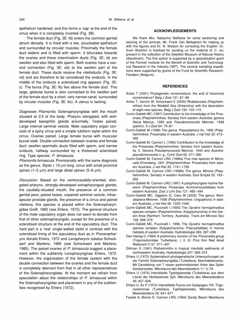
epithelium hardened, and this forms a ‘cap’ at the end of thecirrus when it is completely inverted (Fig. 3B).
The female duct (Fig. 3E: fd) enters the common genitalatrium dorsally. It is lined with a low, anucleated epitheliumand surrounded by circular muscles. Proximally the femaleduct widens and is filled with sperm. It bifurcates towardsthe ovaries and these insemination ducts (Fig. 3E: id) areswollen and also filled with sperm. Both ovaries have a sec-ond connection (Fig. 3E: od) to the swollen part of thefemale duct. These ducts receive the vitelloducts (Fig. 3E:vd) and are therefore to be considered the oviducts. In themiddle of the oviducts a sclerotized ring appears (Fig. 3E:z). The bursa (Fig. 3E: fb) lies above the female duct. Thislarge, globular bursa is also connected to the swollen partof the female duct by a short, very narrow canal, surroundedby circular muscles (Fig. 3E: bs). A uterus is lacking.
Diagnoses Pilamonila. Solenopharyngidae with the mouthsituated at 2/3 of the body. Pharynx elongated, with well-developed basophilic glands proximally. Testes paired.Large internal seminal vesicle. Male copulatory organ con-sists of a spiny cirrus and a simple tubiform stylet within thecirrus. Ovaries paired. Large female bursa with muscularbursal stalk. Double connection between ovaries and femaleduct: swollen spermatic ducts filled with sperm, and narrowoviducts, halfway surrounded by a thickened sclerotisedring. Type species: P. bimascula.Pilamonila bimascula. Provisionally with the same diagnosisas the genus. Stylet ± 15 µm long, cirrus with small proximalspines (1–2 µm) and large distal spines (5–8 µm).
Discussion. Based on the ventrocaudally-oriented, elon-gated pharynx, strongly-developed extrapharyngeal glands,the caudally-situated mouth, the presence of a commongenital pore, paired testes, unpaired seminal vesicle, intrac-apsular prostate glands, the presence of a cirrus and pairedvitellaria, this species is placed within the Solenopharyn-gidae Graff, 1882 (see Ehlers, 1972). The general structureof the male copulatory organ does not seem to deviate fromthat of other solenopharyngids, except for the presence of asclerotised structure at the end of the ejaculatory duct. Thishard part is a ‘real’ single-walled stylet in contrast with thesclerotised lining of the ejaculatory duct as in Procerophar-ynx litoralis Ehlers, 1972 and Lenopharynx tubatus Schock-aert and Martens, 1985 (see Schockaert and Martens,1985). The paired ovaries of P. bimascula suggest a place-ment within the subfamily Lenopharynginae Ehlers, 1972.However, the organisation of the female system with thedouble connection between the ovaries and the female ductis completely aberrant from that in all other representativesof the Solenopharyngidae. At the moment we refrain fromspeculation about the relationships of P. bimascula withinthe Solenopharyngidae and placement in any of the subfam-ilies recognised by Ehlers (1972).
ACKNOWLEDGEMENTS
We thank Mrs. Natascha Steffanie for serial sectioning andstaining of the animals, Mr. Frank Van Belleghem for helping uswith the figures and Dr. N. Watson for correcting the English. Dr.Sven Boström is thanked for sending us the material of C. axi,present in the collection of the Swedish Museum of Natural History(Stockholm). The first author is supported by a specialisation grantof the Flemish Institute for the Benefit of Scientific and Technolog-ical Research in the Industry (IWT). The several sampling expedi-tions were supported by grants of the Fund for Scientific Research-Flanders (Belgium).
REFERENCES
Artois T (2001) Phylogenetic nomenclature: the end of binominalnomenclature? Belg J Zool 131: 87–89
Artois T, Vermin W, Schockaert E (2000) Rhabdocoela (Platyhelm-inthes) from the Weddell Sea (Antarctica) with the descriptionof eight new species. Belg J Zool 130: 103–110
Curini-Galletti MC (1997) Contribution to the knowledge of the Pros-eriata (Platyhelminthes: Seriata) from eastern Australia: generaNecia Marcus, 1950 and Pseudomonocelis Meixner, 1938(partim). It J Zool 64: 75–81
Curini-Galletti M (1998) The genus Polystyliphora Ax, 1958 (Platy-helminthes: Proseriata) in eastern Australia. J nat Hist 32: 473–499
Curini-Galletti M, Cannon L (1995) Contribution to the knowledge ofthe Proseriata (Platyhelminthes: Seriata) from eastern Austra-lia. II. Genera Pseudomonocelis Meixner, 1943 and Acantho-pseudomonocelis n. g.. Contr Zool 65: 271–280
Curini-Galletti M, Cannon LRG (1996a) Five new species of Mono-celis Ehrenberg, 1831 (Platyhelminthes: Proseriata) from east-ern Australia. J nat Hist 30: 1741–1759
Curini-Galletti M, Cannon LRG (1996b) The genus Minona (Platy-helminthes, Seriata) in eastern Australia. Zool Scripta 25: 193–202
Curini-Galletti M, Cannon LRG (1997) A polypharyngeal marine flat-worm (Platyhelminthes, Proseriata, Archimonocelididae) fromeastern Australia. Zool J Linn Soc 121: 485–494
Curini-Galletti MC, Oggiano G, Casu M (2002) The genus Nem-atoplana Meixner, 1938 (Platyhelminthes: Unguiphora) in east-ern Australia. J nat Hist 36: 1023–1046
Curini-Galletti MC, Puccinelli I (1990) The Gyratrix hermaphroditusspecies complex (Platyhelminthes: Kalyptorhynchia) in the Dar-win Area (Northern Territory, Australia). Trans am Microsc Soc109: 368–379
Curini-Galletti MC, Puccinelli I. 1998. The Gyratrix hermaphroditusspecies complex (Kalyptorhynchia: Polycystididae) in marinehabitats of eastern Australia. Hydrobiologia 383: 287–298
Den Hartog C (1964) A preliminary revision of the Proxenetes group(Trigonostomidae, Turbellaria). I, II, III. Proc Kon Ned AkadWetensch C 67: 371–407
Dittman S (1991) Plathelminths in tropical intertidal sediments ofnortheastern Australia. Hydrobiologia 227: 369–374
Ehlers U (1972) Systematisch-phylogenetische Untersuchungen ander Familie Solenopharyngidae (Turbellaria, Neorhabdocoela).Mit Darstellung von 7 neuen psammobionten Arten des SylterSandstrandes. Mikrofauna des Meeresbodens 11: 3–78
Ehlers U (1974) Interstitielle Typhloplanoida (Turbellaria) aus demLitoral der Nordseeinsel Sylt. Mikrofauna des Meeresbodens49: 427–526
Ehlers U, Ax P (1974) Interstitielle Fauna von Galapagos VIII. Trigo-nostominae (Turbellaria, Typhloplanoida). Mikrofauna desMeeresbodens 30: 641–671
Faubel A, Blome D, Cannon LRG (1994) Sandy Beach Meiofauna

Typhloplanoida – Australian East Coast 341
of Eastern Australia (Southern Queensland and New SouthWales). I. Introduction and Macrostomida (Platyhelminthes).Invertebr Taxon 8: 989–1007
Faubel A, Rohde K (1998) Sandy Beach Meiofauna of Eastern Aus-tralia (Southern Queensland and New South Wales). IV. Prose-riata, Plathelminthes. Mitt hamb zool Mus Inst 95: 7–28
Faubel A, Rohde K, Watson NA (1994) Sandy Beach Meiofauna ofEastern Australia (Southern Queensland and New SouthWales). II. Luriculus australiensis, gen. et sp. nov. (Luridae:Dalyelliidae: Platyhelminthes). Invertebr Taxon 8: 1009–1015
Hartenstein V, Dwine KA (2000) Freshwater dalyellid flatworm,Gieysztoria superba sp. nov. (Dalyelliidae: Rhabdocoela) fromsoutheast Queensland, Australia. Memoirs QueenslandMuseum 45: 381–384
Hochberg R, Cannon LRG (2001) A new species of Gieysztoria(Platyhelminthes; Rhabdocoela; Dalyelliidae) from a freshwaterlake in Queensland, Australia. Zootaxa 11: 1–8
Hochberg R, Cannon LRG (2002a) Two new freshwater rhab-docoels, Austrodalyellia gen. nov. and Haplodidymos gen. nov.(Platyhelminthes) from Queensland, Australia. Zootaxa 44: 1–15
Hochberg R, Cannon LRG (2002b) Magnetia queenslandica, a newgenus and new species of typhloplanid flatworm (Platyhelm-inthes: Rhabdocoela) from Magnetic Island in North Queen-sland, Australia. Raffles Bull of Zool 51: 1–6
International Commission on Zoological Nomenclature (1999) Inter-national Code of Zoological Nomenclature. 4th ed, InternationalTrust for Zoological Nomenclature, London
Karling TG (1986) Free-living marine Rhabdocoela (Platyhelm-inthes) from the N. American Pacific coast. With remarks onspecies from other areas. Zool Scr 15: 201–219
Kolasa J, Schwartz SC (1988) Two new Mesostoma species (Tur-bellaria, Rhabdocoela) from Australia. Zool Scr 17: 329–335
Luther A (1943) Untersuchungen an Rhabdocoelen Turbellarien -VII- Über einige Repräsentanten der Familie Proxenetidae.Acta Zool Fenn 38: 3–95
Luther A (1948) Untersuchungen an Rhabdocoelen Turbellarien -VII- Über einige marine Dalyellioida. -VIII- Beiträge zur Kennt-nis der Typhloplanoida. Acta Zool Fenn 55: 3–122
Marcus E (1951) Turbellaria Brasileiros (9). Bol Fac Fil Cienc LetrUniv S. Paulo, Zoologia 16: 5–215
Martens PM, Curini-Galletti MC (1989) Monocelididae and Archi-monocelididae (Platyhelminthes Proseriata) from SouthSulawesi (Indonesia) and Northern Australia with biogeographi-cal remarks. Trop Zool 2: 175–205
Noreña-Janssen C, Faubel A (1992) Revision of the subfamilyMesostominae (Rhabdocoela, Platyhelminthes). Mitt hamb zoolMus Inst 89: 7–47
Riedl R (1954) Neue Turbellarien aus dem mediterranen Felslitoral-Ergebnisse der “Unterwasser Expedition AUSTRIA 1948–1949”. Zool Jb Abt Syst 82: 157–244
Schmarda LK (1859) Neue wirbellosen Thiere. Beobachtet und ges-ammelt auf einer Reise un die Erde 1853 bis 1857 Band 1: Tur-bellarien, Rotatorien und Anneliden. W. Engelmann, Leipzig
Schockaert ER (1996) The Importance of Turbellarians in Ecosys-tems. In “Methods for the Examination of Organismal Diversityin Soils and Sediments” Ed by GS Hall, CAB International,Wallingford, pp 211–225
Schockaert ER, Martens PM (1985) Turbellaria from Somalia. III.Lecithoepitheliata and Typhloplanoida. Monit zool ital (NS)Supple 20: 27–41
Sluys R (1986) First representative of the order macrostomida inAustralia (Platyhelminthes, Macrostomidae). Records of theSouth Australian Museum 19: 399–404
Willems WR, Artois TJ, Vermin WA, Schockaert ER (in press) Revi-sion of the taxon Trigonostomum Schmidt, 1852 (Platyhelm-inthes, Typhloplanoida, Trigonostomidae) with the descriptionof seven new species. Zool J Linn Soc
(Received October 16, 2003 / Accepted December 23, 2003)